The application of liposomes for solubilisation of
hydrophobic drugs
Mathew Louis Steven Leigh
A thesis submitted to the University of London for the degree of Doctor of Philosophy.
Department of Pharmaceutics School of Pharmacy University of London
All rights reserved
INFORMATION TO ALL USERS
The quality of this reproduction is dependent upon the quality of the copy submitted.
In the unlikely event that the author did not send a complete manuscript and there are missing pages, these will be noted. Also, if material had to be removed,
a note will indicate the deletion.
uest.
ProQuest 10105123
Published by ProQuest LLC(2016). Copyright of the Dissertation is held by the Author.
All rights reserved.
This work is protected against unauthorized copying under Title 17, United States Code. Microform Edition © ProQuest LLC.
ProQuest LLC
789 East Eisenhower Parkway P.O. Box 1346
Abstract
The application of liposomes for the solubilisation of lipophilic hydrophobes for intravenous administration has been investigated.
The lipid composition o f four commercially available phospholipids extracted from soya bean and egg sources were studied. The particle size of liposome dispersions generated from pro-liposomes made from these four phospholipids was determined using a variety of sizing techniques. Various factors potentially affecting the particle size such as the degree of agitation, lipid composition and hydration of the liposome were studied. The size distribution of the dispersions without drug were compared to a commercially available intravenous emulsion.
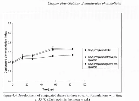
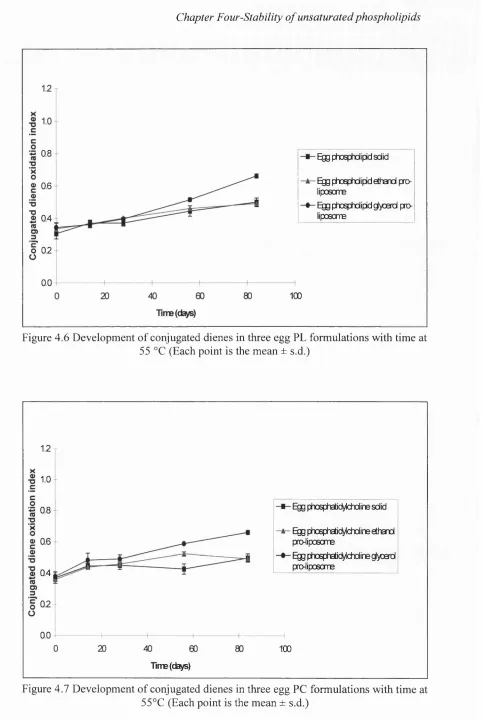
The stability of three placebo anhydrous lipid formulations was examined under accelerated conditions. The harsh conditions induced extensive browning in the soya phospholipid and egg phospholipid samples. Oxidative changes were most pronounced in the soya phosphatidylcholine formulations. Long term stability studies at 40 °C, 20 °C and 4 °C were also carried out on a specific soya phospholipid blend pro liposome formulation. In this particular study, the oxidative status of the lipid remained essentially unchanged throughout the test period.
The model hydrophobe selected for these studies was the immunosupressant cyclosporin A (cyA). Association of cyA was found to be dependent upon a careful balance of the pro-liposome components. Furthermore, association was further improved by hydrating the pro-liposome with an appropriate amount of water.
I wish to thank my supervisor Dr. Kevin Taylor for his helpful comments, constructive advice and guidance throughout my studies. I am also very grateful to Professor J.M Newton and Dr. R. O’Neill, who arranged for me to commence my studies in early
1995, Avithout any bureaucracy.
I thank Dr. Michael Schneider of Lucas Meyer, Germany for providing me with all the phospholipids employed in these studies. Additionally, his expertise on the complex nature of phospholipids was most appreciated.
I thank my fnends in the Pharmaceutics department who managed to put up with me and inspired me with love, fnendship and poetry when it was needed.
I would also like to thank the porters and night porters who allowed me to work at any time of the weekend, day or night without any complaint.
Dedication
Abstract 2
Acknowledgements 3
Dedication 4
Contents 5
List of Tables 14
List of Figures 17
List of Plates 19
List of Abbreviations 21
Chapter 1. Introduction 23
1.1 An introduction to liposomes 24
1.1.1 Liposomes 24
1.1.2 General applications of liposomes 25
1.1.3 Liposome production techniques 26
1.1.3.1 Film hydration 28
1.1.3.2 Emulsion techniques 28
1.1.3.3 Freeze drying: Dried-reconstituted vesicles 29
1.1.3.4 Detergent techniques 30
1.1.3.5 Solvent injection 30
1.1.3.6 Pro-liposome 31
1.1.4 Size reduction techniques 31
1.1.4.1 French Press 32
1.1.4.2 High pressure homogenisation 32
1.1.4.3 Microfluidisation 32
1.1.4.4 Membrane extrusion 33
1.1.4.5 Sonication 33
1.1.4.6 Freeze thaw sonication 33
1.1.5 Liposome formulations for intravenous applications 34
1.2 Parenteral administration 36
1.2.1 Parenteral route 36
1.2.2 Drugs with poor aqueous solubility 38
1.2.3 Hydrophobic drugs 39
1.2.4 Difficulty of dissolving hydrophobic drugs in water 40 1.2.5 Potential dangers of injecting hydrophobes intravenously 40 1.2.6 Formulation restrictions for large volume IV products 41 1.2.7 Terminology used to describe excipients which increase
hydrophobe solubility 42
1.2.8 General properties of an ideal IV solubiliser 43
1.2.9 Solubilising lipophilic hydrophobes 43
1.2.9.1.2 Oil in water emulsions 44 1.2.9.1.3 Bile salt micelles and mixed micelles 44
1.2.9.1.4 Non-ionic micelles 44
1.2.9.2 Systems under investigation to enhance solubility of
hydrophobes 46
1.3 Liposomes as solubilisers 46
1.3.1 Developing novel carrier systems 46
1.3.2 Liposomes as solubilisers for IV water insoluble drugs 48 1.3.3 Advantages of using liposomes as solubilisers for IV drugs 49
1.3.4 Pharmacokinetics of drug and liposome 50
1.3.4.1 Pharmacokinetic distribution of lipophilic hydrophobes 50 1.3.4.2 In vivo distribution of classical liposomes 51 1.3.5 Studies reporting the use of liposomes as solubilisers 52 1.3.6 Manufacturing liposome dispersions just prior to IV administration 53
1.3.7 General aims of project 53
Chapter 2. Composition of unsaturated phospholipids 54
2.1 Introduction 55
2.1.1 Phospholipid structure 5 5
2.1.2 Organisation of phospholipids in aqueous environment 56
2.1.3 Transition temperature 57
2.1.4 Importance of lipid composition in liposome formulations 58
2.1.5 Analysis of phospholipids 59
2.1.6 Selecting phospholipids suitable for solubilising intravenous
hydrophobes 59
2.1.7 Sources and applications of unsaturated phospholipids 60
2.1.8 Aims of composition study 60
2.2 Materials 60
2.2.1 Phospholipids under investigation 60
2.2.2 Materials used in TLC identification of lipids 60
2.2.3 Materials used in ^^P-NMR analysis 61
2.2.4 Materials for zeta potential 61
2.3 Methods 61
2.3.1 ^^P-NMR methodology 61
2.3.2 Thin layer chromatography (TLC) 62
2.3.2.1 TLC separation of PC samples 63
2.3.2.2 TLC separation of PL samples 63
2.3.2.3 Spray/vapour reagents used in TLC identification 64
2.3.3 Zeta potential measurements 64
2.3.4 GC to determine fatty acid profile of phospholipids 65
2.3.5 Purification of egg PC and soya PC 65
2.4 Results and discussion 65
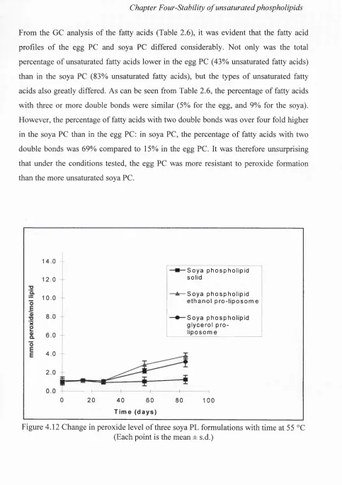
2.4.1 Comparison of soya PC and soya PL 65
2.4.1.1 Identification o f lipids in soya PC and soya PL by TLC 65 2.4.1.2 Results of ^ ^P-NMR analysis of soya PC and soya PL 65 2.4.1.3 Comparing the lipid compositions of soya PC and soya PL 66
2.4.2.3 Comparing the lipid compositions o f egg PC and egg PL 67
2.4.3 Fatty acid profile of the four lipids 68
2.4.4 Zeta potential measurements 70
2.4.5 Detection of saccharides in lipid samples using TLC 70
2.4.6 TLC of purified egg PC and soya PC 71
2.5 Conclusions 71
Chapter 3. Particle size of liposomes generated from pro-liposomes 72
3.1 Introduction 73
3.1.1 Rationale for generating small liposomes 73 3.1.2 Potential dangers of particles in intravenous products 73 3.1.3 Ideal size characteristics for liposomes as solubilisers 75 3.1.4 TPN as a standard for the size distribution 75
3.1.5 Sizing techniques 76
3.1.6 Aims of the particle sizing study 77
3.2 Materials 77
3.2.1 Materials for pro-liposomes 77
3.2.2 Materials for sizing 77
3.3 Methods 78
3.3.1 Cleaning glassware and apparatus 78
3.3.2 Pro-liposomes 78
3.3.2.1 Drug-free pro-liposomes 78
3.3.3.2 Selecting pro-liposome components and manufacturing
pro-liposomes 78
3.3.2.3 Liposome isotonicity and lipid concentration 79
3.3.3 Number of experimental replicates 79
3.3.4 Sample storage after conversion 79
3.3.5 Statistical analysis 79
3.3.6 Protocol for particle sizing 80
3.3.7 Diluting dispersions for particle size measurements 81
3.3.8 Particle sizing techniques 82
3.3.8.1 Visual inspection 82
3.3.8.2 Light microscopy 82
3.3.8.3 Photon correlation spectroscopy 83
3.3.8.4 Laser diffraction 84
3.3.8.5 Coulter Counter 84
3.3.8.6 Electron microscopy 85
3.3.8.7 Turbidity measurements 86
3.4 Studies performed 86
3.4.1 Measuring the particle size distribution of TPN 86
3.4.2 One stage conversion 86
3.4.2.1 Agitation during one stage conversion 87
3.4.2.2 Manipulation of aqueous phase 88
3.4.2.3 Manipulation of pro-liposome 88
3.4.2.3.3 Effect of replacing soya PC with soya PL 89 3.4.3 Effect of dilution on liposome dispersions converted in one stage 89
3.4.4 Two stage conversion 90
3.4.4.1 Effect of Eppendorf mixing during hydration stage 90 3.4.4.2 Effect of varying the amount of water for hydration 90 3.4.4.3 Turbidity of diluted dispersions generated in two stages 91 3.4.4.4 Effect of agitation during dispersion stage 91
3.4.4.5 Effect of lipid composition 92
3.4.4.5.1 Comparing the particle size of dispersions
produced from pure soya PC and pure egg PC 92 3.4.4.5.2 Effect of replacing soya PC with soya PL 92
3.4.4.5.3 Effect of including EPG 93
3.5 Results and discussion 93
3.5.1 Particle size distribution of Intralipid® 93
3.5.2 One stage conversion 94
3.5.2.1 Effect of energy input during single stage conversion 94
3.5.2.2 Manipulation of aqueous phase 98
3.5.2.3 Manipulation of the pro-liposome 99 3.5.2.3.1 Effect of freezing pro-liposome 99 3.5.2.3.2 Effect of varying lipid composition 100 3.5.2.3.3 Effect of replacing soya PC with soya PL 101 3.5.3 Dilution of liposome dispersions converted in one stage 102 3.5.3.1 Liposome dispersions before dilution 102 3.5.3.2 Liposome dispersions after diluting in isotonic glucose 103 3.5.3.3 Liposome dispersions after diluting in deionised water 106
3.5.4 Two stage conversion 107
3.5.4.1 Effect of mixing water to hydrate pro-liposome 107 3.5.4.2 Varying the amount of water added to the pro-liposome 108 3.5.4.3 Turbidity studies of diluted dispersions converted in
two stages 111
3.5.4.4 Effect of agitation during dispersion stage 112 3.5.4.5 Effect of lipid composition on liposomes converted in
two stages 114
3.5.4.5.1 Comparing the particle size distribution of dispersions produced from purified egg PC and
purified soya PC 114
3.5.4.5.2 Effect of replacing soya PC with soya bean PL 115
3.5.4.5.3 Effect of including EPG 117
3.6 Conclusions 119
Chapter 4. Stability of unsaturated phospholipids 121
4.1 Introduction 122
4.1.1 Stability 122
4.1.2 Chemistry of phospholipid breakdown 122
4.1.2.1 Phospholipid hydrolysis 122
4.1.2.2 Phospholipid oxidation 123
4.1.3 Prevention of oxidation 126
4.1.5.2 Liposome integrity 128
4.1.5.3 Drug stability 128
4.1.6 Reported investigations into phospholipid stability 128
4.1.7 Aims of the stability study 129
4.2 Materials 130
4.2.1 Lipids under investigation 130
4.2.2 Materials for conjugation assays 130
4.2.3 Materials for peroxide assay and standard curve 130 4.2.4 Materials for TEARS and TBARS(F) assay and standard curves 130
4.2.5 Materials for ^^P-NMR spectroscopy 131
4.3 Methods 131
4.3.1 Manufacture of samples 131
4.3.2 Analysis of oxidised phospholipid 131
4.3.2.1 Conjugated diene and triene assay 132
4.3.2.2 Hydroperoxide assay 132
4.3.2.3 Thiobarbituric acid reactive substances (TEARS) 134
4.3.2.3.1 TEARS(F) 134
4.3.2.3.2 TEARS 135
4.3.3 Visual appearance of formulations 135
4.3.4 Detection of lyso-phosphatidylcholine 136 4.3.4.1 Sample preparation prior to ^^P-NMR analysis 137 4.3.4.2 Experimental procedure for P-NMR spectroscopy 137
4.3.5 Repeat measurements 137
4.3.6 Data presentation 137
4.3.7 Stress test 137
4.3.7.1 Storage conditions 137
4.3.7.2 Assay time points 138
4.3.8 Accelerated and long term stability test 138
4.3.8.1 Storage conditions 138
4.3.8.2 Assay time points 138
4.4 Results and discussion 138
4.4.1 Comparing formulations at time zero 138
4.4.1.1 Oxidation levels of the different formulations 138 4.4.1.2 Oxidation levels of lipids in solid form 139 4.4.1.3 Visual appearance of the formulations 140
4.4.2 Stress storage stability 140
4.4.2.1 Diene conjugation and triene conjugation ratios 140
4.4.2.2 Hydroperoxide levels 145
4.4.2.3 TEARS formation 148
4.4.2.4 TEARS(F) formation 149
4.4.2.5 Visual appearance of the lipids 154
4.4.2.6 Lyso-phosphatidylcholine level 155
4.4.3 Stability of soya PL blend pro-liposome subjected to accelerated
4.5 Conclusions 163
Chapter 5. Solubilisation of cyclosporin A by liposomes generated
from pro-liposomes 165
5.1 Introduction 166
5.1.1 Selection o f drug for investigation 166
5.1.2 Importance of molecularly dispersing hydrophobes 166
5.1.3 Liposomes and cyclosporin A 166
5.1.4 Location of cyclosporin A within membrane 167 5.1.5 Methods for determining the degree of association 168
5.1.6 Aims of solubilising study 169
5.2 Materials 169
5.3 Methods 170
5.3.1 CyA pro-liposome manufacture 170
5.3.2 Converting cyA pro-liposomes into cyA liposomes in one stage 170 5.3.3 Converting cyA pro-liposomes into cyA liposomes in two stages 171
5.3.4 Cleaning syringes and containers 171
5.3.5 Analytical filtration 171
5.3.5.1 Filtration 172
5.3.5.2 Filter analysis 173
5.3.6 Sizing techniques 174
5.3.7 Statistical analysis 174
5.4 Studies performed 174
5.4.1 Controls
5.4.1.1 Analytical filtration of drug-free egg PL dispersions 174 5.4.1.2 Analytical filtration of drug-free soya PC/EPG dispersions 175 5.4.1.3 Diluting cyA dissolved in ethanol and glycerol 175 5.4.2 One stage conversion of cyA pro-liposomes into cyA liposome
dispersions 175
5.4.2.1 Ethanol cyA pro-liposomes 175
5.4.2.2 Raising ethanol level in glycerol-free cyA pro-liposomes 175 5.4.2.3 CyA pro-liposomes containing glycerol 176 5.4.2.4 Short term storage of cyA liposome dispersions 176 5.4.2.5 Raising the ethanol level of cyA pro-liposomes containing
glycerol 176
5.4.2.6 Raising the lipid: cyA weight ratio 177
5.4.3 Two stage conversion 177
5.4.3.1 Conversion of cyA pro-liposomes in two stages 177 5.4.3.2 Equilibrating hydrated cyA pro-liposomes 177 5.4.3.3 Hydration of cyA pro-liposomes with reduced
levels of water 178
5.4.3.4 Delaying the mixing of pro-liposome with water
for hydration 178
5.4.3.5 Effect of ethanol level on cyA association 179
5.4.3.5.1 High ethanol level 179
5.4.3.5.2 Medium ethanol level 179
5.4.3.6 Medium ethanol formulation with soya PL blend 179
5.4.3.7 Ethanol cyA pro-liposomes 180
5.4.3.10 Blending EPG with soya PC 181
5.4.4 Particle size and cyA association 181
5.4.4.1 Influence of varying the amount of water for hydration 181 5.4.4.2 Influence of varying the ethanol level 182 5.4.4.3 Influence of varying the lipid: cyA ratio 182
5.5 Results and discussion 183
5.5.1 Controls 183
5.5.1.1 Analytical filtration of drug-free liposome dispersions 183 5.5.1.2 Analytical filtration of drug-free dispersions converted
in two stages 184
5.5.1.3 Diluting cyA dissolved in ethanol and glycerol 184
5.5.2 One stage conversion 185
5.5.2.1 Ethanol cyA pro-liposomes 185
5.5.2.2 Raising the ethanol level in ethanol cyA pro-liposomes 187 5.5.2.3 CyA pro-liposomes containing glycerol 188 5.5.2.4 Short term storage of cyA liposome dispersions 189 5.5.2.5 Raising the ethanol level in cyA pro-liposomes containing
glycerol 190
5.5.2.6 Raising the lipid: cyA weight ratio of an egg PL
pro-liposome 190
5.5.3 Two stage conversion 191
5.5.3.1 Conversion of cyA pro-liposomes in two stages 191 5.5.3.2 Equilibrating hydrated cyA pro-liposome 193 5.5.3.3 Hydrating cyA pro-liposomes with reduced levels of water 194 5.5.3.4 Delaying mixing of cyA pro-liposome and
water for hydration 195
5.5.3.5 Role of the level of ethanol for cyA association 196 5.5.3.5.1 High ethanol formulation 196 5.5.3.5.2 Medium ethanol formulation 197 5.5.3.6 Medium ethanol formulation with soya PL blend 197 5.5.3.7 Role of glycerol in two stage conversion 199 5.5.3.8 Lowering the soya PL blend: cyA weight ratio 199 5.5.3.9 Lowering the soya PL blend: cyA weight ratio
without mixing 200
5.5.3.10 Influence of EPG on cyA association 201
5.5.4 Particle size and cyA association 201
5.5.4.1 Influence of varying the amount of water for hydration 201 5.5.4.2 Influence of varying the level of ethanol 203 5.5.4.3 Influence of varying the lipid: cyA ratio 204
5.6 Conclusions 205
Chapter 6. Freeze drying liposome dispersions generated
from pro-liposomes 207
6.1 Introduction 208
6.1.1 Freeze drying 208
6.1.3 Glass transition and glasses 209
6.1.4 Stages o f freeze drying 209
6.1.5 Freeze drying liposomes 210
6.1.6 Desirable properties for freeze dried liposomes 211
6.1.7 Liposome damage during freeze drying 211
6.1.8 Minimising liposome damage during lyophilisation 212
6.1.9 Stabilisers 212
6.1.10 Aims of freeze drying study 214
6.2 Materials 215
6.3 Methods 215
6.3.1 Manufacture of pro-liposomes 215
6.3.2 Producing liposome dispersions 216
6.3.2.1 Liposome size reduction 216
6.3.2.2 Extrusion 217
6.3.4 Freeze drying protocol 218
6.3.4.1 Freezing 218
6.3.4.2 Drying 219
6.3.5 Reconstitution of lyophilised cakes 220
6.3.6 Assessment of freeze dried cakes 220
6.3.6.1 Properties o f cake and reconstituted dispersion 221 6.3.6.2 Particle size o f reconstituted liposome dispersion 221 6.3.6.3 Analytical filtration of reconstituted dispersions 222
6.3.6.4 Statistical analysis 222
6.4 Studies performed 222
6.4.1 Stabilising phospholipid vesicles with saccharides 222 6.4.1.1 Incorporating glucose into liposome dispersions 223 6.4.1.2 Incorporating sucrose into liposome dispersions 223 6.4.2 Varying lipid: sucrose concentration of liposome dispersions 224 6.4.3 Factors affecting size o f reconstituted liposomes 224
6.4.3.1 Half isotonic dispersions 224
6.4.3.2 Influence of freezing history and agitation
during reconstitution 225
6.4.3.3 Concentrating dispersions by adding a reduced
amount of deionised water to the lyophilised cake 225 6.4.4 Effect of freezing regime on liposome size 226 6.4.5 Freeze drying liposomes incorporating sucrose and glycerol 226 6.4.6 Effect of lipid composition on the size of reconstituted dispersions 227
6.4.6.1 Effect of blending soya PL with soya PC 227 6.4.6.2 Effect of blending soya PL with egg PC 227 6.4.6.3 Effect of raising egg PG with soya PC 227 6.4.7 Effect of adding water soluble polymers to liposome dispersions 228 6.4.8 Freeze drying liposomes containing cyA 229
6.4.8.1 Effect of varying cyA levels 229
6.4.8.1.1 CyA liposomes incorporating sucrose 229 6.4.8.1.2 CyA liposomes incorporating glucose 230 6.4.8.2 Effect of varying lipid composition 231 6.4.8.3 Effect of varying lipid concentration and cyA concentration 231
6.5.1.1 Incorporating glucose into soya PL blend liposomes 232 6.5.1.2 Incorporating sucrose into soya PL blend liposomes 234 6.5.2 Effect of varying lipid: sucrose concentrations whilst
maintaining the lipid: sucrose ratio 236
6.5.3 Factors potentially affecting size of reconstituted liposomes 237
6.5.3.1 Half isotonic dispersions 237
6.5.3.2 Influence of freezing history and gentle agitation 238 6.5.3.3 Concentrating the dispersions by adding reduced
amounts of water for rehydration to freeze dried cakes 240 6.5.4 Comparing freezer freezing with liquid nitrogen freezing 240 6.5.5 Lyophilising dispersions incorporating glycerol and sucrose 242 6.5.6 Effect of lipid composition on size of reconstituted dispersions 244 6.5.6.1 Effect of blending soya PL with soya PC 244 6.5.62 Effect of blending soya PL vrith egg PC 247 6.4.6.3 Effect of blending egg PG with soya PC 248 6.5.7 Effect of including polymers in liposome dispersions 249 6.5.8 Freeze drying liposomes containing cyA 251
6.5.8.1 Effect of varying cyA levels 251
6.5.8.1.1 CyA liposomes incorporating sucrose 251 6.5.8.1.2 CyA liposomes incorporating glucose 252 6.5.8.2 Effect of varying lipid composition 253 6.5.8.3 Effect of raising lipid concentration and cyA concentration 254
6.6 Conclusions 255
Chapter 7. Final considerations 257
References 265
L ist o f T ables Chapter 2. 2.1 2.2 2.3 2.4 2.5 2.6 2.7 2.8 2.9 2.10 2.11
Composition of unsaturated phospholipids 54
Sprays and reagents used to locate lipid spots on TLC plate 64 Lipids present in soya PC and soya PL as identified by TLC 65 Chemical shifts and relative molar quantities of the phospholipids
in soya PC as determined by ^^P-NMR 66
Chemical shifts and relative molar quantities of the phospholipids
in soya PL as determined by P-NMR 66
Lipids present in egg PC and egg PL as identified by TLC 67 Chemical shifts and relative molar quantities of the phospholipids
in egg PC as determined by ^^P-NMR 67
Chemical shifts and relative molar quantities of the phospholipids
in egg PL as determined by P-NMR 67
Fatty acid profile of four commercially available unsaturated
phospholipids 68
Reported fatty acid profile of soya PC and egg PC 69 Zeta potential of four commercially available lipids and TPN 70 Lipids present in purified soya PC and purified egg PC 71 Chapter 3. Particle size of liposomes generated from pro-liposomes
3.1 Particle size results of Intralipid® concentrate
3.2 Effect of three different energy inputs during conversion of egg PL pro-liposomes in one stage
3.3 Effect of three different energy inputs during conversion of soya PL pro-liposomes in one stage
3.4 Effect of elevating the aqueous phase temperature during one stage conversion of liposomes
3.5 Effect of converting a firozen pro-liposome in one stage 3.6 Effect of lipid composition on particle size of liposomes
converted in one stage
3.7 Effect of replacing soya PC with soya PL in pro-liposomes converted into liposomes in one stage whilst handshaking vigorously
3.8 Effect of passing egg PL pro-liposome and water between two Eppendorf syringes prior to dispersing in bulk water
3.9 Effect of adding varying amounts of water to hydrate egg PL pro-liposome
3.10 Effect of adding varying amounts of water to hydrate egg PL pro-liposome
3.11 Effect of agitation during dispersion stage
3.12 Effect of egg PC and soya PC on liposome particle size 3.13 Effect of soya PL content on the particle size of liposomes
converted in two stages
3.14 Effect of soya PL content on the macroscopic and microscopic appearance of various soya dispersions
3.15 Number of particles in 90:10 soya PL blend dispersion converted in two stages
Chapter 4. Stability of unsaturated phospholipids 121 4.1 Change in physical appearance o f the three presentations of
four lipids over 84 days at 55 °C 155
Chapter 5. Solubilisation of cyclosporin A by liposomes generated
from pro-liposomes 165
5.1 Filtration of drug-free egg phospholipid liposome dispersions
converted from pro-liposomes in one and two stages 183 5.2 Analytical filtration of soya PC/EPG dispersions without cyA 184 5.3 Recovery of precipitate from ethanol: glycerol: cyA solutions
diluted with deionised water 185
5.4 Association of cyA with liposomes converted in one stage from
an egg PL pro-liposome 185
5.5 Association of cyA with liposomes converted in one stage from
an egg PL pro-liposome 187
5.6 Association of cyA with liposomes formed in one stage from
an egg PL pro-liposome 188
5.7 Association of cyA with liposomes formed in one stage from
an egg PL pro-liposome after storage for 24 hours 189 5.8 Association of cyA with liposomes converted in one stage from
an egg PL pro-liposome with raised ethanol level 190 5.9 Association of cyA with liposomes formed in one stage from
an egg PL pro-liposome with a lipid: cyA weight ratio of 60:1 190 5.10 Effect of adding water for hydration to an egg PL pro-liposome
converted in two stages on cyA association 191 5.11 Effect of delaying the mixing of the water for hydration and
an egg PL pro-liposome for 24 hours on cyA association 194 5.12 Effect of reducing the amount o f water for hydrating
an egg PL pro-liposome converted in two stages on
cyA association 194
5.13 Effect of lowering the egg PL: cyA ratio in pro-liposomes
converted in two stages on cyA association 195 5.14 Effect of mixing varying levels o f water for hydration vdth a
pro-liposome with raised ethanol level on cyA association 196 5.15 Effect of adding water for hydration to an egg PL pro-liposome
converted in two stages on cyA association 197 5.16 Effect of adding water for hydration to a soya PL blend
pro-liposome converted in two stages on cyA association 198 5.17 Effect of adding water for hydration to a glycerol-free soya PL
blend pro-liposome on cyA association 199
5.18 Effect of lowering lipid: cyA ratio on cyA association 200 5.19 Effect of lowering lipid: cyA ratio on cyA association
without mixing 200
5.20 Effect of EPG content on cyA association 201
5.21 Influence of adding water for hydration on cyA association
5.22 Influence of ethanol level in cyA pro-liposomes on the association
of cyA and particle size of liposomes 203
5.23 Influence of lowering lipid level in cyA pro-liposomes on the
association of cyA and particle size of liposomes 204
Chapter 6. Freeze drying liposome dispersions generated
from pro-liposomes 207
6.1 Effect of glucose on particle size of three different soya PL blend
liposome dispersions before and after lyophilisation 232 6.2 Effect of sucrose on the particle size of three different soya PL
blend liposome dispersions before and after lyophilisation 234 6.3 Influence of lipid: sucrose concentration on the particle size
of the liposome dispersions before and after lyophilisation 236 6.4 Particle size of the “half isotonic” liposome soya PL blend
dispersion prior to freeze drying 237
6.5 Effect of the freezing history and inverting the soya PL blend
dispersions during reconstitution on liposome size 238 6.6 Effect of freeze drying on the particle size of liposomes rehydrated
with reduced amounts of water 240
6.7 Effect of freezing history on particle size o f a half isotonic
soya PL blend liposome dispersion after defrosting 241 6.8 Effect of freeze drying on the particle size of half isotonic
liposome dispersions incorporating sucrose and/or glycerol 243 6.9 Effect of freeze drying on the particle size of soya PC liposomes
blended with soya PL 244
6.10 Effect of freeze drying on the particle size of various egg PC
liposomes blended with varying amounts o f soya PL 247 6.11 Effect of freeze drying on the particle size of soya PC liposomes
incorporating various levels o f egg PG 248 6.12 Particle size of soya PL blend dispersion prior to addition of
polymer 249
6.13 Effect of freeze drying on the particle size of soya PL blend
liposomes incorporating various polymers in varying amounts 249 6.14 Effect of freeze drying on the particle size and association of cyA
soya PL blend liposome dispersions incorporating sucrose 251 6.15 Effect of freeze drying on the particle size and association of cyA
soya PL blend liposome dispersions incorporating glucose 252 6.16 Effect of freeze drying on the particle size and association of
two cyA liposome dispersions 253
6.17 Effect of freeze drying on the particle size and association of
two cyA soya PL blend liposome dispersions 254
Chapter 1. Introduction 23 1.1 Generalised structure of a glycerophospholipid molecule 25
Chapter 2. Composition of unsaturated phospholipids 54
2.1 Bilayer arrangement of PC 57
2.2 ^^P-NMR trace of egg PC sample 62
Chapter 3. Particle size of liposomes generated from pro-liposomes 72
3.1 Particle sizing protocol 80
3.2 Turbidity of egg PL dispersion converted in one stage after diluting x200 fold in 50 mg/g glucose, deionised water and
50 mg/g glucose containing Tween 80 105
3.3 Turbidity of egg PL dispersion converted in two stages after
diluting xlOO fold in 50 mg/g glucose and deionised water 112 3.4 Turbidity of purified egg PC dispersion converted in two stages
after diluting xlOO fold in 50 mg/g glucose, deionised water
and 50 mg/g glucose containing Tween 80 115
Chapter 4. Stability of unsaturated phospholipids 121
4.1 Generalised scheme showing hydrolysis reactions of PL 122 4.2 Formation of peroxides in polyunsaturated fatty acid chains 125 4.3 Comparison of the UV spectra for the three anhydrous
soya PC presentations at time zero 139
4.4 Development of conjugated dienes in three soya PL formulations
with time at 55 °C 141
4.5 Development of conjugated dienes in three soya PC formulations
with time at 55 °C 141
4.6 Development of conjugated dienes in three egg PL formulations
with time at 5 5°C 142
4.7 Development of conjugated dienes in three egg PC formulations
with time at 5 5°C 142
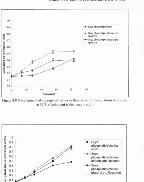
4.8 Development of conjugated trienes in three soya PL formulations
with time at 5 5 °C 143
4.9 Development of conjugated trienes in three soya PC formulations
with time at 55 °C 143
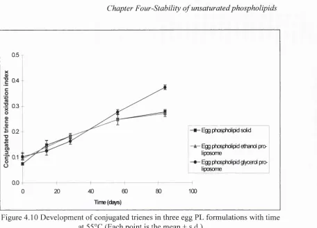
4.10 Development of conjugated trienes in three egg PL formulations
with time at 5 5 °C 144
4.11 Development of conjugated trienes in three egg PC formulations
with time at 55 °C 144
4.12 Change in peroxide level of three soya PL formulations with time
at 55 °C 146
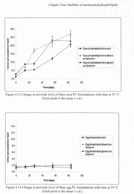
4.13 Change in peroxide level of three soya PC formulations with time
4.14 Change in peroxide level of three egg PL formulations with time
at 55 °C 147
4.15 Change in peroxide level of three egg PC formulations with
time at 55 °C 148
4.16 TEARS value of three soya PL formulations with time at 55 °C 150 4.17 TEARS value of three soya PC formulations with time at 55 °C 150 4.18 TEARS value of three egg PL formulations with time at 55 °C 151 4.19 TEARS value of three egg PC formulations with time at 55 °C 151 4.20 TEARS(F) values of three soya PL formulations with time at 55 °C 152 4.21 TEARS(F) values of three soya PC formulations with time at 55 °C 152 4.22 TEARS(F) values of three egg PL formulations with time at 55 °C 152 4.23 TEARS(F) value of three egg PC formulations with time at 55 °C 153 4.24 Soya PC content of three soya PL formulations over
84 days at 55 °C 157
4.25 Soya PC and soya LPC content of three soya PC formulations
over 84 days at 55 °C 157
4.26 Egg PC and egg LPC content of three egg PL formulations
over 84 days at 55 °C 158
4.27 Egg PC and egg LPC content of three egg PC formulations
over 84 days at 55 °C 158
4.28 ^^P-NMR spectrum of soya PL at time zero 159
4.29 ^^P-NMR spectrum o f soya PL from the glycerol pro-liposome
at 28 days 159
4.30 P-NMR spectrum of soya PL from the ethanol pro-liposome
at 84 days 160
4.31 Development of conjugated dienes in soya PL blend
pro-liposome with time at 40 °C, 20 °C and 4 °C 161 4.32 Development of conjugated trienes in soya PL blend
pro-liposome with time at 40 °C, 20 °C and 4 °C 162 4.33 Development of hydroperoxides in soya PL blend pro-liposome
with time at 40 °C, 20 °C and 4 °C 162
4.34 Development of TEARS in soya PL blend pro-liposome
with time at 40 °C, 20 °C and 4 °C 163
Chapter 5. Solubilisation of cyclosporin A by liposomes generated
from pro-liposomes 165
5.1 Chemical structure of cyA 168
Chapter 3. Particle size of liposomes generated from pro-liposomes 72 3.1 Typical light micrograph of egg PL dispersion converted in one
stage by vigorous handshaking of pro-liposome with excess water 96 3.2 Typical light micrograph of egg PL dispersion converted in one
stage by moderate handshaking of pro-liposome Avith excess water 97 3.3 Typical light micrograph of egg PL dispersion converted in one
stage from pro-liposome without handshaking 97 3.4 Typical light micrograph of soya PL dispersion converted in one
stage from pro-liposome without handshaking 98 3.5 Typical light micrograph of soya PL dispersion converted in one
stage from pro-liposome by vigorous handshaking 101 3.6 Typical freeze fracture replica o f egg PL dispersion formed from
pro-liposome by vigorous handshaking 103
3.7 Typical light micrograph showing the presence of lipid strands
after two fold dilution of egg PL dispersion in 50 mg/g glucose 105 3.8 Typical light micrograph of egg PL liposome dispersion generated
in one stage after two fold dilution in deionised water 107 3.9 Typical freeze fracture replica of egg PL liposome dispersion
converted in two stages by addition of half the weight of water to pro-liposome and subsequent addition to bulk water 111 3.10 Typical freeze fracture replica of egg PL liposome dispersion
converted in two stages by addition of an equal weight of water to pro-liposome and subsequent addition to bulk water 111
Chapter 5. Solubilisation of cyclosporin A by liposomes generated from pro-liposomes
5.1 Typical SEM of cyA powder
5.2 Typical SEM of cyA precipitated from ethanol/glycerol
5.3 Typical light micrograph of precipitate recovered from a liposome dispersion generated in one stage from an egg PL pro-liposome containing cyA
5.4 Typical light micrograph of precipitate recovered from liposome dispersion generated in one stage from an egg PL pro-liposome 5.5 Typical light micrograph showing precipitate strands recovered
from a liposome dispersion converted in two stages from an egg PL pro-liposome
5.6 Typical SEM of fine precipitate and a spherical precipitate
recovered from a liposome dispersion converted in two stages from an egg PL pro-liposome
5.7 Typical SEM of fine precipitate and a section o f an elongated precipitate recovered from a liposome dispersion converted in two stages from an egg PL pro-liposome
5.8 Typical SEM of precipitates recovered from a liposome dispersion converted in two stages from an egg PL pro-liposome
Chapter 6. Freeze drying liposome dispersions generated from
pro-liposomes 207
6.1 Typical light micrograph of a reconstituted soya PL blend
dispersion containing 60 mg/g of lipid and 50 mg/g o f glucose 233 6.2 Typical light micrograph of a reconstituted soya PL blend
dispersion containing 30 mg/g of lipid and 50 mg/g of glucose 234 6.3 Typical light micrograph of a rehydrated soya PL blend dispersion
which was frozen in liquid nitrogen prior to lyophilisation 239 6.4 Typical light micrograph of a rehydrated soya PL blend dispersion
which was frozen in freezer prior to lyophilisation 239 6.5 Typical light micrograph of a soya PC liposome dispersion
reconstituted to a lipid concentration of 120 mg/g and
a sucrose concentration o f 90 mg/g 246
6.6 Typical light micrograph of a liposome dispersion with
soya PC: soya PL weight ratio of 75:25 reconstituted to a lipid
concentration of 120 mg/g and a sucrose concentration o f 90 mg/g 246 6.7 Typical light micrograph of an egg PC liposome dispersion
reconstituted to a lipid concentration o f 120 mg/g
and a sucrose concentration of 90 mg/g 248
ANOVA Analysis of variance
Aq. - Aqueous
BHT - Butylhydroxytoluene
BP - British Pharmacopoeia
B.N. - Batch number
CMAH - Polar alkali TLC solvent system
CMC - Critical micelle concentration
CMH - Non-polar TLC solvent system
cyA - Cyclosporin A
D-90 - 90 % undersize value
DRV - Dried-reconstituted vesicles
DSC - Differential scanning calorimetry
EE - Entrapment efficiency
EM - Electron microscopy
EPG - Egg phosphosphatidylglycerol
FD - Freeze drying
FM - Freezer frozen and gently inverted for one minute FS - Freezer frozen and left to hydrate without shaking
GC - Gas chromatography
GPR - General purpose reagent
GPRO - Glycerol pro-liposome used in stability study
GRAS - Generally regarded as safe
H„ - Hexagonaln phase
HDL - High density lipoprotein
HPLC - High performance liquid chromatography
IV - Intravenous
LA - Ethanol pro-liposome formulation used in stability study
LD - Laser diffraction
LD50 Median lethal dose
LIP - Solid lipid formulation used in stability study
LM - Light microscopy
LUV - Large unilamellar vesicle
LPC - Lyso-phosphatidylcholine
MCT - Medium chain triglyceride
MDA - Malondialdehyde
MLV - Multilamellar vesicle
MPS - Mononuclearphagocyte system
MW - Molecular weight
NA - Not applicable
NaCi - Sodium chloride
NDE - New drug entity
NM - Nitrogen frozen and gently inverted for one minute NS - Nitrogen frozen and left to hydrate without shaking
O.D. - Optical density
PEG - Polyethyleneglycol
PA - Phosphatidic acid
PCS - Photon correlation spectroscopy
PE - Phosphatidylethanolamine
PG - Phosphatidylglycerol
PI - Phosphatidylinositol
P.L - Polydispersity index
PL - Phospholipid
^^P-NMR - Phosphorus nuclear magnetic resonance
PRO - Glycerol pro-liposome used in particle size study
PS - Phosphatidylserine
PVC - Polyvinylchloride
PVP - Polyvinylpyrrolidone
RES - Reticuloendothelial system
REV - Reverse phase evaporation
Rf Rate of flow value
s.d. - Standard deviation
SEM - Scanning electron microscopy
SPLV - Stable plurilamellar vesicle
SUV - Small unilamellar vesicle
TEA - Thiobarbituric acid
TEARS - Thiobarbituric acid reactive substances TEARS(F) - TEARS in the presence of iron
Tc - Transition temperature
Tg - Glass transition temperature
TEP - 1,1,3,3 -T etraethoxypropane
TLC - Thin layer chromatography
TPN - Total parenteral nutrition
USP - United States Pharmacopoeia
UV - Ultra-violet
w/o - Water in oil emulsion
w/o/w - Water in oil in water system
w/w - Weight for weight
w/v - Weight for volume
v/v - Volume for volume
Chapter one- Introduction
1 Introduction
The following introduction is divided into three sections. The first part provides a general introduction to liposomes in the context of intravenous applications. This is followed by a general section relating to the formulation of water insoluble drugs for intravenous administration. These two sections are united in the third section by introducing liposomes as solubilisers for hydrophobic drugs.
1.1 An introduction to liposomes
1.1.1 Liposomes
Liposomes are organised assemblies of phospholipids, which are amphiphilic molecules possessing a hydrophilic headgroup and two hydrophobic hydrocarbon tails (Fig. 1.1). These amphiphiles have the ability to self aggregate into discrete vesicles, which usually have concentric bilayers when equilibrated with excess water (Bangham et al., 1965). These phospholipid bilayers are usually arranged as smectic liquid crystals, i.e. the phospholipids are arranged into distinct layers with their long axis parallel to one another (Florence and Attwood, 198J).
As a consequence of this closed structure, liposomes have the ability to entrap/interact with a diverse variety of drugs with differing properties. Molecules at both ends of the solubility spectrum can be entrapped, as well as drugs with intermediate aqueous solubility. Water soluble compounds can be entrapped within the aqueous core and channels of the liposome. In contrast, hydrophobic materials can be sequestered between the fatty acids o f the phospholipid bilayer, where a hydrophobic hydrocarbon domain exists. Lipophiles with a hydrophilic moiety may span these two regions (Juliano and Stamp, 1979a). Charged species can be electrostatically attached to the surfaces of some types of liposomes (Lee and Schreier, 1993), further illustrating the versatility of liposomes to interact with biologically active materials.
Structurally, liposomes are arbitrarily classified by their size and the number bilayers: if the average diameter of the liposomes exceeds 100 nm they are conveniently classed as large, whilst if the diameter is 100 nm or less, they are referred to as small. A liposome which possesses a single bilayer is known as a unilamellar vesicle, a liposome with a few bilayers (2-3) is classified as oligolamellar and a lipid vesicle with many/several bilayers is classed as multilamellar. As 'will become evident later, the size and
lamellarity are important to control, because these factors may influence the behaviour o f liposome in vivo.
Lipid class Chemical structure
Glycerophospholipid H2C-O-R1
HC-O-R2
I
o
I
il
HjC-O-P-O-basc
I
o
-diacyl: R„ Rj^CO-R dialkyl: R„ R j^ -R lyso: Rj=H
Figure 1.1 Generalised structure of a glycerophospholipid molecule
1.1.2 General applications of liposomes
Due to their structural similarities to cell membranes, liposomes were originally employed in biochemistry as models of cells. However, since the early 1970s, various liposome applications have been investigated in fields diverse as pharmaceutics, vaccination, imaging, agriculture, aquaculture, cosmetics and food technology.
Chapter one- Introduction
The pharmaceutical industry has viewed liposomes with a degree of scepticism over the years for a variety of reasons. One reason has been that the pharmaceutical characterisation/development lagged behind the intensive animal testing of the early 1980s. This meant that even if preclinical liposome formulations appeared successful in animal models, to scale up the manufacturing procedure to reliably produce sufficient quantities for preliminary human investigations was often problematic. The importance
O f
of scaling up has often been underestimated, even though the productionTarge quantities is a prerequisite for most industrial/commercial applications (Ostro, 1988). To appreciate why scaling up has been problematic, one need only reflect on most of the techniques currently used to generate liposomes (section 1.1.3). Secondly, economic considerations may also have contributed to the slow development of liposomal formulations: the raw material phosphatidylcholine, which is the backbone of most liposome formulations, is expensive. This is presumably the main reason why most commercial liposomal applications have focused upon life threatening disease states, particularly systemic microbial infections and cancer, where a higher cost of the product can be justified. Thirdly, the performance of liposomes particularly for systemic targeting may have been overestimated (Poste, 1986). Physical factors such as avoidance of the mononuclear phagocyte system (MPS), extravasation and access to the target site, particularly to solid tumours may limit the efficacy of even the most optimised long circulating liposome systems. The MPS, formerly known as the reticuloendothelial system (RES), is a collection of cells and tissues which play a key role in immunity by engulfing foreign bodies such as bacteria. Some tissues rich in these cells include the liver’s K up^r cells, spleen, bone marrow and lymphoid tissue. Methods preventing the uptake of liposomes by this tissue have been studied to prolong the circulation time with the intent of targeting non-MPS tissues.
1.1.3 Liposome production techniques
Many of the techniques which have evolved for manufacturing liposomes are limited to laboratory scale or small scale production. Hence difficulties may be encountered when these processes are scaled up. The majority of these techniques, although ideal for laboratory scale, are unsuitable for pharmaceutical production. This may be because of toxic residues of solvents/intermediates in the final dispersion or simply the physical impracticalities of the process, when scaling up is attempted.
The main methods employed to form liposomes are outlined below and have been evaluated based on three criterion:
1) Ease o f large scale production.
2) Suitability for parenteral production: the presence of residual solvents and/or contaminants in the final dispersion.
3) Particle size of the liposomes generated.
Sterility has not been listed here, because aseptic filtration o f the final dispersion can be used to prepare a sterile product, if appropriately sized liposomes are produced. Additionally, association of lipophilic hydrophobes has not been included, because it is difficult to assess the association of hydrophobic agents in general. The degree of association seems to be highly dependant upon the individual molecule and its interaction with the bilayer (Cullis et al., 1987). This contrasts with aqueous entrapment, which can be assessed by determining the capture volume of the liposome dispersion (Perrett et al., 1991; Lidgate et al., 1993).
In order to produce a liposome dispersion suitable for intravenous administration, generally a two stage procedure is needed: the first stage involves forming a coarse liposome dispersion, which is usually followed by a modification technique reducing the size and size distribution of the dispersion.
Forming a liposome dispersion is a relatively simple procedure: in the presence of excess water, most phospholipids reorganise themselves to form liposomes. However, to form liposomes with particular characteristics, such as appropriate size, good aqueous entrapment and satisfactory stability, may be rather more challenging.
The main methods for producing liposomes broadly fall into the following categories: 1) Film hydration
2) Emulsion 3) Freeze drying
4) Detergent based methods 5) Solvent injection
Chapter one- Introduction
1.1.3.1 Film hydration
This was the first technique used to form liposomes. It involves the creation of a large surface area for the hydration of phospholipid by formation of a thin film (Bangham et al., 1965). This film is formed by drying down the phospholipid from a volatile organic solvent onto the inner wall of a glass round bottomed flask using a rotating evaporator. The resultant film can easily be hydrated at the appropriate temperature (section 2.1.3) and subsequently dispersed by handshaking or mechanical agitation. The amount of water and degree of shaking added at this stage can influence the type of liposome generated. Generally, without the addition of charged species and agitation, the liposomes are multilamellar in character.
A modification of the film technique was developed by Payne et al. (1986). It involves depositing a phospholipid film onto a selected water soluble substrate such as a saccharide or sodium chloride. Typically this is achieved by evaporating the volatile solvent from an organic solution of phospholipids in the presence of water soluble substrate. As the volatile solvent is removed, the water soluble substrate becomes coated with a film of phospholipid. This coating improves the hydration o f the phospholipid by creating a larger surface area. The particle size of the liposomes hydrated in this manner is reported to be somewhat smaller than those generated by the original film method. In its original form, the film hydration technique is simple, and the most trouble free for laboratory production. However, these techniques are difficult to scale up and control of particle size, particularly for parenterals, is difficult. Hence a post production size reduction technique would normally be required. The type of liposome can also be influenced to some degree by the addition of charged species, which tends to favour fewer number of bilayers (Cowley et al., 1978).
1.1.3. 2 Emulsion techniques
i) Water in oil method (W /0)
A small amount of water containing the aqueous material to be encapsulated is added to phospholipid dissolved in an immiscible organic solvent, e.g. chloroform. The aqueous phase is dispersed into fine droplets by the input of mechanical energy, e.g. sonication. These small water droplets are emulsified at the solvent/water interface by the phospholipid monolayer, which forms the core of the inner leaflet of the bilayer (New, 1990a). To add the outer leaflet of the bilayer, the double emulsion technique is
employed. This is achieved by introducing the emulsified water droplets in the immiscible organic solvent into an aqueous environment. Upon a more gentle mechanical agitation, a w/o/w system is generated. Removal of the organic phase by evaporation results in the formation o f a liposome dispersion, which is predominantly unilamellar in character. The size of the liposomes is largely dependant upon the degree of mechanical input when generating the emulsions, but liposome diameters down to
100 nm have been reported.
ii) Reverse phase evaporation (REV)
This technique involves dissolving the lipid in a water immiscible organic solvent, to which a small volume of the water is added (Szoka and Papahadjopoulos, 1978). This mixture is sonicated and the resultant w/o type emulsion is partially evaporated until a gel-like lamellar structure is obtained. This semi-solid gel is vortexed vigorously until structural collapse occurs and a liposome dispersion is generated. The resultant liposome is about 500 nm in diameter, with improved aqueous entrapment. The liposomes are predominantly unilamellar, though some oligolamellar liposomes are also present.
iii) Stable plurilamellar vesicles (SPLV)
This technique is similar to the w/o method but the drying down is carried out under nitrogen and continuous bath sonication (Gruner et al., 1985). The resultant vesicles, known as stable plurilamellar vesicles (SPLV), are reported to be osmotically stable, because no osmotic gradient exists between the bilayers of the vesicles compared to MLVs. However, in practice this difference between SPLVs and ML Vs may not matter if post-production techniques, such as extrusion, microfluidisation or homogenisation are employed. The processing is likely to result in osmotically homogeneous liposomes, because the liposomes are being disrupted during size reduction.
All of the emulsion techniques described above are unacceptable pharmaceutically, because it is difficult to lower organic solvent residue levels to acceptable limits, particularly in the presence of water.
1.1.3.3 Freeze drying: Dried-reconstituted vesicles (DRV)
Chapter one- Introduction
the SUVs fuse to form MLVs, which have improved aqueous entrapment. Although it is a satisfactory technique for entrapping water soluble materials, it is probably inappropriate for hydrophobic materials for parenteral purposes. It would be illogical to produce SUVs in order to generate larger liposomes which would have to be reduced in size again.
1.1.3.4 Detergent technique
The principit' behind this technique is to form mixed micelles from phospholipid and detergent. For parenteral applications, the detergent employed is typically an anionic bile salt, though for non-pharmaceutical applications a variety of other surfactants, e.g. p-alkyl glycoside, can also be used. This mix can be converted to unilamellar vesicles by lowering the detergent concentration in the micelles by dialysis (Kagawa and Racker, 1971), column chromatography or dilution (Son and Alkan, 1989). Once the detergent level has been lowered sufficiently, the mixed micelles vesiculate into liposomes.
One advantage with this approach is the absence of solvent residues in the final dispersion^ (although for intravenous applications residual detergent could be toxic if the detergent is not carefully selected. Appropriate selection of the conditions enables control o f the average size of the liposomes within a narrow size distribution. Homogeneous liposomes of diameter around 100 nm or less can be produced, the exact size depends upon the lipid concentration and detergent employed. It is reported that 100% association of hydrophobes can be achieved, if the material, e.g. membrane spanning proteins, is intrinsically suited to the hydrophobic region within the membrane. However, if the material is lipophilic but not anchored to the membrane, this technique may not be suitable for this type of drug, because it may disassociate from the membrane during dialysis. Large scale production of liposomes using the detergent technique has been made feasible using cross flow filtration techniques (Schubert,
1996).
1.1.3.5 Solvent injection
The are two main methods employing solvent injection. The first technique carefully introduces a weak ethanolic solution of phosphatidylcholine into water (Batzri and Korn, 1973). The resultant liposome dispersion is composed entirely o f SUVs. The main disadvantage is the dilute concentration of lipid, which can not be increased to above about 2 mg/ml without additional processing.
The second solvent injection technique employs ether as the organic solvent (Deamer and Bangham, 1976). Unlike ethanol, ether is a water immiscible solvent. The liposomes are produced in a similar way to the ethanol method in terms of injection technique. However, instead o f mixing the ether solution with the aqueous phase at room temperature, the ether is vaporised as the ether solution of lipid is injected into a warm aqueous phase heated to 60 °C. The liposomes generated are mostly unilamellar and have a particle size of approximately 100 nm.
The advantage of these procedures is that the liposome dispersion probably does not require any further size reduction and can be filter sterilised. However, these techniques are difficult to scale up, slow to manufacture and produce very dilute liposome dispersions. In the case of the ethanol injection technique, the dispersion may require solvent removal and a procedure to concentrate the liposomes.
1.1.3.6 Pro-liposome
This technique is based upon the aqueous dilution of an ethanolic solution of phospholipid to form liposomes (Perrett et al., 1991 ). The liposomes generated are characteristically MLV in type, though if the blend of lipid and pro-liposome composition are carefully selected, either oligolamellar liposomes or large unilamellar liposomes can be generated. The liposomes are simple to manufacture, do not require the employment of any pharmaceutically unacceptable solvent at any stage and the method is eminently suited for large scale production. The main disadvantage for parenteral applications is the large liposome size.
1.1.4 Size reduction techniques
Many o f the techniques described above may require a further processing step after manufacture to reduce liposome size or enhance the aqueous entrapment. In the context of using liposomes as carriers for parenterals, one of the main concerns is to reduce the size o f the liposomes. Therefore, most manufacturing protocols employ two stage procedures to generate liposomes suitable for intravenous administration.
Chapter one- Introduction
various methods by which this liposome size reduction can be achieved are outlined below:
1.1.4.1 French press
French presses are used to disrupt cells under high pressure. In the case of liposomes, the mechanical ram o f the press forces the liposome dispersion through a narrow orifice, which ruptures the vesicles by shearing. The resultant translucent dispersion consists predominantly of SUVs (Barenholz et al., 1979; Hamilton et al., 1980).
1.1.4.2 High pressure homogenisation
This technology was originally developed for reducing the size o f emulsion droplets in the food industry to prevent creaming in dairy products. It was later employed in pharmaceutics in an allied technology to produce fat emulsions for parenteral nutrition. This technology is the current commercial technique of choice to reduce coarse liposome dispersions to small unilamellar liposomes. The homogeniser reduces the size o f the liposomes via cavitation and shearing during laminar and turbulent flow (Brandi et al., 1990; Bachmann et al., 1993). This is achieved by passing the dispersion through a small knife edged gap under high pressure. After optimising the process, it is possible to generate homogeneous liposomes with average diameters of less than 50 nm within a very narrow size band. These translucent dispersions are eminently suited to aseptic filtration. This direct method of producing liposomes is amenable to large scale processing. Furthermore, scaling up the production is easy: once the parameters for small scale production have been established and optimised, the information can usually be directly transferred to larger scale manufacturing equipment for larger batches.
The main disadvantage is that during use, particulates may be shed from the interaction chamber. Additionally, there is little control over the size of the liposome, only small unilamellar vesicles can be generated.
1.1.4.3 Microfluidisation
This process is similar to high pressure homogenisation, the main difference being the interaction chamber, which relies upon head on collision of the liquid at right angles against a plate of the interaction chamber (Mayhew et al., 1984). The advantage is that high amounts of lipid (up to 20% w/v) can be handled. The disadvantages are the same as high pressure homogenisation.
1.1.4.4 Membrane extrusion
Originally described by Olson et al. (1979) and Szoka et al. (1980), the liposome dispersion is physically extruded through a filter with cylindrical channels of defined diameter. These channelled pores are created by laser etching through a polycarbonate filter. After repeated extrusions, the upper size limit of the dispersion approaches the diameter of the pores. This method is useful for producing small batches of liposomes with a defined size, ranging from 30 nm up to several microns in diameter. If the liposome diameters are 200 nm or less, the dispersions can be readily sterilised by filtration. Although this technique can be scaled up (Schneider et al., 1995), without arranging several filters in parallel, the batch volume is usually restricted to approximately 100 litres (O’Hara, personal communication). This is due to the filter diameter, which can not easily be increased above 47 mm without the filter integrity being compromised.
1.1.4.5 Sonication
There are two types of sonication: probe and bath. Probe ultrasonication employing a titanium probe can be used to produce SUVs between the lowest theoretical limit of about 25 nm up to 80 nm (Huang, 1969). For size reproducibility between batches, it is important to maintain the exact position of the probe. The main disadvantages are possible oxidative and hydrolytic lipid damage, which can occur if the process is not controlled. This is due to the highly energy intensive nature of the process. Secondly, the probe sheds titanium into the dispersion, resulting in fine grey deposits which have to be removed from the dispersion by centrifugation. Bath sonication is less intense than probe sonication and, therefore, the rate o f the size reduction is inferior and final liposome diameter is generally larger.
1.1.4.6 Freeze thaw sonication
This technique involves alternating cycles o f sonication and freezing of a liposome dispersion (Pick, 1981). For large scale production this technique is impractical and is normally applied to enhance the aqueous entrapment of water soluble materials by creating larger unilamellar vesicles (LUVs).
Chapter one- Introduction
investment. The size reduction step should be appreciated in the context of the manufacturing process as a whole. It is just a part of the manufacturing process, further processing steps, such as sterilisation and stabilisation, may also be required.
1.1.5 Liposome formulations for intravenous applications
Many liposome formulations for parenteral applications are intended to alter the bio disposition of biologically active compounds. The parenteral route has been concentrated upon, because this route bypasses the natural barriers of the body. Within parenterals, although many different routes have been examined, most effort has been devoted to the intravenous route (Ostro and Cullis, 1989). From the very beginning, liposomes have been advocated as “magic bullets”, in order to target biologically active materials. Hence, work has focused upon extending the circulation time of liposomes in vivo with the aqueous drug stably entrapped and targeting these vesicles to specific sites. In the early 1980s extensive work was carried out employing liposomes as a targeted carrier system. It was believed that once injected, they would circulate around the bloodstream unnoticed, targeting and delivering the encapsulated drug to the desired site. However, it was discovered that if unsaturated phosphatidylcholine vesicles are employed, the liposomes not only immediately leaked their entrapped material but also rapidly disintegrated in vivo (Gregoriadis, 1988). In order to decrease this leakage and disintegration, two difficulties had to be overcome. Firstly, the stability and circulation time o f liposomes in the bloodstream had to be improved. Secondly, suitable biologically active materials had to be selected which could be effectively entrapped and retained inside the liposome.
Bilayer integrity was greatly aided by the addition of membrane stabilising components, such as the sterol cholesterol (Guo et al., 1980; Allen et al., 1981). Further improvements based upon the same principle, i.e. reducing the membrane mobility and increasing the rigidity, were made by employing saturated phosphatidylcholines. These types o f liposomes with hydrogenated phospholipids and cholesterol are being used to entrap daunorubicin, a toxic cytotoxic agent, in a commercially available product “Daunoxome®” (NeXstar, USA). Although such modifications greatly aid the integrity of the liposome in vivo, they do not confer the liposome with the ability to evade the host’s defence system. The liposome is still viewed as foreign by the immune system: once in the bloodstream, most liposomes are rapidly recognised and engulfed by the
MPS. This efficient clearance system may severely limit the half life of the liposome and the drug entrapped within the liposome.
In order to extend the circulation time of the liposome it was realised the size had to be carefully controlled. It was believed that a liposome diameter o f 100 nm or less maximised the circulation time of the vesicles (Hwang et al., 1980; Allen and Everest, 1983; Proffitt et al., 1983). Further improvements in circulation time of the liposome were made as different surface properties of liposomes were explored. Work in the 1980s employed phosphatidylinositol as a means of altering the properties of the liposome (Kao and Loo, 1980; Gabizon and Papahadjopoulos, 1988). This negatively charged phospholipid was incorporated into the bilayer, thereby projecting inositol groups at the surface of the bilayer, which significantly prolonged the retention of the liposome in the circulation. The mechanism of this prolongation has been attributed to ste fie stabilisation. By reducing liposome opsonisation by the plasma proteins, the liposome is not as quickly detected by the MPS. However, due to the high cost and potential toxicity, this approach was not adopted for commercial applications. Nevertheless, it demonstrated the principle that it was possible to increase the half life of liposomes in the bloodstream. The next major advance in circulation prolongation was the development of liposomes incorporating lipids covalently attached to polymers (Blume and Cevc, 1990; Woodle and Lasic, 1992). Although a variety of different polymers can be employed, the liposomes incorporating PEG-ylated phosphatidylethanolamine were selected for further development. Selection of a specific PEG size and incorporation of PEG-ylated phospholipid between 5-10% greatly improved the circulation time of these liposomes.
Chapter one- Introduction
unentrapped material by column chromatography or centrifugation. This was an inefficient and a wasteful means of entrapping material. Various techniques, such as pH gradient active loading and remote loading (Bally et al., 1985; Hope et al., 1985; Mayer et al., 1990), have greatly improved the loading efficiency of liposomes. These methods exploited the properties of weak bases: by creating a pH gradient between the liposome interior and exterior, it was possible to direct some drugs towards the aqueous interior of the liposome. The uncharged molecule diffused across the membrane, but once inside the aqueous channels of the liposome, the molecule became charged as a result of the change in pH. The charge prevented diffusion of the molecule out of the liposome, thereby entrapping the charged material within the liposome. Further refinements were made using an elegant ammonium sulphate gradient for amphiphiles, such as doxorubicin. This enabled up to 90% of the active material to be entrapped inside the liposome without further purification (Cohen, 1991; Haran et al., 1993).
Although these systems have developed considerably since the first description of the classical liposome in 1965, the challenge, however, of true site specific delivery still remains. Currently the most advanced system being evaluated clinically are the liposomes incorporating PEG-ylated phosphatidylethanolamine with ammonium gradient loaded doxorubicin (Doxil®, Sequus, USA). These liposomes are highly stable in vivo, and have prolonged circulation times. However, they still do not offer true targeting, even if tumour sites may be more permeable than healthy tissue and enable slightly higher amounts of cytotoxic to be delivered. The true specificity required for “targeting” has yet to be achieved. An added intelligent step, which dictates where and when systems should release their load is required, before the term “targeted” can be genuinely applied. Many ideas have been considered and proposed for this programming, e.g. magnetic, fusogenic, pH dependant release, thermo release and antibodies (Straubinger et al., 1988 and Matthay et al., 1989). However, to date there has been little success with these approaches.
1.2 Parenteral administration
1.2.1 Parenteral route
The parenteral route is a general term used to describe the breaching of any dermal/mucosal barrier by injection to administer drugs into the body. The term parenteral is derived from the Greek para and enteron meaning “beyond the intestine”.