University of Pennsylvania
ScholarlyCommons
Publicly Accessible Penn Dissertations
2017
Structural And Functional Studies Of The Human
Telomeric Pot1-Tpp1 Complex And Its Role In
Human Disease
Cory Taylor Rice
University of Pennsylvania, [email protected]
Follow this and additional works at:
https://repository.upenn.edu/edissertations
Part of the
Biochemistry Commons
, and the
Biophysics Commons
This paper is posted at ScholarlyCommons.https://repository.upenn.edu/edissertations/2550
For more information, please [email protected].
Recommended Citation
Rice, Cory Taylor, "Structural And Functional Studies Of The Human Telomeric Pot1-Tpp1 Complex And Its Role In Human Disease" (2017).Publicly Accessible Penn Dissertations. 2550.
Structural And Functional Studies Of The Human Telomeric Pot1-Tpp1
Complex And Its Role In Human Disease
Abstract
Telomeres are essential regions of repetitive DNA at the ends of eukaryotic chromosomes that serve an
indispensable role in the protection and replication of the genome.
Telomeres shorten over time due to nucleolytic degradation, oxidative damage, and through a process known
as the ‘end replication problem,’ which shortens telomeres after every round of genome replication. Critically
short telomeres are linked to cellular senescence, aging, and human disease. Therefore, it is imperative to
understand the molecular mechanisms that regulate and protect telomere length.
Correct regulation of telomere length is key and is facilitated by two telomere capping complexes: shelterin
and CST. The human shelterin complex is composed of TRF1, TRF2, Rap1, TIN2, POT1, and TPP1, binds
single and double stranded telomeric DNA and protects telomeres from degradation and unwanted DNA
repair. The human CST complex, composed of CTC1, Stn1, and Ten1, bind and caps the single-stranded
overhang of telomeres. These two complexes also maintain telomere length by regulating access of telomerase
to telomeres, the reverse transcriptase that extends the length of telomeres.
In humans, the shelterin subcomplex, POT1-TPP1, is thought to directly engage and enhance the processivity
of telomerase. Conversely, CTC1 of the human CST complex interacts with TPP1 inhibiting the activity of
telomerase at telomeres. Furthermore, the CST complex is involved in the recruitment of Pol
to telomeres;
the DNA polymerase involved in the replication of the telomeric C-strand. As a result, the interaction
between the shelterin and CST complexes represents a potential key mechanism for telomere length
regulation that is still not fully understood.
Naturally occurring mutations in patients with rare aging disorders and cancers have been recently linked to
telomere capping complexes. Elucidating the structural and functional consequences of these naturally
occurring mutations will allow us to better understand how dysfunctional telomere maintenance results in
human disease. My dissertation focuses on the functional and structural data of the human POT1-TPP1
complex, which reveals that naturally occurring mutations in the C-terminal domain of POT1 result in partial
disruption of the POT1-TPP1 complex and telomere phenotypes associated with unregulated telomere
length.
Degree Type
Dissertation
Degree Name
Doctor of Philosophy (PhD)
Graduate Group
Biochemistry & Molecular Biophysics
First Advisor
Emmanuel Skordalakes
Keywords
Cancer, Chromosomes, Structural Biology, Telomeres, X-ray Crystallography
Subject Categories
Biochemistry | Biophysics
STRUCTURAL AND FUNCTIONAL STUDIES OF THE HUMAN TELOMERIC POT1-TPP1
COMPLEX AND ITS ROLE IN HUMAN DISEASE
Cory Taylor Rice
A DISSERTATION
in
Biochemistry and Molecular Biophysics
Presented to the Faculties of the University of Pennsylvania
in
Partial Fulfillment of the Requirements for the
Degree of Doctor of Philosophy
2017
Supervisor of Dissertation
_______________________
Emmanuel Skordalakes
Associate Professor of Gene Expression and Regulation
Graduate Group Chairperson
__________________________
Kim A. Sharp, Associate Professor of Biochemistry and Biophysics
Dissertation Committee:
Ronen Marmorstein, Professor of Biochemistry and Biophysics
Paul M. Lieberman, Professor of Microbiology
Gregory D. Van Duyne, Professor of Biochemistry and Biophysics
STRUCTURAL AND FUNCTIONAL STUDIES OF THE HUMAN TELOMERIC POT1-TPP1
COMPLEX AND ITS ROLE IN HUMAN DISEASE
COPYRIGHT
2017
Cory Taylor Rice
This work is licensed under the
Creative Commons Attribution-
NonCommercial-ShareAlike 3.0
License
To view a copy of this license, visit
iii
ACKNOWLEDGMENT
First and foremost, I would like to express my gratitude and thankfulness for having
Emmanuel as a mentor. He has helped guide me through numerous challenges I have faced
during my dissertation. His door was always open to discuss and troubleshoot experiments and
would never hesitate to help me collect or analyze data. His guidance has shaped me into the
scientist that I am today and instilled in me the desire to pursue research at the highest quality
and integrity. For all of these things and many more, I am truly grateful.
I would also like to thank my thesis committee for their intellectual contributions to my
project. Dr. Ronen Marmorstein (chair), Dr. Paul Lieberman, Dr. Gregory Van Duyne, Dr. Kathryn
Ferguson, and Dr. Kim Sharp all provided tremendous support throughout my dissertation and
ensured that I was prepared for the next step in my career. In addition, I would like to thank Dr.
Lieberman’s lab and Dr. Susan Janicki’s lab for their time and resources as I have benefitted
greatly from our collaborations. In addition, Dr. Zhong Deng and Zhuo Wang from the Lieberman
lab and Prashanth Shastrula from the Janicki lab were always very helpful and supportive.
I would also like to thank the BMB department and the friends I have made throughout
graduate school. Specifically, Atrish Bagchi, Christopher Bialas, John Welsh, Catherine
Debrosse, Jarret Remsberg, Joe Jordan, and Matthew Thompson were constantly supportive and
I would like to thank them for our numerous discussions and memories we have made over the
years.
Lastly, but certainly not least, I would like to thank my family for their unconditional love
and support. My mother and father were always there for me and taught me to always challenge
myself in order to achieve great things. I thank my brother and his family for their constant love
and support. Finally, I would like to thank my fiancée, Francesca Tuazon, her parents, and their
cat Arusha, for the life we have built together and their constant love and support that helped me
iv
ABSTRACT
STRUCTURAL AND FUNCTIONAL STUDIES OF THE HUMAN TELOMERIC POT1-TPP1
COMPLEX AND ITS ROLE IN HUMAN DISEASE
Cory Taylor Rice
Emmanuel Skordalakes
Telomeres are essential regions of repetitive DNA at the ends of eukaryotic
chromosomes that serve an indispensable role in the protection and replication of the genome.
Telomeres shorten over time due to nucleolytic degradation, oxidative damage, and through a
process known as the ‘end replication problem,’ which shortens telomeres after every round of
genome replication. Critically short telomeres are linked to cellular senescence, aging, and
human disease. Therefore, it is imperative to understand the molecular mechanisms that regulate
and protect telomere length.
Correct regulation of telomere length is key and is facilitated by two telomere capping
complexes: shelterin and CST. The human shelterin complex is composed of TRF1, TRF2, Rap1,
TIN2, POT1, and TPP1, binds single and double stranded telomeric DNA and protects telomeres
from degradation and unwanted DNA repair. The human CST complex, composed of CTC1,
Stn1, and Ten1, bind and caps the single-stranded overhang of telomeres. These two complexes
also maintain telomere length by regulating access of telomerase to telomeres, the reverse
transcriptase that extends the length of telomeres.
In humans, the shelterin subcomplex, POT1-TPP1, is thought to directly engage and
enhance the processivity of telomerase. Conversely, CTC1 of the human CST complex interacts
with TPP1 inhibiting the activity of telomerase at telomeres. Furthermore, the CST complex is
v
of the telomeric C-strand. As a result, the interaction between the shelterin and CST complexes
represents a potential key mechanism for telomere length regulation that is still not fully
understood.
Naturally occurring mutations in patients with rare aging disorders and cancers have
been recently linked to telomere capping complexes. Elucidating the structural and functional
consequences of these naturally occurring mutations will allow us to better understand how
dysfunctional telomere maintenance results in human disease. My dissertation focuses on the
functional and structural data of the human POT1-TPP1 complex, which reveals that naturally
occurring mutations in the C-terminal domain of POT1 result in partial disruption of the
vi
TABLE OF CONTENTS
ACKNOWLEDGMENT ... III
ABSTRACT ... IV
TABLE OF CONTENTS ... VI
LIST OF TABLES ... IX
LIST OF FIGURES ... X
CHAPTER 1 TELOMERES AND CHROMOSOME END-PROTECTION ... 1
1.1 Function and composition of telomeric DNA ... 2
1.2 Telomerase and telomere replication ... 3
1.3 Telomere length regulation ... 4
1.4 Telomeres and human disease ... 6
1.5 Project goals ... 10
CHAPTER 2 STRUCTURE OF THE HUMAN POT1-TPP1 COMPLEX AND ITS ROLE
IN CANCER ... 11
vii
2.2 Results ... 13
2.2.1 Structure of the human POT1-TPP1 complex ... 13
2.2.2 Structural analysis of POT1C mutations ... 18
2.2.3 Several POT1C mutations partially disrupt the POT1-TPP1 complex ... 21
2.2.4 POT1C mutants do not alter telomerase processivity ... 25
2.2.5 Partial disruption the POT1-TPP1 complex reduces POT1-DNA binding ... 27
2.2.6 POT1C mutations do not affect the localization of POT1 to telomeres ... 29
2.2.7 Partial disruption of the POT1-TPP1 complex results in telomere elongation ... 31
2.2.8 POT1C mutations lead to fragile telomeres ... 33
2.3 Discussion ... 36
2.4 Materials and methods ... 38
2.4.1 Protein expression and purification ... 38
2.4.2 Protein crystallization and structure determination ... 40
2.4.3 Isothermal Titration Calorimetry (ITC) ... 41
2.4.4 Fluorescence Polarization (FP) Assays ... 41
2.4.5 Human Cell Culture ... 42
2.4.6 Western Blot ... 43
2.4.7 Reverse Transcription PCR and quantitative PCR analyses ... 44
2.4.8 Direct telomerase activity assays ... 44
2.4.9 Cell Imaging ... 45
2.4.10 Southern blot analysis ... 46
2.4.11 Fluorescence in situ hybridization (FISH) ... 46
viii
ix
LIST OF TABLES
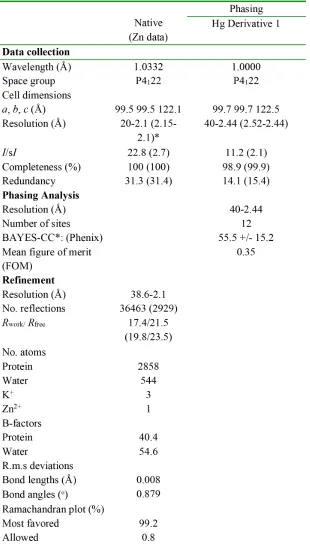
Table 1: Data collection and refinement statistics for POT1C-TPP1(PBD) ... 17
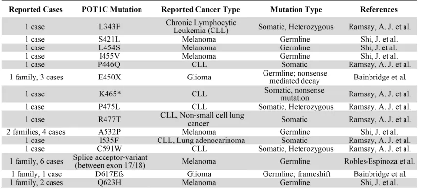
Table 2: Reported cases of POT1C mutations in cancer. ... 21
Table 3: ITC binding data for wild type and mutant POT1C proteins with TPP1(PBD) ... 25
x
LIST OF FIGURES
Figure 1.1 The shelterin and CST complexes cooperate to maintain telomere length. ... 6
Figure 1.2 Primary structures of the human POT1-TPP1 and CST complexes. ... 9
Figure 2.1 Structure of the human POT1-TPP1 complex. ... 16
Figure 2.2 Structural analysis of POT1C mutations. ... 20
Figure 2.3 Expression and purification of POT1C mutants. ... 23
Figure 2.4 ITC binding data for the POT1C and TPP1(PBD) proteins. ... 24
Figure 2.5 Direct telomerase activity assays ... 26
Figure 2.6 POT1-TPP1 telomeric DNA binding assays. ... 28
Figure 2.7 Cell imaging for localization of wild type and mutant POT1 to telomeres. ... 30
Figure 2.8 Southern blot analysis of wild type and mutant POT1. ... 32
1
CHAPTER 1
2
1.1 Function and composition of telomeric DNA
Telomeres are nucleoprotein structures at the ends of eukaryotic chromosomes that are
essential for the protection and replication of the genome (Blackburn and Gall, 1978; de Lange,
2009). Since eukaryotic chromosomes are linear, telomeres appear as double-strand breaks and
activate the DNA damage response (Longhese, 2008; Maser and DePinho, 2004). This presents
the cell with a unique challenge in maintaining genome integrity since unwanted DNA repair at
telomeres leads to chromosome fusions and genomic instability. However, telomere specific
proteins exist to bind and cap telomeres to prevent activation of unwanted DNA repair. Telomere
specific proteins also maintain telomere length, which is essential for the full replication of the
genome and prevents cellular senescence (Effros and Walford, 1984; Greider and Blackburn,
1996).
Telomeres are composed of tandem DNA repeats (TTAGGG in mammals) and consists
of both duplex DNA and a single-stranded overhang (G’-overhang). The 5’ to 3’ strand is referred
to as the G-strand because it contains the G-rich telomeric repeats, whereas the 3’ to 5’ strand is
known as the C-strand. These characteristics result in telomeric DNA forming two well-defined
tertiary structures, G-quadruplexes and T-loops (Doksani et al., 2013; Henderson et al., 1987;
Rhodes and Giraldo, 1995). The G-rich nature of telomeric DNA promotes G-quadruplexes (G4
-DNA), which are formed through Hoogsteen hydrogen bonds when four or more guanine bases
come together to form a planar structure (Henderson et al., 1987; Tran et al., 2011). The
presence of stable G4-DNA interferes with DNA replication and must be resolved for the proper
replication of telomeres (Biffi et al., 2013; Oganesian et al., 2006; Schaffitzel et al., 2001; Zahler
et al., 1991). Several telomere associated proteins have been reported to resolve G4-DNA, such
as the S. cerevisiae Cdc13 protein and C. glabrata CST complex (Lin et al., 2001; Lue et al.,
3
The other tertiary structure of telomeric DNA, T-loops, are generated when the
single-stranded G’-overhang invades the duplex DNA to form a loop like structure (Doksani et al., 2013).
The formation and stabilization of T-loops is promoted by components of the shelterin telomere
capping complex, such as TRF2 and Rap1 (Griffith et al., 1999; Grunstein, 1997; Stansel et al.,
2001). Although T-loops have not been directly identified in yeast, they are a conserved feature
among vertebrates, ciliates, and protozoans (Griffith et al., 1999; Munoz-Jordan et al., 2001; Murti
and Prescott, 1999). As a result, T-loops have been proposed as a general mechanism to protect
telomeres since they sequester, and thus cap, the G’-overhang (de Lange, 2004; Griffith et al.,
1999).
1.2 Telomerase and telomere replication
Due to the linear nature of eukaryotic chromosomes, telomeres gradually shorten after
every round of genome replication (Levy et al., 1992). Telomere shortening, known as the
end-replication problem, is due in part to the requirement of RNA primers (Okazaki fragments) during
lagging strand synthesis (Okazaki et al., 1967). In addition, significant telomere shortening comes
from the generation of the 3’ overhang, which requires exonucleolytic activity to resect back the 5’
end of telomeres (Li et al., 2009; Machwe et al., 2004; Sfeir et al., 2005). Furthermore, since DNA
damage repair is repressed at telomeres, telomeric DNA accumulates damage overtime which is
also known to contribute to telomere shortening (Hewitt et al., 2012; Kawanishi and Oikawa,
2004; Rossiello et al., 2014). Consequently, it is approximated that 50-200 bases of telomeric
DNA is lost with every cell division (Zhao et al., 2011). Over time, telomeres may become
critically short, which results in cellular senescence. Cellular senescence is a non-replicative state
in which cells retain metabolic function but no longer divide and is linked to aging and human
4
To combat telomere shortening, telomeres are extended by telomerase, an enzyme that
specifically replicates telomeres. Telomerase is a stable ribonucleoprotein complex, consisting of
a protein subunit (TERT) and an RNA component (TER). The RNA component serves as the
template for TERT to add repeats of telomeric DNA to the ends of chromosomes. While
telomerase is required to extend the telomeric overhang (G-strand), DNA polymerase alpha
(Polα) is required to synthesize the C-strand, therefore generating duplex telomeric DNA
(Hubscher et al., 2002). The switch from G to C-strand synthesis is tightly regulated, which leads
to the generation of a homogenous length of double stranded telomeric DNA (Blackburn, 2000;
Diede and Gottschling, 1999; Fan and Price, 1997; Vermeesch and Price, 1994). Although the
mechanism is still not fully understood, average telomere length is maintained and regulated by
the shelterin and CST telomere capping complexes (Fan and Price, 1997; Wright et al., 1999;
Zhao et al., 2009; Zhao et al., 2011).
1.3 Telomere length regulation
Correct regulation of telomere length is facilitated by two telomere capping complexes:
shelterin and CST. The human shelterin complex is composed of TRF1, TRF2, Rap1, and TIN2,
which localize to double stranded telomeric DNA, and POT1 and TPP1, which localize to
single-stranded telomeric DNA. Furthermore, POT1 and TPP1 form a stable complex that directly
interacts with telomerase through the N-terminal oligosaccharide/oligonucleotide-binding (OB)
fold of TPP1. This interaction enhances the processivity of telomerase by allowing it to remain on
the overhang and synthesize additional telomeric repeats (Wang and Lei, 2011a; Wang et al.,
2007). However, POT1 has been reported to act as a negative regulator of telomere length,
through binding and sequestering the telomeric overhang from telomerase (Kelleher et al., 2005).
Telomere length is also maintained by the trimeric CST complex, which specifically binds
5
and Ten1 in higher eukaryotes and Cdc13, Stn1, and Ten1 in yeast (Price et al., 2010; Wellinger,
2009). Yeast Cdc13 has been reported to be both a positive and negative regulator of telomerase
activity (Chandra et al., 2001; Evans and Lundblad, 1999; Grandin et al., 2000; Lin and Zakian,
1996). However, the human homolog CTC1 is currently only known to be a negative regulator of
telomerase. While the precise mechanism remains uncharacterized, CTC1 has been reported to
terminate telomere elongation by binding TPP1 and preventing telomerase from accessing the
overhang (Chen, 2012).
Our current understanding of telomere maintenance and replication suggests that
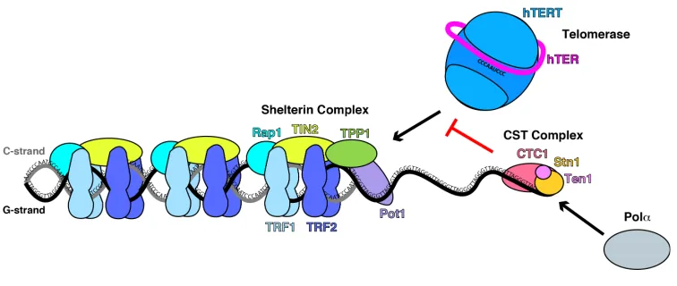
shelterin and CST cooperate to properly maintain telomere length (Figure 1.1). Since telomere
maintenance is critical to genome stability, it is not surprising that mutations in the shelterin and
CST complexes are known to cause human disease. It is well-documented that human telomere
dysfunction causes symptoms of pre-mature aging, pulmonary fibrosis, and bone marrow failure,
as well as an increased incidence of cancer (Anderson et al., 2012; Armanios and Blackburn,
2012; Chen et al., 2013; Gu and Chang, 2013; Keller et al., 2012; Vulliamy et al., 2002; Walne et
al., 2013). Therefore, further insight into the molecular mechanisms of telomere maintenance will
6
Figure 1.1 The shelterin and CST complexes cooperate to maintain telomere length.
TRF1/2, Rap1, and TIN2 of the shelterin complex bind and coat double-stranded telomeric DNA.
The shelterin components POT1 and TPP1 bind single-stranded telomeric DNA and recruit
telomerase to telomeres. CTC1 is reported to bind TPP1 and terminate telomerase activity at the
overhang. In addition, CTC1 and Stn1 are known cofactors of Pola, the DNA polymerase that is
recruited to telomeres to synthesize the C-strand.
1.4 Telomeres and human disease
Telomeres were first implicated in human disease in the early 1990s when telomerase
activity was linked to immortal cell lines and cancer (Kim et al., 1994). Since then the majority of
cancers have been found to have an upregulated activity of telomerase, which suggests that
telomerase and telomere length play a key role in extending the replicative potential of cancer
cells. Not only does misregulation of telomere length increase the risk of cancer, but it also results
in symptoms of pre-mature aging and affects tissues with both high and slow-turnover rates
(Armanios and Blackburn, 2012). Therefore, studying the causes and clinical manifestations of
7
The first disorder to be linked to telomere dysfunction was dyskeratosis congenita (Heiss
et al., 1998). Although a rare disease, affecting roughly 1 in 1 million individuals, dyskeratosis
congenita is defined by signs of premature aging such as skin hyperpigmentation, oral
leukoplakia, and nail dystrophy (de la Fuente and Dokal, 2007; Savage, 1993; Walne and Dokal,
2008). The primary causes of mortality in dyskeratosis patients is bone marrow failure, pulmonary
fibrosis, and cancer. Coats plus syndrome is a similar telomere related disease characterized by
exudative retinopathy, intracranial calcifications, and abnormalities in bone marrow and growth
(Anderson et al., 2012; Armanios, 2012; Keller et al., 2012; Mangino et al., 2012; Polvi et al.,
2012; Romaniello et al., 2012).
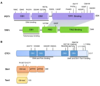
To-date numerous mutations have been found in CTC1 and Stn1 in patients with
dyskeratosis congenita and Coats plus syndrome (Figure 1.2B) (Anderson et al., 2012; Keller et
al., 2012; Polvi et al., 2012; Simon et al., 2016; Walne et al., 2013). The mutations found in CTC1
are throughout the entire protein and have been shown to disrupt key functions of the protein
such as DNA, Stn1, and Polα binding (Figure 1.2B) (Chen et al., 2013; Gu and Chang, 2013).
Two studies have reported short telomere length for patients with CTC1 mutations, however two
additional reports did not observe the same phenotype (Anderson et al., 2012; Keller et al., 2012;
Polvi et al., 2012; Walne et al., 2013). Interestingly, CTC1 mutations when studied in cell culture
lead to elongated telomeres due to the loss of telomerase repression (Chen et al., 2013; Gu and
Chang, 2013). Thus, CTC1 mutations are distinct from classical telomere dysfunction syndromes
that usually lead to short telomeres and may not be directly associated with a loss of overall
telomere length. Mutations in Stn1 are located in the N-terminal OB-fold that is responsible for
binding Ten1 and single-stranded DNA and result in DNA replication defects at both telomeric
and non-telomeric sites (Figure 1.2B) (Bryan et al., 2013; Simon et al., 2016).
Recently, mutations in POT1 and TPP1 have also been identified in patients with chronic
8
(Figure 1.2A) (Bainbridge et al., 2015; Ramsay et al., 2013; Shi et al., 2014; Takai et al., 2016;
Trigueros-Motos, 2014). Mutations which occur in the N-terminal OB-folds of POT1 disrupt the
binding of POT1 to single-stranded telomeric DNA and result in elongated and fragile telomeres
(Bainbridge et al., 2015; Ramsay et al., 2013; Shi et al., 2014; Takai et al., 2016;
Trigueros-Motos, 2014). The telomere length of peripheral blood mononuclear cells isolated from patients
was examined and revealed elongated telomeres for N-terminal POT1 mutations in addition to
the Q623H C-terminal POT1 mutation (Shi et al., 2014). Despite this one study, it is currently
unknown how mutations in the C-terminal domain of POT1 impact the function of POT1 and its
role in telomere length regulation. Although they are predicted to disrupt the assembly of the
POT1-TPP1 complex, there is a lack of structural and function data on the assembly of POT1 and
TPP1. Therefore, structural and functional studies the POT1-TPP1 complex will allow us to better
9
Figure 1.2 Primary structures of the human POT1-TPP1 and CST complexes.
(A) POT1 and TPP1 naturally occurring mutations recently found in patients with chronic
lymphocytic leukemia, familial glioma and melanoma, and Coats plus. POT1 N-terminal mutations
are known to disrupt POT1-DNA binding. However, POT1 C-terminal mutations have only been
predicted to disrupt POT1-TPP1 binding as there is currently a lack of structural and functional
data on the POT1-TPP1 assembly. (B) CTC1 and Stn1 naturally occurring mutations recently
found in patients with dyskeratosis congenita and Coats plus. CTC1 is shown with the predicted
10
1.5 Project goals
To-date, the only structural information we have of human POT1 is of the two N-terminal
OB-folds which mediate POT1-DNA binding. From these high-resolution structures we find that
naturally occurring mutations in the N-terminal OB-folds of POT1 disrupt POT1-DNA binding and
results in telomere elongation and dysfunctional telomere maintenance. There is currently no
structural information on the C-terminal domain of POT1 or how POT1 interacts with TPP1.
Structural and functional studies of the C-terminal domain of POT1 and the POT1-binding domain
of TPP1 would allow us to determine the impact of C-terminal mutations on POT1 function as well
as to gain further insight on how the POT1-TPP1 complex assembles. The goal of this research
is to better understand the mechanism of POT1-TPP1 dependent telomere capping and how
naturally occurring mutations within these proteins contribute to cancer. Through the use of X-ray
crystallography, biochemical and cell-based assays, we wanted to specifically address the
following items:
1. Identify how the C-terminal domain of human POT1 binds and interacts with the
POT1-binding domain of human TPP1.
2. Characterize the C-terminal mutations of POT1 and elucidate their impact on the
function of POT1 in telomere maintenance.
3. Determine how the disruption of the POT1-TPP1 complex contributes to telomere
11
CHAPTER 2
12
Summary
To understand the impact of C-terminal mutations on the assembly of function of the
POT1-TPP1 complex at telomeres, we determined the X-ray crystal structure of the C-terminal
domain of human POT1 (POT1C) in complex with the POT1-binding domain (PBD) of TPP1. We
found that the POT1C forms a bilobal structure consisting of an OB-fold and a holliday junction
resolvase domain. The TPP1(PBD) consists of several loops and helices involved in extensive
interactions with the bilobal POT1C domain. Biochemical data indicates that several of the
naturally occurring mutations in POT1C partially disrupt the POT1-TPP1 complex which, in turn,
disrupts the complex’s ability to bind telomeric DNA efficiently. We also show that the POT1C
mutations result in telomere elongation and the formation of fragile telomeres, which is known to
promote genomic instability that contributes to cancer and disease progression.
2.1 Introduction
The shelterin complex localizes to telomeres through binding both double and
single-stranded telomeric DNA (Lei et al., 2004; Loayza et al., 2004). POT1 is unique from the rest of
the shelterin complex as it is the only component that binds single-stranded telomeric DNA, and
does so with high affinity and specificity (Barrientos et al., 2008; Lei et al., 2004; Loayza et al.,
2004). POT1 binding to telomeric DNA is mediated by its two N-terminal OB-folds and the
remaining C-terminal portion is known to interact with TPP1 (Figure 1.2A) (Lei et al., 2003; Xin et
al., 2007). Furthermore, TPP1 binding to POT1 enhances its telomeric DNA binding affinity by as
much as 10-fold (Abagyan and Totrov, 1994; Nandakumar and Cech, 2012; Taylor et al., 2011;
Wang et al., 2007). Therefore, POT1 can localize to telomeres through binding to telomeric DNA,
13
POT1 binding to telomeres caps and sequesters the G’-overhang, thereby preventing
ATR dependent DNA damage response (DDR) and access of telomerase for telomere elongation
(Denchi and de Lange, 2007; He et al., 2014; Hockemeyer et al., 2005; Tong et al., 2015).
However, during S-phase, the POT1-TPP1 complex assembles at telomeres and actually recruits
telomerase to the telomeric overhang (Wang et al., 2007; Xin et al., 2007; Zhang et al., 2013).
Human TPP1 is known to make direct contact with telomerase via the N-terminal OB-fold of TPP1
(Figure 1.2A) (Nandakumar et al., 2012). POT1 also mediates the proper loading of telomerase to
telomeric DNA by resolving G-quadruplexes; tertiary structures that can impede telomerase
activity (Zaug et al., 2005). As a result, the POT-TPP1 complex can enhance the processivity of
telomerase, while POT1 alone can function as both a positive and negative regulator of
telomerase activity (Colgin et al., 2003; Kelleher et al., 2005; Latrick and Cech, 2010; Lei et al.,
2005; Loayza and De Lange, 2003; Wang and Lei, 2011b; Wang et al., 2007; Zaug et al., 2005).
Though TPP1 binding to POT1 is required for POT1 to function properly at telomeres, the
molecular basis of POT1-TPP1 assembly remains relatively uncharacterized. Therefore, we
sought to crystalize the human POT1-TPP1 complex. In this chapter, I discuss the high-resolution
crystal structure of the human POT1-TPP1 complex and the functional characterization of
naturally occurring mutations of the POT1 C-terminal domain. This research allows for a better
understanding of how some of these mutations disrupt the function of POT1 and contribute to
cancer.
2.2 Results
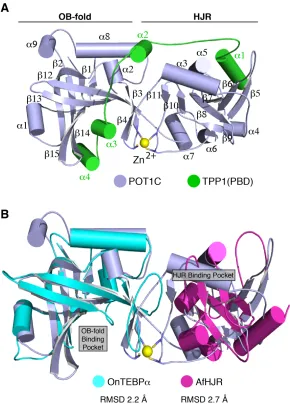
2.2.1 Structure of the human POT1-TPP1 complex
To study the interacting domains of POT1 and TPP1, we used limited proteolysis and
14
(POT1C) and the POT1-binding domain of TPP1 (TPP1(PBD)). POT1C consists of residues
330-634 and TPP1(PBD) of 255-337. After crystal screening and optimization, we solved the structure
of POT1C-TPP1(PBD) by the method of single wavelength anomalous dispersion (SAD) and a
Hg derivative (Table 1). In the structure there was clear electron density for POT1C residues
332-633 and TPP1(PBD) residues 266-326 (Figure 2.1A).
From the structure we found that POT1C consists of an OB-fold and a holliday junction
resolvase domain (HJR) (Figure 2.1A). The canonical binding pocket of the POT1C(OB) is
formed through the organization of six b-strands, a common feature of OB-folds, which forms a
deep and well defined indentation on the surface of the protein and is occupied by the a3 and a4
helices of the TPP1(PBD) (Figure 2.1). The POT1C(OB) is most similar to the third OB-fold of the
Oxytricha Nova Telomere End-Binding Protein alpha subunit (OnTEBPa, PDB ID: 1OTC), with an
RMSD of 2.2 Å (Figure 2.1B). However, an overlay of the TEBP alpha and beta dimer with that of
the POT1-TPP1 structure shows no similarities in the organization of the two heterodimers (PDB
ID: 1OTC (Horvath et al., 1998)).
The POT1C holliday junction resolvase domain (POT1C(HJR)) is an insertion of the
OB-fold and consists of seven antiparallel b-sheets surrounded by five alpha helices (a3-7) (Figure
2.1A). The structure of the POT1C(HJR) is most similar to the Archaeoglobus fulgidus holliday
junction resolvase domain (AfHJR, PDB:ID 2WIW), with an RMSD 2.7Å (Figure 2.1B), but there
are two distinct differences between the POT1 and Af HJRs. One key difference lies within the
organization of the helices present in both HJR domains with four out of the five helices not
overlapping (Figure 2.1B). Another striking difference lies within the DNA binding pocket of the
AfHJR. We find that the DNA binding pocket in the POT1C(HJR) does not align with the pocket in
the AfHJR and is instead occupied by the a3 helix of POT1C (Figure 2.1B).
Interestingly, POT1C contains a Zn2+ ion coordination site mediated by a tetra-cysteine
15
remains to be determined, but it likely contributes in the stability and organization of the POT1C
domains. The organization of the POT1C domains generates a long cylindrical structure with an
extensive surface area for the TPP1(PBD) to bind (Figure 2.1A). We find that the TPP1(PBD)
consists of an extended coil composed of four alpha helices and is organized in an opposite
orientation (N- to C-terminal) to that of POT1C. As a result, we find that the N-terminal helix (a1)
of TPP1(PBD) interacts with the POT1C(HJR) (Figure 2.1A). Whereas the C-terminal portion of
TPP1(PBD), consisting of helices a3 and a4, interacts with the canonical binding pocket of the
POT1C(OB) (Figure. 2.1A). Consequently, all of the helices and loops throughout the TPP1(PBD)
form numerous interactions with POT1C, with many side chains of TPP1 found buried in large
16
Figure 2.1 Structure of the human POT1-TPP1 complex.
(A) Cartoon representation of crystal structure of the POT1C-TPP1(PBD) complex; POT1C is
shown in light blue color with the OB-fold and HJR of POT1C labeled above each motif; the
TPP1(PBD) is shown in green color (PDB ID: 5UN7). The Zn2+ ion (yellow) is coordinated by 4
cysteines (C382, C385, C503 and C506 - stick). (B) Overlay of POT1C and the Oxytricha nova
TEBPa OB-fold (OnTEBPa - cyan color, PDB ID: 1OTC (Horvath et al., 1998)) and the
17
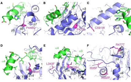
18 2.2.2 Structural analysis of POT1C mutations
Recently, POT1 has been shown to be frequently mutated in cases of chronic
lymphocytic leukemia (CLL) and familial cases of melanoma and glioma (Bainbridge et al., 2015;
Ferrandon et al., 2013; Lin et al., 2010; Ramsay et al., 2013; Robles-Espinoza et al., 2014; Shi et
al., 2014). Mutations in the N-terminal OB-folds of POT1 disrupt DNA binding and result in
aberrant telomere elongation and phenotypes associated with telomere uncapping, such as
chromosome fusions and fragile and missing telomeres (Bainbridge et al., 2015; Ramsay et al.,
2013; Robles-Espinoza et al., 2014; Shi et al., 2014). Although the POT1 C-terminal mutation,
Q623H, has been shown to result in elongated telomere is patients (Shi et al., 2014), the
structural ramifications of this mutation and other mutations in POT1C remain uncharacterized.
The majority of mutations found in the POT1C domain are somatic mutations and are so
far only found in cases of CLL (L343F, P446Q, P475L, R477T and C591W) (Table 2) (Ramsay et
al., 2013). However, the A532P and Q623H mutations are germline and have only been reported
in cases of familial glioma and melanoma (Table 2) (Shi et al., 2014). In the structure we find that
P446 is critical for TPP1 binding, as it lies within a key region of a loop that joins the HJR strands
b6 and b7 and coordinates L271 of the N-terminal helix a1 of TPP1 (Figure 2.2A). Mutating this
proline to glutamine would destabilize this loop and therefore disrupt the pocket in which the
N-terminal helix a1 of TPP1 interacts with the HJR of POT1. On the other hand, P475 is located in
the core of the HJR, in b8, and helps contribute to the fold of the HJR (Figure 2.2B). Therefore,
the proline to leucine mutations would most likely lead to a subtle perturbation of structural
elements of the HJR resulting in the re-organization of the a5 helix, which makes extensive
interactions with the a1 helix of TPP1 (Figure 2.2B).
As for C591, we find this residue is located at the C-terminal a8 helix, which spans the
19
2.2C). Furthermore, the N-terminal portion of the POT1C a8 helix also makes extensive
interactions with the a2 helix of TPP1 as well as forming a pocket for the W293 side chain of
TPP1 to bind. Therefore, a displacement of the a8 helix of POT1C, from the cysteine to
tryptophan mutation, would likely lead to a reorganization in this region of the OB-fold and disrupt
TPP1 binding. From the structure we also find that Q623 is located in the center of the canonical
binding pocket of the POT1C OB-fold, which directly interacts with the backbone of the a3 helix of
TPP1 that is coordinated by the solvent accessible side chains (Figure 2.2D and E). We predict
that the introduction of a larger hydrophobic side chain, such as histidine, would likely lead to the
loss of hydrogen binding that is created by the NE2 of Q623 in POT1 with the carboxyl of L313 in
TPP1 (Figure 2.2D and E). As a result, the Q623H mutation would likely disrupt POT1-TPP1
binding. Lastly, we find that I535 is located in the POT1C(HJR) in a7 with the hydrophobic side
chain being buried into the core of this region in POT1C. The introduction of the larger
phenylaline side chain at I535, such as phenylalanine, would likely lead to the destabilization in
the core of the HJR domain (Figure 2.2F).
Unlike P446Q, P475L, I535F, C591W, and Q623H, it is unclear how the L343F, R477T,
and A532P POT1C mutants contribute to human disease (Figure 2.2E and F). From the structure
we find that L343 comprises part of a loop that connects b1 to a1 in the POT1C (OB) (Figure
2.2E). Although L343 is important for the structural organization of this loop, it does not make
direct contacts with TPP1 and that mutating this leucine to phenylalanine would not likely disrupt
POT1-TPP1 binding (Figure 2.2E). Similarly, we find that R477 is part of b8 and is located at the
opposite face where the a1 helix of TPP1 binds the POT1C(HJR) (Figure 2.2F). R477, unlike
L343, is solvent exposed and does not interact with TPP1, with the nearest point of contact with
TPP1 being approximately 18 Å away. We also find that like R477, A532 is located on the
20
Figure 2.2 Interactions between POT1C and TPP1l; POT1C mutations are highlighted.
(A) The POT1C(HJR) (blue) forms extensive interaction with the leucine rich a1 of TPP1(PBD)
(green). The P446Q mutation site is shown in magenta. (B) P475L mutation site (magenta) is
located in the core of the POT1C(HJR) domain. (C) The C591W mutation (magenta) site in the
a8 helix of the POT1C(OB). The a8 helix makes extensive interactions with the a2 helix of TPP1
and forms a pocket for the W293 side chain of TPP1 to bind (D) The Q623H mutation (magenta)
is located in the core of the POT1C(OB) binding pocket and makes direct contacts with L313 of
TPP1. (E) The POT1C(OB) binding pocket forms extensive interaction with the a3 and a4 helices
of TPP1(PBD). The L343F and Q623H mutations are shown in magenta. (F) The R477T, A532P,
21
Table 2: Reported cases of POT1C mutations in cancer.
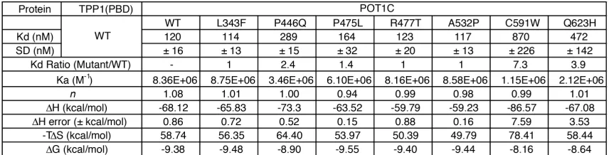
2.2.3 Several POT1C mutations partially disrupt the POT1-TPP1 complex
To determine the impact of POT1C mutations on the assembly of the POT1-TPP1
complex, we measured the binding affinity of each mutant to the TPP1(PBD) using Isothermal
Titration Calorimetry (ITC). For these binding experiments, we overexpressed the POT1C and
TPP1(PBD) constructs we used in crystallographic studies and purified them separately using
three successive steps of purification as described in the methods section of this chapter (Figure
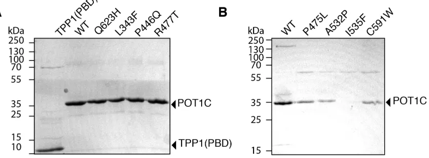
2.3A and B). Of note, all of the mutant POT1C proteins were expressed in sufficient quantities for
the ITC assay, except for the I535F mutant, which did not express stably (Figure 2.3B).
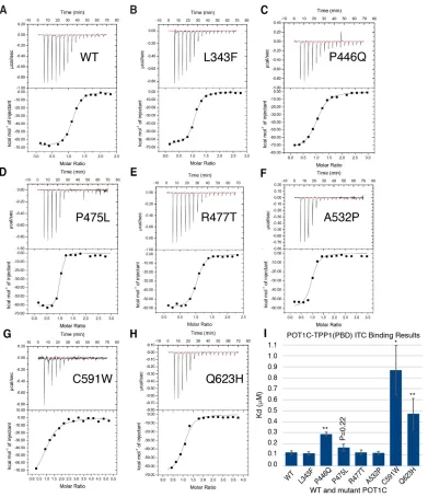
From the ITC assay, we found that the wildtype POT1C and TPP1(PBD) bound with
nanomolar affinity (Kd = 120 ± 16 nM – Figure 2.4A and Table 3). Out of all the POT1C mutants
we tested, only the P446Q, C591W, and Q623H mutants displayed a significant loss of affinity to
TPP1 (2.4, 7.9, and 3.9-fold change, respectively – Figure 2.4C, G, H, I and Table 3). P475L did
22
not statistically significant (P = 0.22, Figure 2.4I). The remaining POT1C mutants, L343F, R477T,
and A532P displayed wildtype TPP1(PBD) affinity (1-fold change – Figure 2.4B, E, F and I and
Table 3).
An analysis of the overall enthalpic and entropic components of wild type POT1C and
TPP1(PBD) binding reveal that is comprised of favorable enthalpic-driven interactions, as
indicated by the negative binding enthalpy (DH = -68.12 kcal/mol), that is typically mediated by
hydrogen-bonding and van der Waals interactions (Table 3) (Velazquez-Campoy et al., 2015).
The increase in entropy is an unfavorable contribution to binding that is usually associated with
conformational restrictions (-TDS = 58.74 kcal/mol) (Table 3). The overall contribution of enthalpic
and entropic components is represented by the change in free energy (DG), where wild type
POT1C-TPP1(PBD) binding displays a negative and favorable free energy change of
approximately -9.38 kcal/mol (Table 3). In contrast, the P446Q, C591W, and Q623H mutants
display a decrease in binding energy (DG = -8.9, -8.16, and -8.64 kcal/mol, respectively)
compared to wild type binding due to an increase in unfavorable conformational changes (-TDS)
23
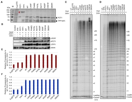
Figure 2.3 Expression and purification of POT1C mutants.
(A) SDS-PAGE of purified TPP1(PBD) and wild type (WT) and mutant POT1C proteins (Q623H,
L343F, P446Q, and R477T) for use in the ITC binding experiments. (B) SDS-PAGE of purified
WT and mutant POT1C proteins (P475L, A532P, I535F, and C591W) for use in the ITC binding
experiments. Of note, the P475L, A532P, and C591W had slight decrease in expression levels
24
Figure 2.4 ITC binding data for the POT1C and TPP1(PBD) proteins.
(A – G), ITC binding data of wild type (WT) and mutant (L343F, P446Q, P475L, R477T, A532P,
C591W and Q623H) POT1C with TPP1(PBD). (I) Bar graph of binding constants (Kd, µM) for WT
and mutant POT1C proteins derived from the fitted ITC data. Two-tailed Student’s t-test with
respect to WT: *P < 0.05, **P < 0.01. The data clearly shows that the POT1C, P446Q, C591W
25
Table 3: ITC binding data for wild type and mutant POT1C proteins with TPP1(PBD)
2.2.4 POT1C mutants do not alter telomerase processivity
To determine if the POT1C mutants disrupted telomerase processivity, we performed
direct telomerase activity assays using super telomerase extracts. Super telomerase extracts
were prepared by lysing HEK293T cells over-expressing human TERT and TER (hTERT and
hTER plasmids were a gift from the Lingner lab) (Cristofari and Lingner, 2006). Super telomerase
extracts were then supplemented with saturating amounts of POT1 (wild type or mutant) and
TPP1. We purified full-length POT1 and a stably expressed N-terminal truncation of TPP1
consisting of residues 87-544 (TPP1(87)) from E. coli to supplement extracts (Figure 2.5A). We
also performed direct assays using cell extracts co-expressing full-length POT1 and TPP1 with
human telomerase in HEK293T cells (Figure 2.5B). A detailed summary of how we carried out the
direct assay reactions is explained in the methods sections of this chapter.
The use of both E. coli purified and co-transfected POT1 and TPP1 proteins with super
telomerase extracts produced increased telomerase processivity only when POT1 and TPP1
were both present (Figure 2.5C and D). Surprisingly, all of the POT1C mutants showed wild type
telomerase processivity, with no statistically significant differences (Figure 2.5C-F). Even the
POT1C mutants, P446Q, C591W, and Q623H, which displayed a significant loss of affinity for
TPP1(PBD) in the ITC assays (Table 3), also had wildtype telomerase processivity within the
26
Figure 2.5 Direct telomerase activity assays
(A) SDS-PAGE gel for the 2 µg of super-telomerase extract (STE) and 150 nM of E. coli purified
full-length POT1 (flPOT1) and TPP1(87) proteins used in the direct assay of panel c. The red
arrow indicates where TERT is expected to run (~130 kDa). (B) Western blot of HEK293T lysates
used in the direct assay of panel d. (C) Telomerase direct activity assay using 2 µg of STE
supplemented with150 nM of purified wild type (WT) or mutant flPOT1 and WT TPP1(87) (D)
Telomerase direct activity assay using 5 µg of STE co-transfected with WT or mutant flPOT1 and
WT full-length TPP1 (flTPP1). The number of telomeric repeats added to the primer are indicated
on the right of the gels C and D. (E and F), Quantification of telomerase processivity of panels C
and D respectively; The values are the average of two independent experiments; error bars
27
2.2.5 Partial disruption the POT1-TPP1 complex reduces POT1-DNA binding
TPP1 binding to POT1 is known to enhance the affinity of POT1 for telomeric DNA
(Nandakumar and Cech, 2012; Taylor et al., 2011). Therefore, we wanted to determine if any of
the POT1C mutations would disrupt the DNA binding affinity of POT1 in the presence of TPP1.
To test this, we performed Fluorescence Polarization (FP) assays using a labeled DNA probe
consisting of three telomeric repeats ((TTAGGG)3, 18mer) and the POT1C and TPP1(PBD)
proteins used for structural and ITC experiments (Figure 2.3A). We also tested wild type and
mutant (P446Q, C591W, and Q623H) full-length POT1 alone in addition to the wild type and
mutant full-length POT1-TPP1 complexes used in the direct telomerase assays (Figure 2.5A).
The FP binding assays show that POT1C, TPP1(87) and the POT1C-TPP1(PBD)
complex do not bind single-stranded telomeric DNA (Figure 2.6A and C). We also found that the
wild type and mutant (P446Q, C591W, and Q623H) full-length POT1 proteins bind
single-stranded telomeric DNA with similar affinity (approximate Kd = 20 nM, Figure 2.6B, D and Table
4). Consistent with previously published reports, the FP assays show that the DNA affinity of
wildtype POT1 is enhanced in the presence of TPP1, where the wild type POT1-TPP1 complex
binds single-stranded telomeric DNA with low nanomolar affinity (Kd = 5.8 ± 0.5 nM, Figure 2.6C
– D and Table 4) (Lei et al., 2004; Wang et al., 2007). Interestingly, the POT1C mutants that have
wildtype TPP1 binding affinity according to the ITC assays (L343F, R477T, and A532P, Table 3)
all display a similar affinity for telomeric DNA as the wildtype POT1-TPP1 complex (Figure 2.6C,
D and Table 4). In contrast, the P446Q, P475L, C591W, and Q623H POT1C mutants, which
disrupt POT1C-TPP1(PBD) binding, all display a decrease in DNA binding affinity for the
28
Figure 2.6 POT1-TPP1 telomeric DNA binding assays.
(A) FP assays of POT1C and POT1C-TPP1(PBD) complex with single-stranded DNA probe
consisting of 3 telomeric repeats (18mer). (B) FP assays of the wild type (WT) and mutant (those
that partially disrupt the POT1-TPP1 complex P446Q, C591W and Q623H) flPOT1 with the
18mer DNA probe. (C) FP assays of the WT and mutant flPOT1 + TPP1(87) complex with the
18mer. (D) Bar graph showing the differences in Kd (nM) between the WT and mutant flPOT1
and flPOT1-TPP1(87) complex. Two-tailed Student’s t-test with respect to WT POT1-TPP1(PBD)
29
Table 4: FP assay binding data for POT1 and TPP1 proteins with telomeric 18mer.
2.2.6 POT1C mutations do not affect the localization of POT1 to telomeres
Since POT1 is known to localize to telomeres through its interaction with TPP1 and DNA,
we wanted to determine if the POT1C mutants were able to localize to telomeres. In collaboration
with Dr. Susan Janicki’s lab, we first co-expressed YFP-tagged wild type and mutant POT1 with
mCherry-tagged TRF2 in HEK293T cells. We then used confocal microscopy on fixed cells
co-expressing the fluorescently labeled POT1 and TRF2 proteins to determine if the POT1C mutants
would co-localize to telomeres with TRF2. We also performed western blot analysis of transfected
HEK293T cells to validate the expression of the wildtype and mutant YFP-tagged POT1 proteins
(Figure 2.7A). Surprisingly, all of the POT1C mutants co-localized with TRF2 (Figure 2.7B). In
conclusion, none of the POT1C mutants we tested disrupt the ability of POT1 to localize to
telomeres. This is consistent with studies of N-terminal POT1 mutants that also disrupt
30
Figure 2.7 Cell imaging for localization of wild type and mutant POT1 to telomeres.
(A) Western blot showing the levels of YFP tagged, wild type (WT) and mutant (L343F, P466Q,
P475L, R477T, A532P, I535F, C591W and Q623H) full length POT1. We used GFP (aGFP) or
POT1 (aPOT1) to detect the levels the YFP-POT1 in each cell line. GAPDH was used as a
loading control. (B) Maximum projection images of co-localization of YFP-POT1 (green) and
Cherry-TRF2 (red) proteins are shown. Merged images include DAPI. Scale bar = 5µm. The
31
2.2.7 Partial disruption of the POT1-TPP1 complex results in telomere elongation
To determine the impact of POT1C mutations on telomere length regulation in cells, we
performed Southern blot analysis on genomic DNA purified from HEK293T cells stably infected
and expressing wild type and mutant full-length POT1 proteins. Endogenous levels of POT1 were
reduced using a shRNA targeting the 3’UTR of POT1 (shPOT1, TRCN0000009837 - Sigma
Mission shRNA library) (Figure 2.8A). HEK293T cells were infected with lentivirus prepared with
shPOT1 (pLKO.1 vector, puromycin resistance) and wildtype or mutant POT1 vectors (pLU
vector, blasticidin resistance) and stably selected using puromycin and blasticidin. For controls,
we utilized cells transfected with empty vectors (pLKO.1 and pLU vectors), wild type POT1 with
the shPOT1, and shPOT1 treated cells with empty pLU vector (Figure 2.8A and B).
We determined by western blot analysis that wild type and mutant full-length POT1 were
expressed at similar levels in the presence of shPOT1 (Figure 2.8B). The Southern blot data
shows significant telomere elongation in the shPOT1 treated cells compared to marginal
increases observed in wild type POT1 cells (Figures 2.8C-F). For the C591W and Q623H POT1C
mutants we observed elongated compared to the wild type POT1 cells after 120 population
doublings (Figures 2.8C-E). In addition, the L343F mutant also displayed a subtle elongation in
telomere length. In contrast we observed wildtype changes in telomere length for the R477T,
32
Figure 2.8 Southern blot analysis of wild type and mutant POT1.
33
Figure 2.8 Southern blot analysis of wild type and mutant POT1 (continued).
(A) Quantitative RT-PCR showing knock down of endogenous POT1 mRNA, normalized to
GAPDH transcript levels, after lentiviral infection of HEK293T cells with shPOT1 and wild type
(WT) Flag-POT1; selection was carried out with puromycin and blasticidin; ± s.d. (n = 3). (B)
Western blot showing WT and mutant POT1 protein expression levels in HEK293T cells
expressing shPOT1 and Flag-POT1. Cells infected with the empty pLU and pLKO.1 lentiviral
vectors are used as controls. (C - E) Southern blots of HEK293T cells expressing shPOT1 and
WT or mutant flPOT1. DNA from 5, 50 and 120 populations doublings (PD) is shown. HEK293T
cells treated with only shPOT1 range from 5, 30, and 70 population doublings. DNA length
standards are indicated along the left of each gel. The white dashed line indicates the baseline
telomere length for the vector control. Green (vector control), yellow (WT and shPOT1 treated
cells), and red (POT1 mutants) dots show mean telomere length at each PD. (F) Quantification of
the mean telomere length (kb) from panel C-Eacross population doublings (PD) was performed
with TeloTool (Gohring et al., 2014). Green (control), yellow (WT and shPOT1 treated cells), and
red (POT1 mutants) dots show mean telomere length at each PD. Black bars indicate the range
of telomere restriction fragments (TRF) at each PD.
2.2.8 POT1C mutations lead to fragile telomeres
N-terminal mutations of POT1, which disrupt POT1-DNA binding are reported to cause
telomere elongation and chromosome fusions, fragile telomeres, and missing telomere signal at
the ends of chromosomes (Ramsay et al., 2013; Robles-Espinoza et al., 2014; Shi et al., 2014).
To examine the integrity of telomeres in cells expressing the POT1C mutants, we preformed
34
cell lines as those used in the Southern blot analysis (Figure 2.8B). Metaphase spreads after 50
population doublings were prepared by fixing chromosomes to microscope slides and hybridizing
the telomeres with a 5′ TelC-Tamra peptide nucleic acid (PNA) probe ((CCCTAA)3-Tamra,
Panagene). Chromosomal DNA was then stained with DAPI and images were taken with Nikon
E600 upright fluorescent microscope.
The frequency of chromosome fusions, fragile telomeres, and telomere free ends were
then recorded as a percentage of all the metaphase chromosomes counted, with an average of
500 chromosomes counted for each sample. The HEK293T cells carrying the empty vectors
displayed an average of 2-3% of chromosomes with fusion, fragile, and missing telomeres,
comparable to the amount observed in the wild type POT1 cell line (Figure 2.9). Elevated levels
of chromosome fusions were only observed in cell lines expressing the L343F (7%) and I535F
(5%) mutants (Figure 2.9). Interestingly, all mutants showed an increased level of fragile
telomeres (L343F, 9%; P446Q, R477T and Q623H, 9%; P475L and C591W, 10%) compared to
the low frequency observed for the empty vector and wild type POT1 controls (approximately 3%)
(Figure. 2.9B and D). We also observed an increase in the occurrence of missing telomeres in the
35
Figure 2.9 Fluorescence in situ Hybridization (FISH) data.
(A)Telomeric FISH metaphase spreads of cells expressing wild type (WT) and mutant (L343F,
P466Q, P475L, R477T, A532P, I535F, C591W and Q623H) flPOT1 proteins after 50 population
doublings (Red, TelC-Tamra; blue, DAPI). Endogenous POT1 levels were reduced with shPOT1.
Telomere fusions (fused chromosome ends), fragile telomeres (multiple telomere signals) and
missing telomeres (no telomere signal) are indicated by red, pink and green arrows respectively.
Scale bar is 5 µm. (B-D), Quantification and examples of telomere fusions, fragile, and missing
telomeres from the metaphase spreads of panel A. Bars indicate the percent of metaphase
events. Error bars are indicating standard deviation. An average of 500 chromosomes was
36
2.3 Discussion
Naturally occurring mutations in POT1 have been linked to human cancer and are
located throughout the entire protein (Figure 1.2B and Table 2) (Bainbridge et al., 2015; Lin et al.,
2010; Ramsay et al., 2013; Robles-Espinoza et al., 2014; Shi et al., 2014). The N-terminal
OB-folds of POT1 mediate DNA binding and mutations in this region of POT1 disrupt POT1-DNA
binding and result in dysfunctional telomere maintenance (Bainbridge et al., 2015; Ramsay et al.,
2013; Robles-Espinoza et al., 2014; Shi et al., 2014). The C-terminal domain of POT1 binds
TPP1 and POT1-TPP1 binding is known to enhance the DNA affinity of POT1 (Abagyan and
Totrov, 1994; Barrientos et al., 2008; Nandakumar and Cech, 2012; Taylor et al., 2011; Wang et
al., 2007). We speculate that mutations which disrupt POT1-TPP1 assembly will indirectly affect
POT1-DNA binding. Furthermore, since the POT1-TPP1 complex also enhances the processivity
of telomerase, we hypothesize that mutations disrupting POT1-TPP1 assembly will decrease
telomerase processivity (Lei et al., 2004; Loayza et al., 2004; Wang et al., 2007).
The structure of the POT1C-TPP1(PBD) complex presented here allows us for the first
time to understand how POT1 and TPP1 interact. We also find that several of the POT1C
mutations are directly or indirectly involved in critical interactions between the POT1C and
TPP1(PBD). ITC and FP binding assays reveal that some of these mutants (P446Q, P475L,
C591W and Q623H) partially disrupt the POT1-TPP1 complex and reduce the affinity of the
complex for telomeric DNA (Table 3 and 4). Furthermore, Southern blot analysis shows longer
telomeres in mutants (C591W and Q623H) which significantly disrupt the POT1-TPP1 assembly
(Figure 2.8). We postulate that partial disruption of the POT1-TPP1 complex prevents the proper
assembly of the complex at telomeres and leads to a misregulation of telomerase access, thus
resulting in longer, fragile telomeres. This is in agreement with a previous report which
demonstrated that reduced affinity of the POT1-TPP1 complex for telomeric DNA leads to
telomere elongation due to persistent access of telomerase to the telomeric overhang (Loayza
37
show elevated levels of missing and fragile telomeres (Figure 2.9), which is consistent with
reports that unregulated telomere length leads to chromosomal abnormalities (Bailey and
Murnane, 2006; Sfeir et al., 2009; Webb CJ, 2013).
Interestingly, none of the POT1C mutations appear to affect telomerase processivity,
even those which significantly reduce the assembly of POT1 and TPP1 (P446Q, C591W, and
Q623H) (Figure 2.5C-F). Cell imaging experiments also show that all of the POT1C mutants still
localize to telomeres (Figure 2.7). Although surprising, a complete deletion of the first N-terminal
OB-fold or the entire C-terminal domain of POT1 still results in localization of POT1 to telomeres
(Barrientos et al., 2008; Liu et al., 2004; Loayza and De Lange, 2003). However, POT1
localization to telomeres is hindered when TPP1 is depleted or when the POT1-TPP1 interaction
is completely disrupted (Liu et al., 2004). In addition, several of the POT1C mutations we
examined (L343F, A532P, and R477T) do not disrupt the assembly of the POT1-TPP1 complex.
The precise role of these mutations is currently unclear and further studies are required to better
understand their impact on the function of the POT1-TPP1 complex and telomere maintenance.
Nevertheless, we observed phenotypes of telomere dysfunction in all of the POT1C
mutants. Specifically, we observed fragile telomeres in all mutants, while in some mutants we
also observed defects in telomere length and missing or fused telomeres. Taken together, the
data clearly shows that there is a confluence of factors that contribute to the disruption of the
POT1-TPP1 complex by mutations found in the C-terminal domain of POT1. It is also worth
noting that, so far, many POT1 mutations are heterozygous and therefore cells still contain one
wild type copy of the POT1 allele (Table 2). Furthermore, POT1 mutations typically occur in
combination with an array of other defective genes known to predispose patients to malignant
disease, such as: CDKN2a, CDK4, BAP1, Notch1, SF3B1, TP53, and ATM (Bainbridge et al.,
38
developing mutations in POT1 contributes to the driving nature and prognosis of malignant
disease among other factors.
Telomere length regulation is a vital for maintaining proper telomere length and
preserving telomere stability (Rivera et al., 2017). Like N-terminal POT1 mutants, we observed
elongated telomere length for the two most severe POT1C mutants (C591W and Q623H) and in
all of the mutants we do observe phenotypes of telomere dysfunction. Although the defects we
observe in POT1C mutants are not severe, dysfunctional telomere maintenance can result in
genome instability overtime, which can contribute to the clonal evolution and development of
cancer (Feldser et al., 2003). Reports have linked elongated telomeres to an increased risk of
adult glioma, melanoma, CLL, and other carcinomas (Anic et al., 2013; Ojha et al., 2016; Walsh
et al., 2015). On the other hand, short telomeres promote genome instability and are also
reported to drive tumor progression and be linked to a poor disease prognosis in CLL patients
(Lin et al., 2010; Lin et al., 2014). There is clearly a link between the misregulation of telomere
length and the dysfunction of telomeres, however the direct mechanism of how this contributes to
the human disease is still not fully understood. Therefore, understanding how POT1 regulates
telomere length will enable us to better understand how telomere length contributes to genome
instability, aging, and the development of cancer.
2.4 Materials and methods
2.4.1 Protein expression and purification
Human POT1C, comprising residues 330-634, was identified via limited proteolysis and
cloned into a pET28b vector containing a N-terminal hexahistidine - pMocr fusion tag, cleavable
by TEV protease. The TPP1(PBD) construct was designed to contain residues 255-337 and was
39
protease. Both POT1C and TPP1(PBD) were overexpressed in Escherichia coli ScarabXpress T7
lac competent cells (Scarab Genomics) at 18ºC and 30ºC for 4 hours, respectively, using 1 mM
IPTG (isopropyl-β-D-thiogalactopyranoside; Gold Biotechnology). The cells were harvested by
centrifugation and lysed in a buffer containing 25 mM Tris-HCl, pH 7.5, 1.0 M KCl, 1.0 M Urea,
5% glycerol, 1 mM phenylmethylsulfonyl fluoride (PMSF), and 1 mM benzamidine (Ni Buffer A)
via sonication. The proteins were purified over a Ni-nitrilotriacetic acid (Ni-NTA) column, buffer
exchanged while on the Ni-NTA column with 25 mM Tris-HCl, pH 7.5, 0.2 M KCl, and 5% glycerol
(Ni Buffer C). The complex was eluted with 300 mM imidazole onto a tandem HS(poros) –
HQ(poros) columns (Applied Biosystems) equilibrated with Ni Buffer C. The HS-HQ columns
were then detached and the POT1C-TPP1(PBD) complex was eluted from the HQ column with a
salt gradient of 0.2 M KCl to 1.0 M KCl. The fusion tags were cleaved by TEV overnight at 4°C
and removed from the sample by an additional step of Poros-HS and HQ. The clean complex was
passed over a Superdex S200 (GE Healthcare) to remove any aggregates.
Full-length wild type and mutant POT1 were cloned into the same vector as POT1C.
TPP1(87-544) was cloned into a pET28b vector containing an N-terminal hexahistidine-MBP
(maltose binding protein) cleavable by TEV protease. POT1 and TPP1 were overexpressed
separately in ScarabXpress cells at 18º C overnight using 1 mM IPTG. The cells were harvested
by centrifugation and lysed in Ni buffer A via sonication. The proteins were first purified over a
Ni-NTA column and then eluted onto an amylose column (New England Biotech) to further purify the
POT1 – TPP1 complex and remove excess POT1 before eluting with a buffer containing 25 mM
Tris-HCl, pH 7.5, 0.5 M KCl, 5% glycerol, 1 mM DTT and 30 mM maltose. The fusion tags were
cleaved with TEV protease overnight at 4oC. The POT1-TPP1 complex was then buffer
exchanged to remove the maltose and passed over an orthogonal Ni-NTA and amylose column
to remove any residue fusion tags and TEV protease from the samples. The purified full-length
40
concentrations of BSA standard and quantified using ImageQuant TL (GE Healthcare) to
determine the concentration of the complex.
2.4.2 Protein crystallization and structure determination
POT1C-TPP1(PBD), crystal screening produced a crystal hit under sitting-drop vapor
diffusion at room temperature in a crystallization buffer containing 2.4 M KCl, 50 mM K/Na
Tartrate, 20 mM BaCl2, and 0.1 M Sodium Citrate, pH 5.5. A longer construct of POT1C
consisting of residues 325-634 produced a different crystal form that belonged to the P1 space
group and diffracted to 3 Å at best. The new crystal form belongs to the P4122 (sg91, 1 copy in
asymmetric unit) and diffracted to 2.1 Å. The native dataset (containing Zn) was collected from 2
crystals at 1.03317 Å wavelength at BL12-2 SSRL to 2.1 Å resolution. The radiation damage was
slowed with a careful absorbed dose estimate, allowing high multiplicity and accumulating a
significant anomalous signal from the present Zn and Sulfurs in the protein. Both the native and
derivative data were processed with XDS (autoxds script at SSRL) with a zero dose correction.
We solved the structure using a single methyl mercury (meHg) derivative using the SAD
approach as implemented in SHELXCDE using the graphical interface of the HKL2MAP software.
SHELXD identified 5 well defined Hg sites and SHELXE extended and optimized the initial
phases to 2.1 Å and generated a preliminary structure of 330 residues with excellent contrast
(0.943) and connectivity (0.843) resulting in FOM of 0.605. Subsequently the model was traced
using 10 cycles of BUCCANEER. The resulting model was almost complete - 358 sequenced
residues. The remaining model was improved in COOT and refined with BUSTER (version
2.10.2). The refinement converged to R/Rfree = 17.3/19.3 % to 2.1 Angstrom resolution. The model
has excellent stereochemistry as examined by MOLPROBITY server
41 2.4.3 Isothermal Titration Calorimetry (ITC)
We carried out ITC experiments on a MicroCal iTC200 (Malvern) using separately
purified TPP1(PDB) and wild type and mutant POT1C proteins. The purified proteins were buffer
exchanged into a buffer containing 25 mM Hepes, pH 7.5, 0.1 M KCl, 5% glycerol, and 1 mM
TCEP. Protein concentration was measured using a Bradford Assay (Bradford, 1976).
TPP1(PBD) at a concentration of 100 µM was injected into a cell containing 10 µM Pot1-C. For
the ITC experiment, the cell of the calorimeter was kept at 25° C (298.15 K), and the volume of
each injection was 2.47 µL with a total of 16 injections. Analysis of the ITC data used the Origin
analysis software (GE Healthcare) to obtain binding constants and ratios. Binding constants (Ka),
reaction stoichiometry (n), binding enthalpy (DH), and entropy change (DS) were obtained from
the Origin analysis software used to fit the ITC data using a one-site binding model. The binding
free energy change (DG) was determined with the following equation:
DG = -RT ln Ka = DH - TDS
2.4.4 Fluorescence Polarization (FP) Assays
We performed FP DNA binding assays using an Envision Xcite Multilabel Plate
Reader (Perkin Elmer). 20 µl binding reactions were carried out in a buffer containing 20 mM
Hepes, pH 7.5, 100 mM KCl, 2 mM MgCl2, 1 mM EDTA, 2 mM DTT, 1 mg/mL BSA, 5% v/v
glycerol, and 75 nM polyT50 competitor (IDT). The 18mer DNA probe
(TTAGGGTTAGGGTTAGGG) was purchased with a 5′ 6-FAM label from IDT. The final probe
concentration used was 2.5 nM, while the POT1-TPP1 protein concentration ranged from 0 to
100 nM or POT1C-TPP1(PBD) concentrations ranged from 0 to 5 µM. The reactions were
incubated at room temperature for 30 minutes and pipetted in triplicate into a black 384 well