R E V I E W
Development and Clinical Prospects of Techniques
to Separate Circulating Tumor Cells from Peripheral
Blood
This article was published in the following Dove Press journal:
Cancer Management and Research
Cheng Tian Xinhua Xu Yuke Wang Dailong Li Haiyan Lu Ziwei Yang
Yichang Central People’s Hospital, First Clinical Medical College of Three Gorges University, Yichang 443000, People’s Republic of China
Abstract: Detection of circulating tumor cells (CTC) is an important liquid biopsy
techni-que that has advanced considerably in recent years. To further advance the development of technology for curing cancer, several CTC technologies have been proposed by various research groups. Despite their potential role in early cancer diagnosis and prognosis, CTC methods are currently used for research purposes only, and very few methods have been accepted for clinical applications because of difficulties, including CTC heterogeneity, CTC separation from the blood, and a lack of thorough clinical validation. Although current CTC technologies have not been truly implemented, they possess high potential as future clinical diagnostic techniques for individualized cancer. Here, we review current developments in CTC separation technology. We also explore new CTC detection methods based on telomer-ase and nanomaterials, such as in vivo flow cytometry. In addition, we discuss the difficulties that must be overcome before CTC can be applied in clinical settings.
Keywords: circulating tumor cells, cancer, liquid biopsy, tumor metastasis, cancer diagnosis
Introduction
Circulating tumor cells (CTC) enter peripheral blood circulation either spontaneously or during cancer treatment. Due to high activity and metastasis potential, CTC are
some-times considered as the primary mechanism of tumor metastasis.1 Thus, they are
important marker of tumor screening, diagnosis, prognosis, and efficacy evaluation.2–6
Therefore, imaging CTC should improve the accuracy of lung cancer screening and early detection. CTC are extremely rare, with only one per 106–107 peripheral blood
leuko-cytes from cancer patients.7 Because of their rarity, research on CTC imaging did not
emerge until the development of the cell search system, a Food and Drug Administration (FDA)-approved medical device for CTC selection and counting. However, challenges
remain in terms of formulating a uniform standard for CTC separation (Table 1).8,9
The heterogeneity of CTC is the primary obstacle in their detection. These cells differ in size, shape, and immunotyping across tissue types. For example, during epithelial-to-stromal transformation (EMT), both epithelial cell adhesion molecule (EpCAM) and cytokeratin (CK) are downregulated resulting in failure of conventional EpCAM-based capturing techniques in detecting CTC subpopulations with more
mesenchyme-like phenotype.10,11 In addition, due to multi-step cell preparation
pro-cesses, circulating tumor cells may be destroyed and fragmented, resulting in
inaccu-rate test results.7
Correspondence: Xinhua Xu First Clinical Medical College of Three Gorges University, Yichang Central People’s Hospital, 180 Yiling Main Road, Yichang 443000, People’s Republic of China
Email [email protected]
Cancer Management and Research
Dove
press
open access to scientific and medical research
Open Access Full Text Article
Cancer Management and Research downloaded from https://www.dovepress.com/ by 118.70.13.36 on 27-Aug-2020
T able 1 Cir culating T umor Cells W ere Selected Based on Surface Analysis
Isolation Method
CellSear ch EasySep MA CS IMS STMBs HTMSU CTC-Chip GEDI Chip
CTC enrichment
EpC AM CD45 EpC AM EpC AM EpC AM, HER2, EGFR EpC AM Anti-EpC AM
functionalized micr
oposts and chip structure Immunocapture and micr ofluidics
CTC detection Immunofluorescence analysis
for CK, CD45, and D API
Immunofluorescence analysis
for CK, CD45, and D API
Immunofluorescence analysis
for CK, CD45, and D API
Immunofluorescence analysis
for CK, CD45, and D API
Immunofluorescence analysis
for CK, CD45, and D API
Immunofluorescence analysis
for CK, CD45, and D API
Immunocytochemical analysis
or
R
T-PCR
Immunofluorescence analysis
for PSMA, CD45, D API Viability na 92.59% Viable Mostly viable 85% 80% Viable Viable Recovery 42%, 85% na 79±10% 96% > 60% Purity 0.1%, 1.4% 42% 84±3% 100% 52–67% 68% Thr oughput 9 mL/h 1–4 mL/h na 1–2 mL/h 1–2 mL/h
Sample volume
9 mL 0.5–2 mL 1 mL 1 mL 2.7 mL 1 mL K ey features FD A appr oved for advanced breast, pr ostate, and colorectal cancers; ferr ofluid nanoparticles; Easy-to-use batch separation; high backgr ound P os/neg enrichment; high surface area to volume; difficult to use with whole blood Low backgr ound leuk ocytes (not tested on clinical samples) Uses antibodies simultaneously; low sample volume Single-step separation; low volu mes of blood;
conductivity-based enumeration
Micr ovortex increases the efficiency; various CTC-specific antigens can be used Functional assa ys in situ; size and collision inclination dependency References T alasaz AH77 Nolan J75 Miltenyi S40 Xiong K43 Lu N-N25 Adams AA52, Dharmasiri U53, Dharmasiri U78 Nagrath S61 Gleghorn JP59
Cancer Management and Research downloaded from https://www.dovepress.com/ by 118.70.13.36 on 27-Aug-2020
The detection of CTC has three main steps: 1) blood sample preparation for tumor cell separation, 2) antibody staining or DNA probe detection, and 3) imaging of cells. Improved cell separation technology allows us to obtain complete CTC for biological characterization and
func-tional analysis (Figure 1). In this review, we focus on the
development of CTC separation technique, particularly the advantages and disadvantages of various separation meth-ods. We then summarize the areas in which separation technique still requires improvement and the current lim-itations of its clinical application. Finally, we examine the potential future directions of CTC use in clinical settings.
Cell Separation Methods
Surface Antigen-Based Separation
Circulating tumor cells are recognized by their round or oval morphology, along with the presence of surface antigens epithelial cell adhesion molecule (EpCAM) and cytokeratin
(CK), as well as the absence of CD45.12 Separation methods
using surface antigens can be divided into positive and nega-tive selection. The most representanega-tive posinega-tive-selection method is the cell search system. Negative selection generally involves removing unnecessary cells to indirectly capture
CTC. For example, all CD45+ peripheral blood cells can be excluded to leave only the CD45-CTC. However, this method
usually leads to low purity.13–16 Another technique called as
EasySep (developed by Stemcell) is an immunomagnetic method that separates CTC through negative enrichment using a mixture of CD45-containing antibodies and differ-ently sized magnetic beads. With an average recovery of 42.23%, EasySep can produce living cells without markers and also be used for subsequent downstream analysis.
The prognosis of patients with breast, prostate, or
col-orectal cancers is related to CTC count.17–19 After
reveal-ing this potential, the FDA approved the cell search system as the only technology for clinical CTC detection. This immunocytochemistry method is based on magnetic immunity and staining. First, EpCAM antibody beads are used to enrich CTC, which are then extracted from blood under a strong magnetic field. Next, tumor cells are fixed and identified through fluorescent-stained keratin. A semi- automatic four-color fluorescence microscope is used to detect stained cells, and those with the tumor cytological
characteristics are identified as CTC.20 However, a related
method is superior to simply using EpCAM antigen to
enrich CTC21 at least in case of lung cancer. Because
Figure 1 Graphic illustration of three major steps in a CTC assay. Step 1. Sample preparation: in order to isolate and detect CTCs at a high frequency, the blood sample should be pretreated to remove the erythrocytes and/or leukocytes as much as possible to provide a low interference background. Step 2. Cell enrichment: CTC can be detected by multiple methods, depending on the different theories, they are divided into seven groups: surface antigen based, physical property based, nanotechnology based, electrophoresis based, micro-devices, telomerase, Density-based and in vivo flow cytometry. Step3. Cell analysis: At present, the research on the biological characteristics and functional analysis of circulating tumor cells mainly involves two aspects: genome/transcriptome analysis and in vitro culture. We can be understood through genetic analysis to guide clinical treatment, and by vitro culture can further understand the biological characteristics of CTC.
Cancer Management and Research downloaded from https://www.dovepress.com/ by 118.70.13.36 on 27-Aug-2020
EpCAM is expressed mostly in lung cancers that originate from epithelial cells, anti-EpCAM antibodies can be used in CTC, and profile kits to detect CTC in peripheral blood. However, the use of CellSearch device for CTC detection was discontinued a long time ago owing to severe pro-blems in clinical application.
Another surface antigen useful for CTC analysis is melanoma cell adhesion molecule (MCAM). When used in combination with EpCAM as a cell-search enrichment marker, CTC detection rate is improved from 18%
(EpCAM only) to 25%.22 However, we require more
data to conclude whether changes to MCAM+ CTC during localized or metastatic cancer treatment are related to clinical outcomes.
A recently developed method uses MET gene to detect
amplified tumor cells in a rapid, noninvasive, sensitive, and
specific way.23 c-MET is a protein product encoded by the
MET proto-oncogene, which is a hepatocyte growth factor
receptor with tyrosine kinase activity and is associated with a variety of oncogene products and regulatory proteins. The sensitivity of this method to the cells highly expressing
CMET was 40–80%, and specificity to CMET-negative
cells was 100%. Given that CMET+ CTC and MET
ampli-fication are highly correlated, the former is a potential bio-marker for predicting individualized treatment for patients
with cancers that highly express CMET (eg, gastric,
color-ectal, and renal cancers). Technologies using other surface
markers (eg, EGFR, HER2, and MUC1) have also been
developed, along with antibodies that target stem cells and
stromal markers.24 In the integrated immunomagnetic
separation system, chemical coupling is used to prepare Strep-tag-II-derived immunoglobulin G (IgG). This is then reversibly loaded on Strep-tag-combined magnetic beads (stmbs) to fix antibodies, including anti-EpCAM, anti-
HER2, and anti-EGFR. Using different antibodies that can
capture more CTC subgroups simultaneously, the technique
results in high capture efficiency (79%) for IgG stmbs.25
Epithelial markers, such as CK 8, 18, 19, and EpCAM, are the commonly used biomarkers for CTC identification, although they could identify only tissue origin and not the biological behavior (benign vs malignant). It is known that tumor cells could decrease or lose epithelial marker expression during metastasis/dissemination, causing sig-nificant heterogeneity, however, researchers have not been able to identify a universal tumor antigen which can trap all potential CTC that fail to express epithelial
markers.7 This limitation invalidates the CTC
immuno-magnetic enrichment and staining procedures, reducing
the application of cell search systems in several cancers,
including lung, gastric, and liver.11,25,26 In addition, only
the screening and isolation tables would be available for any cancers that affect the epithelial-to-stromal transfor-mation, because surface antigens such as EpCAM, keratin, MCAM, and c-Met may cease being expressed during this process. However, negative selection enrichment has the disadvantage of potential contamination by CD45 + cells that are not completely separated, lowering the sample specificity and purity. However, further cleaning would cause cell destruction and fragmentation, thereby increas-ing the cost while reducincreas-ing the efficiency.
Telomerase-Based Detection
Recent research has identified a special reverse transcriptase in tumor cell nuclei that increases telomere length repeat-edly. This characteristic suggests that the reverse transcrip-tase, or the telomerase, may be a potential biomarker and prognostic indicator of tumors. Telomerase activity is reac-tivated in almost all the tumor cells, but never in normal
cells.27 Therefore, telomerase activity should be a viable
option for detecting CTC in cancers without EpCAM expression, including prostate, bladder, colon, breast, and
brain cancers.28 For example, while most of the available
methods cannot detect CTC in prostate cancer, analysis of telomerase activity successfully achieved high detection rate
(75%) and specificity (100%).29 Telomerase activity has also
been successfully used to identify 100%, 84%, 73%, and
90% of patients with stage IV ovarian cancer,30 stage IV
breast cancer,31 stage IIIB/IV NSCLC (no small cell lung
cancer),32 and metastatic bladder cancer,31 respectively.
However, the telomere-based method has a major limitation in that the peripheral blood cells must be split, precluding
any other further analysis.33
Electrophoresis-Based Separation
The electrophoretic method involves using dielectrophor-esis (DEP) field flow to isolate CTC from normal periph-eral blood cells through their morphological and biophysical differences (eg, membrane capacitance,
shape, size, and conductivity).34 Previous studies have
shown that differences in conductivity between normal blood cells and CTC depend on the effective surface area of the cell membrane, which in turn influences the
mem-brane capacitance.35 Isolating cells with different dielectric
phenotypes using DEP clarifies the range in conductivity
between cancer cells and normal blood cells (Table 2).36
Another study proposed the use of staggered electrode
Cancer Management and Research downloaded from https://www.dovepress.com/ by 118.70.13.36 on 27-Aug-2020
structure to capture CTC, resulting in 92% efficiency in isolating human lung cancer cell line NCI-H1975 at high
flux (flow rate of 6 mL/h).37 A study isolating CTC using
side electrophoresis processed 5 mL of samples continu-ously within 2 h, with 85% recovery rate and >90% CTC
purity.38 Finally, two-dimensional electrophoresis
success-fully isolated human breast cancer cells and colorectal cancer cells.
Taken together, these results showed that DEP is capable of screening normal blood cells, tumor cells, malignant cell clusters, and malignant cell groups with different heterogeneity in peripheral blood. However, DEP also has obvious disadvantages. Processing time is too long, resulting in low efficiency. In addition, inappropriate cell concentration in samples easily causes contamination of CTC from normal cells, reducing pur-ity. Finally, the specific buffer used by DEP has a strong correlation with cell survival rate, suggesting a strong confounding factor that would influence interpretation of results.
Nanotechnology-Based Separation
Nanoparticles are similar in size to cell membrane pores. Additionally, covering nanoparticles with CTC-specific antibody can increase the surface area for CTC binding. These characteristics suggest that techniques using nano-particles can promote cellular interaction, enhance cell adhesion, and isolate CTC, greatly improving efficiency, purity, sensitivity, and repeatability in malignant cell cap-ture. In the past few decades, nanotechnology research has greatly advanced, resulting in tools such as the
multifunc-tional immunomagnetic nano carrier platform (Table 3).
However, nanotechnology methods also suffer from high contamination from normal blood cells. To address this
issue of high contamination, Wang et al39 developed
a CTC capture platform that combines silicon dioxide nanoparticles (SINP) with antibodies. The platform has anti-Epcam-coated SINP substrate and a PDMS (poly- dimethylsiloxane) chip to increase cellular surface area. The PDMS chip also has a serpentine mixed channel that promotes CTC and lining.
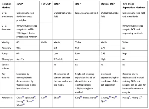
Table 2 Circulating Tumor Cells Were Selected Based on DEP
Isolation Method
eDEP TWDEP cDEP iDEP Optical DEP Two Steps
Separation Methods
CTC enrichment
Dielectrophoresis field-flow assist principles
Dielectrophoretic field
Dielectrophoretic field Dielectrophoretic
field
Dielectrophoretic field and microfluidic
CTC detection
Immunofluorescence analysis for ASPL- TFE3 type 1 fusion protein and vimentin
Immunofluorescence analysis, PCR and sequencing methods
Viability 0.9 Viable Viable Viable Viable Viable
Recovery 0.85 0.8 0.75 0.71 na
Purity 0.9 Low Low 0.92 High
Throughput 5mL/2h 2.2 mL/h na High na
Sample volume
na na na na
Key features
Separated by electrophoresis; demonstrated by fluorescence in situ hybridization
The absence of contact between the electrodes and the media
Single-cell trapping; separation based on the cell size and electrophoresis; a high-throughput method
Size-based separation; higher resolution of the cell separation
Requires CD45 depletion and manual staining; Different antigens can be used for immunofluorescence analysis
References Chan,79 Vykoukal80,
Huang,81 Moon,82
Cheng83
Cen84 Zhao85 Kang86, Bhattacharya87 Chuang,88 Wu89,
Qin90
Huang91, Huang C92
Cancer Management and Research downloaded from https://www.dovepress.com/ by 118.70.13.36 on 27-Aug-2020
Another enrichment technology based on immunomag-netic nanoparticles is the magimmunomag-netic cell separation system
(MACS).7,40 This method involves high-gradient magnetic
separation to capture CTC that are labeled using magnetic nanoparticles coupled with EpCAM antibodies. Although there are studies using MACS to capture CTC from
per-ipheral blood of metastatic cancer patients,41,42 the
techni-que is more suitable for tissue samples than for whole
blood samples.43 Biomimetic immunomagnetics (IMS)
were introduced recently to address the problem of poor MACS detection in whole blood. This technology formed magnetic corpuscles through camouflage magnetic nanoclusters with white blood cell membrane fragments. As a result, adsorption of non-specific white blood cells in peripheral blood samples was inhibited, reducing the con-tamination of CTC by background cells. Although this equipment has not been tested in a clinical setting, experi-ments revealed a near-complete lack of background cells,
as a major advantage.43,44
The use of immunomagnetic nanocarriers can achieve
high capture rates in cancers lacking EpCAM
expression.45 Moreover, the different nano-materials
avail-able for linings (eg, quartz,46 polymers,47 and gold48)
allow flexibility that improve CTC capture efficiency of EpCAM-expressing cancers and detection efficiency of low EpCAM-expressing malignant tumors. The recently developed nanoparticle HBCTC chip can be coated with surface markers including EpCAM, HER2, and EGFR, or a mixture of the three, achieving >90% capture efficiency
for low EpCAM-expressing cells (such as the MDA-MB -231 line). The thiol exchange reaction then releases cap-tured cells from nanoparticles, allowing for subsequent CTC molecular analyses, such as next-generation RNA
sequencing and cell culture.49,50
Methods that improve nanomaterial adhesion to cancer cells over normal cells include a nano-surface subjected to
reactive ion etching.51 The nanotube array has also been
developed as a sensitive biodetector for CTC detection. Compared with an anti-EpCAM-modified planar substrate, the anti-EpCAM-modified conducting polymer nanotube has a CTC capture rate of 70% and a cell survival rate of 97%.
Although nanotechnology has significantly improved capture efficiency, little data are available on how nanos-tructured substrates influence the interaction between tumor cells and normal peripheral blood cells. Moreover, the purity of CTC extracted via nanomaterials still needs improvement.
Micro-Device-Based Separation
Micro-devices are any tools created through micro- processing technology and chemical synthesis for specific molecular biological test functions in a very small area. Numerous studies show that micro-devices have short processing time, simple operation, and high separation
efficiency.7 Currently available micro-devices include the
microsingular chip,52,53 microfilter,54–56 micro EDAR
(sensitive decision aliquot ranking) cytometer,57 and
microGEDI (geometrically enhanced differential
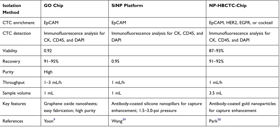
Table 3 Circulating Tumor Cells Were Selected Based on Chip
Isolation Method
GO Chip SiNP Platform NP-HBCTC-Chip
CTC enrichment EpCAM EpCAM EpCAM, HER2, EGFR, or cocktail
CTC detection Immunofluorescence analysis for
CK, CD45, and DAPI
Immunofluorescence analysis for CK, CD45, and DAPI
Immunofluorescence analysis for CK, CD45, and DAPI
Viability 0.92 87–93%
Recovery 91–95% 0.95 91–92%
Purity High
Throughput 1–3 mL/h 1 mL/h 1 mL/h
Sample volume 1 mL 1 mL 3.5 mL
Key features Graphene oxide nanosheets;
easy fabrication; high purity
Antibody-coated silicone nanopillars for capture enhancement; 1.5–3.0-psi pressure
Antibody-coated gold nanoparticles for capture enhancement
References Yoon9 Wang39 Park50
Cancer Management and Research downloaded from https://www.dovepress.com/ by 118.70.13.36 on 27-Aug-2020
T able 4 Cir culating T umor Cells W ere Selected Based on Micr o-De vices Micr opost Chip Micr ofilter Micr osinusoidal Chip Mag Sweeper Micr opillar Chip Micr ocrescent Chip Micr o wall Chip Micr oGEDI Chip Micr o vortex Chip
Nanopillar Chip
CTC enrichment
Antibody
based
Cell
size
Antibody/aptamer based Antibody based Antibody based
Cell size and deformability Cell size and deformability PSMA/HER2 (+ size selection)
Antibody based Antibody based
CTC detection
0.85 Isolation method 0.94 0.71 0.8 0.92 0.95 Viability ~99% na na na na na na Recovery > 60% 86–90% 90–97% > 50% na 80–100% > 95% Purity 9%, 50% na 1 51–100% na 83–89% na 62–74% 0.14 0.14 Thr oughput > 387~609 > 142 > 260 > 120 > 480 > 575 > 450 > 550 > 300~420 > 550
Sample volume
2.7 mL 7.5 mL 1 mL 9 mL 1 mL 1–3 mL 1 mL 1 mL 4 mL 1 mL References Stott 49
, Nagrath
61 Zheng, Lin, Zheng 54 − 56 Adams, 52 Dharmasiri 53 Nagrath 61 , T
alasaz, Ameri
4 , 9 Helzer 9 T an 6 , 9, T an 7 Mohamed 9 Kirby 58 Gleghorn 59 Stott 49 W ang 39
Cancer Management and Research downloaded from https://www.dovepress.com/ by 118.70.13.36 on 27-Aug-2020
immunocapture) chip (Table 4).58,59 Among these, the high-throughput microsampling unit is a microfluidic device that uses surface-immobilized monoclonal antibo-dies to separate CTC from the blood. The device releases
unlabeled live CTC via trypsin.52 Next, the CTC chip
promotes antibody attachment through geometrical arrangement of 78,000 anti-EpCAM antibody
microcol-umns with the surface area of ~970 mm2 60 and fluid
flow rate.61 Gedi combines positive enrichment (using
antibody-coated microcolumn) with hydrodynamic chro-matography to maximize the streamline distortion of its geometry, reducing non-specific leukocyte adhesion and
capturing high-purity CTC.59 In comparison to the
detec-tion sensitivity of the cell search system (96%),
microfilter,55 microGEDI chip,58 and microedar cell
analyzer57 have sensitivities of 75%, 94%, and 100%,
respectively, whereas these four methods have specificities of 16%, 22%, 0%, and 0%.
Micro-devices benefit CTC biological characterization as they maintain high efficiency and purity, while they also ensure CTC integrity and activity. We note that micro- devices using surface antigen recognition have higher purity than the cell search system, but require more processing time. However, neither method can recognize CTC without related surface antigens, meaning that they will lose key malignant cells. Additionally, size-based CTC-screening micro-devices have faster processing speed than cell search systems, but insufficient purity. Though including the deformability para-meters in size-based screening can further improve the purity, this increase is limited.
Microfluidics have also been combined with aptamers, single-stranded RNA, DNA, or peptide molecules with high specificity and affinity for specific target molecules to improve the purity. But these technologies have signifi-cant limitations. Screening requires a median of ~10 h. This longer processing time may affect the CTC survi-val, as well as the stability of cellular immunophenotype and genotyping. Thus, micro-devices may not be condu-cive for further CTC research.
Separation Based on Differences in Cell
Density
Separating cells based on buoyancy is one of the oldest isolation methods. Also called as density gradient
centri-fugation or equal density gradient centricentri-fugation.7
Buoyancy-based methods have the advantages of low cost, simple to operate, and independent of specific antigen
expression. Whole blood can be divided into three layers:
plasma layer, denuclearized cell layer, and CTC layer.62
However, these methods have low efficiency and purity of this method is low due to a large number of hematopoietic monocytes. The development of Oncoquick with a porous
membrane63 improved recovery rate to 87%.64 CTC purity
still remains very low using buoyancy-based techniques, barely reaching to 1%, even with the most advanced ver-sions. A new centrifugal microfluidics platform was devel-oped to address such problems. First, CTC were bound to anti-EpCAM-labeled beads to distinguish them from nor-mal blood cells. Next, CTC were precipitated and sepa-rated under the density gradient medium, thereby improving the separation purity. Despite an improvement through the use of EpCAM, the lack of a standardized CTC density range may still result in CTC being lost
during extraction.66
Another buoyancy-based device is the Accucycle sys-tem. Through a unique separation tube, blood is separated into three layers hematocrit, plasma, white blood cells, and platelets. The CTC are processed with row classification in the Cytefinder, an automatic scanning digital microscope and image analysis system. The average recovery rate of tumor cells is 90% using this method. Clinical trials reveal that after CTC enrichment, Accucycle can be used for
genome analysis of a single CTC.64
The main advantage of buoyancy-based CTC separa-tion is simplicity, resulting in viable cells that can be used for subsequent downstream experiments. These methods are also cost-effective and do not require much enrichment time. However, buoyancy-based methods are highly prone to contamination by other peripheral blood cells. In addi-tion, the size and density range of different CTC subtypes are unknown, meaning that some CTC may not be detected.
Separation Based on Cell Size
Cell-size-based methods mainly involve microfiltration and
therefore do not depend on surface antigen expression.67
ISET is the most suitable representative of these
techniques.68 In ISET, pre-fixed peripheral blood is filtered
using an 8 μm pore membrane. Next, CTC are immunos-tained for cell counting and morphological analysis. Enrichment of CTC was identified using ISET, mixed with epithelial and stromal phenotypes in the peripheral blood of
NSCLC patients for the first time.69 This outcome further
demonstrated the significance of EMT and the mechanism of metastasis. Another study using ISET detected CTC in 31
Cancer Management and Research downloaded from https://www.dovepress.com/ by 118.70.13.36 on 27-Aug-2020
patients with uveal melanoma by ISET, along with a single CTC or CTC cell cluster in 17 others. These results con-firmed that CTC predicts poor prognosis in patients with uveal melanoma. Although not all the CTC are larger than normal blood cells, ISET has a >90% capture rate. Another major advantage is that ISET can separate epithelial cells
without destroying cell morphology.68 However, it and the
other cell-size-based methods may miss the smaller tumor cells. More research using different cell lines and tumor types are required to determine the application scope and size threshold of ISET. In addition, the technique’s specifi-city and purity must be increased.
In vivo Flow Cytometry
In vivo flow cytometry (IVFC) allows the quantitative analysis of circulating cells. The technique involves laser scanning of surface blood vessels to detect cells via var-ious visualization methods, including fluorescence excita-tion and emission, photoacoustic effects, and photothermal
effects.70 The biggest advantage of IVFC is that the blood
collection and treatment are not required.71,72 At present,
fluorescent and photoacoustic IVFC are the most widely
used.73 Fluorescent IVFC is approximately 1.8 times more
sensitive than conventional whole blood flow cytometry.72
This technique helps to identify the effects of other treat-ments, through fluorescence IVFC, as in case where sor-afenib was revealed to reduce CTC count and lung
metastasis in patients with advanced liver cancer.74
Another study has used IVFC to observe the interactions between circulating breast cancer cells and dendritic cells. Mixed fluorescence and photoacoustic IFVC was also used
successfully to detect GFP + breast cancer cells75 and to
identify apoptotic CTC in vivo. After androgen depriva-tion therapy, patients with prostate cancer were monitored using IVFC, revealing that the therapy reduced CTC count in peripheral blood. One use of fluorescent IVFC in an orthotopic liver cancer transplantation model and subcuta-neous prostate cancer model demonstrated that the number of CTC clusters increased with primary tumor develop-ment. Thus, IVFC allowed researchers to confirm the key role of CTC clusters in tumor metastasis.
Nevertheless, IVFC has a clear disadvantage: its
detec-tion speed is 1 μL/min,76 while ~5 L/min blood passes
through human blood vessels. Therefore, IVFC is too slow for clinical needs. However, as seen in the examples provided, this technique is particularly useful for CTC detection and counting, which should be valuable for clinical monitoring and prognosis evaluation. The fact
that CTC in circulating blood are not purified means that IVFC is not conducive to the detection of CTC molecular typing and the study of biological characteristics.
Conclusions
The detection and isolation of CTC have progressed in recent years. Advancements have been mainly driven by interdisciplinary research involving cancer biology, oncol-ogy, cell physics, materials science, chemistry, nanotech-nology, and bioengineering. The diversification of CTC detection methods greatly improved the efficiency and reduced the cost. Unfortunately, every available detection method has major flaws. The cell search system involving surface antigen screening and enrichment possess the ease of automation and high specificity. Yet its clinical applica-tion has been hindered by low sensitivity and inability to detect all cancer types. In addition, most current CTC isolation methods require multi-step cell preparation, lead-ing to CTC loss or damage, thus adversely affectlead-ing detec-tion efficiency, accuracy, and sensitivity. The difficulty of extracting complete and viable CTC means further analy-sis is frequently impossible, thereby limiting the utility of these techniques for clinical decision-making.
The availability of high-throughput DNA sequencing means the whole genome and transcriptome of a single CTC can be obtained. These data allow increased attention to CTC biological characteristics, along with more exploration of genetic informatics and phenotypic func-tions (eg, through genome/transcriptome analysis and in vitro culture). These experiments require high molecular integrity and cell survival rate of isolated CTC, highlight-ing the importance of improvhighlight-ing CTC separation technol-ogy. Therefore, future research on CTC detection should first focus on acquiring basic knowledge on CTC, specifi-cally understanding the phenotypic changes accompanying the epithelial-stromal transformation. Insight into this pro-cess will help researchers improve the CTC separation efficiency, survival rate, and integrity while developing new methods. Additionally, new capture mechanisms should be explored to reduce blood cell contamination while improving survival and molecular integrity of pur-ified CTC. Finally, antigen markers should be combined to increase cell-capture specificity and solve the issue of CTC heterogeneity. We also require in-depth research to address the experimental protocols, such as determination of detec-tion timing and cut-off values, as well as standardizadetec-tion
of specific techniques. Finally, we recommend
Cancer Management and Research downloaded from https://www.dovepress.com/ by 118.70.13.36 on 27-Aug-2020
investigations to clarify the consistency of CTC biological characteristics.
Although there are problems to be solved, CTC analy-sis is a simple and feasible liquid biopsy technology that has gained serious attention and has achieved major suc-cess. With further development, CTC-based diagnosis should have considerable value in individualized treatment of patients with cancer.
Acknowledgments
This study was funded by Hubei natural science founda-tion project (2011CDB330, 2014CFB312) and Hubei pro-vincial health department research project (JX4B52).
Disclosure
Xinhua Xu reports a licensed patent (ZL201920055267.9): A special petri dish for magnetic trap. The authors report no other potential conflicts of interest in this work.
References
1. Strilic B, Offermanns S. Intravascular survival and extravasation of tumor cells. Cancer Cell. 2017;32(3):282–293. doi:10.1016/j. ccell.2017.07.001
2. Sharma S, Zhuang R, Long M, et al. Circulating tumor cell isolation, culture, and downstream molecular analysis. Biotechnol Adv. 2018;36 (4):1063–1078. doi:10.1016/j.biotechadv.2018.03.007
3. Paget S. Paget,Stephen paper reproduced from the Lancet, 1889. Cancer Metastasis Rev. 1989;8(2):98–101.
4. Wiley HE, Gonzalez EB, Maki W, Wu MT, Hwang ST. Expression of CC chemokine receptor-7 and regional lymph node metastasis of B16 murine melanoma. J Natl Cancer Inst. 2001;93(21):1638–1643. doi:10.1093/jnci/93.21.1638
5. Ming Y, Li Y, Xing H, et al. Circulating tumor cells: from theory to nanotechnology-based detection. Front Pharmacol. 2017;8. 6. Esmaeilsabzali H, Beischlag TV, Cox ME, Parameswaran AM,
Park EJ. Detection and isolation of circulating tumor cells: principles and methods. Biotechnol Adv. 2013;31(7):1063–1084. doi:10.1016/j. biotechadv.2013.08.016
7. Ferreira MM, Romani VC, Jeffrey SS. Circulating tumor cell technologies. Mol Oncol. 2016;10(3):374–394. doi:10.1016/j. molonc.2016.01.007
8. Bunger S, Zimmermann M, Habermann JK. Diversity of assessing circulating tumor cells (CTCs) emphasizes need for standardization: a CTC Guide to design and report trials. Cancer Metastasis Rev.
2015;34(3):527–545. doi:10.1007/s10555-015-9582-0
9. Krebs MG, Hou J-M, Ward TH, Blackhall FH, Dive C. Circulating tumour cells: their utility in cancer management and predicting outcomes. Ther Adv Med Oncol. 2010;2(6):351–365. doi:10.1177/ 1758834010378414
10. Wicha MS, Hayes DF. Circulating tumor cells: not all detected cells are bad and not all bad cells are detected. J Clin Oncol. 2011;29 (12):1508–1511. doi:10.1200/JCO.2010.34.0026
11. Allard WJ, Miller MC, et al. Tumor cells circulate in the peripheral blood of all major carcinomas but not in healthy subjects or patients with nonmalignant diseases. Clin Cancer Res. 2004;10:6897–6904. doi:10.1158/1078-0432.CCR-04-0378
12. Tibbe AGJ, Miller MC, Terstappen LWMM. Statistical considera-tions for enumeration of circulating tumor cells. Cytometry Part A.
2007;71A(3):154–162. doi:10.1002/cyto.a.20369
13. Hvichia GE, Parveen Z, Wagner C, et al. A novel microfluidic plat-form for size and deplat-formability based separation and the subsequent molecular characterization of viable circulating tumor cells. Int J Cancer. 2016;138(12):2894–2904. doi:10.1002/ijc.30007 14. Yang L, Lang JC, Balasubramanian P, et al. Optimization of an
enrichment process for circulating tumor cells from the blood of head and neck cancer patients through depletion of normal cells. Biotechnol Bioeng. 2009;102(2):521–534. doi:10.1002/bit.22066 15. Lara O, Tong XD, Zborowski M, Chalmers JJ. Enrichment of rare
cancer cells through depletion of normal cells using density and flow-through, immunomagnetic cell separation. Exp Hematol.
2004;32(10):891–904. doi:10.1016/j.exphem.2004.07.007
16. Baccelli I, Schneeweiss A, Riethdorf S, et al. Identification of a population of blood circulating tumor cells from breast cancer patients that initiates metastasis in a xenograft assay. Nat Biotechnol. 2013;31(6):539–U143. doi:10.1038/nbt.2576
17. Hayes DF. Circulating tumor cells at each follow-up time point during therapy of metastatic breast cancer patients predict progression-free and overall survival. Clin Cancer Res. 2006;12 (14):4218–4224. doi:10.1158/1078-0432.CCR-05-2821
18. Danila DC, Heller G, Gignac GA, et al. Circulating tumor cell number and prognosis in progressive castration-resistant prostate cancer. Clin Cancer Res. 2007;13(23):7053–7058. doi:10.1158/ 1078-0432.CCR-07-1506
19. Cohen SJ, Punt CJA, Iannotti N, et al. Relationship of circulating tumor cells to tumor response, progression-free survival, and overall survival in patients with metastatic colorectal cancer. J Clin Oncol.
2008;26(19):3213–3221. doi:10.1200/JCO.2007.15.8923
20. Moon DH, Lindsay DP, Hong S, Wang AZ. Clinical indications for, and the future of, circulating tumor cells. Adv Drug Deliv Rev.
2018;125:143–150. doi:10.1016/j.addr.2018.04.002
21. Mostert B, Kraan J, Bolt-de Vries J, et al. Detection of circulating tumor cells in breast cancer may improve through enrichment with anti-CD146. Breast Cancer Res Treat. 2010;127(1):33–41. doi:10.1007/s10549-010-0879-y
22. Onstenk W, Kraan J, Mostert B, et al. Improved circulating tumor cell detection by a combined EpCAM and MCAM cell search enrichment approach in patients with breast cancer undergoing neoadjuvant chemotherapy. Mol Cancer Ther. 2015;14(3):821–827. doi:10.1158/ 1535-7163.MCT-14-0653
23. Zhang T, Boominathan R, Foulk B, et al. Development of a novel c-MET-Based CTC detection platform. Mol Cancer Res. 2016;14 (6):539–547. doi:10.1158/1541-7786.MCR-16-0011
24. Satelli A, Brownlee Z, Mitra A, Meng QH, Li S. Circulating tumor cell enumeration with a combination of epithelial cell adhesion molecule and cell-surface vimentin-based methods for monitoring breast cancer therapeutic response. Clin Chem. 2015;61(1):259–266. doi:10.1373/clinchem.2014.228122
25. Lu -N-N, Xie M, Wang J, et al. Biotin-triggered decomposable immunomagnetic beads for capture and release of circulating tumor cells. ACS Appl Mater Interfaces. 2015;7(16):8817–8826. doi:10. 1021/acsami.5b01397
26. Coumans FA, Ligthart ST, Uhr JW, Terstappen LW. Challenges in the enumeration and phenotyping of CTC. Clin Cancer Res. 2012;18 (20):5711–5718. doi:10.1158/1078-0432.CCR-12-1585
27. Roh JI, Sung YH, Lee HW. Clinical implications of antitelomeric drugs with respect to the nontelomeric functions of telomerase in cancer. Onco Targets Ther. 2013;6:1161–1166. doi:10.2147/OTT. S50918
28. Macarthur KM, Kao GD, Chandrasekaran S, et al. Detection of brain tumor cells in the peripheral blood by a telomerase promoter-based assay. Cancer Res. 2014;74(8):2152–2159. doi:10.1158/0008-5472. CAN-13-0813
Cancer Management and Research downloaded from https://www.dovepress.com/ by 118.70.13.36 on 27-Aug-2020
29. Fizazi K, Morat L, Chauveinc L, et al. High detection rate of circu-lating tumor cells in blood of patients with prostate cancer using telomerase activity. Ann Oncol. 2007;18(3):518–521. doi:10.1093/ annonc/mdl419
30. Sapi E, Okpokwasili NI, Rutherford T. Detection of telomerase-positive circulating epithelial cells in ovarian cancer patients. Cancer Detect Prev. 2002;26(2):158–167. doi:10.1016/S0361-090X(02)00034-X 31. Soria JC, Gauthier LR, Raymond E, et al. Molecular detection of
telomerase-positive circulating epithelial cells in metastatic breast cancer patients. Clin Cancer Res. 1999;5(5):971–975.
32. Gauthier LR, Granotier C, Soria JC, et al. Detection of circulating carcinoma cells by telomerase activity. Br J Cancer. 2001;84 (5):631–635. doi:10.1054/bjoc.2000.1662
33. Hong B, Zu Y. Detecting circulating tumor cells: current challenges and new trends. Theranostics. 2013;3(6):377–394. doi:10.7150/thno.5195 34. Dabighi A, Toghraie D. A new microfluidic device for separating
circulating tumor cells based on their physical properties by using electrophoresis and dielectrophoresis forces within an electrical field. Comput Methods Programs Biomed. 2019;185:105147. doi:10.1016/ j.cmpb.2019.105147
35. Gascoyne PR, Shim S, Noshari J, Becker FF, Stemke-Hale K. Correlations between the dielectric properties and exterior morphol-ogy of cells revealed by dielectrophoretic field-flow fractionation. Electrophoresis. 2013;34(7):1042–1050. doi:10.1002/elps.201200496 36. Wang XB, Huang Y, Gascoyne PRC, Becker FF, Holzel R, Pethig R.
Changes in friend murine erythroleukemia cell-membranes during induced-differentiation determined by electrorotation. Biochimica Et Biophysica Acta-Biomembranes. 1994;1193(2):330–344. doi:10.1016/ 0005-2736(94)90170-8
37. Kim SH, Ito H, Kozuka M, Hirai M, Fujii T. Localization of low-abundant cancer cells in a sharply expanded microfluidic step-channel using dielectrophoresis. Biomicrofluidics. 2017;11:5. doi:10.1063/1.4998756
38. Cheng IF, Huang W-L, Chen T-Y, Liu C-W, Lin Y-D, Su W-C. Antibody-free isolation of rare cancer cells from blood based on 3D lateral dielectrophoresis. Lab Chip. 2015;15(14):2950–2959. doi:10.1039/C5LC00120J
39. Wang S, Liu K, Liu J, et al. Highly efficient capture of circulating tumor cells by using nanostructured silicon substrates with integrated chaotic micromixers. Angewandte Chemie-Int Ed. 2011;50 (13):3084–3088. doi:10.1002/anie.201005853
40. Miltenyi S, Muller W, Weichel W, Radbruch A. High-gradient mag-netic cell-separation with MACS. Cytometry. 1990;11(2):231–238. doi:10.1002/cyto.990110203
41. Giordano A, Gao H, Anfossi S, et al. Epithelial-mesenchymal transi-tion and stem cell markers in patients with HER2-positive metastatic breast cancer. Mol Cancer Ther. 2012;11(11):2526–2534. doi:10.1158/1535-7163.MCT-12-0460
42. Pluim D, Devriese LA, Beijnen JH, Schellens JHM. Validation of a multiparameter flow cytometry method for the determination of phosphorylated extracellular-signal-regulated kinase and DNA in cir-culating tumor cells. Cytometry Part A. 2012;81A(8):664–671. doi:10.1002/cyto.a.22049
43. Xiong K, Wei W, Jin Y, et al. Biomimetic immuno-magnetosomes for high-performance enrichment of circulating tumor cells. Adv Mater.
2016;28(36):7929–7935. doi:10.1002/adma.201601643
44. Rodriguez PL, Harada T, Christian DA, Pantano DA, Tsai RK, Discher DE. Minimal “Self” peptides that inhibit phagocytic clear-ance and enhclear-ance delivery of nanoparticles. Science. 2013;339 (6122):971–975. doi:10.1126/science.1229568
45. Wu C-H, Huang -Y-Y, Chen P, et al. Versatile immunomagnetic nanocarrier platform for capturing cancer cells. ACS Nano. 2013;7 (10):8816–8823. doi:10.1021/nn403281e
46. Lee S-K, Kim G-S, Wu Y, et al. Nanowire substrate-based laser scanning cytometry for quantitation of circulating tumor cells. Nano Lett. 2012;12(6):2697–2704. doi:10.1021/nl2041707
47. Hong WY, Jeon SH, Lee ES, Cho Y. An integrated multifunctional platform based on biotin-doped conducting polymer nanowires for cell capture, release, and electrochemical sensing. Biomaterials.
2014;35(36):9573–9580. doi:10.1016/j.biomaterials.2014.08.027 48. Zhai -T-T, Ye D, Zhang Q-W, Wu Z-Q, Xia X-H. Highly efficient
capture and electrochemical release of circulating tumor cells by using aptamers modified gold nanowire arrays. ACS Appl Mater Interfaces. 2017;9(40):34706–34714. doi:10.1021/acsami.7b11107 49. Stott SL, Hsu C-H, Tsukrov DI, et al. Isolation of circulating tumor
cells using a microvortex-generating herringbone-chip. Proc Natl Acad Sci U S A. 2010;107(43):18392–18397. doi:10.1073/ pnas.1012539107
50. Park MH, Reategui E, Li W, et al. Enhanced isolation and release of circulating tumor cells using nanoparticle binding and ligand exchange in a microfluidic chip. J Am Chem Soc. 2017;139 (7):2741–2749. doi:10.1021/jacs.6b12236
51. Chen W, Weng S, Zhang F, et al. Nanoroughened surfaces for efficient capture of circulating tumor cells without using capture antibodies. ACS Nano. 2013;7(1):566–575. doi:10.1021/nn304719q 52. Adams AA, Okagbare PI, Feng J, et al. Highly efficient circulating
tumor cell isolation from whole blood and label-free enumeration using polymer-based microfluidics with an integrated conductivity sensor. J Am Chem Soc. 2008;130(27):8633–8641. doi:10.1021/ ja8015022
53. Dharmasiri U, Balamurugan S, Adams AA, Okagbare PI, Obubuafo A, Soper SA. Highly efficient capture and enumeration of low abundance prostate cancer cells using prostate-specific membrane antigen aptamers immobilized to a polymeric microfluidic device. Electrophoresis.
2009;30(18):3289–3300. doi:10.1002/elps.200900141
54. Zheng S, Lin H, Liu JQ, et al. Membrane microfilter device for selective capture, electrolysis and genomic analysis of human circu-lating tumor cells. J Chromatogr A. 2007;1162(2):154–161. doi:10.1016/j.chroma.2007.05.064
55. Lin HK, Zheng S, Williams AJ, et al. Portable filter-based micro-device for detection and characterization of circulating tumor cells. Clin Cancer Res. 2010;16(20):5011–5018. doi:10.1158/1078-0432. CCR-10-1105
56. Zheng S, Lin HK, Lu B, et al. 3D microfilter device for viable circulating tumor cell (CTC) enrichment from blood. Biomed Microdevices. 2011;13(1):203–213. doi:10.1007/s10544-010-9485-3 57. Schiro PG, Zhao M, Kuo JS, Koehler KM, Sabath DE, Chiu DT.
Sensitive and high-throughput isolation of rare cells from peripheral blood with ensemble-decision aliquot ranking. Angew Chem Int Ed Engl. 2012;51(19):4618–4622. doi:10.1002/anie.201108695 58. Kirby BJ, Jodari M, Loftus MS, et al. Functional characterization of
circulating tumor cells with a prostate-cancer-specific microfluidic device. PLoS One. 2012;7(4):e35976. doi:10.1371/journal. pone.0035976
59. Gleghorn JP, Pratt ED, Denning D, et al. Capture of circulating tumor cells from whole blood of prostate cancer patients using geometrically enhanced differential immunocapture (GEDI) and a prostate-specific antibody. Lab Chip. 2010;10(1):27–29. doi:10.1039/B917959C 60. Ma Y, Hao S, Wang S, et al. A combinatory strategy for detection of
live CTCs using microfiltration and a new telomerase-selective adenovirus. Mol Cancer Ther. 2015;14(3):835–843. doi:10.1158/ 1535-7163.MCT-14-0693
61. Nagrath S, Sequist LV, Maheswaran S, et al. Isolation of rare circu-lating tumour cells in cancer patients by microchip technology. Nature. 2007;450(7173):1235–U1210. doi:10.1038/nature06385 62. Diamond E, Lee GY, Akhtar NH, et al. Isolation and characterization
of circulating tumor cells in prostate cancer. Front Oncol.
2012;2:131. doi:10.3389/fonc.2012.00131
63. Chung J, Issadore D, Ullal A, Lee K, Weissleder R, Lee H. Rare cell isolation and profiling on a hybrid magnetic/size-sorting chip. Biomicrofluidics. 2013;7(5):54107. doi:10.1063/1.4821923
Cancer Management and Research downloaded from https://www.dovepress.com/ by 118.70.13.36 on 27-Aug-2020
64. Campton DE, Ramirez AB, Nordberg JJ, et al. High-recovery visual identification and single-cell retrieval of circulating tumor cells for genomic analysis using a dual-technology platform integrated with automated immunofluorescence staining. BMC Cancer. 2015;15. 65. Park JM, Kim MS, Moon HS, et al. Fully automated circulating tumor
cell isolation platform with large-volume capacity based on lab-on- a-disc. Anal Chem. 2014;86(8):3735–3742. doi:10.1021/ac403456t 66. Rikkert LG, van der Pol E, van Leeuwen TG, Nieuwland R,
Coumans FAW. Centrifugation affects the purity of liquid biopsy-based tumor biomarkers. Cytometry A. 2018;93(12):1207–1212. doi:10.1002/ cyto.a.23641
67. Hosokawa M, Hayata T, Fukuda Y, et al. Size-selective microcavity array for rapid and efficient detection of circulating tumor cells. Anal Chem. 2010;82(15):6629–6635. doi:10.1021/ac101222x
68. Vona G, Sabile A, Louha M, et al. Isolation by size of epithelial tumor cells - A new method for the immunomorphological and molecular characterization of circulating tumor cells. Am J Pathol.
2000;156(1):57–63. doi:10.1016/S0002-9440(10)64706-2
69. Lederlin M, Tredaniel J, Priollet P. [Why screen for lung cancer in patients with arterial disease?]. J Mal Vasc. 2015;40(6):359–364. French. doi:10.1016/j.jmv.2015.07.001
70. Hartmann C, Patil R, Lin CP, Niedre M. Fluorescence detection, enumeration and characterization of single circulating cells in vivo: technology, applications and future prospects. Phys Med Biol.
2017;63(1):01TR01. doi:10.1088/1361-6560/aa98f9
71. Novak J, Georgakoudi I, Wei X, Prossin A, Lin CP. In vivo flow cytometer for real-time detection and quantification of circulating cells. Opt Lett. 2004;29(1):77–79. doi:10.1364/OL.29.000077 72. Fan ZC, Yan J, Liu GD, et al. Real-time monitoring of rare
circulat-ing hepatocellular carcinoma cells in an orthotopic model by in vivo flow cytometry assesses resection on metastasis. Cancer Res. 2012;72 (10):2683–2691. doi:10.1158/0008-5472.CAN-11-3733
73. Suo Y, Gu Z, Wei X. Advances of in vivo flow cytometry on cancer studies. Cytometry A. 2019.
74. Yan J, Fan Z, Wu X, et al. Circulating tumor cells are correlated with disease progression and treatment response in an orthotopic hepato-cellular carcinoma model. Cytometry A. 2015;87(11):1020–1028. doi:10.1002/cyto.a.22782
75. Nolan J, Nedosekin DA, Galanzha EI, Zharov VP. Detection of apoptotic circulating tumor cells using in vivo fluorescence flow cytometry. Cytometry Part A. 2019;95A(6):664–671. doi:10.1002/ cyto.a.23642
76. Pera V, Tan X, Runnels J, Sardesai N, Lin CP, Niedre M. Diffuse fluorescence fiber probe for in vivo detection of circulating cells. J Biomed Opt. 2017;22:3. doi:10.1117/1.JBO.22.3.037004
77. Talasaz AH, Powell AA, Huber DE, et al. Isolating highly enriched populations of circulating epithelial cells and other rare cells from blood using a magnetic sweeper device. Proc Natl Acad Sci U S A.
2009;106(10):3970–3975. doi:10.1073/pnas.0813188106
78. Dharmasiri U, Njoroge SK, Witek MA, et al. High-throughput selection, enumeration, electrokinetic manipulation, and molecular profiling of low-abundance circulating tumor cells using a microfluidic system. Anal Chem. 2011;83(6):2301–2309. doi:10.1021/ac103172y
79. Chan KL, Gascoyne PRC, Becker FF, Pethig R. Electrorotation of liposomes: verification of dielectric multi-shell model for cells. Biochimica Et Biophysica Acta-Lipids Lipid Metabol. 1997;1349 (2):182–196. doi:10.1016/S0005-2760(97)00092-1
80. Vykoukal DM, Gascoyne PRC, Vykoukal J. Dielectric characteriza-tion of complete mononuclear and polymorphonuclear blood cell subpopulations for label-free discrimination. Integrative Biol.
2009;1(7):477–484. doi:10.1039/b906137a
81. Huang Y, Yang J, Wang XB, Becker FF, Gascoyne PRC. The removal of human breast cancer cells from hematopoietic CD34 (+) stem cells by dielectrophoretic field-flow-fractionation. J Hematother Stem Cell Res. 1999;8(5):481–490. doi:10.1089/ 152581699319939
82. Moon H-S, Kwon K, Kim S-I, et al. Continuous separation of breast cancer cells from blood samples using multi-orifice flow fractionation (MOFF) and dielectrophoresis (DEP). Lab Chip. 2011;11 (6):1118–1125. doi:10.1039/c0lc00345j
83. Cheng IF, Chen T-Y, Lin Y-D, et al. A Novel Dielectrophoresis-Based Microfluidic Chip for Antibody-Free Isolation of Circulating Tumor Cells from Blood. 2015.
84. Cen EG, Dalton C, Li YL, Adamia S, Pilarski LM, Kaler K. A combined dielectrophoresis, traveling wave dielectrophoresis and electrorotation microchip for the manipulation and characterization of human malignant cells. J Microbiol Methods. 2004;58(3):387–401. doi:10.1016/j.mimet.2004.05.002
85. Zhao K, Peng R, Li D. Separation of nanoparticles by a nano-orifice based DC-dielectrophoresis method in a pressure-driven flow. Nanoscale. 2016;8(45):18945–18955. doi:10.1039/C6NR06952E 86. Kang KH, Kang YJ, Xuan XC, Li DQ. Continuous separation of
microparticles by size with direct current-dielectrophoresis. Electrophoresis. 2006;27(3):694–702. doi:10.1002/elps.200500 558
87. Bhattacharya S, Chao T-C, Ariyasinghe N, et al. Selective trapping of single mammalian breast cancer cells by insulator-based dielectrophoresis. Anal Bioanal Chem. 2014;406(7):1855–1865. doi:10.1007/s00216-013-7598-2
88. Chuang C-H, Huang Y-W, Wu Y-T. Dielectrophoretic chip with multilayer electrodes and micro-cavity array for trapping and pro-grammably releasing single cells. Biomed Microdevices. 2012;14 (2):271–278. doi:10.1007/s10544-011-9603-x
89. Wu L, Yung L-YL, Lim K-M. Dielectrophoretic capture voltage spectrum for measurement of dielectric properties and separation of cancer cells. Biomicrofluidics. 2012;6:1. doi:10.1063/ 1.3690470
90. Qin Y, Wu L, Schneider T, et al. A Self-Digitization Dielectrophoretic (SD-DEP) chip for high-efficiency single-cell cap-ture, on-demand compartmentalization, and downstream nucleic acid analysis. Angewandte Chemie-Int Ed. 2018;57(35):11378–11383. doi:10.1002/anie.201807314
91. Huang C, Liu H, Bander NH, Kirby BJ. Enrichment of prostate cancer cells from blood cells with a hybrid dielectrophoresis and immunocapture microfluidic system. Biomed Microdevices. 2013;15 (6):941–948. doi:10.1007/s10544-013-9784-6
92. Huang C, Santana SM, Liu H, Bander NH, Hawkins BG, Kirby BJ. Characterization of a hybrid dielectrophoresis and immunocapture microfluidic system for cancer cell capture. Electrophoresis.
2013;34(20–21):2970–2979. doi:10.1002/elps.201370191
Cancer Management and Research downloaded from https://www.dovepress.com/ by 118.70.13.36 on 27-Aug-2020
Cancer Management and Research
Dove
press
Publish your work in this journal
Cancer Management and Research is an international, peer-reviewed open access journal focusing on cancer research and the optimal use of preventative and integrated treatment interventions to achieve improved outcomes, enhanced survival and quality of life for the cancer patient.
The manuscript management system is completely online and includes a very quick and fair peer-review system, which is all easy to use. Visit http://www.dovepress.com/testimonials.php to read real quotes from published authors.
Submit your manuscript here: https://www.dovepress.com/cancer-management-and-research-journal
Cancer Management and Research downloaded from https://www.dovepress.com/ by 118.70.13.36 on 27-Aug-2020