서 론
갑상선암은 국내 전체암의 2위, 여성에서 첫 번째로 많이 발 생하는 암으로 [1] 그 중에서도 유두상암이 95.6%로 가장 많 은 빈도를 차지한다 [2]. 갑상선암의 수술 전 병기결정은 외과 의가 수술범위를 결정하는 데 있어서 많은 도움을 줄 수 있다. 가장 흔히 사용되는 갑상선암의 병기분류는 대부분의 암과 마 찬가지로 TNM (TNM병기분류로 T는 종양의 크기 및 범위, N은 임파선전이유무, M은 원격전이유무) 병기분류이다. 현재 2010년에 7th Edition TNM staging이 발표된 상태로, 따라서 본 종설에서는 American Joint Committee on Cancer (AJCC)가 제시한 제7차 개정판에 따른 갑상선의 TNM 병기 분류에 대해 살펴보고자 한다 (Table 1).1. T staging
T staging은 기본적으로 갑상선 암의 크기와 주변조직의 침 범 정도에 따라 병기가 달라진다. 따라서 정확한 갑상선 암의 크기 측정과 갑상선 피막을 비롯한 주변의 연조직 (soft tissue), 근육, 기도, 식도, 신경 등의 침범여부를 판단하여 T 병기를 결정하는 것이 중요하고 이에 따라 수술 방법과 범위가 달라지게 된다. 현재 나와 있는 여러 지침들에 의하면 갑상선암의 크기가 1 cm보다 작고, 개수가 한 개, 갑상선 내부에 있고 주변 림프절의 전이가 의심되지 않는 저위험군 환자의 경우에는 한쪽엽절제 술 (lobectomy)를 권고하고 있고 (Fig. 1), 반대로 암의 크기가 1 cm 보다 크거나, 다병소성 또는 양측성, 피막 바깥으로의 침 범이 있는 경우, 림프절 전이나 원격전이가 있는 경우, 가족력 이나 두경부에 방사선 조사력의 과거력이 있는 경우, 45세 이 상인 경우에서는 재발확률이 높다고 알려져 있기 때문에 [3, 4] 갑상선 전절제술 (total thyroidectomy)이 권고된다 [5-8]. 또한 주변 띠근육 (strap muscle)이나 흉쇄유돌근 (sternocleidomastoid muscle)을 침범한 경우 침범된 근육을 포함하는 전체 절제가 필요하다. 따라서 수술 전 영상검사를 통해서 위에 언급한 암의 크기, 다병소성, 주변조직 침범여부 등의 다양한 항목들을 파악해야 정확한 병기결정이 이루어져 서 수술범위가 결정되고 재발 등의 예후를 예측할 수 있다.갑상선암의 병기결정
김자영∙김수진∙김은경
곽진영
연세의대 영상의학교실J Korean Soc Ultrasound Med 2011; 30: 113-122
Received January 24, 2011; Revised March 21, 2011; Accepted April 21, 2011.
Address for reprints :
Jin Young Kwak, MD, Department of Radiology, Yonsei University College of Medicine, 50 Yonsei-ro, Seodaemoon-gu, Seoul 120-752, Korea.
Tel. 82-2-2228-7400 Fax. 82-2-393-3035 E-mail: [email protected]
Thyroid Cancer Staging
Ja Young Kim, MD, Soo Jin Kim, MD, Eun-Kyung Kim, MD, Jin Young Kwak, MD
Department of Radiology, Yonsei University College of MedicineThe current prevalence of thyroid cancer in women is high. Likewise, other cancers and thyroid cancer have been based on the TNM classification system. Staging of thy-roid cancer has an important role in determining the extent of surgical excision and lymph node dissection, planning the adjuvant therapy after surgery and predicting the recurrence rate and the prognosis of patients. Ultrasonography is the basic imaging modality to identify the tumor size and the extent of lymph node metastasis. More recently, computed tomography, magnetic resonance imaging and positron emission tomography provide additional help for the staging of thyroid cancer. So, this article describes the 7th edition of the TNM staging of thyroid cancer, as proposed by the American Joint Committee on Cancer, and the details of radiologic evaluation of the T, N and M stages.
1) 초음파
초음파는 수술 전 갑상선검사에 있어 첫 번째로 행해지는 검 사방법이며 가장 손쉽고 비침습적이며 방사선 노출 없이 할 수 있는 검사이며 최근 기계의 발전으로 고해상도의 영상을 얻을 수 있기 때문에 중요도가 높은 검사이다 [9, 10]. 초음파가 전 산화단층촬영술 (CT)과 자기공명영상 (MRI)에 비해 뛰어난 점들은 병변의 정확한 위치와 크기 측정이 더 용이하고 다병소 성, 미세한 피막바깥 침범, 미세석회화 존재유무를 더 잘 볼 수 있고 또한 3 mm 이하의 작은 병변도 잘 발견할 수 있다는 데 있다 [11-16]. 특히 유두암종은 다병소성이 26~31%정도로 다른 암종에 비해 높다고 알려져 있기 때문에 수술 전 다병소성 여부를 초 음파로 확인하는 것이 중요하다 [17]. 미국갑상선 협회진료권고안 (American thyroid association management guideline) [5]에서도 모든 환자에 대해 수술 전 암이 존재하는 갑상선 엽 의 반대쪽 갑상선엽과 림프절을 초음파로 살펴보는 것을 권고 하고 있는데 다병소성이 확인되면 수술범위가 달라지기 때문 이다. 이러한 다병소성, 양측성을 진단하는 데 있어 초음파가 CT보다 우월하다는 연구결과가 최근 최 등의 연구에서 보고되 고 있으며 [12], Shetty 등[15]의 연구에서도 초음파에 비해 CT가 결절을 발견하는 민감도가 낮고 악성을 의심할 만한 특 징적인 CT소견이 없다는 한계점을 지적하였다. 또한 King 등 [13]의 연구에서는 원발성 병변, 다병소성, 림프절 전이를 찾 는데 MRI보다 초음파가 성적이 우수하다고 보고하였다. 갑상선 피막 바깥으로의 침습여부를 보는 데에도 초음파는 좋은 영상기법이다 (Fig. 2). Shimamoto 등[14]의 연구에서는 Table 1. TNM Staging in the 7th Edition of the American Joint Committee on Cancer (2010)
Primary tumor (T)
All categories may be subdivided: (s) solitary tumor and (m) multifocal tumor (the largest determines the classification). TX Primary tumor cannot be assessed
T0 No evidence of primary tumor
T1 Tumor 2 cm or less in greatest dimension limited to the thyroid T1a Tumor 1 cm or less, limited to the thyroid
T1b Tumor more than 1 cm but not more than 2 cm in greatest dimension, limited to the thyroid T2 Tumor more than 2 cm but not more than 4 cm in greatest dimension, limited to the thyroid
T3 Tumor more than 4 cm in greatest dimension limited to the thyroid, or any tumor with minimal extrathyroid extension (e.g., extension to sternothyroid muscle or perithyroid soft tissues)
T4a Moderately advanced disease.
Tumor of any size extending beyond the thyroid capsule to invade subcutaneous soft tissues, larynx, trachea, esophagus, or recurrent laryngeal nerve
T4b Very advanced disease.
Tumor invades prevertebral fascia or encases carotid artery or mediastinal vessels All anaplastic carcinomas are considered T4 tumors
T4a Intrathyroidal anaplastic carcinoma
T4b Anaplastic carcinoma with gross extrathyroid extension Regional lymph nodes (N)
Regional lymph nodes are the central compartment, lateral cervical, and upper mediastinal lymph nodes NX Regional lymph nodes cannot be assessed
N0 No regional lymph node metastasis N1 Regional lymph node metastasis
N1a Metastasis to Level VI (pretracheal, paratracheal, and prelaryngeal/Delphian lymph nodes)
N1b Metastasis to unilateral, bilateral, or contralateral cervical (Levels I, II, III, IV or V) or retropharyngeal or superior mediastinal lymph nodes (Level VII)
Distant metastasis (M)
M0 No distant metastasis (no pathologic M0; use clinical M to complete stage group) M1 Distant metastasis
갑상선 바깥으로의 침범여부를 평가했을 때 초음파가 민감도 62.9%, 특이도 97.6%의 결과를 보였고, 미세유두암을 대상으 로 한 곽 등의 연구에서도 병변의 25% 이상이 주변의 갑성선 피막과 닿아있는 경우를 침범이 있다고 간주했을 때 민감도 65.2%, 양성예측도 70.7%의 결과를 보였다 [11]. 하지만 초음파 영상은 기도, 식도침범 여부를 보기 어렵고 크기가 큰 갑상선 암에 평가에 한계가 있다는 단점이 있어 이 러한 경우에는 추가적인 CT나 MRI검사가 도움을 줄 수 있다.
2) CT, MRI
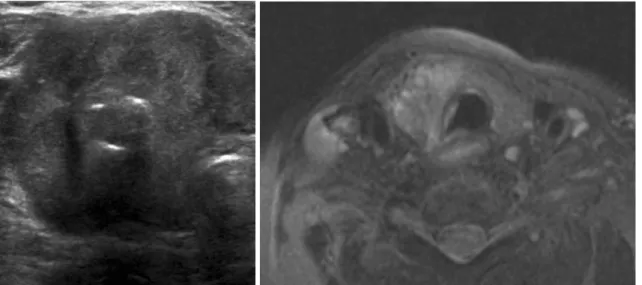
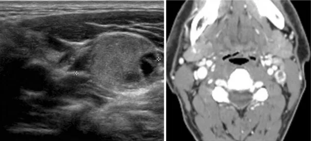
CT나 MRI는 기도, 식도 등의 주변장기로의 침범이 의심되 는 경우, 경동맥의 침범여부, 초음파의 해상도를 넘어서는 크 기가 큰 갑상선 암의 평가에 있어서 특히 유용하다 [13, 18]. 특히 MRI는 조직의 대조도가 높은 검사방법으로 Wang 등 [19]의 연구에 따르면 갑상선 암의 식도 침범을 판단하는 데에 MRI 정확도가 91%정도로 높았다고 보고하고 있으며 기도침 범을 판단하는 데에 정확도가 90%에 이른다고 보고하였다 [20]. 또한 갑상선 암의 수술병기 결정에 있어 초음파와 MRI 를 직접 비교한 연구에서도 기도침범 유무의 진단에서 MRI가 더 우수하다고 보고하고 있다 [13] (Fig. 3). 그러나 아직까지 CT, MRI는 T 병기결정에서 추가적인 도움 을 주는 영상기법이며 모든 환자에서 시행하는 것은 권고하지 않는다 [5].3) TNM staging 7th edition의 변화
이번 7차 개정에 따르면 갑상선 암의 T병기분류에서 바뀐 것은 T1 병기이다. 이전에 하나의 T1 병기가 7차에서는 좀더 세분화되어 T1a와 T1b로 분류된다. 그 기준은 종양의 크기이 며 1 cm을 기준으로 1 cm이하인 경우 T1a, 1 cm보다 크고 2 cm보다 작은 경우를 T1b로 정하고 있다. 또한 T 범주를 단발 성 병변과 다발성 병변으로 세분화하였으며 이전에 쓰던 절제 가능한 (respectable), 절제불가능한 (unresectable)을 의미하 였던 T4a, T4b의 용어를“moderately advanced”와“very advanced.”로 바꾸었다 [21]. 이처럼 T1병기가 세분화된 이 유는 갑상선암의 크기가 1 cm보다 클 경우 갑상선 전절제술이 권고되고 있으며, 이는 한쪽엽절제술을 시행한 경우에 비해 재 발률과 사망률을 줄일 수 있다고 보고되었기 때문이다 [22]. 반면에 암의 크기가 1 cm보다 작고, 단일병소, 갑상선 내부에 있고 주변 림프절의 전이가 의심되지 않는 저위험군 환자의 경 우에는 한쪽엽절제술을 고려할 수 있다. 따라서 기존의 T1병 기가 1 cm을 기준으로 세분화된 것은 이러한 배경에 있다.2. N staging
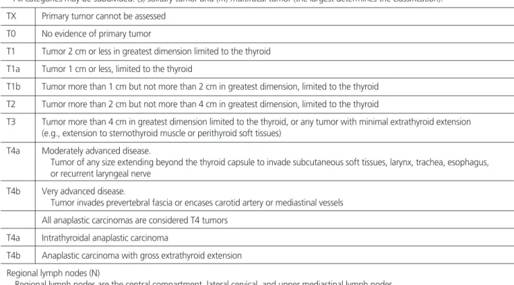
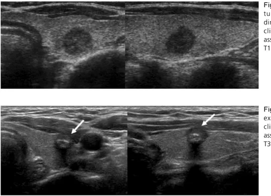
림프절 전이 여부는 수술범위의 결정에 상당히 중요하고 전 이가 있을 경우 국소재발의 위험성을 높이기 때문에 그 예측이 중요하다. 갑상선암의 림프절 전이가 예후에 영향을 미치는 가 에 대해 논란이 많으며 [23-26] 그에 따라 예방적 림프절 절 제에 대한 것도 논란이 있는 부분이나 수술 전 만져지는 림프 절이나 초음파에서 발견된 림프절은 제거해 주어야 한다. 현재 갑상선 암 환자에서 전이가 의심되는 림프절이 있을 경 우에 수술적으로 제거하는 것이 지침 (guideline)이고 이를 위 해 수술 전에 림프절전이를 예측하는 것은 상당히 중요하다. 림프절의 분류방법은 AJCC의 분류 시스템을 토대로 하고 있으며 림프절 위치를 해부학적 구조들을 기준으로 구역을 나 Fig. 1. Left papillary thyroid cancer, tumor size is 1.0 cm in greatest dimension, limited to the thyroid, clinical staging T1a lesion. Final assignment was surgically confirmed T1.Fig. 2. Left thyroid mass with focal extrathyroidal extension (arrows), clinical staging T3 lesion. Final assignment was surgically confirmed T3.
누어 표시하고 있다. Level I의 림프절은 턱밑 (submental)과 악하 (submandibular)위치이다. Level II, III, IV의 림프절은 내경정맥과 경동맥을 따라 각각 경부의 상부, 중간, 하부로 나 눈다. 그 기준은 설골의 상부는 level II, 설골과 윤상연골 (cricoid cartilage) 사이는 level III, 그리고 윤상연골 하부는 level IV이다. 특히 초음파에서 level II, III, IV를 구분하는 방법 은 경동맥이 내경동맥과 외경동맥으로 분지하는 그 상부가 level II, 경동맥의 분지한 위치에서 갑상선이 시작되는 부위가 level III, 그리고 갑상선이 위치하는 부위와 그 아래쪽은 level IV로 구분한다 [27]. Level V는 흉쇄유돌근을 기준으로 그 후 방인 후경부를 말하며 Level VI는 전후두부 (prelaryngeal), 전기도부 (pretracheal), 그리고 기관 주위 (paratracheal) 위 치를 의미한다.
1) 중심경부림프절 (Central cervical lymph node)
갑상선 유두암의 경우에는 level VI의 중심구획 림프절 (central compartment)이 59.1%로 가장 흔하게 림프절 전이 가 일어나는 곳이며 양측 전부 전이림프절이 있는 경우가 42.2%를 차지한다 [28]. 또한 눈에 보이지 않는 림프절전이 (occult metastasis)가 일어나는 비율이 60-80%로 높으며, 수술 전 검사를 통해 이 부위의 전이여부를 정확히 파악하는 것이 어렵기 때문에 [29] 전체갑상선을 절제할 경우에는 중심 림프절절제술 (central neck dissection)을 권고하고 있다 [28]. 대부분의 기관에서 중심구획의 림프절 전이는 갑상선 절 제 수술 시에 수술 범위에 포함되지만 저위험군 환자에서 반대 측 중심구획림프절을 수술하느냐에 대해서는 논란이 있으므로 초음파로 자세히 검사하는 것이 중요하다 [30]. 현재 미국갑상 선 협회진료권고안에서도 [5] 예방적 중심림프절절제술의 경 우 T3나 T4의 진행된 유두암종에서 시행될 수 있다고 권장하 고 있다.2) 외측경부림프절 (Lateral cervical lymph node)
Level I-V의 림프절의 경우에는 수술 전 신체검사(physical examination)나 영상의학 검사들을 통해 전이가 있는지 여부 를 조사하는 것이 중요한데 이는 수술범위와 림프절 절제여부 를 결정해야 되기 때문이다. 특히 level II-V의 전이여부는 수 술범위 결정에 중요한 위치이다. 대부분의 갑상선암 재발이 경 부에서 일어나고 그 중 림프절 전이가 60�75% [31, 32] 정도 를 차지한다고 알려져 있으므로 림프절 전이가 있다면 최초 수 술 시 전이 림프절을 같이 수술하는 것이 중요하다 (Fig. 4).
3) 초음파
초음파는 N staging을 위해 처음으로 하게 되는 검사방법이 며 단순초음파를 통해 림프절의 크기와 모양, 내부 구조나 석 회화 침착 등의 형태를 관찰하고 도플러 초음파를 통해 림프절 내의 혈관분포의 특성을 확인할 수 있다 [33, 34]. 정상적인 림프절은 초음파상에서 타원형의 림프절 내부에 고에코의 문(hilum)을 가지며 혈관은 문을 따라 분포하고 있으 므로 도플러 상에서 문 주위로 혈관분포가 관찰된다. 반면, 전 이 림프절의 경우에는 국소 또는 전반적인 고에코, 미세 또는 거 대 석 회 화 , 낭 성 변 화 , 주 변 혈 관 신 생 (peripheral vascularization)등의 이상 혈관 소견, 그리고 구형 모양 등으로 초음파상에서 보일 수 있다 [34-38] (Fig. 5). Leboulleux 등 [39]은 초음파상의 림프절의 낭성변화와 고에코성 점적 A BFig. 3. 74-year-old female with papillary thyroid carcinoma with tracheal invasion.
A. A large mass containing calcification replace the right lobe of thyroid. US cannot show the full extent of the mass because of air shadowing.
(hyperechoic punctuation)은 전이를 강하게 의심할 수 있는 소견이며 반면에 구형모양, 저에코성, 문의 소실은 단독 기준 으로는 전이를 의심하는데 특이도가 떨어진다고 보고하였다. 초음파는 CT나 MRI에 비해 정상적인 문의 구조, 혈관성 (vascularity), 작은 석회화, 그리고 크기가 2-3 mm정도로 작 은 이상 림프절을 보는데 CT나 MRI보다 유용하다. 특히 최근 초음파 기계의 발전과 더불어 림프절 전이의 정확도는 이전보 다 높아졌으며, 박 등[16]의 연구에 따르면 N staging의 초음 파의 정확도는 71.3%로 보고하고 있고 김 등[40]의 연구에서 도 림프절 전이에 대한 초음파의 진단적 민감도 51%, 특이도 92%, 정확성 77%로 보고하고 있다. 그러나 초음파의 한계점 은 검사의에 의존적이고 종격동 림프절은 초음파로 검사가 불 가능하다는 점이다.
4) CT, MRI
갑상선 초음파 시에 양측 갑상선 엽과 더불어 양측 경부에 림프절을 잘 살펴보는 것이 중요하며 필요에 따라서 CT, MRI, 양전자방출 단층촬영술 (PET) 등의 추가적인 검사의 도움을 받을 수도 있다. 최근 ATA guideline에서는 CT와 MRI가 기본 적으로 권장되지 않으나 추가적인 도움을 받을 수 도 있다고 언급하였다.CT는 경부 림프절 전체를 볼 수 있는 검사방법으로 초음파
A B
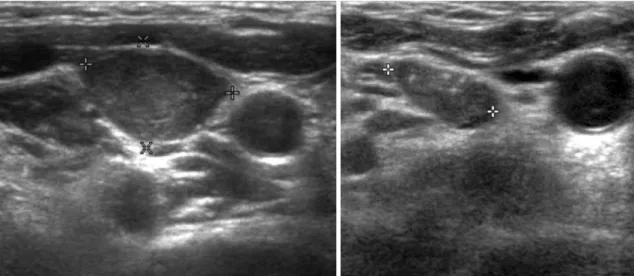
Fig. 4.32-year-old female with papillary carcinoma and metastatic lymph nodes
A.Initial ultrasonography shows hyperechoic lymph nodes at right level IV, which were confirmed as metastatic lymph nodes by fine needle aspiration cytology.
B. 2 years after surgery, follow-up ultrasonography shows enlarged hyperechoic lymph nodes with punctate calcification at right level IV, which were confirmed as metastatic lymph nodes.
A B
Fig. 5. 68-year-old male with papillary thyroid carcinoma and metastatic lymph nodes. Transverse ultrasonography shows (A) hyperechoic, punctuate calcification, cystic change of lymph nodes at right level IV and (B) calcified lymph nodes at right level III, which were confirmed as metastatic lymph nodes.
에코의 한계상 보이지 않거나 검사가 제한적인 부위의 림프절 을 평가하는데 도움을 준다. 악성림프절은 CT상에서 낭성변 화, 다양한 석회화 침착, 테두리 조영증강 (rim enhancement) 또는 비균질 조영증강(heterogenous enhancement) 등의 소 견을 보이는 것으로 보고되고 있다 [40-43] (Figs. 6, 7). 최 근에 초음파와 CT검사를 같이 한 경우에 경부림프절 전이 진 단의 정확성을 높인다는 연구결과들이 보고되고 있으며 이 두 가지 영상기법을 통해 효과적인 수술 전 병기 결정이 이루어 질 수 있다 [12, 40, 43]. MRI 검사는 상대적으로 초음파나 CT에 비해 제한적으로 행 해진다. 악성림프절은 MRI상에서 T2 강조영상에서 고신호강 도를 보이며 CT에서와 같이 테두리 조영증강 또는 비균질 조 영 증강을 보인다. 특히 림프절 내부에 괴사가 있는 경우 T1WI에서 저신호강도를 보이고 T2WI에서 고신호강도를 보 인다 [41, 42].
5) 세포검사
이렇게 초음파를 비롯한 영상검사를 통해 전이가 의심되는 림프절이 발견되는 경우 수술 전 초음파를 이용한 세포검사가 필요하다. 초음파를 이용한 세침흡인 검사 (ultrasonography guided fine needle aspiration cytology)는 림프절 전이를 진 단하는데 유용한 검사이지만 민감도는 70~90%정도이며 일부A B
Fig. 7. 34-year-old female with papillary thyroid carcinoma and metastatic lymph nodes. A.US image shows a hyperechoic node with puntate calcifications at the level IV of left neck. B. CT image shows heterogenous enhancement of node.
A B
Fig. 6.36-year-old male with papillary thyroid carcinoma and metastatic lymph nodes. A.US image shows a hyperechoic node with cystic change at the level II of left neck. B.CT image shows rim enhancement of node.
5-10%는 비진단적 (non-diagnostic), 6-8%는 위음성 (false negative)의 결과도 포함되게 된다. 최근에 많은 연구들을 통해 세포검사로 얻은 미세침세척액 에서 티로글로불린(thyroglobulin)을 측정하는 것이 림프절 전 이여부를 판단하는데 높은 민감도와 특이도를 가진다고 알려 져 있다 [44-47]. 초음파를 이용한 세침흡인 검사와 티로글로 불린 측정을 병행할 경우 민감도가 97~100%정도로 높아지며 [42] 진단의 정확도를 높일 수 있다 [44, 46]. 전 등[48]의 연 구에서는 크기가 작은 림프절의 경우에 진단에 더욱 도움이 된 다고 보고하였고 Cignarelli 등[49]의 연구에서는 림프절 중 낭 성 변성이 있는 경우에 세포검사만 할 경우 불충분한 검체를 얻기 쉽지만 티로글로블린을 측정함으로써 진단의 민감도를 높일 수 있다고 보고하였다.
3. M staging
M staging은 원격전이 여부에 따라서 병기를 분류한다. 갑상 선암은 대부분이 다른 암에 비해 예후가 좋은 것으로 알려져 있으나 드물게 원격전이가 있는 경우는 예후가 나쁠 수 있다. 분화성 갑상선암의 원격전이는 전체의 4-19% 정도로 보고되 고 있으며 [50, 51], 암의 종류 중에는 여포암과 유두암의 경우 에 원격전이가 많이 발생하고 원격전이 부위는 폐와 뼈가 가장 흔한 것으로 알려져 있다. 그러나 알려진 생존율의 측면에서 볼 때 갑상선암 환자가 원격전이가 발견되더라도 방사성 요오 드 치료 등을 통해 완치가 가능하며 특히 폐 전이의 경우에 치 료효과가 좋은 것으로 김 등[1]의 연구에서 보고하고 있다. 또 한 갑상선암의 원격전이가 발견되더라도 10년 이상 생존율이 26% [52], 15년 이상 생존율이 24%로 [50] 다른 암에 비해 예후가 좋은 편이다. 특히 나이가 젊은 유두암 환자의 원격전 이의 경우에 더욱 예후가 좋은 것으로 보고되고 있다 [53, 54]. 현재 방사선 요오드 전신스캔 (I-131 whole body scan)과 혈청 티로글로불린 측정이 갑상선 암의 전이와 재발을 검사하 는데 이용되고 있다. 이 검사방법들은 정상조직이 남아 있을 때도 양성의 결과가 나오기 때문에 기본적으로 전체 갑상선 절 제술과 방사선 요오드 치료를 받은 후 시행되어야 의미가 있 다. 특히 방사선 요오드는 진단과 치료에 동시에 사용되고 있 는데 전신 스캔을 통해 원격전이 여부를 볼 수도 있고 치료를 통해서 수술 후 남아있는 갑상선 조직을 제거해서 재발을 줄이 는 역할을 하고 있다 [55, 56]. 최근에는 PET을 통한 원격전이의 예측이 도움을 주고 있다. 방사선 요오드 전신스캔은 특이도가 높은 검사이나 민감도가 상대적으로 낮기 때문에 위음성의 가능성이 높다. 특히 분화가 나쁜 갑상선암의 경우에는 위음성의 결과가 나올 확률이 높은 데 이러한 경우 임상적으로 전이가 의심된다면 추가적인 양전 자방출 단층촬영술을 통해 원격전이나 재발여부를 진단할 수 있다 [57, 58]. 또한 전이가 의심되는 부위의 해부학적인 위치 선정 (localization)을 통해 진단의 정확성과 치료에 도움을 줄 수 있다 [59].4. Staging grouping
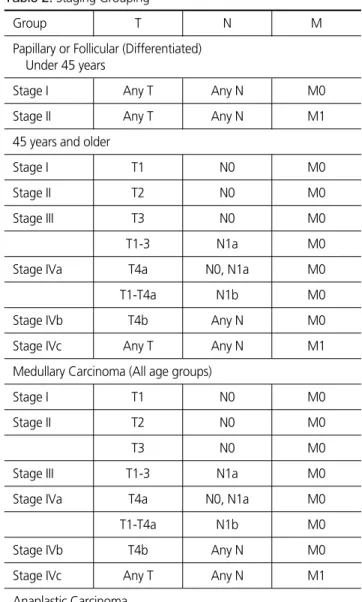
갑상선암의 병기는 TNM병기분류에 기본을 두고 있으나 조 직형과 진단 당시의 연령이 예후에 영향을 미친다고 알려져 있 기 때문에 이점을 염두하여 분류법을 제시하고 있다. 유두암과 여포암의 경우에는 45세를 기준으로 하고 있으며 환자의 나이 에 따라 예후가 다르기 때문에 병기분류를 나이에 따라 다르게 하고 있다 (Table 2).5. TNM병기분류의 제한점
AJCC 가 제시한 TNM 병기분류는 가장 널리 사용되는 병기 Table 2.Staging GroupingGroup T N M
Papillary or Follicular (Differentiated) Under 45 years
Stage I Any T Any N M0 Stage II Any T Any N M1 45 years and older
Stage I T1 N0 M0
Stage II T2 N0 M0
Stage III T3 N0 M0
T1-3 N1a M0 Stage IVa T4a N0, N1a M0
T1-T4a N1b M0 Stage IVb T4b Any N M0
Stage IVc Any T Any N M1 Medullary Carcinoma (All age groups)
Stage I T1 N0 M0
Stage II T2 N0 M0
T3 N0 M0 Stage III T1-3 N1a M0
Stage IVa T4a N0, N1a M0
T1-T4a N1b M0 Stage IVb T4b Any N M0
Stage IVc Any T Any N M1 Anaplastic Carcinoma
All anaplastic carcinomas are considered Stage IV Stage IVa T4a Any N M0 Stage IVb T4b Any N M0 Stage IVc Any T Any N M1
분류이지만 독립적인 예후인자들에 대한 고려가 빠져있다는 단점이 있다. 따라서 TNM 병기분류를 통해서는 정확한 예후 예측에 한계가 있으며 재발을 예측할 수 없다.
TNM 병기분류 외에 이러한 예후인자들을 고려한 병기분류 로 MACIS (Metastases, Age, Completeness of resection, Invasion, Size, Mayo clinic system), EORTC (European Organization for Research and Treatment of Cancer), 등이 사용되고 있으며 주로 원격전이, 환자나이, 종양의 범위 등을 고려하여 갑상선암의 예후예측과 추적검사에 도움을 주고 있 다.
결 론
TNM병기분류는 갑상선 암뿐만 아니라 모든 암에서 치료의 기준이 되며 예후를 예측하는 중요한 요소이다. 수술 전에 이 러한 TNM병기분류를 올바로 제시해 주는 것이 영상의학적으 로 중요하며 가장 핵심적인 역할을 하는 초음파를 비롯하여 CT, MRI, PET 등의 추가적인 영상검사를 통해 병변의 범위, 림프절전이, 원격전이 유무 등을 진단하는 것이 필요하다.요 약
갑상선암은 여성에서 높은 유병율을 가지고 있으며 그 중에서도 유두상암이 대부분을 차지한다. 다른 암과 마찬 가지로 갑상선암에서도 TNM 병기분류를 기본으로 하고 있다. 갑상선암의 병기결정은 외과의가 수술범위를 결정하 는데 기준이 되며, 수술 후 보조치료 여부, 재발 등의 환자 의 예후를 예측할 수 있는 중요한 요소이다. 초음파는 갑상 선암의 평가에 가장 기본적인 검사이며 병변의 크기와 범 위, 림프절 전이유무 등을 파악하여 병기결정에 유용하다. 그 외 최근에는 전산화 단층 촬영술, 자기공명영상, 전신 양전자 방사 단층 촬영 등의 검사를 통해 병기결정에 추가 적인 도움을 얻고 있다. 따라서 본 종설에서는 AJCC가 제 시한 제7차 개정판에 따른 갑상선의 TNM 병기분류와, 각 각의 세부적인 T, N, M 병기의 영상의학적 평가방법에 대 해 기술하고자 한다.References
1. Ministry for Health WaFA. Annual Report of cancer inci-dence (2007), cancer prevalence (2007) and survival (1993-2007) in Korea. 2009
2. Center GNCI. Thyroid cancer: incidence and histological pattern in Korea. 2009
3. Hay ID, Bergstralh EJ, Goellner JR, Ebersold JR, Grant CS. Predicting outcome in papillary thyroid carcinoma:
devel-opment of a reliable prognostic scoring system in a cohort of 1779 patients surgically treated at one institution during 1940 through 1989. Surgery 1993;114:1050-1057
4. Hay ID, Thompson GB, Grant CS, et al. Papillary thyroid carcinoma managed at the Mayo Clinic during six decades (1940-1999): temporal trends in initial therapy and long-term outcome in 2444 consecutively treated patients. World J Surg 2002;26:879-885
5. Cooper DS, Doherty GM, Haugen BR, et al. Revised American Thyroid Association management guidelines for patients with thyroid nodules and differentiated thyroid cancer. Thyroid 2009;19:1167-1214
6. Ito Y, Tomoda C, Uruno T, et al. Clinical significance of metastasis to the central compartment from papillary mi-crocarcinoma of the thyroid. World J Surg 2006;30:91-99 7. Sundram F, Robinson BG, Kung A, et al.
Well-differentiat-ed epithelial thyroid cancer management in the Asia Pacific region: a report and clinical practice guideline. Thyroid 2006;16:461-469
8. Bilimoria KY, Bentrem DJ, Ko CY, et al. Extent of surgery affects survival for papillary thyroid cancer. Ann Surg 2007;246:375-381
9. Kouvaraki MA, Shapiro SE, Fornage BD, et al. Role of pre-operative ultrasonography in the surgical management of patients with thyroid cancer. Surgery 2003;134:946-954 10. Stulak JM, Grant CS, Farley DR, et al. Value of
preopera-tive ultrasonography in the surgical management of initial and reoperative papillary thyroid cancer. Arch Surg 2006;141:489-494
11. Kwak JY, Kim EK, Youk JH, et al. Extrathyroid extension of well-differentiated papillary thyroid microcarcinoma on US. Thyroid 2008;18:609-614
12. Choi JS, Kim J, Kwak JY, Kim MJ, Chang HS, Kim EK. Preoperative staging of papillary thyroid carcinoma: com-parison of ultrasound imaging and CT. AJR Am J Roentgenol 2009;193:871-878
13. King AD, Ahuja AT, To EW, Tse GM, Metreweli C. Staging papillary carcinoma of the thyroid: magnetic reso-nance imaging vs ultrasound of the neck. Clin Radiol 2000;55:222-226
14. Shimamoto K, Satake H, Sawaki A, Ishigaki T, Funahashi H, Imai T. Preoperative staging of thyroid papillary carci-noma with ultrasonography. Eur J Radiol 1998;29:4-10 15. Shetty SK, Maher MM, Hahn PF, Halpern EF, Aquino SL.
Significance of incidental thyroid lesions detected on CT: correlation among CT, sonography, and pathology. AJR Am J Roentgenol 2006;187:1349-1356
16. Park JS, Son KR, Na DG, Kim E, Kim S. Performance of preoperative sonographic staging of papillary thyroid carci-noma based on the sixth edition of the AJCC/UICC TNM classification system. AJR Am J Roentgenol 2009;192:66-72 17. Lee J, Rhee Y, Lee S, et al. Frequent, aggressive behaviors of thyroid microcarcinomas in korean patients. Endocr J 2006;53:627-632
thyroid nodules: comparison with ultrasonography. Radiat Med 2004;22:346-353
19. Wang J, Takashima S, Matsushita T, Takayama F, Kobayashi T, Kadoya M. Esophageal invasion by thyroid carcinomas: prediction using magnetic resonance imaging. J Comput Assist Tomogr 2003;27:18-25
20. Wang JC, Takashima S, Takayama F, et al. Tracheal inva-sion by thyroid carcinoma: prediction using MR imaging. AJR Am J Roentgenol 2001;177:929-936
21. AJCC cancer staging manual. New York: Springer, 2009 22. Sanders LE, Cady B. Differentiated thyroid cancer:
reex-amination of risk groups and outcome of treatment. Arch Surg 1998;133:419-425
23. Shaha AR. Thyroid cancer: extent of thyroidectomy. Cancer Control 2000;7:240-245
24. Mazzaferri EL. Long-term outcome of patients with differ-entiated thyroid carcinoma: effect of therapy. Endocr Pract 2000;6:469-476
25. McHenry CR, Rosen IB, Walfish PG. Prospective manage-ment of nodal metastases in differentiated thyroid cancer. Am J Surg 1991;162:353-356
26. Akslen LA, Haldorsen T, Thoresen SO, Glattre E. Survival and causes of death in thyroid cancer: a population-based study of 2479 cases from Norway. Cancer Res 1991;51:1234-1241
27. Cheung YC, Wan YL, Lui KW, Lee KF. Sonographically guided core-needle biopsy in the diagnosis of superficial lymphadenopathy. J Clin Ultrasound 2000;28:283-289 28. Li Z, Qu XC, Cheng B. The characteristics of papillary
thy-roid cancer lymph node metastasis and the clinical signifi-cance of central region cervical lymph node dissection. Zhonghua Wai Ke Za Zhi 2008;46:1407-1409
29. van den Brekel MW, Castelijns JA. What the clinician wants to know: surgical perspective and ultrasound for lymph node imaging of the neck. Cancer Imaging 2005;5 Spec No A:S41-49
30. Son YI, Jeong HS, Baek CH, et al. Extent of prophylactic lymph node dissection in the central neck area of the pa-tients with papillary thyroid carcinoma: comparison of limited versus comprehensive lymph node dissection in a 2-year safety study. Ann Surg Oncol 2008;15:2020-2026 31. Watkinson JC, Franklyn JA, Olliff JF. Detection and
surgi-cal treatment of cervisurgi-cal lymph nodes in differentiated thy-roid cancer. Thythy-roid 2006;16:187-194
32. Biscolla RP. Cervical lymph nodes metastases in patients with differentiated thyroid cancer. Arq Bras Endocrinol Metabol 2007;51:813-817
33. Wang S, Zhang R, Shu J, et al. Diagnostic value of ultra-sonography in cervical lymph node metastasis. Lin Chuang Er Bi Yan Hou Ke Za Zhi 1999;13:352-353
34. Tschammler A, Ott G, Schang T, Seelbach-Goebel B, Schwager K, Hahn D. Lymphadenopathy: differentiation of benign from malignant disease--color Doppler US as-sessment of intranodal angioarchitecture. Radiology 1998;208:117-123
35. Kessler A, Rappaport Y, Blank A, Marmor S, Weiss J, Graif M. Cystic appearance of cervical lymph nodes is character-istic of metastatic papillary thyroid carcinoma. J Clin Ultrasound 2003;31:21-25
36. Vassallo P, Wernecke K, Roos N, Peters PE. Differentiation of benign from malignant superficial lym-phadenopathy: the role of high-resolution US. Radiology 1992;183:215-220
37. Ahuja AT, Chow L, Chick W, King W, Metreweli C. Metastatic cervical nodes in papillary carcinoma of the thyroid: ultrasound and histological correlation. Clin Radiol 1995;50:229-231
38. Kuna SK, Bracic I, Tesic V, Kuna K, Herceg GH, Dodig D. Ultrasonographic differentiation of benign from malignant neck lymphadenopathy in thyroid cancer. J Ultrasound Med 2006;25:1531-1537
39. Leboulleux S, Girard E, Rose M, et al. Ultrasound criteria of malignancy for cervical lymph nodes in patients fol-lowed up for differentiated thyroid cancer. J Clin Endocrinol Metab 2007;92:3590-3594
40. Kim E, Park JS, Son KR, Kim JH, Jeon SJ, Na DG. Preoperative diagnosis of cervical metastatic lymph nodes in papillary thyroid carcinoma: comparison of ultrasound, computed tomography, and combined ultrasound with computed tomography. Thyroid 2008;18:411-418
41. Som PM, Brandwein M, Lidov M, Lawson W, Biller HF. The varied presentations of papillary thyroid carcinoma cervical nodal disease: CT and MR findings. AJNR Am J Neuroradiol 1994;15:1123-1128
42. Krestan C, Herneth AM, Formanek M, Czerny C. Modern imaging lymph node staging of the head and neck region. Eur J Radiol 2006;58:360-366
43. Ahn JE, Lee JH, Yi JS, et al. Diagnostic accuracy of CT and ultrasonography for evaluating metastatic cervical lymph nodes in patients with thyroid cancer. World J Surg 2008;32:1552-1558
44. Pacini F, Fugazzola L, Lippi F, et al. Detection of thy-roglobulin in fine needle aspirates of nonthyroidal neck masses: a clue to the diagnosis of metastatic differentiated thyroid cancer. J Clin Endocrinol Metab 1992;74:1401-1404
45. Frasoldati A, Toschi E, Zini M, et al. Role of thyroglobulin measurement in fine-needle aspiration biopsies of cervical lymph nodes in patients with differentiated thyroid can-cer. Thyroid 1999;9:105-111
46. Mikosinski S, Pomorski L, Oszukowska L, et al. The diag-nostic value of thyroglobulin concentration in fine-needle aspiration of the cervical lymph nodes in patients with dif-ferentiated thyroid cancer. Endokrynol Pol 2006;57:392-395
47. Cunha N, Rodrigues F, Curado F, et al. Thyroglobulin de-tection in fine-needle aspirates of cervical lymph nodes: a technique for the diagnosis of metastatic differentiated thy-roid cancer. Eur J Endocrinol 2007;157:101-107
thy-roglobulin measurement in fine-needle aspiration for diag-nosing metastatic cervical lymph nodes from papillary thy-roid cancer: correlations with US features. Korean J Radiol 2009;10:106-111
49. Cignarelli M, Ambrosi A, Marino A, et al. Diagnostic utili-ty of thyroglobulin detection in fine-needle aspiration of cervical cystic metastatic lymph nodes from papillary thy-roid cancer with negative cytology. Thythy-roid 2003;13:1163-1167
50. Ruegemer JJ, Hay ID, Bergstralh EJ, Ryan JJ, Offord KP, Gorman CA. Distant metastases in differentiated thyroid carcinoma: a multivariate analysis of prognostic variables. J Clin Endocrinol Metab 1988;67:501-508
51. Shaha AR, Shah JP, Loree TR. Differentiated thyroid can-cer presenting initially with distant metastasis. Am J Surg 1997;174:474-476
52. Shoup M, Stojadinovic A, Nissan A, et al. Prognostic indi-cators of outcomes in patients with distant metastases from differentiated thyroid carcinoma. J Am Coll Surg 2003;197:191-197
53. Sampson E, Brierley JD, Le LW, Rotstein L, Tsang RW. Clinical management and outcome of papillary and follicu-lar (differentiated) thyroid cancer presenting with distant metastasis at diagnosis. Cancer 2007;110:1451-1456
54. Casara D, Rubello D, Saladini G, Gallo V, Masarotto G, Busnardo B. Distant metastases in differentiated thyroid cancer: long-term results of radioiodine treatment and sta-tistical analysis of prognostic factors in 214 patients. Tumori 1991;77:432-436
55. Samaan NA, Schultz PN, Hickey RC, et al. The results of various modalities of treatment of well differentiated thy-roid carcinomas: a retrospective review of 1599 patients. J Clin Endocrinol Metab 1992;75:714-720
56. Mazzaferri EL, Jhiang SM. Differentiated thyroid cancer long-term impact of initial therapy. Trans Am Clin Climatol Assoc 1995;106:151-168
57. Grunwald F, Menzel C, Bender H, et al. Comparison of 18FDG-PET with 131iodine and 99mTc-sestamibi scintig-raphy in differentiated thyroid cancer. Thyroid 1997;7:327-335
58. Grunwald F, Schomburg A, Bender H, et al. Fluorine-18 fluorodeoxyglucose positron emission tomography in the follow-up of differentiated thyroid cancer. Eur J Nucl Med 1996;23:312-319
59. Shammas A, Degirmenci B, Mountz JM, et al. 18F-FDG PET/CT in patients with suspected recurrent or metastatic well-differentiated thyroid cancer. J Nucl Med 2007;48: 221-226