ISSN (Print) : 2320 – 3765 ISSN (Online): 2278 – 8875
I
nternational
J
ournal of
A
dvanced
R
esearch in
E
lectrical,
E
lectronics and
I
nstrumentation
E
ngineering
(An ISO 3297: 2007 Certified Organization)
Website: www.ijareeie.com
Vol. 6, Issue 6, June 2017
A Survey on Retinal Image Blood Vessel
Segmentation
Payal Bhujangrao Nimbhorkar , Prof. S. S. Patil
Dept. of Electronics and Telecommunication, Sinhgad College of Engineering, Pune, India.
ABSTRACT: Computer based automatic blood vessel segmentation is an efficient way to segments the retinal blood
vessel. Retinal blood vessels plays and important role in diagnosing eye related diseases. The main function of Retinal blood vessel is to carry fresh blood from heart to eye and then deoxygenated blood back to heart from eye. Many diseases may effect these blood vessel and leads to eye blindness. But the early stage observation of this blood vessel structure through retinal images can help diagnosing eye sight related problems. Many techniques have been proposed for automatic segmentation of retinal blood vessel. This paper is presenting a review of some previously proposed techniques or methods for segmentation of retinal blood vessel.
KEYWORDS: Segmentation; Retinal Blood Vessel; Image Processing
I. INTRODUCTION
Eye is the one of the most important organ in our body, called as organs of vision. Blood vessel extraction is very important as many eye diseases are recognized by inspecting the blood vessel. These blood vessel carry fresh oxygenated blood from heart to feed nutrition’s to tissues and cells present in retina and then carry the deoxygenated blood from eye to heart. There are mainly two types of blood vessels arteries those carry fresh blood and vein those carry oxygenated blood.
The eye is a window to the retinal vascular system which is uniquely accessible for the non-invasive, in vivo study of a continuous vascular bed in humans.Retinal blood vessels have been shown to change in diameter, branching angles or tortuosity, as a result of a disease, such as:
Hypertension
Diabetes Mellitus
Retinopathy of Prematurity (ROP)
Diabetic retinopathy is a disorder of the retinal vessel that eventually develops to some degree in nearly all patients with long-standing diabetes mellitus. Contributes 4.8% of the 37 million cases of blindness throughout the worldMost Common cause of bilateral severe visual loss in working age group in USA recent study in urban population in south India estimates prevalence of DM in adult population as high as 28% & the prevalence of DR in diabetics to 18%.Retinal blood vessel have a range of different sizes.Multiscale techniques have been developed to provide a way to isolate information about objects in an image by looking for geometric features at different scales.
ISSN (Print) : 2320 – 3765 ISSN (Online): 2278 – 8875
I
nternational
J
ournal of
A
dvanced
R
esearch in
E
lectrical,
E
lectronics and
I
nstrumentation
E
ngineering
(An ISO 3297: 2007 Certified Organization)
Website: www.ijareeie.com
Vol. 6, Issue 6, June 2017
III. LITERATURE SURVEY
Marin et al. [1] developed a supervised strategy for the segmentation of blood vessels. Original image is first preprocessed to remove vessel central light reflex issue and diminish inappropriate illumination in the image. Noise smoothing is done utilizing the Gaussian kernel. Vessel Enhancement is done utilizing the morphological Top Hat change. Each pixel is characterized to be either non-vessel pixel or a vessel pixel depending on the 7-D feature vector. The feature vector consists of two moment invariants based features and five gray level based features. Moment invariant based features provide flexibility to the algorithm in a sense that the features are not affected by translation, rotation or shifting of an image. Just two of the seven Hu's moments are utilized as utilizing all the moments actually reduces the accuracy of the algorithm. The 7-D feature vector is given as input to the multilayer feed forward neural network classifier for the classification of the pixels. Post processing performs a very important role of filling the gaps between vessel pixels and by removing the falsely detected isolated pixels. Performance evaluation is done on STARE and DRIVE databases utilizing the built up measurements like specificity, affectability, ppv (called positive prescient esteem), npv (called negative prescient esteem) and precision. The strategy gives high normal exactness of 0.9489 consolidated for both STARE and DRIVE databases which is similar to other effectively existing strategies on the premise of normal precision. Region under the bend acquired is 0.9678 which is far superior to the effectively existing strategies. The strategy is straightforward yet is computationally concentrated. Execution can in any case be expanded. Inkaew et al. [2] performed extraction/division of retinal veins utilizing angle introduction. The technique is not affected by low contrast or improper illumination as it does not depend on the gray level intensity directly. First derivative operators in both x and y direction are utilized to discover Gradient vectors which are then standardized. The units vectors either separate or merge from picture highlights until and unless the components are not brighter/darker than the foundation. Unit gradient vectors are utilized to discover angle introductions. Utilizing gray level discontinuity magnitude different image features especially linear and circular features are found in gradient orientations. As the features are of variable sizes like the width or diameter of a blood vessel, first order derivative, sobel operator is used at three different scales. The results obtained using various scales are integrated to obtain the final gradient orientation image. Blood vessels are segmented utilizing thresholding and morphological operations. The assessment of the calculation, for execution, is done on STARE and DRIVE databases. Metrics used for comparison between automated segmented image obtained by the proposed technique and the ground truth pictures are ROC (Collector Operating Curve), AUC (Area under Curve) and MAA (Maximum Average Accuracy). The algorithm gives true positive rate and true negative rate of 0.8774 and 0.8267 separately. The restriction of this work is that patches or blobs are not identified by this calculation in anomalous pictures furthermore, should be additionally inspected.
Akram et al. [3] proposed a technique for segmenting the blood vessels in retinal images for detecting proliferative diabetic retinopathy. In proliferative diabetic retinopathy, PDR, new blood vessels starts growing (called neovascularization) in the retina which ruins the vision seriously. Vessel improvement assumes a key part in PDR as it is exceptionally important to upgrade thin and less unmistakable vessels. Vessel enhancement is done utilizing Gabor Wavelets as of now examined in. Multi-layered Thresholding system is utilized for the segmentation of blood vessels. Main task that is performed in this work is finding of new blood vessels which are found by computing the entropy and thickness of every last vessel of the divided picture utilizing a 15 *15 cover. This strategy is called sliding window system. Execution assessment is done on DRIVE and STARE databases. Normal precision of the strategy is exceptionally equivalent to effectively existing comparable techniques as it gives a normal exactness of 0.9469 and 0.9502 in DRIVE and STARE databases separately.
ISSN (Print) : 2320 – 3765 ISSN (Online): 2278 – 8875
I
nternational
J
ournal of
A
dvanced
R
esearch in
E
lectrical,
E
lectronics and
I
nstrumentation
E
ngineering
(An ISO 3297: 2007 Certified Organization)
Website: www.ijareeie.com
Vol. 6, Issue 6, June 2017
gotten to be 0.9254. Even though the results are competitive with other existing methods but if this method is used with supervised training might provide better results.
Ocbagabir et al. [5] developed a new algorithm called Star Networked Pixel Tracking algorithm, a rule based algorithm, for segmentation and detection of blood vessels in retinal images. Blood vessel enhancement is done utilizing the Adaptive Histogram Equalization alongside the morphological operations. Commotion is evacuated utilizing the middle sifting method. This algorithm is capable of detecting the pixel orientation in the eight directions has the ability of recognizing the vessel pixels from the non-vessel pixels. It looks at every single pixel of picture with its neighbor pixels which are at 45 degree to each other. On the off chance that a zero pixel is found in the area then the first pixel is set apart as nonvessel pixel generally the calculation denote the present pixel as vessel pixel and proceed onward to the following pixel. Same strategy is connected to the next pixel but only in seven directions because one direction is already known from where the movement has taken place. Each and every pixel is marked as non-vessel or a vessel pixel in the whole image. Once vessel map is obtained, local enhancement of the image is done using the intensity and standard deviation that identify the neighbourhood of thin and small vessels. Automatic thresholding is applied to the standardized retinal picture to remove the veins. Execution assessment is done on the DRIVE database.. Mehrotra et al. [6] proposed a technique for retinal blood vessels discovery in view of scientific morphology and KCN clustering. Novel Edge Preserving Filter (NEPF) and Gaussian smoothening is connected to evacuate the motivation clamor what’s more, Gaussian clamor separately. Non uniform illumination is overcome using an equalization algorithm. Both top-hat and bottom-hat transformations are applied to get the candidates for blood vessels. KCN (Kohonen Clustering Network) clustering procedure is then connected to the resultant picture which makes two groups, vein and non-vein groups. Euclidean separation is utilized as a part of the development of groups. Assessment of the strategy was done on DRIVE database. The technique provides high accuracy and sensitivity of 0.9802 and .9912 respectively. FPF (False Positive Fraction) was found to be .0415.
Binooja.B.R et al. [7] The automatic Artery/Vein classifications of retinal images are essential for the estimation of vascular changes. This technique utilizes force highlights and extra data removed from the diagram for the separation of Artery/Vein. Feed forward neural network is utilized to distinguish sickness like Diabetic Macular Edema as direct or extreme. This proposed framework has better exactness, speed and great execution.
SangmeshBiradar et al. [8] This writing overview proposed different methodologies for recognizing and dividing the veins and optic plate. We have attempted to cover later and early writing identified with division calculations and systems. Quicker division can be accomplished through the multi-scale picture handling technique. The most critical application of segmentation is radiological diagnostic framework. Progresses in radiological imaging framework result in huge number of patient pictures. Handling of these pictures, quick division calculations required. One approach to do quick division is by creating parallel calculations.
Malak T. Bantan et al. [9] in this paper, a segmentation strategy for the retinal blood vessel is proposed. We show the methodology of pre-processing and blood vessels segmentation. It is a combination of efficient morphological operations, image enhancement, and noise reduction systems. Classification Error is measured between our outcomes and the ground truth also, discovered sensibly adequate. Moreover, the calculation will be adjusted later on to bring about better division on account of diabetic retinopathy.
ISSN (Print) : 2320 – 3765 ISSN (Online): 2278 – 8875
I
nternational
J
ournal of
A
dvanced
R
esearch in
E
lectrical,
E
lectronics and
I
nstrumentation
E
ngineering
(An ISO 3297: 2007 Certified Organization)
Website: www.ijareeie.com
Vol. 6, Issue 6, June 2017
IV. SEGMENTATION
Manual segmentation of retinal blood vessel is a difficult task. As vascular structure is tree like complex structure. It may took hours to segment blood vessel manually. Manual segmentation may not provide the actual sized blood vessel, there may a difference in actual blood vessels and segmented vessels. Also if there are two different observers observing the same retinal image. Then there may some bias in two different results. But as automatic segmentation took only few minutes or seconds. It is difficult to get 100% accuracy with automatic segmentation, but its better to have results close to 100% than to wait for hours.
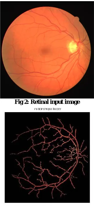
Fig 2: Retinal input image
Fig 3: Segmentation of blood vessels.
V. NEED AND SIGNIFICANCE
Need for computer based automatic retinal blood vessel segmentation has following reasons.
ISSN (Print) : 2320 – 3765 ISSN (Online): 2278 – 8875
I
nternational
J
ournal of
A
dvanced
R
esearch in
E
lectrical,
E
lectronics and
I
nstrumentation
E
ngineering
(An ISO 3297: 2007 Certified Organization)
Website: www.ijareeie.com
Vol. 6, Issue 6, June 2017
With automatic segmentation of blood vessels eye related disease can be treated at early stages.
Automatic segmentation can also help in retinal based recognition tasks.
VI. CONCLUSION
A lot of research has been done in blood vessel till now. And this area has been reached too far till now. Many new techniques and methods has been discussed previously in literature review. There were many good techniques, those segments with very good accuracy in less interval of time. But still these techniques lacks in few aspects. There is no doubt that computer based automatic segmentation cannot reach to 100% in accuracy but it is possible to reach close to 100%. On the bases of literature there are too many areas that can be improve a bit to improve the accuracy. Improvement in blood vessel segmentation accuracy is possible but it needs good knowledge under better guidance. This paper gives a brief introduction about blood vessel segmentation and need of blood vessel segmentation. Also this paper provides a brief survey on previous and recent research on blood vessels segmentation. Various methods and techniques has been discussed above in literature for segmenting blood vessel. The motive of this paper is to introduce the previous and present segmentation techniques and to provide the interested parties literature for research on this topic.
REFERRENCES
[1] Diego Marín, Arturo Aquino*, Manuel Emilio Gegúndez-Arias, andJosé Manuel Bravo, “A New Supervised Method for Blood Vessel Segmentation in Retinal Images by Using Gray-Level and Moment Invariants-Based Features”, IEEE Trans. Medical Imaging, Vol. 30, no. 1,Jan. 2011.
[2] Danu inkaew, RashmiTurior, BunyaritUyyanonvara, Toshiaki Kondo,” Automatic Extraction of Retinal Vessels Based on Gradient Orientation Analysis”, IEEE Eighth International Joint Conference on Computer Science and Software Engineering (JCSSE), pp. 102-107, 2011
[3] M. U. Akram, I. Jamal, A. Tariq and J. Imtiaz, “Automated Segmentation of Blood Vessels for Detection of Proliferative Diabetic Retinopathy”, IEEE-EMBS International Conference on Biomedical and Health Informatics, China, Jan. 2012.
[4] I. Lazar and A. Hajdu, “Segmentation of Vessels in Retinal Images Based on Directional Height Statistics”, 34th Annual International Conference of the IEEE EMBS, USA, Sep. 2012.
[5] H. Ocbagabir, I. Hameed, S. Abdulmalik and D. B. Buket, “A Novel Vessel Segmentation Algorithm in Color Images of the Retina”, IEEE Applications and Technology Conference in Systems, pp. 1-6, May.2013.
[6] A. Mehrotraet. al,,”Blood Vessel Extraction For Retinal Images Using Morphological Operator and KCN Clustering”, IEEE International Advance Computing Conference (IACC), 2014.
[7] Binooja. B.R, Nisha. A.V “Diabetic Macular Edema Detection byArtery/Vein Classification Using Neural Network neural network”, International Journal of Engineering and Technical Research (IJETR) ISSN: 2321-0869 (O) 2454-4698 (P), Volume-3, Issue-7, July 2015.
[8] Mr.SangmeshBiradar , Prof. A.S.Jadhav “A Survey on Blood Vessel Segmentation and Optic Disc Segmentation of Retinal Images”,. International Journal of Advanced Research in Computer and Communication EngineeringVol. 4, Issue 5, May 2015.
[9] Malak T. Bantan “Auto-segmentation of Retinal Blood Vessels Using Image Processing”.