MODALITIES
:
FUNCTIONAL ACTIVATION AND
CONNECTIVITY DURING TASK AND REST
Submitted by Anselm Doll
At the Graduate School of Systemic Neurosciences
of Ludwig-Maximilians-Universität Munich
June 2015
Oral defense: 12.10.2015
Supervisors: Dr. Afra Wohlschläger, Dr. Christian Sorg & Dr. Virginia Flanagin
1. Acknowledgements ... 4
2. Abbreviations ... 5
3. Abstract ... 6
4. Overview ... 7
5. Introduction ... 8
5.1 Practical definition of mindfulness and its psychological effects... 8
5.2 Mindfulness improves emotion regulation ... 9
5.3 Emotions as situated conceptualization ... 10
5.4 Emotion regulation and its interaction with mindfulness ... 12
5.5 Learning effects of mindfulness meditation ... 21
5.6 Resting state networks involved in mindfulness training ... 24
5.7 Changed resting state connectivity in psychiatric disorders – exemplified in borderline personality disorder ... 26
5.8 Summary and aims of the thesis ... 26
6. Paper one: Mindful attention to breath regulates emotions via increased amygdala-prefrontal cortex connectivity ... 28
7. Paper two: The effects of mindful attention to breath on aversive prediction error-based learning in the striatum ... 55
8. Paper three: Mindfulness is associated with intrinsic functional connectivity between default mode and salience networks ... 76
9. Paper four: Shifted intrinsic connectivity of central executive and salience network in borderline personality disorder ... 104
10. General Discussion ... 118
10.1 Mindful attention to breath regulates emotions via increased amygdala-prefrontal cortex connectivity ... 118
10.4 Shifted intrinsic connectivity of central executive and salience network in borderline personality
disorder ... 122
10.5 Involvement of the amygdala and the putamen in focused attention meditation ... 123
10.6 Overlap between mindfulness networks and the DMN – mindwandering vs. self-monitoring ... 124
10.7 Mindfulness trains the activation of dorsal prefrontal cortex in response to mindwandering ... 125
10.8 Rumination and resting state networks ... 126
10.9 Mindfulness affects functional networks: a possible pathway of therapeutic effects of mindfulness .. 128
10.10 The overlap of connectivity effects of mindfulness and borderline personality disorder ... 128
10.11 Learning effects of mindfulness: possible neural pathway for anti anxiety effects of mindfulness .... 129
11. Conclusion ... 130
12. References ... 131
13. List of publications ... 147
14. Eidesstattliche Versicherung/Affidavit ... 148
15. Author contributions ... 149
1.
A
CKNOWLEDGEMENTS
This document would not have been possible without the help of many helping hands and bright
minds. I am especially thankful to my supervisor Christian Sorg, who has taught me great deals
about neuroscience, thinking, soccer, and my character. Thank you for motivating me to do
better and helping me improve my thinking, writing and perspective taking. I also want to thank
Afra Wohlschläger and Virginia Flanigin, for their motivational attitude.
My special thanks go to Prof. Zimmer, Prof. Förstl and Prof. Bäuml for their financial
support. I would also like to extend my thanks to the Elite Netzwerk Bayern for their financial
assistance. My further thanks goes to Marie-Luise Brandi, for reading this dissertation, giving me
great constructive feedback, and sharing her humor. I am very glad to have worked in an
extraordinary group of young scientist, where the atmosphere is incomparably supportive and
helpful. Some of these colleagues I want to thank specifically. For the great help with analyses
techniques and the fun times I have had I want to thank Satja Mulej Bratec. The same goes for,
Andrei Manoliu, who so much supported me in both scientific and personal questions.
I am sincerely grateful to Britta Hölzel, for her amazing way of carefully pointing at
weaknesses in my work and having great ideas for corrections. Furthermore, I want to thank,
Christine, Joyce, Boucard for her help during my experiments, and many chai lattes. I would like
to offer my special thanks to the technicians and staff of the neuroradiology department,
especially Josette and Daniel, and the TUM Neuro Imaging Centre for their assistance in data
collection. I would further like to thank my mother, Elisabeth, and my dear friends, Fabian,
Christoph, Hagen, Annette and Benni for supporting me in finding my way in difficult times and
helping me to finish what I started. Finally, I want to thank the participants of my studies for
2.
A
BBREVIATIONS
ACC - anterior cingulate cortex PPI - psycho-pysiological interaction
AI - anterior insula PV - passive viewing
aMCC - anterior medial cingulate cortex ROI - region of interest
aPE - aversive prediction error rs-fMRI - resting state-functional magenteic
ATB - attention to breath resonance imaging
BOLD - blood oxygen level dependent SN - salience network
BPD - borderline personality disorder SVA - small volume analysis
CEN - central executive network TPJ - temporo-parietal junction
CS - conditioned stimulus US - unconditioned stimulus
CPZ - chlorpromazine VLPFC - ventrolateral prefrontal cortex
dACC - dorsal anterior cingulate cortex VMPFC - ventromedial prefrontal cortex
DLPFC - dorsolateral prefrontal cortex VTA - ventral tegmental area
DMN - default mode network
DMPFC - dorsomedial prefrontal cortex FA - focused attention
FMI - Freiburg Mindfulness Inventory
fMRI - functional magnetic resonance imaging FWE - family wise error
HC - healthy controls
iFC - intrinsic functional connectivity ICA - independent component analysis ICN - intrinsic connectivity network
intra-iFC - intrinsic functional connectivity within networks inter-iFC - between networks intrinsic functional connectivity MAAS - Mindful Attention and Awareness scale
MBSR - mindfulness based stress reduction MD - major depression
MNI - Montreal Neurological Institute mPFC - medial prefrontal cortex OM - open monitoring
P - prediction
PCC - posterior cingulate cortex PE - prediction error
3.
A
BSTRACT
Mindfulness, i.e., attention to present moment experience without judgment, has beneficial
effects on different aspects of behavior and cognition. For example, it improves
psychopathological symptoms related to emotion regulation, anxiety and interpersonal
functioning. However, very little is known about the neural correlates of these effects. Existing
neuroimaging experiments have shown that both mindfulness and psychiatric disorders manifest
as distinct, but partly overlapping patterns in brain activation and functional connectivity in
various networks of the brain. Therefore, understanding of the neural correlates of mindfulness
and its effect on emotion regulation and learning may elucidate how mindfulness affects
cognition, particularly in case of psychiatric disorders. The present dissertation aims at
investigating the neural correlates of mindfulness effects on emotion regulation and learning. To
this end, I report the results of studies investigating the effect of mindful focused attention to
breath on brain activations and functional connectivity during (1) exposure to stimuli with
negative valence and (2) classical conditioning. Furthermore, I report studies that investigated
(3) the correlates of mindfulness disposition in the brai s i t i si et o k a hite tu e a d (4)
compared it with changes in this architecture in patients with borderline personality disorder,
who are known to profit from mindfulness-based treatment. The included studies allow for the
following conclusions: (1) mindful focused attention leads to reduced activation of the amygdala
during stimulation with aversive pictures and (2) reduced prediction error related signaling in
right putamen during conditioning. (3) Trait mindfulness is associated with the functional
connectivity between key cognitive networks, i.e., the default mode, salience and central
executive networks. (4) Borderline patients showed aberrant within- and between-network
connectivity among default mode and salience network which overlapped with effects of
mindfulness in dorso-medial prefrontal cortex. These results provide evidence for a potential
neural pathway of how mindful focused attention affects emotional processing and learning.
Furthermore the results suggest neural mechanisms by which mindfulness may support the
treatment of psychiatric disorders, with the example of borderline personality disorder,
presumably by improving emotion regulation abilities, rumination tendency and relearning of
4.
O
VERVIEW
The present Ph. D. dissertation was written in semi-cumulative form. It comprises a general
introduction, which is followed by the reports of four brain imaging experiments in form of
manuscripts for publication. At the time of thesis submission, one of these papers had been
su itted to NeuroImage pape one), one is prepared for submission (paper two), one is
under review in F o tie s i Hu a Neu os ie e paper three) and one has been published in
F o tie s i Hu a Neu os ie e (paper four). The joint discussion summarizes and interprets
5.
I
NTRODUCTION
The concept of mindfulness has spread from Buddhist tradition into psychotherapy and
neuroscience. The aim of this thesis is to connect these fields of research by investigating the
neural correlates of mindfulness in the context of its uses in psychotherapy. To this end, the
introduction will shed light on the neural effects of mindfulness in the context of emotion
regulation and learning, and then take a systems view on the intrinsic brain networks that are
involved in mindfulness and borderline personality disorder as an example of an affective brain
disease.
Since a model of regulation of emotions cannot stand without a theory about emotional
processing, I will first present a recent neuroscientific model of emotions and outline traditional
approaches for emotion regulation. Next, I will compare these strategies with those provided by
mindfulness practice. The role mindfulness plays in psychotherapy is closely related to relearning
of emotional responses and regulation strategies. Therefore, the second part of the introduction
is dedicated to the neuroscientific basis of classical conditioning and the possible mechanisms
how mindfulness may influence learning. The third part will introduce an intrinsic brain network
perspective on the processes involved in mindfulness and show, why the study of network
interactions may be beneficial in explaining neural effects of mindfulness especially in the
context of affective disorders.
5.1
P
RACTICAL DEFINITION OF MINDFULNESS AND ITS PSYCHOLOGICAL
EFFECTS
A group of leading scientists in the field has developed a practical definition and
operationalization for mindfulness (Bishop, Lau, & Shapiro, 2004) that can be generalized to
different traditions of mindfulness practice. In this description, mindfulness is seen as a
self-regulatory mechanism that monitors attention and orients it towards present experience
without judgment. Mindfulness encompasses the way how something is done rather that what
exactly is done. Thus, mindfulness is a meta-awareness skill that can be achieved by mental
training. As this training looks back on over two millennia of tradition, multiple techniques to
develop this skill have been proposed and are still taught. However, all techniques include
instructions on how to allocate attention that can be subdivided into two categories: focused
Slagter, Dunne, & Davidson, 2008). FA is mostly used to teach novices as it trains the essential
skill in mindfulness: staying in the present moment without judgment of good or bad. It involves
three crucial abilities of regulating attention: monitoring the focus of attention, disengaging
from distracting objects of attention and redirecting attention back to the initial object of
attention. With sufficient training, these skills are becoming more and more effortless. In
addition, the urge to act on emotions or thoughts, termed emotional reactivity, is reduced with
training (e. g., Britton, Shahar, Szepsenwol, & Jacobs, 2012) and substituted with mere
observation.
FA has been subject to more extensive research than OM. This may be due to the fact
that in contrast to FA, OM is more vaguely defined as sustaining the monitoring feature of
attention without engaging it towards a specific object. In other words, OM entails sustaining
moment to moment awareness to any experience in the present (A. Lutz, Slagter, et al., 2008).
Research into the psychological effects of mindfulness has shown increases in attentional
performance, especially selective attention (C. G. Jensen, Vangkilde, Frokjaer, & Hasselbalch,
2012) that might be related to more effective usage of brain resources (Slagter et al., 2007).
However, a meta analysis on the psychological effects of mindfulness concluded that the
attentional effects were only moderate in magnitude, and that meditation has even stronger
effects on personal relationships and emotion regulation (Sedlmeier et al., 2012).
5.2
M
INDFULNESS IMPROVES EMOTION REGULATION
Mindfulness has been shown to have beneficial effects on emotion regulation. Emotion
regulation describes a variety of strategies that influence the generation of emotions and how
these are experienced and expressed (Gross & Thompson, 2007). Evidence for a beneficial effect
of mindfulness on emotion regulation is available from both correlational studies using
questionnaires (Hill & Updegraff, 2012; Sedlmeier et al., 2012) and those using experimentally
controlled conditions (e. g., Arch & Craske, 2006; J. Lutz et al., 2013). The beneficial effects of
mindfulness include increased emotional differentiation (Hill & Updegraff, 2012), improved
reappraisal skills (Sedlmeier et al., 2012), relaxation (reviewed in Hölzel et al., 2011), and
decreased emotional reactivity after meditation (Arch & Craske, 2006). Given these effects of
mindfulness on emotion regulation it is unsurprising that psychotherapists included mindfulness
practice in a number of structured psychotherapy manuals for disorders of emotion regulation
(e. g., for borderline personality disorder, see Linehan, 1993a, for bipolar disorder, see Williams
emotion regulation, it is necessary to get a grip on the processes that underlie emotional
processing in general, which I will review next.
5.3
E
MOTIONS AS SITUATED CONCEPTUALIZATION
The framework of emotion regulation is embedded into a model of emotion generation.
Although most research into emotion regulation has been based on the process model of
emotion generation (Gross, 1998), the most extensive model that can be related to the current
fi di gs i eu os ie e has ee fo ulated Li d uist a d olleagues i a o st u ti ist
odel of e otio ge e atio (for a review, see Lindquist, Wager, Kober, Bliss-Moreau, &
Barrett, 2012, Fig.1). The advantage of the constructivist model of emotion generation is that it
accounts for the complexity of the emotional experience. In the model, emotions are based on
fou so alled ps hologi al p i iti es , hi h a e s alle uildi g lo ks of og iti e
functioning: (1) core affect, (2) situated conceptualization of core affect, (3) language and (4)
executive attention. Each of these psychological primitives is defined by the processes it
implements and by various associated brain regions. While affect in psychology is a term used
for anything emotional, core affect describes the mental representations of bodily changes that
can be experienced as positive or negative feelings with some degree of arousal. Core affect is
associated with brain regions that, among other things, implement visceral control and are
involved in processing of salience related to the internal homeostasis: the amygdala, medial
orbitofrontal cortex, insula, anterior cingulate cortex, thalamus, hypothalamus, bed nucleus of
stria terminalis, basal forebrain and the periaqueductal gray (Lindquist et al., 2012). In short,
core affect attaches a label of bodily value to an object. The second psychological primitive,
situated conceptualization then connects this label to prior history and experience with the
object, in order to categorize it or make meaning of it. As such, this system comprises regions
involved in memory and personal value like ventromedial prefrontal cortex (VMPFC),
dorsomedial PFC (DMPFC), medial temporal lobe (hippocampus, entorhinal cortex,
parahippocampal cortex), and posterior cingulate cortex (PCC) /retrosplenial area, also called
the default mode network (e. g., Raichle et al., 2001). Through these regions and in combination
with the fourth psychological primitive, executive attention, an object and its associated core
affect are combined with prior experience. These effortless and automatic processes result in
one experience that could have the quality of a physical symptom (e. g., a rumbling stomach), a
feeling (e. g., tiredness), or a discrete emotion (e. g., surprise vs. fear). In other words,
situated conceptualization network will make inferences about what may have triggered an
emotion that has been identified in another person, enabling us to experience empathy or
produce a theory of mind (for a neuroscientific review, see Schurz, Radua, Aichhorn, Richlan, &
Perner, 2014). The third process in the model, language or emotional words, works closely
together with the conceptualization process in order to make emotional categories of what is a
very complex phenomenological process, i.e., emotions. This process involves the right anterior
temporal lobe and ventro-lateral PFC (VLPFC). While the anterior temporal lobe combines
heteromodal information into a representation of a complex category, the VLPFC is associated
with semantic processing and categorization of objects. As such, emotional words serve as
placeholders for a combination of various experiences, memories and core affects. The fourth
psychological primitive, executive attention guides and enhances certain interoceptive feelings
and representations of external objects to create a moment of here and now, sustaining the
combination of external objects, core affect, and situated conceptualization into one concept of
an emotion. Executive attention recruits the dorso-lateral PFC (DLPFC) and VLPFC (reviewed in
Lindquist et al., 2012) with lateral parietal regions (Corbetta & Shulman, 2002). In summary,
different aspects of emotions are represented in a variety of brain regions. The experience of an
emotion is produced by executive attention effortless combining representations of an object
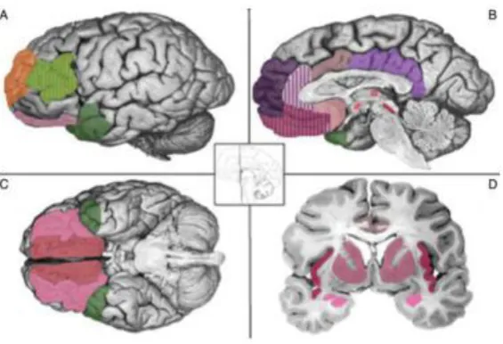
Figure 1. The constructivist model of emotion generation.
The figure shows brain regions that are hypothesized to be involved in the generation of emotion
through four psychological primitives. Core affect (pink): amygdala, insula, medial orbitofrontal
cortex, anterior cingulate cortex, thalamus, hypothalamus, bed nucleus of the striaterminalis,
basal forebrain and periaequedictal grey. Conceptualization (purple): ventromedial prefrontal
cortex, dorsomedial prefrontal cortex, medial temporal lobe, posterior cingulate
cortex/retrosplenial area. Language (green): ventrolateral prefrontal cortex & anterior temporal
lobe. Executive attention (orange): dorso-lateral prefrontal cortex & ventrolateral prefrontal
cortex. Image taken from Lindquist et al. (2012), reprinted with the permission of Cambridge
university press 2012.
5.4
E
MOTION REGULATION AND ITS INTERACTION WITH MINDFULNESS
5.4.1
T
YPES OF BENEFICIAL EMOTION REGULATION STRATEGIESThe most prominent beneficial strategies in the literature have been cognitive in nature, such as
reappraisal and distraction. In studies evaluating the effectiveness of emotion regulation
strategies, reappraisal and distraction are generally contrasted with expressional suppression,
that has been shown to be associated with higher emotional, social and cognitive costs (Gross &
John, 2003; Gross, 2002). The neural correlates of cognitive emotion regulation strategies have
been investigated in a number of studies (e. g., Kanske, Heissler, Schönfelder, Bongers, & Wessa,
Blechert, Sheppes, Rydstrom, & Gross, 2011). Based on these studies, the authors of a recent
meta-a al sis p opose i the heu isti odel of e otio egulatio that og iti e egulatio is
achieved through a three-step-process that first, signals the need to regulate, then down
regulates an emotion and finally, generates a new, regulated, emotional state (Kohn et al., 2013,
Fig. 2). In their heuristic model of emotion regulation an emotion is signaled from subcortical
regions through anterior medial cingulate cortex (aMCC) into ventro-lateral prefrontal cortex
(VLPFC) and insula cortex. These structures evaluate salience and signal the need to regulate to
dorso-lateral prefrontal cortex (DLPFC). Then, DLPFC is thought to induce a reenaction of the
emotional scene through imagination of motoric, somatosensory and language processing and
coherent verbalization via pre-motor areas, angular gyrus and superior temporal gyrus. This new
emotional scene leads to a generation of a new emotion in striatum and amygdala either directly
or guided through aMCC (Kohn et al., 2013). Thus, this mechanism could represent a general
pathway of emotion regulation or be restricted only to cognitive strategies. To investigate this,
studies have used also mindfulness based emotion regulation strategies.
Figure 2. The heuristic model of emotion regulation (Kohn et al., 2013).
Regulation proceeds in three steps. a) VLPFC and insula signal the need for regulation. b) DLPFC
induce a reenaction of the emotional scene through motor, sensorymotor and language
processing areas, c) striatum and amygdala generate a new regulated emotional state. Image
taken from Kohn et al. (2013), reprinted with permission of Elservier.
5.4.2
M
INDFULNESS BASED EMOTION REGULATION STRATEGIESStudies that evaluate mindfulness based strategies have appeared in recent years focusing on
Lieberman, 2007; Modinos, Ormel, & Aleman, 2010; Schulze et al., 2011), following mindfulness
training (Farb et al., 2007; Goldin & Gross, 2010; Hölzel et al., 2013; Taylor et al., 2011) and
during non-judgmental awareness of the present moment (J. Lutz et al., 2013). As described
earlier, mindfulness provides two definable strategies that can be used as regulation strategies
in emotional situations: focused attention (FA) and open monitoring (OM). Emotion regulation
based on mindfulness seems to affect emotion regulation on multiple levels. Strategies may
have a relaxing effect on the organism (Baer, 2003), might serve as retrieval cue for other
cognitive strategies like reappraisal (Treanor, 2011), and can ultimately change one´s attitude
towards emotions altogether as described in detail below (reviewed in Chambers, Gullone, &
Allen, 2009). Initially, focused attention, for example, on the sensations of breathing, might help
disengaging attention from an aversive feeling or a negative thought, thereby facilitating
emotion regulation. This strategy can, for instance, be used when first confronted with an
aversive stimulus in order to calm one´s mind. Later, reappraisal strategies might come into play.
Once a meta-awareness of one´s thoughts is established, other viewpoints of an emotional
situation become apparent, which can allow for refocusing on the emotional stimulus for open
monitoring practice. This is supported by an improved ability for reappraisal that is associated
with dispositional mindfulness (Modinos et al., 2010) and that seems to go hand in hand with
mindfulness ability (Garland, Gaylord, & Fredrickson, 2011; Garland, Gaylord, & Park, 2009).
Another effect of mindfulness training lies in the reduction of expressional suppression
which by itself could positively affect well-being as it can be replaced by more beneficial
strategies (Chambers et al., 2009). Mindfulness training also improves emotional differentiation
and reduces negative emotional liability (Hill & Updegraff, 2012). Finally, long-term mindfulness
training changes one´s attitude towards emotions: by default most emotions are seen as a
phenomenon that requires us to act upon it. However, observing rather than acting upon one´s
emotions, can teach that emotions will arise and disappear without any action required.
Through this experience, the meditator can understand that emotions do not have to be acted
upon, but are simply a phenomenon of the mind. Instead, one can decide which emotions are
helpful to one´s goals and only strengthen those selectively (Chambers et al., 2009). These
multi-layered effects are difficult to control for as mindfulness is only complete if it entails all its
aspects, i. e., control of attention to monitoring of thoughts, sensations and feelings, and
development of a non-reactive and observant attitude towards emotions. Multiple tools for
measuring different facets of mindfulness have been developed and are currently in use (e. g.,
Brown & Ryan, 2003; Murakami et al., 2012; Stro, Michalak, & Heidenreich, 2011; Walach,
functional magnetic resonance imaging have been used in attempts to understand the neural
correlates of the construct of mindfulness during meditation and its effect on emotion
regulation, which will be reviewed in the next paragraphs.
5.4.3
N
EURAL CORRELATES OF MINDFULNESSResearch on mindfulness is faced with a number of problems that have led to different
approaches in identifying brain regions involved in mindfulness. First and foremost, there is no
gold-standard control condition for any mindful state. This problem worsens with increasing
expertise in mindfulness, as it is assumed that highly trained individuals do not fall back into a
o al ode of ai fu tio since their brains already have undergone plastic changes
related to mindfulness training (Hölzel et al., 2008). Studies indicate that there is a difference in
brain activation for beginners and experts (Brefczynski-Lewis, Lutz, Schaefer, Levinson, &
Davidson, 2007; Brewer et al., 2011; Taylor et al., 2011). Therefore, researchers have tried to
study mindfulness naïve subjects or those with only moderate amount of training. In addition to
studying beginners and experts, other studies have tried to define a personal trait or skill in
mindfulness through questionnaires and correlated those scores with brain activation and
behavior.
5.4.3
N
EURAL CORRELATES OF MINDFULNESS MEDITATION IN BEGINNERSA number of studies investigated the neural correlates of mindfulness in beginner meditators. As
described above, the technique in teaching mindfulness to novices involves FA to one stimulus,
for instance the sensations of breathing. Focused attention in novices is related to activation of a
large network of regions including fronto-parietal attention network, regions of the medialPFC,
insula and subcortical areas (Brefczynski-Lewis et al., 2007; Dickenson, Bferkman, Arch,
Liebermann, & Lieberman, 2013; Farb, Segal, & Anderson, 2013; Hasenkamp,
Wilson-Mendenhall, Duncan, & Barsalou, 2012). There is also evidence, for a deactivation of areas of the
default mode network during FA (Farb et al., 2013). Dickenson et al. (Dickenson et al., 2013)
found that focused breathing compared to mindwandering activated a network of structures
located in the temporoparietal junction (TPJ), insula, the DMPFC, and dorsal anterior cingulate
cortex (dACC), together with deactivation of regions of the default mode network (DMN, for a
non-meditators were asked to concentrate on a visual stimulus, they activated a big network of
regions including fronto-parietal cortices, the insula, thalamic nuclei, basal ganglia, and the
cerebellum (Brefczynski-Lewis et al., 2007). In addition, Farb and colleagues (2013) found that
after 10 days of intense meditation training, participants deactivated DMPFC during FA and
showed changed connectivity between the posterior insula and DMPFC during focus on body
sensations. In a methodologically interesting study, Hasenkamp and colleagues (2012) were able
to show that meditation involves four different phases that each is associated with activation of
different brain regions. Focus on the sensation of breathing was associated with activation in
DLPFC. During mind wandering, participants showed activation of the DMN. Awareness of mind
wandering was associated with activation of areas of the salience network (Seeley et al., 2007),
and shifting attention back to the sensation of breathing required activation of the inferior
parietal and DLPFC. Together, these data indicate that focused attention in beginners is
associated with activation of parietal and frontal areas involved in the direction of attention,
together with the insula, the ACC and subcortical structures.
5.4.4
N
EURAL CORRELATES OF MEDITATION IN EXPERT MEDITATORSExpert meditators are people who have been practicing meditation from 1.5 to 60 years. Most
studies recruited their subjects from monasteries or other institutions that implement
meditation in their daily lives. For these individuals, meditation is one of the most trained
activities and therefore presumably an easy state to achieve and sustain. Less brain regions
seem to be necessary and activation is usually seen in subcortical regions. For example,
Bearentsen and colleagues (2010) found that the onset of meditation was characterized by
activation of the putamen and the supplementary motor area together with a deactivation of
occipital regions. During sustained meditation, a network of regions in the right hemisphere
showed deactivations including the insula, middle temporal gyrus, and precentral gyrus. The
authors concluded that the onset of meditation was implemented in
cortico-striatal-thalamico-cortical loops. Interestingly, the authors also found a network involved in attentional control
that showed increased functional connectivity during meditation (Baerentsen et al., 2010).
Another study by Guleria, Kumar, Kishan, and Khetrapal (2013) investigated meditation based on
mantra citation compared to visual identification of shapes. Results showed increased activation
of left DLPFC, inferior frontal gyrus, supplementary motor area and the precuneus. The authors
interpreted their activation as control of attention and working memory (Guleria et al., 2013).
areas involved in sustained attention as a function of meditation expertise. It is difficult
however, to compare studies of expert meditators as the traditions and techniques of
meditation differ from each other. The details in technique might have less impact on activations
in beginners than in experts, because beginners are faced with more basal problems like
sustaining attention.
With a higher consistency, studies have found deactivations of regions belonging to the
DMN (Brefczynski-Lewis et al., 2007; Brewer et al., 2011; Hasenkamp et al., 2012; Ives-Deliperi,
Solms, & Meintjes, 2011). For example, Brefczynski-Lewis et al. (2007) found deactivations of the
default mode network during concentration meditation (similar to FA). Similarly, Ives-Deliperi et
al. (2011) found decreases in activation regions of the DMN for OM meditation compared to
random generation of numbers. In detail, they found deactivations in both anterior and
posterior midline structures of the DMN, which are related to mind wandering, together with an
activation of the PCC. Brewer and colleagues (2011) found that experienced meditators
deactivated regions within the DMN independently of the used meditation technique. However,
in contrast to most other studies, Hölzel et al. (2007) found greater activation of medial PFC
/anterior cingulate regions in experienced meditators compared to non-meditators when
comparing meditation with an arithmetic task. Hasenkamp et al. (2012) found that expert
meditators showed a decreased likelihood of mind wandering during focused attention together
with decreased activation in prefrontal, insula and subcortical regions during shifting of
attention back to the sensations of breathing. In addition, they found that for experts, the
VMPFC showed a quick return to baseline activation after a switch of attention was initiated,
while beginners showed sustained activation even after initiation of a switch. The VMPFC is
involved in processing of personal value (Legrand & Ruby, 2009). The authors speculated that
the return to baseline in activation of this region might be related to the non-judgmental stance
in mindfulness that for beginners is often hard to maintain after realization of mind wandering
(Hasenkamp et al., 2012). In summary, although studies show a remarkable amount of
inconsistencies, expertise in meditation is characterized by a deactivation of areas of the DMN
and the insula that could be related to reduced judgmental processing, together with more
efficient top-down control of sustained attention in prefrontal regions.
5.4.5
N
EURAL CORRELATES OF PRACTICE TIME AND EXPERTISEIn an attempt to bridge the findings from beginners and experts and follow the trajectory in the
practice time. In an activation likelihood estimation meta-analysis, the authors distinguished
between beginners and expert meditators (Tomasino, Fregona, Skrap, & Fabbro, 2012). Their
analysis indicated the superior parietal lobule as overlapping area between beginners and
experts. In addition, beginners showed activation of the superior medial gyrus and inferior
parietal lobule, while experts relied on activation in supplementary motor area and
supramarginal gyrus, which supports the hypothesis of reduced need for executive control in
experts. In addition, Brefcynski-Lewis et al. (2007) found an expertise related negative
correlation between hours of practice and brain activation in a number of regions, including
inferior frontal gyrus, middle frontal gyrus and DLPFC, bilateral insula, posterior superior
temporal gyrus and left globus pallidus during meditation. The authors interpreted these results
as an indication of increased processing efficiency due to increased proficiency with time.
Interestingly, the authors also presented distracting sounds during meditation and found that
activation in medial PFC and amygdala was negatively correlated with hours of practice. These
results underline the interpretation of regions involved in executive functions and attention
regulation showing plasticity effects related to skill-learning for mindfulness (Brefczynski-Lewis
et al., 2007). Similarly, Hasenkamp and colleagues (2012) found a negative correlation between
mindfulness practice and activation in a variety of regions during the shift of attention from
mind wandering back to the sensation of breathing. This might indicate a greater ease for the
reallocation of attention with practice. In summary, results suggest that with expertise,
activations within regions involved in executive and attentional control show a reduction of
activation during meditation, allowing for a longer, more effortless and less distractible form of
meditation.
5.4.6
N
EURAL CORRELATES OF TRAIT MINDFULNESSAs noted above, meditation practice results in skill-learning related plasticity. Yet, the concept of
mindfulness does not only relate to meditation practice, but can also be measured using
self-rating questionnaires (Brown & Ryan, 2003; Murakami et al., 2012; Stro et al., 2011; Walach et
al., 2006). However, the results should be interpreted with caution, since experts from the
philosophical field of mindfulness have criticized these attempts at measuring mindfulness as
incomplete and far from Buddhist understandings, advising towards interview approaches
instead (Grossman & Van Dam, 2011). Nevertheless, a number of studies have used
questionnaires to find correlates of mindfulness ability or trait mindfulness. Interestingly, these
the amygdala during a variety of tasks. Creswell et al. (2007) found more widespread activation
of prefrontal regions together with reduced activation of the amygdala associated with higher
trait mindfulness during affect labeling. Similarly, Frewen and colleagues (2010) observed a
correlation of trait mindfulness with activation of the amygdala and medial PFC during
emotional imagery. In another experiment, Modinos et al. (2010) found that trait mindfulness
was related to activation of the DMPFC during reappraisal of negative emotional stimuli and
Brown and colleagues (2013) found lower late positive potentials, a marker of emotional
arousal, in more mindful individuals in response to emotional pictures. In addition, Way and
colleagues (2010) found that trait mindfulness was negatively correlated with activation of areas
involved in self-referential processing and the amygdala. Together, these data indicate that trait
mindfulness is associated with lower emotional reactivity and improved emotion regulation
ability that could be mediated by lower amygdala responsively and increased activation of the
medial PFC.
5.4.7
N
EURAL CORRELATES OF MINDFUL EMOTION REGULATIONOne of the strongest effects of mindfulness training is an increase in emotional competence
(Sedlmeier et al., 2012). For example, Burg and Wolf (2012) devised a task that tests the ability
to stay focused on the present moment. They found that this ability correlated with decreased
frequency of brooding rumination. As outlined above, mindfulness strategies can be divided into
FA and OM, which both have been shown to effectively regulate emotions (Arch & Craske, 2006;
J. Lutz et al., 2013). These regulation effects have also been studied using fMRI. Studies mostly
report neural differences before and after an 8 week training course in mindfulness based stress
reduction (MBSR) and employ FA together with aversive emotional stimulation. Overall, studies
report regulatory activations in the right insula and medial PFC or the ACC (Farb et al., 2007; P.
Goldin, Ziv, Jazaieri, & Gross, 2012; Zeidan et al., 2011, but see Haase et al., 2014) and some a
reduction in amygdala activation for novices and a reduction in DMN activation for experts
(Desbordes et al., 2012; Taylor et al., 2011). For example, Farb and colleagues (2010) studied
sadness provoking films and found that participants that had followed an 8 week course of
MBSR were resistant against sadness related deactivations in insula, right lateral PFC, subgenual
ACC and activations in language areas, and the precuneus. The authors also noted a correlation
between strength of deactivation of the right insula and depression scores. These results suggest
that mindfulness training alters the cortical reactions to sadness including more interoceptive
load, in which a devise makes it harder and uncomfortable to breathe in order to induce stress,
showed reduced right insula and ACC activation (Haase et al., 2014). These studies emphasize
the role of the insula and the ACC, two hubs of the salience network, which is hypothesized to
play a role in alerting attention to stimuli with importance for homeostatic salience (Seeley et
al., 2007). Another study, involved breath focused meditation in the presence of painful
stimulation (Zeidan et al., 2011). After four days of minimal mindfulness training, activation in
the orbitofrontal cortex was associated with reductions in pain unpleasantness. Additionally, the
same authors found that activation in a network of medial frontal, orbitofrontal, and
dorso-medial frontal regions was associated with a reduction in anxiety after breathing meditation
(Zeidan, Martucci, Kraft, McHaffie, & Coghill, 2014). These studies indicate the involvement of
medial PFC and orbitofrontal cortex in emotional processing during FA meditation.
In an influential study, Farb et al. (2007) investigated the neural correlates of two
distinct forms of self reference: narrative and experiential focus. Narrative focus was defined as
the default setting of the mind, while experiential focus emphasizes current experience and is
promoted by mindfulness training. Results suggest that experiential focus is associated with a
deactivation of midline structures and activation of a right lateralized attention network
together with visceral processing regions like the right insula. In addition, they noted that the
narrative focus was characterized by a coupling between the right insula and medial PFC that
was reduced during experiential focus in participants that had participated in MBSR training.
Other studies reported an additional deactivation within the amygdala for novices that was
related to lower aversive valence scores (Desbordes et al., 2012; Taylor et al., 2011).Thus, it
seems that mindfulness related emotion regulation is linked to deactivations in the amygdala
and activations in insula, DMPFC and orbitofrontal cortex, which might be associated with
experiential focus.
As noted above, the neural correlates of mindfulness seem to change with the amount
of practice. This observation seems to hold true also for effects on emotion regulation. For
example, in one study, experienced meditators showed deactivation within the hubs of the
default mode network when presented with emotions during a mindful state, while beginners
showed activation in a fronto-parietal network together with amygdala suppression (Taylor et
al., 2011). How can we explain this difference in brain activations between experts and novices?
One possible answer was given in a review on attentional allocation during mindful emotion
regulation (Chiesa, Serretti, & Jakobsen, 2013).The authors suggested that novices use top-down
attentional control during mindfulness, while experts rely on bottom-up attentional
mindfulness strictly teaches observant attention even to negative experiences instead of
avoidance. Amygdala deactivation might be a transient effect only seen in novices that may be
due to strong top-down control of attention and focus of attentional resources on perceptional
input with continuous monitoring of mind wandering (Brefczynski-Lewis et al., 2007; Hölzel et
al., 2011). According to this idea, continuous practice could lead to more efficient monitoring,
leaving additional resources for the full experience of emotions. In summary, the neural
correlates of focused attention revolve around deactivation of the amygdala with activations in
fronto-parietal regions and right insula in beginners and deactivation of the DMN in experts.
OM is a strategy that is generally employed only by advanced mindfulness practitioners
as it requires greater skill in monitoring of mind wandering. Only one study has investigated the
neural correlates of emotion regulation effects of OM in meditation naive subjects and also
found amygdala deactivation (J. Lutz et al., 2013). However, no study has directly investigated
the neural correlates of emotion regulation of aversive pictures by FA in early mindfulness
practitioners during the acquisition of their focused attention skill. In paper one, I report a study,
which tried to elucidate the neural processes that underlie these emotion regulatory effects of
FA in participants receiving only two weeks of 20 minutes daily training in FA.
5.5
L
EARNING EFFECTS OF MINDFULNESS MEDITATION
Another aspect of the emotional experience and its regulation is learning. Regulation strategies
are learned throughout life from one´s parents and peers (Gross & Thompson, 2007). For
example, a mother might reinterpret an emotional situation for her child in order to change its
emotions, thereby tea hi g og iti e ha ge I k o ou a e s a ed of that barking dog, but it
was just barking at the pigeons over there, not at you. . A supportive and accepting philosophy
of parents towards emotions has been shown to positively affect children´s emotional coping
efficacy (Gross & Thompson, 2007). Effective emotional coping and emotion regulation
strategies are associated with lower incidents of affective disorders like depression and anxiety
disorders, while patients suffering from an affective disorder mostly employ ineffective
regulation strategies like rumination and avoidance (Aldao, Nolen-Hoeksema, & Schweizer,
2010).
Mindfulness training has been shown to have a beneficial effect on anxiety and other
emotion regulation disorders (Baer, 2003; Goldin & Gross, 2010; Hofmann, Sawyer, Witt, & Oh,
2010). While one of these effects is improved emotional competence as described above, it has
being a retrieval cue for emotion regulation strategies and extinction cues (Hölzel et al., 2011;
Treanor, 2011). Additionally, a recent review summarized current evidence that shows how
mindfulness and extinction processes overlap, but pointed out that evidence is still scarce (Tang,
Hölzel, & Posner, 2015). Only one study exist that investigated the neural effects of mindfulness
training on learning (Kirk, Montague, Mascaro, & Montague, 2015). Thus, questions remain
about how mindfulness may interact with learning and which processes exactly are targeted by
mindfulness. The next paragraphs will deal with an imaging perspective of the neuroscience
behind the most basic learning processes and the possible interactions between mindfulness
and learning.
5.5.1
P
REDICTION ERRORS AS THE BASIS OF LEARNING AND LEARNING OFEMOTION REGULATION
One major component of learning is the ability to predict future states of the world. This
remarkable ability possessed by both animals and humans alike has been thought to be the task
of the telencephalon (Friston, 2005). According to this theory, the brain tries to reduce surprises
by predicting sensory inputs and comparing the predictions to actual sensory information
resulting in an prediction error signal that shapes learning for future events (den Ouden, Kok, &
de Lange, 2012). Evidence in favor of this theory has been found in perception and action as well
as in language, memory, cognitive control and motivational value processing (reviewed in den
Ouden et al., 2012). One of the most basic learning processes is classical conditioning, in which a
conditioned stimulus (CS), for instance a bell, is associated with an unconditioned stimulus (US),
for example, food. Researchers have been trying to predict learning related processes using
computational models (reviewed by Niv & Schoenbaum, 2008). The Rescorla-Wagner model
(Rescorla & Wagner, 1972) is one of the oldest and simplest models for classical conditioning
that is still used in neuroscience today. The model assumes that an organism tries to predict
future rewards and punishments from previous experience. Probabilities of events (Predictions,
V) are computed based on earlier made prediction errors (PE) and real events (R). PEs are
computed based on the Rescorla-Wagner rule shown in formula 1 and 2.
Formula 1: Vt = Pt-1 × Ut
In these formulas, Vt is the predicted outcome of trial t and Rt is the actual outcome of
trial t. The parameter U stands for different CS that can be provided in an experiment.
Furthermore, Pt stands for the update of prediction at trial t, which depends on the prediction
error (PEt in formula 3) from the previous trial t- ultiplied the lea i g ate λ . The learning
rate determines how much information is retained from one trial to another, thus determining
the speed of learning. The PE of trial t is the difference between the prediction (Vt) and the real
outcome (Rt), described in formula 3:
Formula 3: PEt = (Rt– Vt).
The model can explain learning during acquisition, extinction, and a variety of learning
phenomena like blocking (Kamin, 1969), overshadowing (Reynolds, 1961) and conditioned
inhibition (Konorski, 1949; Rescorla & Lolordo, 1965). The most important parameter in models
that predict learning is the PE that gives the difference between a prediction and the actual
outcome. This difference (PE) will then change future predictions in order to make them more
accurate. Using these learning models, imaging experiments have been able to identify brain
regions that implement specific mathematical processes namely, computations related to the
prediction error.
5.5.2
B
RAIN REGIONS CODING PREDICTION ERRORSPrediction error signals have been found in almost every brain region (reviewed by Niv &
Schoenbaum, 2008). However, the main structures implied in reward and punishment prediction
error signaling during conditioning are the striatum and the anterior PFC (for a meta-analysis,
see J. Garrison, Erdeniz, & Done, 2013). The striatum, for instance, coded the prediction error for
visual prediction of a discrimination task (den Ouden, Daunizeau, Roiser, Friston, & Stephan,
2010) and coded incidental learning of predictive auditory cues in a visual detection task (den
Ouden, Friston, Daw, McIntosh, & Stephan, 2009). During classical conditioning, conditioned
stimuli can be paired either with a reward or punishment. Prediction error signals belonging to
these events have been shown to be coded by three different sources of cells in rats
(Bromberg-Martin, Matsumoto, & Hikosaka, 2010). Activation of these cells within the substantia nigra and
tegmentum is related to prediction errors indicating either reward, punishment or both (i.e.,
salience; Bromberg-Martin et al., 2010; Lammel et al., 2012; Lammel, Ion, Roeper, & Malenka,
(Delgado, Li, Schiller, & Phelps, 2008; J. Garrison et al., 2013; J. Jensen et al., 2007; Metereau &
Dreher, 2013). These studies indicate that a possible modulation of prediction errors will
probably be apparent within the striatum signaling PE signals related to reward, punishment and
salience.
5.5.3
M
ODULATION OF PREDICTION ERRORSAssuming that mindfulness training might influence learning related processes, this should be
apparent in PE signaling during classical conditioning in the striatum. Indeed, fMRI experiments
have shown that other emotion regulation strategies can modulate PE signals within the
striatum (Delgado, Gillis, & Phelps, 2008; Staudinger, Erk, Abler, & Walter, 2009; Staudinger, Erk,
& Walter, 2011). In their study, Delgado, Gillis & Phelps (2008) used a classical conditioning
paradigm training subjects to expect a monetary reward following a blue square but not
following a yellow square. Further, subjects were asked to either simply attend or regulate their
emotions related to the squares (their expectation). During the regulation condition, both skin
conductance measurements and PE signaling within the striatum were reduced compared to the
attend condition. Their data indicate that emotional distancing from a conditioned stimulus can
influence learning related computations within the striatum (Delgado, Gillis, et al., 2008). In
another recent experiment, Kirk, Montague, Mascaro and Montague (2015) showed that
mindfulness also can modulate reward PE signals during conditioning. However, similar evidence
is missing for punishment related data (i.e., for an aversive PE) especially for mindfulness based
emotion regulation strategies. In paper two, I report an experiment, in which we posed the
question whether mindful attention to breath can influence aversive PE related signaling within
the striatum during aversive classical conditioning.
5.6
R
ESTING STATE NETWORKS INVOLVED IN MINDFULNESS TRAINING
As described above, mindfulness training has been shown to recruit a number of brain regions.
These include mainly prefrontal, parietal and subcortical brain areas (Creswell et al., 2007;
Dickenson et al., 2013; Frewen et al., 2010; Hasenkamp et al., 2012). For example, Dickenson et
al. (2013) found activations in DMPFC, ACC, insula, and TPJ during a controlled focused breathing
task. This pattern of wide spread brain regions warrants a model of brain function that focuses
Sporns, 2009). Neural networks have been identified that serve as the neural basis for a variety
of cognitive processes, for example, the executive attention network (Corbetta & Shulman,
2002) or the default mode network (Raichle et al., 2001). These findings combined with the
advent of the resting state analysis of brain function have led to the definition of a number of
intrinsic networks that show a similar pattern during rest and goal directed behavior (Smith et
al., 2009).These intrinsic brain networks are usually defined by coherent synchronized ongoing
activation during rest in frequencies below 0.1 Hertz and form functional connectivity networks
when analyzed by correlational methods (Raichle et al., 2001). Resting state networks have
shown changes in connectivity related to learning (Lewis, Baldassarrea, Committeria, Romania, &
Corbetta, 2009) and in psychopathology (e. g., in depression, see Sheline, Price, Yan, & Mintun,
2010, or borderline personality disorder, Doll et al., 2013). As such, resting state connectivity
may serve as a biomarker for changes in brain function (Greicius, 2008).
Recently, meditation by attention to breath has been shown to involve different brain
states, that are associated with distinct cognitive processes and patterns of brain activations
(Hasenkamp et al., 2012). These brain activations were located within three different brain
networks that can be identified also during resting state: the central executive attention network
(Dosenbach et al., 2007) was associated with focus on the breath, the default mode network was
associated with mind wandering and the salience network (Seeley et al., 2007) was activated
during awareness of mind wandering. A resting state analysis of the same subjects revealed
differences in functional connectivity in meditators with high vs. low experience in meditation
(Hasenkamp & Barsalou, 2012). The authors described increases in connectivity for individuals
with more experience within attentional networks and between lateral parietal attention
regions and medial prefrontal cortex (Hasenkamp & Barsalou, 2012). The mean mindfulness
experience of groups in the studies cited above ranged between 450 to 3066 h of meditation,
i.e., participants were clear experts in mindfulness. Few studies have investigated connectivity
changes in mindfulness within the default mode, salience and central executive networks
(Berkovich-Ohana, Glicksohn, & Goldstein, 2013; Hasenkamp & Barsalou, 2012; Kilpatrick et al.,
2011; Shaurya Prakash, De Leon, Klatt, Malarkey, & Patterson, 2013; Taylor et al., 2013). These
studies reported increased connectivity within the medial PFC (Hasenkamp & Barsalou, 2012),
and between medial PFC and the posterior insula (Brewer et al., 2011). However, most studies
focused on within network connectivity. Only two studies systematically reported between
network connectivity changes related to mindfulness (Froeliger et al., 2012; Kilpatrick et al.,
2011). This is surprising since recent results indicate between network interactions as critical for
meditators (Hasenkamp & Barsalou, 2012). In the study reported in paper three, we reasoned
that changes in connectivity might already be present after much less experiences and thus
analyzed resting state brain connectivity after only two weeks of 20 minutes daily training in
attention to breath.
5.7
C
HANGED RESTING STATE CONNECTIVITY IN PSYCHIATRIC DISORDERS
–
EXEMPLIFIED IN BORDERLINE PERSONALITY DISORDER
Borderline personality disorder (BPD) is a pervasive disorder that affects social relations, self
image, impulsive behavior, emotional processing and their regulation (Leichsenring, Leibing,
Kruse, New, & Leweke, 2011). It is associated with substantial suffering for the patients (Oldham,
2007) and produces immense treatment costs (Torgersen, 2005). Mindfulness is one essential
part of the dialectic behavioral therapy that has been proven effective in the treatment of
borderline personality disorder (Kliem, Kröger, & Kosfelder, 2010). The therapy comprises
modules that teach the observant and non judgmental attitude of mindfulness using attentional
exercises that focus on the perception of body and mind (Linehan, 1993b). As described above,
mindfulness is associated with changes in connectivity network architecture. Similarly,
borderline personality disorder has been shown to be characterized by changes in connectivity
networks within the DMN, CEN (Wolf et al., 2011). It has been suggested that the network
architecture of connectivity networks may be a biomarker of psychiatric disorders (Greicius,
2008). However, the connectivity within the SN and the inter-iFC between above mentioned key
cognitive networks, i.e., DMN, SN and CEN, has not been investigated. In paper four, I report a
study that investigated this question focusing a unifying theory of resting state networks that are
affected in affective disorders (Menon, 2011). A better understanding of the interactions of
these networks may not only help the comprehension of the mechanisms behind borderline
personality disorder, but also how mindfulness may support the treatment of this disorder.
5.8
S
UMMARY AND AIMS OF THE THESIS
In summary, mindfulness is an old tradition that only recently has caught the attention of
cognitive neuroscientists. The aim of this dissertation is to provide a comprehensive and in
depth investigation of brain processes involved in mindfulness for novices in order to generate
affective disorders. The interactions of mindfulness and psychotherapy will be relevant, in three
subfields of neuroscience: emotion regulation, learning and resting state connectivity, which
each poses different questions: (1) what are the neural correlates of FA and OM meditation and
their effects on emotional and attentional processing? (2) How do neural processes between
mindfulness meditation and learning interact? (3) What are the effects of mindfulness on
functional connectivity, particularly between key cognitive networks? The present dissertation
aims at answering parts of these questions with three experiments that are reported next in
manuscript form. After that I report a study on the inter iFC of key cognitive networks in BPD,
which will serve as an example on how resting state networks that are involved in mindfulness
6.
P
APER ONE
:
M
INDFUL ATTENTION TO BREATH REGULATES
EMOTIONS VIA INCREASED AMYGDALA
-
PREFRONTAL CORTEX
CONNECTIVITY
This paper comprises the report of an experiment that investigated the neural correlates of
mindful breathing effects on visual-emotional stimulation. The paper was written as a
manuscript meant for publication in a brain imaging journal and has been submitted to
NeuroImage at the ti e of thesis su issio . The follo i g autho s e e i ol ed i the
creation of this manuscript: Anselm Doll, Britta K. Hölzel, Satja Mulej Bratec, Christine C.
Boucard, Xiyao Xie, Afra Wohlschläger and Christian Sorg. The author of this thesis contributed
to the idea of the experiment (together with C. Sorg and A. Wohlschläger) and performed both
data acquisition (together with C. C. Boucard) and –analysis (together with S. Mulej Bratec).
Writing all parts of the manuscript was also performed by the author of the thesis, while
Title:
M
INDFUL ATTENTION TO BREATH REGULATES EMOTIONS VIA
INCREASED AMYGDALA
-
PREFRONTAL CORTEX CONNECTIVITY
Authors and Affiliations:
Anselm Doll1,2,5,6, Britta K. Hölzel1,5, Satja Mulej Bratec1,5,6, Christine Boucard4,5, Xiyao Xie1,5, Afra Wohlschläger1,4,5,6, Christian Sorg1,2,3,5
Department of 1Neuroradiology, 2Psychiartry, 3Nuclear Medicine, 4Neurology, 5TUM-Neuro Imaging Centre of Klinikum rechts der Isar, Technische Universität München TUM; 6Graduate School of Systemic Neurosciences, Ludwig-Maximilians-University, Munich, Germany
Corresponding Author:
Christian Sorg, Dept. of Neuroradiology and Psychiatry, Klinikum rechts der Isar der Technischen
Universität München, Ismaninger Str. 22, 81675 München, Germany, Tel.: +49 (0)89 4140-4665,
[email protected]
Key Words:
Attention-to-breath, mindfulness, emotion regulation, amygdala, fMRI
A
BSTRACT
Mindfulness practice is beneficial for emotion regulation; however, the neural mechanisms
underlying this effect are poorly understood. The current study focuses on effects of
attention-to-breath (ATB) as a basic mindfulness practice on aversive emotions at behavioral and brain
levels. A key finding across different emotion regulation-strategies is the modulation of
amygdala and prefrontal activity. It is unclear how ATB relevant brain areas in the prefrontal
cortex integrate with amygdala activation during emotional stimulation. Given this background,
we investigated how ATB-induced brain activation patterns in amygdala and prefrontal areas
change when emotional stimulation is applied. We proposed that, during emotional stimulation,
ATB down-regulates activation in the amygdala and increases its integration with prefrontal
regions. To address this hypothesis, 26 healthy controls were trained in mindfulness-based
attention-to-breath meditation for two weeks and then stimulated with aversive pictures during
both attention-to-breath and passive viewing while undergoing fMRI. Data were controlled for
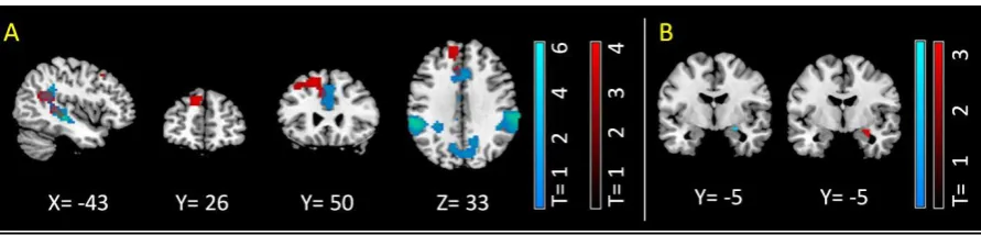
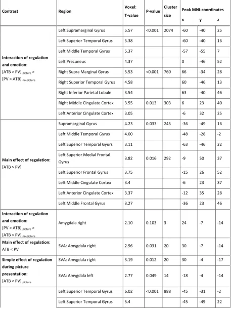
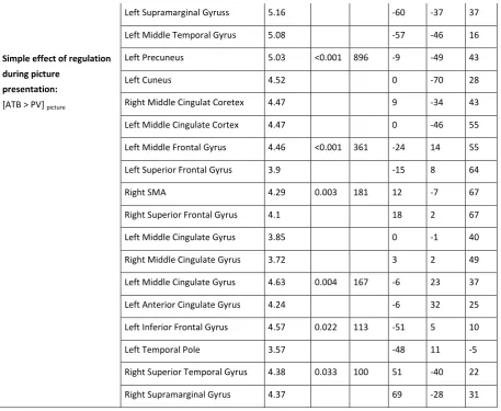
breathing frequency. Results indicate that (1) ATB was effective in regulating aversive emotions.
(2) Left dorso-medial prefrontal cortex was associated with ATB in general. (3) A fronto-parietal
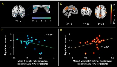
network was additionally recruited during emotional stimulation. (4) ATB down regulated
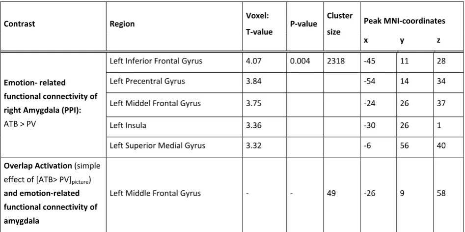
amygdala activity and increased amygdala-prefrontal integration, with such increased
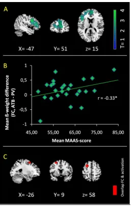
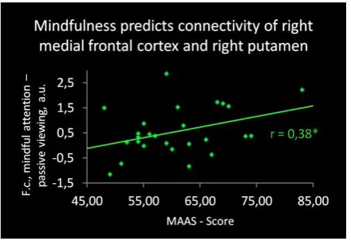
integration being associated with mindfulness ability. Results suggest amygdala-dorsal prefrontal
I
NTRODUCTION
Mindfulness practice is beneficial for the ability to regulate emotions (Goldin and Gross, 2010;
Hölzel et al., 2013; Taylor et al., 2011). For example, only 15 minutes of focused
attention-to-breath (ATB) practice reduces negative emotions of subsequently shown aversive pictures (Arch
and Craske, 2006). Mindfulness refers to attending to present moment experience and allowing
emotions and thoughts without judgment (Bishop et al., 2004). Mindfulness practice is
associated with a reduction in psychiatric symptoms particularly in disorders of emotion
regulation such as major depressive or anxiety disorders (Aldao et al., 2010; Baer, 2003;
Hofmann et al., 2010). Emotion regulation describes a variety of strategies that can influence
which emotions arise, when and how long they occur, and how these emotions are experienced
and expressed (Gross and Thompson, 2007). Imaging experiments have revealed aberrant brain
mechanisms underlying deficiencies in emotion regulation, e.g., in depression (Heller et al.,
2009; Johnstone et al., 2007), anxiety (Etkin and Wager, 2007), and bipolar disorder (Phillips et
al., 2008). Due to the relevance of mindfulness practice for emotion regulation, particularly in
neuropsychiatric disorders of impaired emotion regulation (Goldin and Gross, 2010; Hayes and
Feldman, 2004; Way et al., 2010), it is important to understand the neural mechanisms of the
effects of mindfulness practice on emotion regulation. The current study focuses on the effects
of mindfulness practice on aversive emotions at the level of behavior and brain activation by
using ATB as a paradigm for mindfulness practice. ATB is a basic technique of focused attention
meditation practice often used first to teach novices in mindfulness practice, and is also typically
applied by trained mindfulness practitioners in order to regulate their emotions in stressful
situations even years after training (Kabat-Zinn et al., 1987). Therefore, the analysis of neural
correlates of ATB effects on emotions appears a relevant paradigm to elucidate brain
mechanisms important for the link between mindfulness practice and emotion regulation.
Several studies have investigated the neural correlates of focused attention meditation
in general, and ATB in particular (Brefczynski-Lewis et al., 2007; Dickenson et al., 2013;
Hasenkamp and Barsalou, 2012; Hölzel et al., 2007). These studies reported the involvement of
brain regions related to attentional control, such as activation of a fronto-parietal network in
novice meditators (Dickenson et al., 2013), greater activation of anterior cingulate regions in
experienced meditators compared to non-meditators (Hölzel et al., 2007), and an inverted
u-shaped curve of brain activity in areas involved in sustained attention as a function of meditation
expertise (Brefczynski-Lewis et al., 2007). Few studies investigated neural correlates of focused
emotional contexts, these studies showed relatively increased activation of temporo-parietal
junction, pre- and postcentral gyri, right insula, medial and lateral dorsal PFC (Farb et al., 2007;
Taylor et al., 2011) and – in most cases - down regulation of amygdala activity (Farb et al., 2007;
Taylor et al., 2011; Zeidan et al., 2011). However, while these studies indicate the involvement of
prefrontal areas and the amygdala in emotion regulation effects of ATB, some points remain
unclear. First, it is incompletely understood how brain states of ATB such as increased prefrontal
and decreased amygdala activations interact with emotional stimulation. ATB is a typical state of
mindfulness practice, which might be modulated by emotional stimulation on the one hand and
act on stimulation processes on the other. So far, no study has directly compared activations
during stimulation with those of no stimulation. Second, it is unclear how, during emotional
stimulation, ATB relevant fronto-parietal areas integrate with amygdala activation. Amygdala
activity is involved in negative emotions and, during emotion regulation, this activity is directly
or indirectly controlled by prefrontal cortex activity (Davidson, 2002; Sotres-Bayon et al., 2004).
Since ATB has potential effects on breathing frequency, which might itself exert an influence on
emotion regulation, we controlled all data for breathing frequency. Previously, only one study
controlled for breathing frequency during meditation (Zeidan et al., 2011).
In the current study, we focused on these questions about neural mechanisms of ATB in
the context of emotion regulation. We hypothesized first that brain activation effects of ATB and
visual-emotional stimulation interact in prefrontal areas and amygdala. Secondly, we propose
that, during emotional stimulation, ATB down-regulates activation in the amygdala and increases
its integration with prefrontal regions, independently of effects on breathing frequency. To test
these two hypotheses, healthy participants were stimulated with aversive pictures during
functional MRI and instructed to either mindfully attend to their breath or to passively view the
images. To ensure that participants were able to focus their attention on the breath, this
technique was trained for two weeks before the experiment. Neural outcome measures were
both blood-oxygenation-level-dependent (BOLD) signals for brain activation and emotion-related
BOLD functional connectivity for amygdala integration during visual-emotional processing (i.e.,
psycho-physiological interaction analysis, PPI). The relationship of brain activation with both
regulation success on emotional valence ratings and dispositional mindfulness scores was
assessed via correlation analysis. ATB effects on breathing frequency were controlled via linear