Kejal Kantarci, MD, MS
Nirubol Tosakulwong
Timothy G. Lesnick, MS
Samantha M. Zuk
Jeffrey L. Gunter, PhD
Carey E. Gleason, PhD
Whitney Wharton, PhD
N. Maritza Dowling, PhD
Prashanthi Vemuri, PhD
Matthew L. Senjem, MS
Lynne T. Shuster, MD
Kent R. Bailey, PhD
Walter A. Rocca, MD,
MPH
Clifford R. Jack, Jr., MD
Sanjay Asthana, MD
Virginia M. Miller, PhD
Correspondence to Dr. Kantarci: [email protected]
Supplemental data at Neurology.org
Effects of hormone therapy on brain
structure
A randomized controlled trial
ABSTRACT
Objective:
To investigate the effects of hormone therapy on brain structure in a randomized,
double-blinded, placebo-controlled trial in recently postmenopausal women.
Methods:
Participants (aged 42
–
56 years, within 5
–
36 months past menopause) in the Kronos
Early Estrogen Prevention Study were randomized to (1) 0.45 mg/d oral conjugated equine
estro-gens (CEE), (2) 50
mg/d transdermal 17b-estradiol, or (3) placebo pills and patch for 48 months.
Oral progesterone (200 mg/d) was given to active treatment groups for 12 days each month. MRI
and cognitive testing were performed in a subset of participants at baseline, and at 18, 36, and
48 months of randomization (n
5
95). Changes in whole brain, ventricular, and white matter
hyperintensity volumes, and in global cognitive function, were measured.
Results:
Higher rates of ventricular expansion were observed in both the CEE and the
17b-estra-diol groups compared to placebo; however, the difference was significant only in the CEE group
(
p
5
0.01). Rates of ventricular expansion correlated with rates of decrease in brain volume (
r
5
2
0.58;
p
#
0.001) and with rates of increase in white matter hyperintensity volume (
r
5
0.27;
p
5
0.01) after adjusting for age. The changes were not different between the CEE and 17b-estradiol
groups for any of the MRI measures. The change in global cognitive function was not different
across the groups.
Conclusions:
Ventricular volumes increased to a greater extent in recently menopausal women
who received CEE compared to placebo but without changes in cognitive performance. Because
the sample size was small and the follow-up limited to 4 years, the findings should be interpreted
with caution and need confirmation.
Classification of evidence:
This study provides Class I evidence that brain ventricular volume
increased to a greater extent in recently menopausal women who received oral CEE compared
to placebo.
Neurology®2016;87:887–896GLOSSARY
CEE5conjugated equine estrogens;FLAIR5fluid-attenuated inversion recovery;KEEPS5Kronos Early Estrogen Pre-vention Study;WHIMS5Women’s Health Initiative Memory Study;WMH5white matter hyperintensity.
Hormone therapy with conjugated equine estrogens (CEE) and medroxyprogesterone acetate
ini-tiated later in menopause increased the risk of dementia in the Women’s Health Initiative
Memory Study (WHIMS).
1Whether alternative formulations of hormone therapy can preserve
neuronal integrity and decrease the risk of dementia when administered early in menopause
remains controversial.
2–10Determining the effects of hormone therapy during the early
postmen-opausal years on the risk of dementia would require decades of follow-up. However, noninvasive
imaging markers related to cognitive health have been suggested as short-term surrogate outcomes
to assess the effects of menopausal hormone therapy on the brain. Thus, age-associated changes in
From the Departments of Radiology (K.K., S.M.Z., J.L.G., P.V., M.L.S., C.R.J.), Health Sciences Research (N.T., T.G.L., K.R.B., W.A.R.), Internal Medicine (L.T.S.), Neurology (W.A.R.), and Surgery and Physiology and Biomedical Engineering (V.M.M.), Mayo Clinic, Rochester, MN; Department of Medicine (C.E.G., S.A.), School of Medicine and Public Health, University of Wisconsin and Geriatric Research, Education and Clinical Center, William S. Middleton Memorial, Veterans’Hospital, Madison, WI; Department of Neurology (W.W.), Emory University, Atlanta, GA; and Department of Biostatistics and Medical Informatics (N.M.D.), University of Wisconsin, Madison.
Go to Neurology.org for full disclosures. Funding information and disclosures deemed relevant by the authors, if any, are provided at the end of the article. The Article Processing Charge was paid by the authors.
brain structure on MRI can be used to measure
the effects of estrogens on the brains of
post-menopausal women.
The Kronos Early Estrogen Prevention Study
(KEEPS) tested the hypothesis that menopausal
hormone therapy administered early after the
onset of menopause would slow progression of
atherosclerosis.
11However, hormone therapy in
KEEPS did not affect progression of
atheroscle-rosis
12or cognitive function
13within the 4 years
of hormone therapy. We report here the results
of an ancillary study to KEEPS, conducted to
determine the effects of oral CEE and
transder-mal 17
b
-estradiol therapy on changes in
struc-tural brain MRI over 4 years. The ventricular
volume change was chosen as the primary
out-come measure because it was the most reliable
measure of the change in brain structure
associ-ated with aging and cognitive function.
14The
secondary outcome measures included changes
in whole brain and white matter hyperintensity
(WMH) volumes.
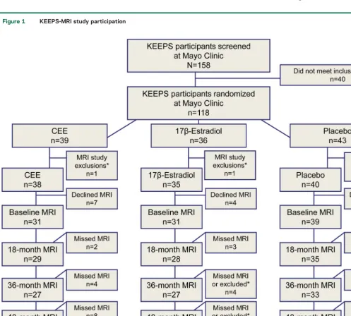
METHODS Participants.KEEPS was a multicenter, random-ized, double-blinded, placebo-controlled clinical trial in recently menopausal women (n5727). Participants enrolled in KEEPS were between 42 and 59 years of age, within 5 to 36 months past their last menses, and were in good cardiovascular health.11An ancillary MRI study to KEEPS was conducted from February 2006 through August 2011 at the Mayo Clinic to investigate the effects of hormone treatment on brain structure. Exclusion criteria for enrollment in the ancillary KEEPS-MRI study were MRI contraindications for safety and neurologic disorders.
Baseline MRI examinations and cognitive testing were com-pleted following randomization but with concealed allocation and before treatment initiation. The treatments were as follows: (1) oral CEE (0.45 mg/d; Premarin, Pfizer, New York, NY); (2) transdermal 17b-estradiol (17b-estradiol skin patch, 50mg/d; Climara, Bayer HealthCare Pharmaceuticals Inc., Wayne, NJ); or (3) placebo pills and patch. Estrogens were administered through 2 different routes, oral or transdermal, because of the increased risk of venous thrombosis with oral estrogens. It was hypothesized that transdermal 17b-estradiol would have a differ-ent effect on risk factors for atherosclerosis and thromboembolic disease compared to oral CEE. Progesterone was given orally (micronized progesterone, 200 mg/d; Prometrium, AbbVie Inc., North Chicago, IL) for 12 days at the beginning of each month to both active treatment groups to protect the endome-trium. Follow-up MRI examinations and cognitive testing were performed at 18 months (estrogen-only phase), 36 months (pro-gesterone phase; to determine whether there would be an effect on the trajectory of change in outcome measures due to proges-terone), and at 48 months (estrogen-only phase).11
Standard protocol approvals, registrations, and patient consents. The present study (NCT00154180; https://clinicaltrials.gov/ct2/show/NCT00154180) was approved by the Mayo Clinic institutional review board (no. 224104). All participants provided written informed consent.
Cognitive testing.A confirmatory factor analysis was used to assess the underlying structure of baseline cognitive data from the KEEPS cognitive and affective study, and to derive summary scores (n5662).15Using standard criteria for model fit, the cognitive variables were summarized in 4 specific independent domains and a general domain representing global cognitive function.16
Magnetic resonance imaging.MRI studies were performed on a single 1.5-tesla system, with an 8-channel phased-array coil (GE Healthcare, Waukesha, WI). A 3-dimensional, magnetization-prepared rapid-acquisition gradient echo sequence was acquired for volumetric analysis, and fluid-attenuated inversion recovery (FLAIR) MRI was acquired for quantification of WMH volume. Changes in ventricle and whole brain volumes were calculated automatically from each registered magnetization-prepared rapid-acquisition gradient echo scan pair using the boundary shift integral and expressed in cubic centimeters of volume change from baseline for each follow-up time point as previously described.17The boundary shift integral is designed to monitor brain structural changes and treatment effects in clinical trials of Alzheimer disease.18
WMH volumes were derived from semiautomated segmenta-tion of FLAIR images as previously described.19Briefly, WMH on FLAIR images were segmented using an automated slice-based seed initialization and region growing method. A trained image analyst (S.Z.), blinded to treatment group as well as to the sequence of the follow-up scans, inspected the segmented WMH mask overlaid on the FLAIR image. Every segmented image was visually compared to the unprocessed FLAIR image and false-positive WMH labels resulting from artifacts were edi-ted to be excluded from the WMH mask. The WMH masks on follow-up scans were also visually compared to the baseline WMH mask for consistent editing of the artifacts. One of the baseline FLAIR scans failed quality control and was therefore excluded from the analysis. Longitudinal change in WMH was expressed in cubic centimeters of volume change from baseline for each follow-up MRI.
Statistical analysis. Baseline characteristics were compared among groups using analysis of variance followed by Tukey pair-wise tests, 2-samplettests, or Fisher exact tests, as appropriate. Changes in the outcomes from baseline were shown using plots of mean values and their associated 95% confidence intervals at each time point. Associations between the outcome variables at 48 months were shown using scatterplots with Pearson correlation coefficients adjusted for age. In a secondary analysis, the percent change in the whole brain, ventricular, and WMH volumes over 18, 36, and 48 months in CEE and 17b-estradiol groups was compared to placebo byttests.
reporting these results. Furthermore, results from the 2 hormone therapy groups were not pooled because of the differences in formulations and route of administration.
For our primary analysis of ventricular volumes using linear mixed models, witha50.05 and the smallest group size of 29 for conservative estimates, there was 80% power to detect a differ-ence in slopes over time between groups of 0.296 cm3per year. For example, for a woman starting at the mean baseline ventricular volume of 19.6 cm3, a 1.5% increase could be detected per year because of treatment (a 2.3% increase in the volume after 18 months, 4.5% after 36 months, and 6.0% after 48 months).
RESULTS
All women enrolled in KEEPS at Mayo
Clinic Rochester (n
5
118) were invited to
participate in the ancillary MRI study. Five
partici-pants were excluded because of neurologic disorders
or MRI contraindications; 12 women declined
par-ticipation. Eligible women underwent a baseline MRI
(n
5
101) before starting treatment. MRI data were
analyzed for the 95 participants who repeated at least
one MRI examination at 18 (n
5
92), 36 (n
5
87),
or 48 (n
5
79) months (figure 1). Baseline global
cognitive performance and whole brain volumes were
not different among treatment and placebo groups.
However, ventricular volumes were larger in the 17
b
-estradiol treatment group (
p
5
0.025) and WMH
volumes were larger in the CEE treatment group
Figure 1 KEEPS-MRI study participation
(
p
5
0.008) compared to placebo at baseline after
adjusting for total intracranial volume (table 1). None
of the participants had silent infarcts on baseline and
follow-up MRI examinations.
Whole brain volumes decreased in the CEE (
p
5
0.004) and the 17
b
-estradiol (
p
5
0.002) treatment
groups, but not in the placebo group (
p
5
0.09) over
48 months. However, the decline in whole brain
vol-ume was not significantly different between the placebo
and treatment groups (CEE and 17
b
-estradiol).
Ven-tricular volumes increased in both the treatment (CEE
and 17
b
-estradiol) and the placebo groups over 48
months (
p
,
0.001). Increases in ventricular volumes
were greater only in the CEE treatment group
com-pared to the placebo group (
p
5
0.01). WMH volume
increased in the CEE (
p
5
0.004) and 17
b
-estradiol
(
p
5
0.002) groups, but not in the placebo group (
p
5
0.42) over 48 months. However, the increase in WMH
volumes was not different when comparing the CEE
(
p
5
0.10) and 17
b
-estradiol (
p
5
0.06) groups to the
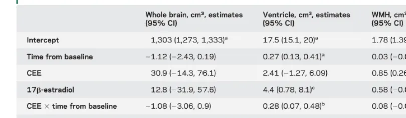
placebo group (figure 2). The results of mixed-effects
models are summarized in table 2. Changes in any of
the MRI measures did not differ between the CEE and
17
b
-estradiol groups (table e-1 at Neurology.org).
Because baseline WMH volumes were higher in
the CEE group than in the placebo group, we tested
whether baseline WMH modified the treatment
3
time interactions by adding baseline WMH and
base-line WMH
3
time to the mixed models. The
param-eter estimates and
p
values for treatment
3
time were
then compared with and without the baseline
adjust-ment to see if they differed. The primary interest was
in possible effects of baseline WMH on the
signifi-cant CEE vs placebo difference in ventricular volume
change. After adjusting for baseline WMH, increases
in ventricular volumes in the CEE treatment group
remained greater than in the placebo group (
p
5
0.011), but did not reach statistical significance in
Table 1 Baseline characteristics of the participants
CEE (n529) 17b-Estradiol (n530) Placebo (n536)
Age, y 53 (52, 54) 53 (52, 54) 53 (52, 54)
Education
High school or less 2 (7) 1 (4) 3 (8)
Some college/college graduate 19 (70) 18 (64) 22 (61)
Some graduate/graduate 6 (22) 9 (32) 11 (31)
Smoking status
Nonsmoker 15 (68) 12 (55) 24 (73)
Smoker (past or current) 7 (32) 10 (45) 9 (27)
Time past menopause, mo 21 (17, 25) 20 (17, 23) 17 (14, 20)
Treatment onset past baseline MRI, d 13 (3, 23) 20 (10, 30) 28 (7, 49)
APOEe4 carrier 4 (15) 13 (45) 7 (21)
Migraines 3 (10) 0 (0) 4 (11)
Global cognitive function scores 20.19 (20.48, 0.10) 0.16 (20.12, 0.44) 0.14 (20.08, 0.36)
Mean systolic blood pressure, mm Hg 122 (117, 127) 120 (114, 126) 122 (118, 126)
Mean diastolic blood pressure, mm Hg 77 (74, 80) 73 (70, 76) 76 (74, 78)
Waist circumference, cm 87 (82, 92) 83 (79, 87) 84 (80, 88)
Body mass index, kg/m2 28 (26, 30) 26 (25, 27) 27 (26, 28)
Coronary arterial calcification present 4 (14) 3 (10) 4 (11)
Carotid intima-media thickness 0.71 (0.67, 0.75) 0.72 (0.68, 0.76) 0.71 (0.68, 0.74)
Low-density lipoprotein, mg/dL 134 (124, 144) 137 (125, 149) 130 (119, 141)
High-density lipoprotein, mg/dL 59 (54, 64) 63 (58, 68) 60 (55, 65)
Triglycerides, mg/dL 93 (78, 108) 101 (81, 121) 91 (76, 106)
Whole brain volume, cm3 1,334 (1,300, 1,368) 1,316 (1,285, 1,347) 1,302 (1,269, 1,335)
Ventricular volume, cm3 19.6 (16.8, 22.4) 22.1 (18.7, 25.5)a 17.6 (15.5, 19.7)
White matter hyperintensity volume, cm3 2.7 (2.1, 3.3)b 2.3 (1.8, 2.8) 1.8 (1.6, 2.0)
Abbreviation: CEE5conjugated equine estrogens. Data are n (%) or mean (95% confidence interval).
the 17
b
-estradiol group compared with the placebo
group (
p
5
0.06) (table e-2).
Percent increases in ventricular volume from
base-line to 18, 36, and 48 months were greater in the
CEE group but not in the 17
b
-estradiol group
com-pared to placebo at all time points. On the contrary,
percent increases in WMH volume from baseline
were greater in the CEE group only at 48 months
and in the 17
b
-estradiol group only at 18 months
compared to placebo (table 3).
Global cognitive function did not change in the
treatment or the placebo group, and there were no
differences in global cognitive change between CEE,
17
b
-estradiol, and placebo groups over 48 months
(figure 2).
Over the 48 months of follow-up, the increase in
ventricular volume correlated with the decline in
whole brain volume (
r
5 2
0.58;
p
#
0.001) and
with the increase in WMH volume (
r
5
0.27;
p
5
0.01) after adjusting for age.
In the CEE group, increases in ventricular volume
at 18 months correlated with timing of initiation of
treatment in relation to the onset of menopause, after
adjusting for age. Women who initiated hormone
therapy later in menopause had greater increases in
ventricular volume at 18 months (
r
5
0.40;
p
5
0.03). This association was weaker at 36 months
(
r
5
0.35;
p
5
0.07) and at 48 months (
r
5
0.26;
p
5
0.24). By contrast, there were no correlations
between increases in ventricular volume and timing
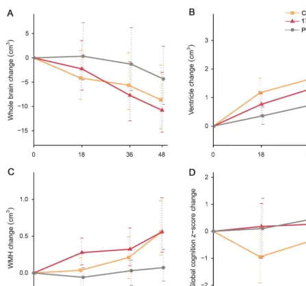
Figure 2 Changes in the MRI raw value measurements and global cognitive function in treatment groupsMean change (cm3) and 95% confidence intervals at 18, 36, and 48 months for whole brain volume (A), ventricular volume (B), and WMH volume (C). Mean
change in global cognitionzscores and 95% confidence intervals at 18, 36, and 48 months (D) is displayed. Increases in ventricular volumes were greater in the CEE treatment group than in the placebo group (p50.01), but not in the 17b-estradiol group compared to the placebo group (p50.09). CEE5
of initiation of 17
b
-estradiol treatment, after
adjust-ing for age.
DISCUSSION
The major finding of this study is that
the rates of increase in ventricular volumes were
greater in recently menopausal women who received
oral CEE therapy compared to placebo over 4 years
without significant differences in changes in global
cognition. Although the rates of changes in MRI
measures did not differ between the CEE and
17
b
-estradiol groups, the rates of increase in
ventric-ular volume did not reach statistical significance in
the 17
b
-estradiol group, compared to placebo,
prob-ably because of our limited sample size and short
follow-up. Furthermore, increases in ventricular
volume over 18 months of treatment were greater
in the CEE group if hormone therapy was initiated
later in menopause.
It is difficult to compare the findings of the
pres-ent study with the findings of other studies in the
lit-erature, because the earlier studies were conducted in
older women, and the structural MRI assessments
were cross-sectional and not longitudinal, and were
performed many years after initiation of hormone
therapy. However, given these limitations, greater
rates of structural brain changes with CEE therapy
compared to placebo in the present study are
consis-tent with the finding of lower regional brain volumes
in women who initiate hormone therapy at age 65
years or older in the WHIMS-MRI, when the
struc-tural MRI changes were observed after the study
med-ications were stopped.
20Our findings also agree with
observational studies that found larger ventricular
vol-umes in women using hormone therapies
21,22but
contradict other studies that showed larger brain
volumes in women who used postmenopausal
hor-mone therapy compared to women who did not.
23–26Although measurements of both whole brain and
ventricular volumes are surrogates for the change in
global brain structure and correlate with each other,
differences between CEE treatment and placebo
groups were statistically significant when comparing
the rates of ventricular volume change but not the
rates of whole brain volume change. This may be
attributable to the differences in noise and
measure-ment variability. Indeed, smaller sample sizes are
needed to detect treatment effects using ventricular
volume change compared to whole brain volume
change.
14Brain volumes quantified on MRI decrease and
ventricular volumes increase during physiologic
aging.
27The decrease in brain volume accelerates in
cognitively normal older adults several years before
they develop mild cognitive impairment
14or
demen-tia.
28Although structural MRI changes are adequate
surrogate markers for tracking the progression of
pre-clinical neurodegenerative brain pathology many
years before the onset of cognitive decline, they may
not be ideal markers for detecting the effects of
spe-cific treatments that might modify brain function or
pathology.
29Evidence of hormonal effects on brain
structure in younger women is limited to studies in
premenopausal women. Premenstrual decreases in
estrogen and progesterone levels are associated with
increased ventricular volumes,
30and postmenstrual
increases in estrogen levels are associated with
preser-vation of ventricular structure and even increases in
hippocampal volume.
31Furthermore, larger gray
mat-ter volumes were found in oral contraceptive users
compared to nonusers.
32Both cyclic increases in
Table 2 Annual change in imaging markers on mixed-effects modelWhole brain, cm3, estimates
(95% CI)
Ventricle, cm3, estimates
(95% CI)
WMH, cm3, estimates
(95% CI)
Intercept 1,303 (1,273, 1,333)a 17.5 (15.1, 20)a 1.78 (1.39, 2.17)a
Time from baseline 21.12 (22.43, 0.19) 0.27 (0.13, 0.41)a 0.03 (20.04, 0.09)
CEE 30.9 (214.3, 76.1) 2.41 (21.27, 6.09) 0.85 (0.26, 1.43)b
17b-estradiol 12.8 (231.9, 57.6) 4.4 (0.78, 8.1)c 0.58 (20.007, 1.16)
CEE3time from baseline 21.08 (23.06, 0.9) 0.28 (0.07, 0.48)b 0.08 (20.015, 0.18)
17b-estradiol3time from
baseline 2
1.14 (23.1, 0.81) 0.18 (20.03, 0.39) 0.09 (20.002, 0.19)
Abbreviations: CEE5conjugated equine estrogens; CI5confidence interval; WMH5white matter hyperintensity. Linear mixed-effects models with time from baseline, treatment group, and their interaction as predictors, whole brain, ventricle, and WMH volume change as outcomes, and random subject-specific intercepts and slopes are reported for the CEE, 17b-estradiol, and placebo groups. Coefficient estimates with their associated 95% CIs and categories of signifi-cance are reported for these models. Treatment3time interactions were of interest because any treatment effects would likely manifest as a change in the trajectories over time.
endogenous hormones and exogenous administration
of hormones appear to preserve ventricular volumes
and increase brain volumes in premenopausal
women, in contrast to the findings of the present
study with exogenous administration of hormones
in postmenopausal women. Therefore, transient
retention of sodium and water, which has been
impli-cated in the premenstrual increases in ventricular
vol-ume when endogenous estrogen is low, would not
explain the finding of increased ventricular volume
in postmenopausal women who received estrogens.
WMH volume gradually increased only in the
CEE and 17
b
-estradiol treatment groups but not
the placebo group. These increases in WMH volumes
over 48 months correlated with the increases in
ven-tricular volumes, suggesting that similar mechanisms
may be driving both of these structural changes.
Accelerated volumetric changes in brain structure
and WMH are associated with cognitive decline in
older adults, including ischemic small vessel disease
and Alzheimer disease.
33A relationship between
lon-gitudinal increases in WMH and ventricular volumes
has been reported in a cohort with atherosclerosis,
slightly older than the participants of the
KEEPS-MRI study (mean age 53 vs 57 years) and with a high
percentage of men, suggesting that cerebral small
vessel disease may underlie the parallel increases in
WMH and ventricular volumes.
34Although the
pathologic underpinnings of WMH and ventricular
volume increases in women with good
cardiovascu-lar health is unclear, there is evidence that the
WMH increases in participants of the
KEEPS-MRI study were associated with thrombogenic
mi-crovesicles in the blood at baseline.
19Thrombogenic
microvesicles are related to the progression of
atherosclerosis.
35In the CEE group, ventricular volumes increased
less in women who initiated hormone therapy earlier
than later in menopause, albeit only during the first
18 months. This association gradually weakened at
36 and 48 months of treatment. Since the mean time
to starting CEE was 20 months past menopause
(range: 5
–
36), the window in which hormone therapy
causes less ventricular expansion appears to be limited
to approximately 3 years into menopause. This time
window may be associated with a gradual loss of
estrogen receptor
a
in the postmenopausal brain.
36Women participating in the KEEPS-MRI study
were in good cardiovascular and neurologic health,
and well educated, which may limit generalization
of findings to a broader population. However, in this
homogeneous population of relatively healthy
women, hormone therapy effects on brain structure
were independent of concurrent cardiovascular or
neurologic diseases. Furthermore, the sample size
and the MRI changes in brain volumes were small,
Table 3 Percentage of change in imaging markers Months Whole brain volume Ventr icular volume White matte r hyp erintensities CEE mean change, % Plac ebo mean change, % Mean % differen ce (95% CI) p Value CEE mean change, % Pla cebo mean chang e, % Mean % diff erence (95% CI) p Value CEE mean chang e, % Placebo mean change, % Mean % difference (95% CI) p Value 18 2 0.3 0.034 2 0.34 ( 2 0.91, 0.24) 0.25 6.42 2.11 4.31 (1.5 7, 7) 0.003 3.88 2 1.56 5.45 ( 2 5.2, 16.1 ) 0.31 36 2 0.42 2 0.09 2 0.33 ( 2 0.97, 0.32) 0.31 9.35 3.96 5.39 (1.5 8, 9.2) 0.006 10.7 5.61 5.07 ( 2 11.3, 21.4) 0.54 48 2 0.64 2 0.33 2 0.31 ( 2 1, 0.39) 0.38 12.3 5.84 6.5 (2.27, 11) 0.003 26.7 2.6 24.1 (1.88, 46.4) 0.03 Months 17 b -Estrad iol mean change, % Placebo mean change, % Mean % diff erence (95% CI) p Value 17 b -Estrad iol mean chang e, %
Placebo mean change,
limiting statistical power. Therefore, the changes
observed in the 17
b
-estradiol group compared to
pla-cebo may reach statistical significance with a longer
duration of the study. Further investigation including
larger numbers of participants and longer follow-up is
warranted. Even though the treatment and placebo
groups did not differ at baseline regarding
cardiovascu-lar risk factors, the CEE group had greater WMH
volume than the placebo group. Some of the
differ-ences observed across groups at baseline are not
sur-prising given the relatively small number of women
included in the MRI ancillary study. Randomization
does not guarantee a balanced allocation across
treat-ment groups when the numbers are small. We found
no evidence that greater WMH volume at baseline
influenced the findings on ventricular volume change.
We acknowledge, however, that post hoc statistical
comparisons are subject to biases.
Among older women who received estrogen
treat-ment in the WHIMS-MRI, lower regional brain
vol-ume was associated with greater cognitive decline or
dementia.
37By contrast, we observed a change in
brain structure over 4 years of hormone therapies that
did not correlate with changes in cognitive function
of recently menopausal women. The interpretation of
this absence of an association between imaging
find-ings and cognitive findfind-ings remains uncertain. On
one hand, it is possible that the women who
experi-enced structural brain changes with hormone
thera-pies early in menopause will develop cognitive decline
during an extended follow-up, and if proven, the
imaging findings would be useful early surrogates of
future clinical events. On the other hand, it is possible
that these brain changes will subside after cessation of
hormone therapy, and women who had structural
brain changes during the 4 years of postmenopausal
hormone therapy will not have an increased risk of
cognitive decline over a longer follow-up.
38In the
WHIMS-MRI studies, even though the brain
vol-umes were smaller in women who received CEE
ther-apies compared to placebo years after the end of the
treatment phase, the rates of decline in brain volumes
remained similar to the placebo group in the years
following the cessation of CEE therapies.
39Thus, the
short-term effects of estrogen treatment on the
vas-cular system and on the brain may be different from
long-term effects, and even opposed. Participants of
the KEEPS-MRI study are being followed to
deter-mine whether these changes in brain structure are
persistent after 4 years of hormone treatment and
whether women with structural brain changes will
develop clinically detectable cognitive decline.
AUTHOR CONTRIBUTIONS
Dr. Kantarci: design or conceptualization of the study, data collection, analysis and interpretation of the data, drafting the manuscript, study funding. Ms. Tosakulwong: analysis or interpretation of the data, revising the manuscript. Mr. Lesnick: analysis or interpretation of the data, revising the manuscript. Ms. Zuk: analysis or interpretation of the data, revising the manuscript. Dr. Gunter: analysis or interpretation of the data, revising the manuscript. Dr. Gleason: data collection, analysis or interpretation of the data, revising the manuscript. Dr. Wharton: data collection, analysis or interpretation of the data, revising the manuscript. Dr. Dowling: analysis or interpretation of the data, revising the manu-script. Dr. Vemuri: analysis or interpretation of the data, revising the manuscript. Mr. Senjem: analysis or interpretation of the data, revising the manuscript. Dr. Shuster: analysis or interpretation of the data, revis-ing the manuscript. Dr. Bailey: analysis or interpretation of the data, revising the manuscript. Dr. Rocca: analysis or interpretation of the data, revising the manuscript. Dr. Jack: analysis or interpretation of the data, revising the manuscript. Dr. Asthana: data collection, analysis or interpretation of the data, revising the manuscript. Dr. Miller: design or conceptualization of the study, data collection, analysis or interpreta-tion of the data, revising the manuscript, study funding.
STUDY FUNDING
This study is funded by the Aurora Foundation to the Kronos Longevity Research Institute and the NIH (NS66147, AG029624, and AG44170). The funding sources had no role in study design, collection, analysis,
Comment:
Hormone therapy in the early postmenopausal stage—
Safe for the brain?
Recent clinical trials, including the Women’s Health Initiative Memory Study of Younger Women,1 provide reassurance that hormone therapy (HT)
confers no cognitive risk when taken early in the postmenopausal period. In contrast, results from this ancillary neuroimaging study from the Kronos Early Estrogen Prevention Study (KEEPS)2suggest a potential adverse effect of HT—
increased ventricular volume—in women randomized to conjugated equine estro-gen (CEE) early in the postmenopausal stage. These findings deserve consider-ation because brain changes related to Alzheimer disease are detectable earlier than neuropsychological changes.
This neuroimaging study is unique because it was embedded in a blinded ran-domized controlled trial, included 2 HT formulations, and repeatedly assessed mul-tiple neuroimaging outcomes over a relatively long 4-year follow-up. Despite these strengths, the findings should be interpreted with caution because sample sizes were quite small and there were some differences in baseline neuroimaging measures across treatment groups. Critically, despite an emphasis on the adverse CEE outcomes, the ventricular volume findings in the transdermal 17b-estradiol group were similar to the CEE group but the effect just missed statistical significance (p50.06). Indeed, there were no differences in ventricular volume between CEE and 17b-estradiol groups, and their ventricular volumes were nearly identical at the end of the study.
Continued follow-up in KEEPS is warranted to determine the reproducibility of the cognitive and neuroimaging findings as women age and to address an issue of critical clinical importance that KEEPS is uniquely positioned to address. HT re-mains the standard treatment for moderate to severe vasomotor symptoms in women with no contraindications to HT. Approximately 28.5% of postmenopausal women younger than 55 years experience moderate to severe vasomotor symptoms, and these symptoms interfere with health- and work-related quality of life.3
Increased awareness of sex difference in Alzheimer disease underscores the need to identify sex-related risk factors for cognitive decline and dementia.
1. Espeland MA, Shumaker SA, Leng I, et al. Long-term effects on cognitive function of postmenopausal hormone therapy prescribed to women aged 50 to 55 years. JAMA Intern Med 2013;173:1429–1436.
2. Kantarci K, Tosakulwong N, Lesnick TG, et al. Effects of hormone therapy on brain structure: a randomized controlled trial. Neurology 2016;87:887–896.
3. Gartoulla P, Worsley R, Bell RJ, Davis SR. Moderate to severe vasomotor and sexual symptoms remain problematic for women aged 60 to 65 years. Menopause 2015;22: 694–701.
Pauline M. Maki, PhD
From the Departments of Psychiatry and Psychology, University of Illinois at Chicago. Study funding: No targeted funding reported.
interpretation, or decision to submit this paper. The corresponding author had full access to all the data in the study and had final respon-sibility for the decision to submit for publication.
DISCLOSURE
K. Kantarci serves on the data safety monitoring board for Pfizer Inc., Takeda Global Research & Development Center, Inc. She is funded by the NIH (R01AG040042, principal investigator [PI], R21 NS066147 [PI], Mayo Clinic Alzheimer’s Disease Research Center/pro-ject 1 P50 AG16574/P1 [PI], P50 AG44170/proCenter/pro-ject 2 [PI], and R01 AG11378 [coinvestigator]). N. Tosakulwong, T. Lesnick, S. Zuk, J. Gunter, C. Gleason, W. Wharton, and N. Dowling report no disclo-sures relevant to the manuscript. P. Vemuri is funded by the NIH. M. Senjem holds stock in Gilead Sciences, Inc., Inovio Pharmaceuticals, Medtronic, Oncothyreon, Inc., and PAREXEL International. L. Shuster is funded by the NIH (P50 AG44170/core B [PI]). K. Bailey is funded by the NIH (P50 AG44170/core B [coinvestigator]). W. Rocca reports no disclosures relevant to the manuscript. C. Jack provides consulting services for Eli Lilly. He is funded by the NIH (R01-AG011378, RO1-AG037551, U01-HL096917, U01-AG032438, U01-AG024904) and Alexander Family Alzheimer’s Disease professorship of the Mayo Foun-dation. S. Asthana is funded by the NIH. V. Miller is funded by the NIA (P50 AG44170 [PI]). Go to Neurology.org for full disclosures.
Received June 17, 2015. Accepted in final form April 22, 2016.
REFERENCES
1. Shumaker SA, Reboussin BA, Espeland MA, et al. The Women’s Health Initiative Memory Study (WHIMS): a trial of the effect of estrogen therapy in preventing and slowing the progression of dementia. Control Clin Trials 1998;19:604–621.
2. Espeland MA, Shumaker SA, Leng I, et al. Long-term effects on cognitive function of postmenopausal hormone therapy prescribed to women aged 50 to 55 years. JAMA Intern Med 2013;173:1429–1436.
3. LeBlanc ES, Janowsky J, Chan BK, Nelson HD. Hor-mone replacement therapy and cognition: systematic review and meta-analysis. JAMA 2001;285:1489–1499. 4. Prentice RL, Manson JE, Langer RD, et al. Benefits and
risks of postmenopausal hormone therapy when it is initiated soon after menopause. Am J Epidemiol 2009;170:12–23. 5. Sherwin BB. Estrogen and memory in women: how can
we reconcile the findings? Horm Behav 2005;47:371–375. 6. Waring SC, Rocca WA, Petersen RC, O’Brien PC, Tangalos EG, Kokmen E. Postmenopausal estrogen replacement therapy and risk of AD: a population-based study. Neurology 1999;52:965–970.
7. Whitmer RA, Quesenberry CP, Zhou J, Yaffe K. Timing of hormone therapy and dementia: the critical window theory revisited. Ann Neurol 2011;69:163–169. 8. Yaffe K, Sawaya G, Lieberburg I, Grady D. Estrogen
ther-apy in postmenopausal women: effects on cognitive func-tion and dementia. JAMA 1998;279:688–695. 9. Zandi PP, Carlson MC, Plassman BL, et al. Hormone
replacement therapy and incidence of Alzheimer disease in older women: the Cache County Study [see comment]. JAMA 2002;288:2123–2129.
10. Rocca WA, Grossardt BR, Shuster LT. Oophorectomy, estrogen, and dementia: a 2014 update. Mol Cell Endo-crinol 2014;389:7–12.
11. Harman SM, Brinton EA, Cedars M, et al. KEEPS: the Kronos Early Estrogen Prevention Study. Climacteric 2005;8:3–12.
12. Harman SM, Black DM, Naftolin F, et al. Arterial imag-ing outcomes and cardiovascular risk factors in recently
menopausal women: a randomized trial. Ann Intern Med 2014;161:249–260.
13. Gleason CE, Dowling NM, Wharton W, et al. Effects of hormone therapy on cognition and mood in recently post-menopausal women: findings from the KEEPS Cognitive and Affective Study. PLoS Med 2015;12:e1001833. 14. Jack CR Jr, Shiung MM, Gunter JL, et al. Comparison of
different MRI brain atrophy rate measures with clinical disease progression in AD. Neurology 2004;62:591–600. 15. Dowling NM, Gleason CE, Manson JE, et al. Character-ization of vascular disease risk in postmenopausal women and its association with cognitive performance. PLoS One 2013;8:e68741.
16. Gleason CE, Dowling NM, Wharton W, et al. Effects of hormone therapy on cognition and mood in recently post-menopausal women: findings from the randomized, con-trolled KEEPS–Cognitive and Affective Study. PLoS Med 2015;12:e1001833; discussion e1001833.
17. Gunter JL, Shiung MM, Manduca A, Jack CR Jr. Meth-odological considerations for measuring rates of brain atro-phy. J Magn Reson Imaging 2003;18:16–24.
18. Fox NC, Cousens S, Scahill R, Harvey RJ, Rossor MN. Using serial registered brain magnetic resonance imaging to measure disease progression in Alzheimer disease: power calculations and estimates of sample size to detect treatment effects [see comment]. Arch Neurol 2000;57:339–344. 19. Raz L, Jayachandran M, Tosakulwong N, et al.
Thrombo-genic microvesicles and white matter hyperintensities in postmenopausal women. Neurology 2013;80:911–918. 20. Resnick SM, Espeland MA, Jaramillo SA, et al.
Postmen-opausal hormone therapy and regional brain volumes: the WHIMS-MRI Study. Neurology 2009;72:135–142. 21. Greenberg DL, Payne ME, MacFall JR, Provenzale JM,
Steffens DC, Krishnan RR. Differences in brain volumes among males and female hormone-therapy users and non-users. Psychiatry Res 2006;147:127–134.
22. Luoto R, Manolio T, Meilahn E, et al. Estrogen replace-ment therapy and MRI-demonstrated cerebral infarcts, white matter changes, and brain atrophy in older women: the Cardiovascular Health Study. J Am Geriatr Soc 2000;48:467–472.
23. Boccardi M, Ghidoni R, Govoni S, et al. Effects of hormone therapy on brain morphology of healthy post-menopausal women: a voxel-based morphometry study. Menopause 2006;13:584–591.
24. Eberling JL, Wu C, Haan MN, Mungas D, Buonocore M, Jagust WJ. Preliminary evidence that estrogen protects against age-related hippocampal atrophy. Neurobiol Aging 2003;24:725–732.
25. Ha DM, Xu J, Janowsky JS. Preliminary evidence that long-term estrogen use reduces white matter loss in aging. Neurobiol Aging 2007;28:1936–1940.
26. Lord C, Buss C, Lupien SJ, Pruessner JC. Hippocampal volumes are larger in postmenopausal women using estro-gen therapy compared to past users, never users and men: a possible window of opportunity effect. Neurobiol Aging 2008;29:95–101.
27. Sowell ER, Peterson BS, Thompson PM, Welcome SE, Henkenius AL, Toga AW. Mapping cortical change across the human life span. Nat Neurosci 2003;6:309–315. 28. Kaye JA, Swihart T, Howieson D, et al. Volume loss of the
29. Fox NC, Black RS, Gilman S, et al. Effects of Abeta immunization (AN1792) on MRI measures of cerebral volume in Alzheimer disease. Neurology 2005;64:1563– 1572.
30. Grant R, Condon B, Lawrence A, et al. Is cranial CSF volume under hormonal influence? An MR study. J Comput Assist Tomogr 1988;12:36–39.
31. Protopopescu X, Butler T, Pan H, et al. Hippocampal structural changes across the menstrual cycle. Hippocam-pus 2008;18:985–988.
32. Pletzer B, Kronbichler M, Aichhorn M, Bergmann J, Ladurner G, Kerschbaum HH. Menstrual cycle and hor-monal contraceptive use modulate human brain structure. Brain Res 2010;1348:55–62.
33. Young VG, Halliday GM, Kril JJ. Neuropathologic corre-lates of white matter hyperintensities. Neurology 2008;71: 804–811.
34. Kloppenborg RP, Nederkoorn PJ, Grool AM, et al. Cere-bral small-vessel disease and progression of brain atrophy: the SMART-MR Study. Neurology 2012;79:2029–2036.
35. Jayachandran M, Litwiller RD, Owen WG, et al. Charac-terization of blood borne microparticles as markers of pre-mature coronary calcification in newly menopausal women. Am J Physiol Heart Circ Physiol 2008;295: H931–H938.
36. Zhang QG, Han D, Wang RM, et al. C terminus of Hsc70-interacting protein (CHIP)-mediated degradation of hippocampal estrogen receptor-alpha and the critical period hypothesis of estrogen neuroprotection. Proc Natl Acad Sci USA 2011;108:E617–E624.
37. Espeland MA, Tindle HA, Bushnell CA, et al. Brain vol-umes, cognitive impairment, and conjugated equine estro-gens. J Gerontol A Biol Sci Med Sci 2009;64:1243–1250. 38. LaCroix AZ, Chlebowski RT, Manson JE, et al. Health outcomes after stopping conjugated equine estrogens among postmenopausal women with prior hysterectomy: a random-ized controlled trial. JAMA 2011;305:1305–1314. 39. Coker LH, Espeland MA, Hogan PE, et al. Change in
brain and lesion volumes after CEE therapies: the WHIMS-MRI studies. Neurology 2014;82:427–434.