ISSN(Online): 2320-9801
ISSN (Print): 2320-9798
I
nternational
J
ournal of
I
nnovative
R
esearch in
C
omputer
and
C
ommunication
E
ngineering
(An ISO 3297: 2007 Certified Organization)
Vol. 4, Issue 10, October 2016
A Review on Detection of Abnormality in
Endoscopic Image using Image Processing
Technique
Shrikant D.Kale1, Dr.Sanket B.Kasturiwala2
M.E Student, Dept of Electronics and Telecommunication, Sipna C.O.E.T., Amravati, India1
Asst. Professor, Dept of Electronics and Telecommunication, Sipna C.O.E.T., Amravati, India 2
ABSTRACT: With invent of faster and accurate devices imaging in the field of Medical has been undergoing a revolution in the past decade. For such devices corresponding software is required which in turn has provided a major requirement for new diverse algorithms in signal and image processing. Many digital image processing techniques are used in the medical practices for image analysis which used as diagnostic tools and quite often provides insight into the inner working of the process under study. The commonly found abnormalities in endoscopic images are cancer tumours’, ulcers, bleeding due to internal injuries, etc. The segmented method is used to segment the tumours, abnormal regions and cancerous growth in the human esophagus. In our proposed work, a method for detecting possible presence of abnormality in the endoscopic images is presented. An algorithm is to develop to perform the segmentation, classification and analysis of medical images, especially the endoscopic images of hollow organ in Gastro-Intestinal tract for the identification and analysis of commonly occurring abnormalities in it.
KEYWORDS: Endoscopy, Tumors, Esophagus, Morphology, Segmentation.
I. INTRODUCTION
With the invention of the endoscope, the gastroenterology can be easily detected. The physician examines the images silently. The endoscope allows the physician to identify, to understand the abnormality physically not by imagination. Endoscopy means looking inside and typically refers to the internal analysis of the body for medical purpose using an endoscope, an instrument used to examine interiors of the void organs or hollow space of the body. Most of the Medical devices examine the subject externally but endoscopes are inserted in to the hollow part of the organs. There are different types of the endoscopy that are used by the physician. These types used are depends on the organs under examine and the procedure and abnormality which is claim by patient. The procedure of the endoscopy is determine by the doctor or surgeon and the patient may be fully conscious or under general anaesthesia. The physician can refer the endoscope where the abnormality detection cannot possible by direct line of sight. The shapes and colours that are seen by us every day produces by the spectrum of visible rays. White light image (WLI) based on all spectrum of visible ray. However, it is often not enough possible to identify the existence or the degree of the any change such as scratch in WLI because it is difficult the understand the difference between the abnormality and the background dead cells in the endoscopic images. Thus, engineers and endoscopists put effort to develop new algorithms used in the smart devices to find out and characterize the abnormality in the endoscopic image easily. Now a day, an endoscopist uses a devices based on the algorithms which used diverse imaging techniques. Such image processing techniques are based on specific spectrums of light and fluorescence, as well as visible ray. Furthermore, magnifying and processed endoscopic images and confocal laser, endomicroscopy brought the world new perspectives which were totally different from non-magnified and unprocessed images and let us estimate histological changes according to specific images.
ISSN(Online): 2320-9801
ISSN (Print): 2320-9798
I
nternational
J
ournal of
I
nnovative
R
esearch in
C
omputer
and
C
ommunication
E
ngineering
(An ISO 3297: 2007 Certified Organization)
Vol. 4, Issue 10, October 2016
subject by subject cannot interpreted properly and non-suitability for comparative evaluation. Also the visual detection of abnormality in images, it may time consuming. Hence a microprocessor based devices with divers algorithm will help significantly in the quantitative analysis and classification of abnormalities find in the endoscopic images which increase in the overall efficiency in the diagnosis of subjects. Such smart diagnosis based on computer algorithm in endoscopy includes the steps likes endoscopic image acquisition, image pre-processing, parametric feature extraction, and classification and identifying threshold i.e. the value which decides the abnormality. A number of algorithms have been proposed to develop the methods for automated diagnosis for the detection of abnormality in the endoscopic images. The presence of abnormality such as the bleeding, growth of tumours, presence of polyps, polypoid lesions and cancerous growth will lead to the surface marks and texture in the endoscopic image. For the analysis of such endoscopic images, morphological watershed segmentation will easily identifies such texture produces due to abnormality. On the basis of such feature, further analysis becomes easier as compared to the texture find out after simple edge analysis. Morphological watershed segmentation includes the concepts such as detection of discontinuity in the image, thresholding and region of processing and separately produces more stable segmentation results, including the time continuous segmentation boundaries.
II. LITERATURE SURVEY
Erzhong Hu et al [1] proposed a modified anomaly detection method, by which both known and unknown anomalies in capsule endoscopy images of small intestine are expected to be detected. To achieve this goal, they introduces feature extraction using a nonlinear colour conversion and Higher order Local Auto Correlation (HLAC) Features, and makes use of image partition and subspace method for anomaly detection. Experiments are implemented among several major anomalies with combinations of proposed techniques. As the result, the proposed method achieved 91.7% and 100% detection accuracy for swelling and bleeding respectively, so that the effectiveness of proposed method is demonstrated.
Erzhong Hu et al [2] proposed an anomaly detection method for capsule endoscopy images captured within the range of small intestine is described. Aiming to realize the anomaly detection, this paper takes the advantage of Higher order Local Auto Correlation features and subspace method using PCA (Principal Component Analysis). The proposed method is validated over capsule endoscopy image sets and its effectiveness is demonstrated by experimental results.
Krishnan, S.M. et al [3] proposed a method for the detecting the possible presence of abnormality during the endoscopy of the lower gastrointestinal system is presented. Image contours corresponding to haustra creases in the colon are extracted and curvature of each contour is computed after nonparametric smoothing. Zero crossings of curvature along the contour are then detected. The presence of abnormality is identified when there is a contour segment between two zero crossings having the opposite curvature signs to those of the two neighbouring contour segments. Results show that the proposed method for detecting the possible presence of abnormality such as polyps and tumors is feasible
Deepti Shikha and B.V. Dhandra [4] proposed a mathematical morphology provides a number of important images processing operations and become the foundation of biomedical computing. The data extracted from images continues to be a fundamental technique for achieving scientific progress in experimental, clinical, biomedical, and behavioural research. Image segmentation is an important component of image analysis, which partitions the whole image under study into various disjoint regions based on potential features such as gray value, colour, texture, etc. An algorithm was developed to perform the segmentation, classification and analysis of medical images, especially the endoscopic images for the identification of commonly occurring throat cancer abnormalities. It was observed that the proposed segmentation generates larger number of regions in the abnormal images as compared to normal. Further, it was seen that a large number of segmented regions generated in normal images due to the presence of noise such as lumen regions, bright spots generated by the reflection of light sources, etc.
ISSN(Online): 2320-9801
ISSN (Print): 2320-9798
I
nternational
J
ournal of
I
nnovative
R
esearch in
C
omputer
and
C
ommunication
E
ngineering
(An ISO 3297: 2007 Certified Organization)
Vol. 4, Issue 10, October 2016
extended minima transform and image imposition techniques. The output of the overall process indicates whether the endoscopic image is normal or abnormal based on the number of watershed regions present in
the image. Future work can be carried out on the analysis of the regions and their shape, size and boundary generated by the proposed method and is under consideration.
S. M. Krishnan et al [6] proposed a new method of computerized detection of the possible presence of abnormality from endoscopic images is presented in this paper. An abnormality is detected from the curvature change of a haustra creases contour. Results show that the proposed method is feasible in detecting the polyps or tumors present along the creases of colon haustra. In the proposed algorithm, false edges due to illumination reflection are eliminated through curvature analysis. Noise effects of digital curve are dealt with by applying adaptive Gaussian smoothing through dynamic support region determination. Successful detection of abnormalities will assist endoscopist in-training and in addition, will enable the assessment of pre and post-operation status of the colon.
P.S. Hiremath et al [7] proposed that the preprocessed endoscopic color images are segmented using color segmentation based on 3-intervals around mean RGB values. The zero-crossing method of edge detection is applied on the gray scale image corresponding to the segmented image. For the large contours, the Gaussian smoothing is performed for eliminating the noise in the curve. The curvature for each point of the curve is computed considering the support region of each point. The possible presence of abnormality is identified, when curvature of the contour segment between two zero crossing has the opposite curvature signs to those of such neighboring contour segments on the same edge contours. The experimental results show successful abnormality detection in the test images using this proposed method.
Adam Brzeski et al [8] proposed six different groups, to which algorithms’ components were assigned: colour techniques, reflecting features of pixels as individual values, texture techniques, considering spatial dependencies between pixels, contour techniques for edges and contours, segmentation techniques for dividing images into meaningful regions, decision mechanisms for final interpretation of the image and other techniques that do not match any of the introduced groups. Authors conclude that the algorithms could be still improved by applying more complete sets of techniques to address the importance of visual features of endoscopic bleeding. Also, improvement is possible in the area of decisive classifiers.
Ravindra S. Hegadi et al [9] proposed intelligent scissors is one of the tools for segmentation. Here the segmentation of endoscopic images is presented using the intelligent scissors. This method is used to segment the tumor, abnormal regions and cancerous growth in the human esophagus. Several methods implemented in the recent past have yielded good results. But this method is simpler. Here a seed point is selected then the cost matrix is constructed which gives the costs of the neighboring points. Using Dijkstra‟s algorithm, the nearest point falling in the
same region is selected. The proposed method has shown encouraging results in segmenting the abnormal parts from esophegal endoscopic images.
Suvidha Sawant et al [10] proposed detection resists the accurate determination of tumor. To avoid that, they uses computer aided method for segmentation (detection) of endoscopy tumor based on the combination of algorithms. This method allows the segmentation of tumor tissue with accuracy and reproducibility comparable to manual segmentation. In addition, it also reduces the time for analysis. At the end of the process the tumor is extracted from the astrointestinal tract image. The wireless capsule endoscopy images are extracted by gray level co-occurrence matrix (GLCM) & principal component analysis (PCA) and wavelet transform (DWT) which are characterize multi resolution property of images. After performing, the probabilistic neural network (PNN) based feature selection classify the type of tumor.
R. Kwitt et al [11] proposed encoding is based on recent advances in scene recognition, where semantic modeling of image content has gained considerable attention over the last decade. While the semantics of scenes are mainly comprised of environmental concepts such as vegetation, mountains or sky, the semantics of endoscopic imagery are medically relevant visual elements, such as polyps, special surface patterns, or vascular structures. The proposed semantic encoding differs from the representations commonly used in endoscopic image analysis (for medical decision support) in that it establishes a semantic space, where each coordinate axis has a clear human interpretation. It is also shown to establish a connection to Riemannian geometry, which enables principled solutions to a number of problems that arise in both physician training and clinical practice.
ISSN(Online): 2320-9801
ISSN (Print): 2320-9798
I
nternational
J
ournal of
I
nnovative
R
esearch in
C
omputer
and
C
ommunication
E
ngineering
(An ISO 3297: 2007 Certified Organization)
Vol. 4, Issue 10, October 2016
The possible presence of abnormality is identified, when curvature of the contour segment between two zero crossings has the opposite curvature signs to those of such neighboring contour segments on the same edge contours. The nearest neighbor classifier is used to classify the images as normal or abnormal. The experiment based on the proposed method is carried out on 50 normal and 50 abnormal endoscopic images and the results are encouraging.
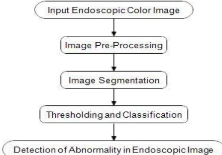
III.PROPOSED WORK
A. DESIGN CONSIDERATIONS: Endoscopic image acquisition.
Image preprocessing i.e. to convert image in standard format.
Image segmentation.
Classification of image
Applying threshold to the abnormality
On the basis of threshold identifying image as normal and abnormal.
B. DESCRIPTION OF THE PROPOSED ALGORITHM:
Endoscopy uses diverse algorithm of image analysis provides images better than that of the other medical tests and in many cases endoscopy is consider as prior to the other imaging techniques such as traditional x-rays. A physician may use an endoscopy as a means to analyse and characterised the possible abnormality in the void organ such as digestive tract. Endoscopy may used to identify an abnormality such as swallowing difficulties, nausea, vomiting, reflux, bleeding, indigestion, abdominal pain, chest pain, change in bowel habits, gastric ulcers, eso varices and many more. Usually the physician treats the subject to identify the abnormality by visual interpretation of endoscopic image. This process may time consuming and the interpretation may vary with respect to physician. On the contrary if this process of analysis of hallow organ, hallucination and exact interpretation of endoscopic images done by diverse algorithm which drives the computer assisted tools will assist the physician for accurate identification of the abnormality in the endoscopic images. Generally in medical practice it is seen that the endoscopic image is interpreted by the experienced physician . The Cuts or in medical jargon Lesion is the most often occurring abnormality in the Gastro-Intestinal tract. The different disorders like Erosive duodenitis, Gastric metaplasia, Diverticula, Mucosal ulcer, space occupying lesions, gastric ulcers, Eso varices and etc are most common lesion in Gastro-Intestinal tract. There are different types of endoscopy, among them Esophago-gastro-duodenoscopy (abbreviated as EGD) used to examined such abnormalities.
In the proposed method, the color endoscopic image is first acquired then it is pre-processed and segmentation technique is applied in the extracted region. The number of watershed region interpreted the image as being abnormal or normal. The proposed design flow is as shown below.
ISSN(Online): 2320-9801
ISSN (Print): 2320-9798
I
nternational
J
ournal of
I
nnovative
R
esearch in
C
omputer
and
C
ommunication
E
ngineering
(An ISO 3297: 2007 Certified Organization)
Vol. 4, Issue 10, October 2016
Input Endoscopic Colour Image :
The input image of endoscopy is colour image shown as below. Here are some few sample images of captured endoscopy as normal and abnormal.
.
Fig. 2 Example of Normal Endoscopic Image Fig. 3 Example of Abnormal Endoscopic Image
Image Preprocessing
The obtained endoscopic image contains the other unused information such as date, name of subject or physician or any information that are not the region of interest. Such region must be filter and the image must convert into standard environment which are applicable for all the captured endoscopic image. Generally it is observed that in images captured through medical or any other image capturing devices may introduce the noise like salt & pepper and speckle noise. In proposed work such noises are removed and then by colour enhancement over saturate the region of interest. Lastly, colour space conversion is used to find out the pixel location. Some technique such as LAB colour space conversion and HSL used to enhance the desired location in the image.
Image Segmentation
The image obtained after the above proposed pre-processing process is converted in to gray scale image. After gray scale conversion the compliment of this images is formed. Since, the intensity value in a gray scale images are ranging from 0 to 255, the complement of this image is computed by using some proposed formulae and algorithm. In the proposed work at the output image N it is expected that dark areas in the images become faint having high degree of brightness where as light areas in the image become darker. The gray scale image is then segmented using morphological watershed segmentation technique that combines morphological operations such as extended minima and morphological gradient with watershed flooding algorithm provides local minima creating number of basins and desired pixel index get found and the binarisation provides the region of interest i.e.we will get the extracted region i.e. abnormal and normal region in endoscopic image.
Classification and Thresholding
Image classification and thresholding is needed to interpret the endoscopic image as normal or abnormal. The identified threshold value is based on segmented pixel index found in the region of interest in endoscopic image.
IV.CONCLUSION AND FUTURE WORK
ISSN(Online): 2320-9801
ISSN (Print): 2320-9798
I
nternational
J
ournal of
I
nnovative
R
esearch in
C
omputer
and
C
ommunication
E
ngineering
(An ISO 3297: 2007 Certified Organization)
Vol. 4, Issue 10, October 2016
techniques can be successfully utilized in construction of abnormality detection algorithm. The algorithm is specially proposed for identify the abnormality in the endoscopic images, which are measured by the expert physicians on his ground. In this way the detection algorithm can reach higher level of abstraction by building a clear and controllable judgment which are basing on a set of understandable parameters derived from the endoscopic images, which could be driven by a set of rules proposed in the algorithm or a random approach. In other words, a shift to the cognitive approach and expert systems looks logical and as well possible here. However, detection of the abnormality is well consider by the physician as the visual features can be well study and may silently considered by the physicians during the examination of the images. The researcher expect that the combination of the techniques proposed in this paper with his best knowledge of endoscopy and application of the endoscopy which achieves higher level of quality for the abnormality detection methods. The proposed could finally lead to including them as one of the clinical procedures in the analysis of abnormality in endoscopic images.
V. ACKNOWLEDGMENT
The proposed work is well discussed with and well guided by and endoscopic images are provided by Dr Amit Kavimandan
REFERENCES
1. Erzhong Hu, Nosato H. ,Sakanashi H.,Murakawa M.,“A modified anomaly detection method for capsule endoscopy images using nonlinear color conversion and Higher order Local Autocorrelation (HLAC)”, Engineering in Medicine and Biology Society (EMBC), 35th Annual International Conference of the IEEE,pp.5477-5480,2013.
2. Erzhong Hu, Nosato H., Sakanashi H.,Murakawa M.,“Anomaly detection method for capsule endoscopy images using Higher order Local AutoCorrelation features”, IEEE International Conference on Systems, Man and Cybernetics (SMC),pp.2289-2293,2012.
3. Krishnan S.M., Nanyang Technol.,Yang X.,Chan K.L.,Kumar S.,“Engineering in Medicine and Biology Society”,Proceedings of the 20th Annual International Conference of the IEEE ” ,Vol.2,29 Oct-1Nov 1998, Hong Kong.
4. Deepti Shikha and B.V. Dhandra,“Abnormality detection in Endoscopic images of Throat cancer by Morphological Operations” , Indian Streams Research Journal, Vol - I , ISSUE – IV,pp.2230-2243,May 2011
5. B.V.Dhandra, Ravindra Hegadi, Mallikarjun Hangarge, V.S.Malemath,“Analysis of Abnormality in Endoscopic images using Combined HSI Color Space and Watershed Segmentation”,IEEE,pp.5243-5246,2006.
6. S.M.Krishnan, X.Yang, K.L.Chan, S.Kumar and P.M.Y.Goh,“Intestinal abnormality detection from endoscopic images”, Proceedings of the 20th Annual International Conference of the IEEE Engineering in Medicine and Biology Society, Vol. 20, pp.895-898,1998. 7. P.S.Hiremath,B.V.Dhandra, Ravindra Hegadi, and G.G. Rajput,“Abnormality Detection in Endoscopic Images Using Color Segmentation
and Curvature Computation”,N.R. Pal et al. (Eds.): ICONIP 2004, LNCS 3316, pp. 834.841, 2004.
8. Adam Brzeski, Adam Blokus, Jan Cychnerski,“An Overview of Image Analysis Techniques in Endoscopic Bleeding Detection”, International Journal of Innovative Research in Computer and Communication Engineering, Vol. 1, Issue 6,pp.1350-1357,August 2013. 9. Ravindra S. Hegadi,Shailaja S. Halli,Arpana Kop, “Segmentation of Abnormal Region from Endoscopic Images using Intelligent
Scissors”,IJCA Special Issue on Recent Trends in Image Processing and Pattern Recognition,pp.89-96,2010
10. Suvidha Sawant, M. S. Deshpande, “ Tumor Recognition in Wireless Capsule Endoscopy Images.”,IOSR Journal of Electrical and Electronics Engineering, Volume 9, Issue 3 Ver. I, pp. 104-109, 2014.
11. R. Kwitt, N. Vasconcelos , N. Rasiwasia , A. Uhl , B. Davis , M. Häfner , F. Wrba, “Medical Image Analysis”, Medical image analysis, pp.1415-1422,2012.
12. B.V.Dhandra, Ravindra Hegadi, “Active Contours without Edges and Curvature Analysis for Endoscopic Image Classification ” , International Journal of Computer Science and Security, Volume (1): Issue (1) ,pp.19-32,2011
BIOGRAPHY
Shrikant D. Kaleis a student of Master of Engineering in the Electronics & Telecommunication Department, Sipna College of Engineering and Technology, S.G.B.A University, Amravati, MS, India. His research interests in Digital Image Processing Techniques.