99
© 2018 by the Serbian Biological Society How to cite this article: Petrušić M, Obreht Vidaković D, Lazić S, Radnović D, Knežević P. Prevalence and genetic variability of Plesiomonas shigelloides in temperate climate surface waters of the Pannonian Plain. Arch Biol Sci. 2018;70(1):99-108.
Prevalence and genetic variability of
Plesiomonas shigelloides
in temperate climate
surface waters of the Pannonian Plain
Milivoje Petrušić1, Dragana Obreht Vidaković2, Sava Lazić3, Dragan Radnović1 and Petar Knežević1,*
1Department of Biology and Ecology, Faculty of Sciences, University of Novi Sad, Trg Dositeja Obradovica 2, 21000 Novi
Sad, Serbia
2Department of Forest and Conservation Sciences, University of British Columbia, 3027-2424 Main Mall, Vancouver, BC
Canada V6T 1Z4
3Department of Virology, Scientific Veterinary Institute ‘‘Novi Sad’’, Rumenački put 20, 21000 Novi Sad, Serbia
*Corresponding author: [email protected]
Received: May 30, 2017; Revised: July 17, 2017; Accepted: July 26, 2017; Published online: August 24, 2017
Abstract: Plesiomonas shigelloides, a Gram-negative bacterium and the causative agent of intestinal diseases and
extraintes-tinal infections in humans and animals, is most frequently found in aquatic environments in tropical or subtropical areas.
The present study was designed to establish the prevalence and genetic variability of P. shigelloides in surface waters (lakes,
rivers, ponds, inlets and canals) located in a temperate climate zone, namely the Pannonian Plain of the northern part of Serbia and southern part of Hungary. The strains were isolated directly by plating samples on inositol-brilliant green-bile agar with neutral red or phenol red as indicators. Our results indicate that phenol red effectively facilitates differentiation of
P. shigelloides from other bacteria. A number of samples were enriched using alkaline peptone water broth, peptone inositol-bile broth and tetrathionate broth. The recovery of the isolates was more successful with the first medium. Out of a total of
51 water samples collected from 28 different locations, 22 samples (43.1%) were found positive for P. shigelloides. Among
the 37 isolated strains, 34 were from lakes (Šatrinci, Ludaš, Panonija, Krivaja, Pecs, Kapetanski rit, Pavlovci, Kovácsszénája, Dobrodol, Vranjaš, Borkovac, Hermann Ottó, Sot, Šelevrenac, Zobnatica, Palić, Orfüí, Jarkovci, Čonoplja) and 3 were from rivers (Danube, Sava). The strains were identified by phenotypic characteristic or by the VITEK2 system and confirmed by PCR using 23S rRNA species-specific oligos. The strains showed a high genetic variability, displaying a variety of RAPD
profiles. Our results reveal for the first time a high prevalence of genetically diverse P. shigelloides populations in surface
waters located in the temperate climate of central and southeastern Europe.
Key words: Plesiomonas shigelloides; surface waters; temperate climate; isolation; RAPD-PCR
INTRODUCTION
Plesiomonas shigelloides is a Gram-negative, rod-shaped bacterium found in aquatic environments and aquatic hosts, including fish, amphibians, shellfish, as well as in various terrestrial animals [1-5]. P. shigel-loides is a facultatively anaerobic, motile (by means of lophotrichous flagella), catalase- and oxidase-positive bacterium, capable of producing indole from tryp-tophan and acid without gas, from D-glucose, m-inositol, maltose and trehalose [6]. The temperature range for P. shigelloides growth is between 8 and 44°C, while optimal growth occurs in the range of 37-38°C [7,8]. Due to a specific phenotype, the bacterium has
been renamed several times: C27 (1947) [9], Pseu-domonas shigelloides (1954); Escherichia sonnei (1956);
Pseudomonas michigani (1959), Aeromonas shigel-loides; Pleisomonas shigelloides (1960); Fergusonia shig-elloides (1963); Scatomonas michigani (1964), Vibrio shigelloides (1971) [10,11] and Proteus shigelloides [12]. For a long time, it was classified as a member of the family Vibrionaceae, along with Aeromonas, as they share many phenotypic features. However, phyloge-netic analyses of 5S rRNA, 16S rRNA and 23S rRNA sequences indicates that Plesiomonas shigelloides is more closely related to the family Enterobacteriaceae,
it is currently classified as the only oxidase-positive lophotrichous member of Enterobacteriaceae.
Most reports on the isolation of P. shigelloides
from environmental samples are from countries situ-ated in tropical or subtropical areas [1,16-23]. The high incidence of P. shigelloides in Southeast Asia has even given the adjective “Asian” to this bacterium, al-though it is also frequently found in freshwaters of Africa and South America [24-26]. However, there have been several successful isolations of P. shigelloides
from environments in temperate latitudes, i.e. in the Czech Republic and the Netherlands [3,27,28], as well as cold climates, in Sweden [4,29].
The bacterium is a concern from both human and animal health prospects. In humans, P. shigelloides has been mainly implicated in gastrointestinal infections, with gastroenteritis as the most common condition [21,30-33]. Although the symptoms associated with gastrointestinal illness due to P. shigelloides typically include diarrhea, abdominal pain, nausea, chills, fever, headaches and vomiting [34,35], P. shigelloides has also been isolated from persons exhibiting no apparent symptoms [36,37]. Interestingly, there are an increas-ing number of reports describincreas-ing human gastroenteri-tis caused by P. shigelloides among immunocompetent populations [20,21,23,38,39]. Extraintestinal infec-tions such as septicemia, meningitis, osteomyelitis, cellulitis, septic arthritis, endophthalmitis, spontane-ous bacterial peritonitis and acute cholecystitis are less common and occur primarily in immunoincompetent or immunocompromised patients [21,40-43]. In ani-mals, P. shigelloides is most frequently found in fish as a pathogen, causing hemorrhaging around the vent, protruded the anus and inappetence [44]. Additional symptoms in fish include emaciation, reddening of the anus with yellow exudation, petechial hemorrhages in the internal muscle wall and occasionally the ac-cumulation of ascitic fluid in the peritoneal cavity [45,46]. Van Damme and Vandepitte [16] noted that
P. shigelloides may be a member of the microbiota of the gastrointestinal tract in warm-water fish, possibly acting as a reservoir of infections.
Although P. shigelloides is primarily found in fresh-waters, the most important vector for its transmission to humans seems to be food, particularly seafood [34,35,47]. De Mondino et al. [2] found a high
inci-dence of P. shigelloides in the polluted salt-water envi-ronments of Rio de Janeiro City and suggested that the contamination may be due to excretion by humans and domestic animals. Thereare anumberof casereportsin the literature confirming illness caused by P. shigelloides
following the consumption of oysters [31,34], fish [40] and crabs [48]. Other modes of transmission include contaminated drinking water [49], vegetables [50] as well as contact with amphibians and reptiles [51].
The aim of this study was to investigate for the first time the prevalence and genetic variability of P. shigelloides in surface waters in a region with a tem-perate climate, i.e. the Pannonian Plain, situated in the northern part of Serbia and the southern part of Hungary. The presence of this bacterium outside of tropical regions can be additional confirmation of the consequences of global warming; the results will indicate if the examined waters are reservoirs for P. shigelloides infections.
MATERIALS AND METHODS
Sampling sites and isolation
equal volumes (25 mL) of water samples with a double-strengthened tetrathionate broth without iodine (TTB; 0.25% casein peptone (Torlak, Serbia), 0.25% meat peptone (Torlak, Serbia), 0.1% bile salts (Torlak, Ser-bia), 1% CaCO3, 3% Na2S2O3, pH 8.4), alkaline peptone water (APW; 1% peptone (Torlak, Serbia), 1% NaCl, pH 8.4) [52] or peptone inositol bile broth (PIBB; 1% inositol, 0.823% Na2HPO4, 0.5% NaCl, 0.5% peptone, 0.15% bile salts No. 3, 0.12% NaH2PO4, pH 7.6) [17].
Bacterial cultures
The reference type strain P. shigelloides ATCC14029 served as a positive control for preliminary identifica-tion tests and the PCR identificaidentifica-tion method based on 23S rRNA, while the reference type strain Aeromonas
hydrophila ATCC7966 was used as a negative control for the PCR. For examination of usability of IBB with two different pH indicators, beside P. shigelloides and
A. hydrophila, two bacterial species were also used:
K. pneumoniae ATCC 31488 and P. mirabilis ATCC 35659. In addition, P. shigelloides ATCC51903 was used in the random amplification of polymorphic DNA (RAPD) analysis. The strains were grown on Luria-Bertani (LB) agar and LB broth under aerobic conditions at 37°C for 24 h.
Identification of environmental isolates
P. shigelloides identification was performed by subject-ing inositol fermentsubject-ing colonies to several prelimi-nary tests: Gram staining, oxidase, catalase, indole production, gelatin hydrolysis, citrate utilization and carbohydrate fermentation tests (glucose, maltose, sorbitol, sucrose and arabinose). The identification of some strains was confirmed by the VITEK2 system (BioMérieux, France). All strains identified by pheno-type as P. shigelloides were additionally confirmed by the PCR method. Genomic DNA was isolated from LB broth overnight cultures using the GeneJet Genomic DNA purification kit (Thermo Scientific, Lithuania). The concentration and purity of the isolated DNA was determined using a BioSpec-nano spectropho-tometer (Shimadzu Corporation, Japan). The PCR was conducted using PS23FW3 primer (5ʹ-CTC CGA ATA CCG TAG AGT GCT ATC C-3ʹ) and PS23RV3 primer (5ʹ-CTC CCC TAG CCC AAT AAC ACC TAA A-3ʹ), designed for a specific amplification of a region of the P. shigelloides 23S rRNA [53]. PCR reactions were performed as described [53] using a thermocy-cler (Biometra, TProfessional, Germany). The PCR products were analyzed on 2% agarose gel, stained with ethidium bromide and visualized under UV light using a BioDoc Analyze (Biometra, Germany). All PCR reactions were conducted in duplicate. The refer-ence strains of Plesiomonas shigelloides ATCC51903 and Aeromonas hydrophila ATCC7966 served as a positive and negative control, respectively.
RAPD fingerprinting
An initial comparison of strains was conducted using a set of 16 decamer oligos (OPA 02, OPA 07, OPA 08, OPB 06, OPB 11. OPB 12, OPB13, OPC 04, OPE 09,
Ta bl e 1. Sa m ple o rig in, m et ho d o f i so la tio n a nd i so la te d s tra in s o f P. s hi ge llo id es fr om s ur face wa ter s Sa m pl e orig in Sa m plin g s ite M ark o n the ma p GPS c oo rdina tes N o. o f s am pl es p er s ite (n=54) (N o. o f dir ec tly p la te d/ enri che d s am pl es) M edi a* (me di a f ro m w hi ch s tr ains w er e iso la te d a re und erline d) St ra in is ol at ed (n=37) La kes Ša tr in ci , S erb ia 18 46°04'5"N, 19°55'0"E 2 (1/1) PD A/ APW+P D A 2S HD (n=43) Lud aš, S erb ia 25 46°50'0"N, 19°49'0"E 2 (2/0) PD A 1LD , 2LD Pa no ni ja, S erb ia 1 45°44'1"N, 19°31'4"E 2 (2/0) PD A 6P AND , 4P AND Kr iva ja, S erb ia 21 45°50'1"N, 19°29'5"E 2 (2/0) PD A 1KD , 8KD Pe cs, H un ga ry 3 46°09'3"N, 18°08'2"E 2 (2/0) PD A 1P ECD , 4P ECD Ka pеt an sk i r it, S erb ia 24 46°02'2"N, 19°56'3"E 2 (2/0) PD A 2KRD , 5KRD , 6KRD Pa vlo vci , S erb ia 15 45°04'2"N, 19°47'5"E 3 (2/1) PD A/ APW+P D A 2P D Ko vács szén áj a, H un ga ry 5 46°10'4"N, 18°06'5"E 2 (2/0) PD A 1K O VD , 4K O VD D ob ro do l, S erb ia 17 45°02'3"N, 19°56'4"E 2 (1/1) PD A/ APW+P D A 2D D , 5D D Vra nj aš, S erb ia 13 45°05'4"N, 19°35'5"E 1 (1/0) PD A 2VD , 8VD Bo rk ovac, S erb ia 16 45°02'3"N, 19°49'1"E 2 (1/1) PD A/ APW+P D A 2B D , 4B D H er m an n Ott ó, H un ga ry 4 46°10'3"N, 18°07'5"E 2 (2/0) PD A 6HERD , 9HERD So t, S erb ia 11 45°09'3"N, 19°20'2"E 2 (2/0) PD A 7S D Še le vr en ac, S erb ia 19 45°04'3"N, 19°59'5"E 2 (2/0) PD A 3S HELD Zo bn at ic a, S erb ia 28 45°50'2"N, 19°37'5"E 2 (2/0) PD A 1ZD , 3ZD , 5ZD Pa lić, S erb ia 27 46°05'1"N, 19°45'3"E 2 (2/0) PD A 1P AD Or fűi , H un ga ry 2 46°08'4"N, 18°09'2"E 2 (2/0) PD A 2O RFD , 14O RFD Ja rk ov ci , S erb ia 20 45°02'5"N, 20°01'3"E 2 (2/0) PD A 2JD , 5JD Č on op lja, S erb ia 22 45°51'1"N, 19°17'5"E 2 (2/0) PD A 4CHD Beš en ov o, S erb ia 14 45°06'2"N, 19°42'4"E 2 (2/0) PD A -Kr va vo , S erb ia 26 46°05'4"N, 19°46'1"E 2 (2/0) PD A -Ti kva ra, S erb ia 9 45°14'2"N, 19°22'6"E 1 (1/1) PD A/(APW/T TB/P IB B)+P D A -Riv ers D an be , S erb ia 7 45°14'2"N, 19°26'5"E 1 (1/1) PD A/( APW /T TB/P IB B)+ PD A D BPM1 (n=3) Ti sza, H un ga ry 23 46°14'6"N, 20°09'2"E 1 (1/1) PD A/(APW/T TB/P IB B)+P D A -Sa va, S erb ia 12 44°54'4"N, 19°45'3"E 1 (1/1) PD A/ APW+P D A D737A2 Po nds (2) Cv rcin a b ara, S erb ia 10 45°14'3"N, 19°23'0"E 2 (2/2) PD A/(APW/T TB/P IB B)+P D A -In le t (1) Ba ger (D an ub e r iv er), S erb ia 8 45°14'2"N, 19°24'4"E 1 (1/1) PD A/( APW /T TB/P IB B)+ PD A D BPM2 Ca na ls (2) D an ub e-T isa-D an ub e, S erb ia 6 45°17'2"N, 19°47'2"E 2 (2/2) PD A/(APW/T TB/P IB B)+P D A -*P D A– P lesio m on as dif fer en tia l a ga
r; APW – A
lka lin e P ep to ne W at er ; T
TB – T
et ra thio na te B ro th w ith ou t I odin e; P IB
B – P
OPM 10, OPM 18, OPN 01, OPN 02, KO 1, K 15, NO 11) in order to identify suitable primers for the RAPD fingerprinting. Based on the performance, five primers were selected: OPA 07, OPB 11, OPM 10, OPN 02 and K 01. The RAPD PCR reaction was performed in a 20-μL volume containing 2.5 μL 10x Dream Taq Green Buffer with 20 mM MgCl2, 1 μL DreamTaq polymerase (Thermo Scientific, Lithuania), 0.5 μL 10 mM dNTPs, 25 ng of corresponding primer and about 100 ng of template DNA. The amplification was carried out in a thermocycler (Biometra, TProfessional, Germany) with the following temperature program: one cycle of initial denaturation at 94°C for 5 min followed by 40 cycles, including denaturation at 94°C for 2 min, annealing at 36°C for 1 min, extension at 72°C for 2 min, and a final extension step at 72°C for 10 min, followed by cooling to 4°C. The obtained DNA fragments were separated by electrophoresis on a 2% agarose gel containing ethid-ium bromide along with 1 kb DNA Ladder (Thermo Scientific, Lithuania) and visualized under UV light using a BioDoc Analyze Systems (Biometra, Germany). All PCR reactions were conducted in duplicates.
The bands were scored in a binary mode with 1 indicating its presence and 0 its absence. A pairwise ge-netic similarity matrix was calculated from binary data using Jaccard’s coefficient. The similarity matrix was subjected to cluster analysis by unweighted paired group method using arithmetic average (UPGMA). Bootstrap analysis, with 100 repetition value, was used to assess the tree topology robustness. These analyses were carried out using D-UPGMA [54, 55] and TreeGraph 2 [56].
RESULTS
Out of 51 water samples collected from different water sources, 22 samples (40.7%) were positive for P. shigel-loides. The list of isolated strains describing the inocula-tion procedure and media used are shown in Table 1. Out of the 37 positive isolates, 34 were from lakes and 3 from rivers, with no positive samples from ponds and canal waters. According to preliminary physiological, biochemical and morphological tests, 37 colonies ob-tained from various sampling sites or using different procedures were chosen for molecular identification. All the P. shigelloides suspected isolates subjected to PCR amplification with primers specific for P. shigel-loides 23S rRNA generated a product of expected
mo-lecular size 284 bp (Fig. 2). Of the 37 positive isolates, 34 were from lakes and 3 were from rivers, with no positive samples from ponds and canal waters.
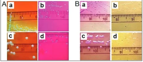
For P. shigelloides isolation from surface water, PDA was used with or without previous enrichment on APW, TTB and PIBB. PDA with neutral red or phenol red was successfully used for this purpose re-sulting in the isolation of 28 strains (75.7%) by spread-ing 0.5 mL of samples directly onto the agar plates. The application of these two indicators revealed that phenol red seems to be a more suitable medium as it facilitates P. shigelloides differentiation from other bacteria by producing a more obvious medium color change (Fig. 3). Of the 13 samples subjected to the enrichment procedure, the presence of the bacterium was detected in 7 samples with 9 strains (24.3%)
iso-Fig. 3. Colony growth of different bacteria on Plesiomonas
dif-ferential agar (PDA). A – PDA containing the indicator phenol
red; only colonies of P. shigelloides ATCC14029 (a) turned the
medium to a yellow color while Aeromonas hydrophila ATCC7966
(b), Klebsiella pneumoniae ATCC31488 (c), and Proteus mirabilis
ATCC35659 (d) did not change the color of the medium (orange
or red); B – PDA containing the indicator neutral red; this
in-dicator did not allow for differentiation between P. shigelloides
ATCC14029 (a) and K. pneumoniae ATCC31488 (c), and A.
hy-drophila ATCC7966 (b), and P. mirabilis ATCC35659 (d) did not change the color of the medium.
Fig. 2. PCR identification of selected P. shigelloides strains. M – 100 bp marker; strain designations are indicated above the
electrophoregram; the strain P. shigelloides ATCC 51903 was a
positive control, and Aeromonas hydrophila ATCC 7966 was a
lated. No positive samples were detected after enrich-ment with TTB and PIBB due to a massive presence of competing microorganisms.
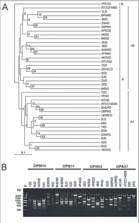
The RAPD analyses of 37 strains with five differ-ent primers resulted in 649 amplified fragmdiffer-ents. The number of detected loci varied between primers from 0 to 14 (OPB11). The fragment size varied, ranging from about 150 bp to about 4.3 kb. The total number of polymorphic fragments by strain was between 8 for strain 2PD and 35 for strain 2BD. The UPGMA dendrogram based on Jaccard’s genetic similarity coef-ficients showed unusual subclustering among different strains (Fig. 4). According to the analysis, most isolates of P. shigelloides, including the two reference strains ATCC51903 and ATCC14029, were classified into group A, whereas the strain 1PECD isolated from Lake Pecs (Hungary) forms a unique group B. The strains from group A are clustered into two subgroups, A1 and A2, and both were further divided into several subc-lades with low bootstrap values. Subgroup A1 contains ATCC14029 along with 14 other strains, all from dif-ferent lakes, except the isolates 2ORFD and 14ORFD from Lake Orfűi (Hungary), and 3ZD and 1ZD from Lake Zobnatica (Serbia). Although these isolates were from the same samples, different RAPD patterns were obtained. Group A2 consists of 22 strains, including ATCC51903. The isolates 5KRD, 6KRD and 2KRD from the Lake Kapetanski Rit (Serbia) were represent-ed by different RAPD patterns along with the isolates 4PAND and 6PAND from the Lake Panonija (Serbia).
DISCUSSION
Numerous studies have shown a high incidence of
P. shigelloides in tropical and subtropical regions. In studies conducted in Brazil [2], Bangladesh [17], Japan [1] and Nigeria [24], the prevalence of the bacterium in pond and river-water samples ranged from 7.4% to 25%. Depending on the study, PDA, taurocholate-tellurite-gelatin agar (TTGA), SS agar, DHL agar and xylose lysine deoxycholate (XLD) agar were used. Moreover, the isolation procedures involved filtration or centrifugation of water samples, with or without using enrichment broths such as APW, gram negative broth and peptone broth. In the mentioned studies, relatively high sample volumes were processed, varying from 50 to 500 mL (in most instances it was 100 mL).
There is a certain absence of publications report-ing the isolation of P. shigelloides in freshwater en-vironments in cold and temperate climates. Among the few publications, a successful isolation has been reported from the continental climate rivers and lake water samples in Slovakia [53], as well as from sam-ples taken in a subpolar region of Sweden [4,29]. In the analysis of cold climate freshwater samples from two lakes and two different sites of a river in Swe-den, Krovacek et al. [4] determined the presence of
Fig. 4. Genetic variability of 37 P. shigelloides strains isolated from
surface waters of Pannonian Plain. A – dendrogram of P.
shigel-loides genetic variability is constructed by the UPGMA method based on the presence/absence of RAPD-PCR fragments amplified using five different primers (OPA 07, OPB 11, OPM 10, OPN 02 and K 01). Bootstrap values based on 100 replications are shown
at the nodes. B – An example of obtained RAPD-PCR profiles
using OPM10, OPB11, OPN02 and OPA07 oligos. P. shigelloides
P. shigelloides after APW enrichment in all examined samples. In the study, 400 mL from each water sam-ple was mixed with 40 mL of 10x concentrated APW, which was a significantly higher sample volume than we used. The same media and method were used in the survey conducted by Gonzáles-Rey et al. [53] who revealed the presence of P. shigelloides in one of the six lakes in the northern Sweden. Taking into account previous reports and the results of the present study, APW appears to be the enrichment medium of choice for P. shigelloides isolation.
When comparing our results with all the results published by other authors mentioned above, it is ob-vious that we have detected a higher percentage of positive samples, although many strains were isolated directly from samples, without an enrichment proce-dure. Differences in the performance of P. shigelloides
isolation in the abovementioned studies can be the result of various media application. Several studies have shown that P. shigelloides grows on a variety of media, including the MacConkey [31,43,57-59], SS agar [1,31,60], modified salmonella-shigella agar with inositol substitution for lactose [60], deoxycholate ci-trate (DC) agar [59], XLD agar [57], Hektoen enteric (HE) agar [57,58], MSDS agar [61], Endo agar [4] and PDA agar [62].
Aside from the used media, another explanation for the successful isolation of P. shigelloides directly from samples in our study could be global warming. The detected abundance of P. shigelloides in geograph-ical regions with temperate climates such as the Czech Republic [3,28], the Netherlands [27] and now Ser-bia and Hungary, is not the only fact supporting this. Namely, most of the previous studies were conducted during the second half of the last century when global warming occurred, and there is a significant temporal distance in comparison to our study.
The conducted RAPD-PCR analysis revealed a notable genetic variability among all 37 isolates as none of the isolates had the same RAPD pattern. Ac-cordingly, even the strains isolated from the same sampling sites were not clonally identical. The den-drogram did not reveal geographical relations among strains since isolates from spatially unrelated samples were grouped in the same cluster. The results of our RAPD analysis are in accordance with the findings
of Gu et al. [63] who used the RAPD technique to determine genetic variability among 26 isolates from fresh water (10 isolates), fish (6 isolates) and human clinical isolates (10 isolates) and found a high genetic variability among most of the isolates while none of the isolates had the same composite RAPD profile. Similarly, among 24 strains belonging to nine O:H serovars isolated from humans, animals and the envi-ronment, 22 different RAPD-PCR patterns were found [64]. The results of high genetic variability of P. shig-elloides are not very surprising since the species has been proven to have high levels of nucleotide diversity (the nucleotide difference between strains is about 1.49%) [65]. This is the result of frequent homologous recombination in housekeeping genes, which affects
P. shigelloides alleles and nucleotides 7 and 77 times more frequently than mutations, respectively [65]. Moreover, P. shigelloides stands out among members of the Enterobacteriaceae, as recombination in other species is much less frequent [66-68].
CONCLUSIONS
Acknowledgments: This work was supported by the Ministry of Education, Science and Technological Development of the Repub-lic of Serbia, Grant OI 172058 and the Hungary-Serbia IPA Cross-Border Cooperation Program (HUSRB 2103/214/250; NNAA). Author contributions: MP and PK wrote the manuscript. MP carried out the experiments, analyzed the data and interpreted the results. DOV contributed to the RAPD-PCR analysis. SL and DR contributed to the interpretation of the results. PK designed and supervised the study, analyzed the data and interpreted the results. All authors are responsible for the revision of the manu-script and proofreading.
Conflict of interest disclosure: The authors declare that they have no competing interests.
REFERENCES
1. Arai T, Ikejima N, Itoh T, Sakai S, Shimada T, Sakazaki R.
A survey of Plesiomonas shigelloides from aquatic
envi-ronments, domestic animals, pets and humans. J Hyg. 1980;84(02):203-11.
2. Mondino S, Nunes M, Ricciardi I. Occurrence of
Plesiomo-nas shigelloides in water environments of Rio de Janeiro city. Mem I Oswaldo Cruz. 1995;90(1):1-4.
3. Aldova E, Melter O, Chyle P, Slorasek M, Kodym, P.
Ple-siomonas shigelloides in water and fish. Cent Eur J Publ Health. 1999;7:172-5.
4. Krovacek K, Eriksson L, González-Rey C, Rosinsky J, Ciznar I. Isolation, biochemical and serological characterisation of Plesiomonas shigelloides from freshwater in Northern Europe. Comp Immunol Microb. 2000;23(1):45-51. 5. Matsuyama R, Kuninaga N, Morimoto T, Shibano T, Sudo A,
Sudo K, Asano M, Suzuki M, Asai T. Isolation and
antimi-crobial susceptibility of Plesiomonas shigelloides from great
cormorants (Phalacrocorax carbo hanedae) in Gifu and Shiga
prefectures, Japan. J Vet Med Sci. 2015;77:1179-81.
6. Janda, J. Family I. Enterobacteriaceae, genus XXVII.
Plesi-omonas Habs and Schubert 1962. In: Brenner D, Krieg NR, Staley JT, editors. Bergey’s manual of systematic bacteriol-ogy. Vol. 2. 2nd ed. New York: Springer; 2005. p 740-4. 7. Rouf M, Rigney M. Growth Temperatures and
Tem-perature Characteristics of Aeromonas. Appl Microbiol.
1971;22(4):503-6.
8. Miller M, Koburger J. Tolerance of Plesiomonas shigelloides
to pH, Sodium Chloride and Temperature. J Food Protect. 1986;49(11):877-9.
9. Ferguson W, Henderson N. Description of Strain C27: A
Motile Organism with the Major Antigen of Shigella sonnei
Phase I. J Bacteriol. 1947;54(2):179-81.
10. Koburger, J. Plesiomonas shigelloides. In: Doyle M, editor.
Foodborne Bacterial Pathogens. New York: Marcel Dekker; 1989. p. 311-26.
11. Hendrie M, Shewan J, Veron M. Aeromonas shigelloides
(Bader) Ewing et al: A proposal that it be transferred to the
genus Vibrio. Int J Sys Bacteriol. 1971;21:25-7.
12. MacDonell M, Swartz D, Ortiz-Conde B, Last G, Colwell R. Ribosomal RNA phylogenies for the vibrio-enteric group of eubacteria. Microbiol Sci. 1986;3:172-8.
13. East A, Allaway D, Collins M. Analysis of DNA encoding 23S rRNA and 16S-23S rRNA intergenic spacer regions
from Plesiomonas shigelloides. FEMS Microbiol Lett.
1992;95(1):57-62.
14. Martinez-Murcia A, Benlloch S, Collins M. Phylogenetic
Interrelationships of Members of the Genera Aeromonas
and Plesiomonas as Determined by 16S Ribosomal DNA Sequencing: Lack of Congruence with Results of DNA-DNA Hybridizations. Int J Syst Bacteriol. 1992;42(3):412-21. 15. Ruimy R, Breittmayer V, Elbaze P, Lafay B, Boussemart O,
Gauthier M, Christen R. Phylogenetic Analysis and
Assess-ment of the Genera Vibrio, Photobacterium, Aeromonas, and
Plesiomonas Deduced from Small-Subunit rRNA Sequences. Int J Syst Bacteriol. 1994;44(3):416-26.
16. Van Damme L, Vanderpille J. Frequent isolation of
Edwardsi-ella tarda and Plesiomonas shigelloides from healthy Zairese freshwater fish; A possible source of sporadic diarrhea in the tropics. Appl Environ Microbiol. 1980;39:475-79.
17. Islam M, Alam M, Khan S. Distribution of Plesiomonas
shigelloides in Various Components of Pond Ecosystems in Dhaka, Bangladesh. Microbiol Immunol. 1991;35(11):927-32.
18. Obi C, Coker A, Epoke J, Ndip R. Aeromonas and
Plesiomo-nas species as bacterial agents of diarrhoea in urban and
rural areas of Nigeria: antibiogram of isolates. Cent Afr J Med. 1995;41(12):397-403.
19. Shigematsu M, Kaufmann M, Charlett A, Niho Y, Pitt T. An
epidemiological study of Plesiomonasshigelloides diarrhoea
among Japanese travellers. Epidemiology and Infection. 2000;125(3):523-30.
20. Wong T, Tsui H, So M, Lai J, Lai S, Tse C, Ng T.
Plesiomo-nas shigelloides infection in Hong Kong: retrospective study of 167 laboratory-confirmed cases. Hong Kong Med J. 2000;6(4):375-80.
21. Tseng H, Liu C, Li W, Su S, Lee C. Characteristics of
Plesi-omonas shigelloides infection in Taiwan. J Microbiol Immu-nol Infect. 2002;35:47-52.
22. Maluping R, Lavilla-Pitogo C, DePaola A, Janda J, Krovacek
K, Greko C. Antimicrobial susceptibility of Aeromonas spp.,
Vibrio spp. and Plesiomonas shigelloides isolated in the Philip-pines and Thailand. Int J Antimicrob Ag. 2005;25(4):348-50. 23. Chen X, Chen Y, Yang Q, Kong H, Yu F, Han D, Zheng S,
Cui D, Li L. Plesiomonas shigelloides Infection in Southeast
China. PLoS ONE. 2013;8(11):e77877.
24. Kwaga J, Adesiyun A, Bello C, Abdullahi S. Occurrence of
Plesiomonas shigelloides in humans and water in Zaria, Nige-ria. Microbiologica 1988;11:165-67.
25. Canosa A, Pinilla G. Bacteriological eutrophication indica-tors in four Colombian water bodies (South America). Lakes Reservoirs: Res Manage. 1999;4(1):23-7.
26. Gibotti A, Saridakis H, Pelayo J, Tagliari K, Falcao D.
Preva-lence and viruPreva-lence properties of Vibrio cholerae non-O1,
Aeromonas spp. and Plesiomonas shigelloides isolated from Cambe Stream (State of Parana, Brazil). J Appl Microbiol. 2000;89(1):70-5.
27. Medema G, Schets C. Occurrence of Plesiomonas
shigelloi-des in surface water: relationship with faecal pollution and
28. Bardon J, Plesiomonas shigelloides and its serovars in animals in the Czech Republic – Region Moravia. Cent Eur J Publ Heal. 1999;1:47-9.
29. Gonzalez-Rey C, Svenson S, Eriksson L, Ciznar I, Krovacek K. Unexpected finding of the “tropical” bacterial pathogen
Plesiomonas shigelloides from lake water north of the Polar Circle. Polar Biol. 2003;26(8):495-9.
30. Reinhardt J, George W. Plesiomonas shigelloides-associated
diarrhea. JAMA- J Am Med Assoc. 1985;253(22):3294-5.
31. Holmberg S, Farmer J. Aeromonas hydrophila and
Plesiomo-nas shigelloides as Causes of Intestinal Infections. Clin Infect Dis. 1984;6(5):633-9.
32. Holmberg S, Wachsmuth I, Hickman-Brenner F, Blake
P, Farmer J. Plesiomonas Enteric Infections in the United
States. Ann Inter Med. 1986;105(5):690.
33. Murray P, Baron E, Jorgensen J, Landry M, Pfaller M, Yolken R. Manual of Clinical Microbiology. 8th ed. Herdon, VA, USA: American Society for Microbiology; 2003.
34. Rutala W, Sarubbi F, Finch C, Maccormack J, Steinkraus G. Oyster-associated outbreak of diarrhoeal disease
possibly caused by Plesiomonas shigelloides. Lancet.
1982;319(8274):739.
35. Kain K, Kelly M. Clinical features, epidemiology, and
treat-ment of Plesiomonas shigelloides diarrhea. J Clin Microbiol.
1989;27(5):998-1001.
36. Bhat P, Shanthakumari S, Rajan D. The characterization
and significance of Plesiomonas shigelloides and Aeromonas
hydrophila isolated from an epidemic of diarrhoea. Indian J Med Res. 1974;62(7):1051-60.
37. Vandepitte J, Makulu A, Gatti F. Plesiomonas shigelloides
Sur-vey and possible association with diarrhoea in Zaire. Ann Soc Belg Med Trop. 1974;54(6):503-13.
38. Bodhidatta L, Serichantalergs O, Sornsakrin S, McDaniel P, Mason C, Srijan A. Case-Control Study of Diarrheal Disease Etiology in a Remote Rural Area in Western Thailand. Am J Trop Med Hyg. 2010;83(5):1106-9.
39. Escobar J, Bhavnani D, Trueba G, Ponce K, Cevallos W,
Eisenberg J. Plesiomonas shigelloides Infection, Ecuador,
2004–2008. Emerg Infect Dis. 2012;18(2):322-4.
40. Ingram C, Morrison A, Levitz R. Gastroenteritis, sepsis, and
osteomyelitis caused by Plesiomonas shigelloides in an
immu-nocompetent host: case report and review of the literature. J Clin Microbiol. 1987;25(9):1791-3.
41. Paul R, Siitonen A, Kärkkäinen P. Plesiomonas shigelloides
bacteremia in a healthy girl with mild gastroenteritis, J Clin Microbiol. 1990;28:1445-6.
42. Brenden R, Miller M, Janda J. Clinical Disease Spectrum and
Pathogenic Factors Associated with Plesiomonas shigelloides
Infections in Humans. Clin Infect Dis. 1988;10(2):303-16.
43. Rolston K, Hopper R. Diarrhoea due to Plesiomonas
shigel-loides to cancer patients. J Clin Microbiol. 1984;20:597-8. 44. Austin B, Austin D. Bacterial fish pathogens. 4th ed.
Chich-ester: Springer; 2007.
45. Cruz J, Saraiva A, Eiras J, Branco R, Sousa J. An outbreak of Plesiomonas shigelloides in farmed rainbow trout, Salmo gairdneri Richardson, in Portugal. B Eur Assoc Fish Pat. 1986;8(1):20-2.
46. Klein B, Kleingeld D, Bohm K. First isolations of
Plesiomo-nas shigelloides from samples of cultured fish in Germany. B Eur Assoc Fish Pat. 1993;13:70-2.
47. Miller M, Koburger J. Plesiomonas shigelloides: An Oppor-tunistic Food and Waterborne Pathogen. J Food Protect. 1985;48(5):449-57.
48. Claesson B, Holmlund D, Lindhagen C, Mätzsch T.
Plesi-omonas shigelloides in acute cholecystitis: a case report. J Clin Microbiol. 1984;20:985-7.
49. Van Houten R, Farberman D, Norton J, Ellison J, Kiehlbauch
J, Morris T, Smith P. Plesiomonas shigelloides and Salmonella
serotype Hartford infections associated with a contaminated water supply—Livingston County, New York, 1996. Morb Mortal Wkly Rep. 1998;47:394-6.
50. Monge R, Arias-Echandi M, Utzinger D. Presence of
cyto-toxic Aeromonas and Plesiomonas shigelloides in fresh
veg-etables. Rev Biomed. 1998;9:176-80.
51. Jagger TD. Plesiomonas shigelloides: a veterinary perspective.
Infect Dis Rev. 2000;2:199–210.
52. Freund S, Koburger J, Wei C. Enhanced Recovery of
Plesi-omonas shigelloides following an Enrichment Technique. J Food Protect. 1988;51(2):110-2.
53. González-Rey C, Svenson S, Bravo L, Rosinsky J, Ciznar I,
Krovacek K. Specific detection of Plesiomonas shigelloides
isolated from aquatic environments, animals and human diarrhoeal cases by PCR based on 23S rRNA gene. FEMS Immunol Med Microbiol. 2000;29(2):107-13.
54. Garcia-Vallve S, Palau J, Romeu A. Horizontal gene trans-fer in glycosyl hydrolases intrans-ferred from codon usage in Escherichia coli and Bacillus subtilis. Mol Biol Evol. 1999;16(9):1125-34.
55. DendroUPGMA: A dendrogram construction utility. [Inter-net]. Tarragona (Spain): Universitat Rovira i Virgili (URV); 2002 [cited 2017 Mar 1]. Available from: http://genomes.urv. cat/UPGMA/index.php
56. Stöver B, Müller K. TreeGraph 2: Combining and visual-izing evidence from different phylogenetic analyses. BMC Bioinformatics. 2010;11:7.
57. Penn R, Giber D, Knoop F, Preheim L. Plesiomonas
shigel-loides overgrowth in the small intestine. J Clin Microbiol. 1982;15:869-72.
58. Pitarangsi C, Echeverria P, Whitmire R, Tirapat C, Formal S, Dammin GJ, Tingtalapong M. Enteropathogenicity of
Aeromonas hydrophila and Plesiomonas shigelloides: preva-lence among individuals with and without diarrhea in Thai-land. Infect Immun. 1982;35(2):666-73.
59. Huq M, Islam M. Microbiological and clinical studies in
diarrhoea due to Plesiomonas shigelloides. Indian J Med Res.
1983;77:793-7.
60. Tsukamoto T, Kinoshita Y, Shimada T, Sakazaki R. Two
epi-demics of diarrhoeal disease possibly caused by Plesiomonas
shigelloides. J Hyg. 1978;80(02):275-80.
61. Sakazaki R, Balows A. The genera Vibrio, Plesiomonas,
62. Schubert R. Ueber den Nachweis von Plesiomonas
shigelloi-des Habs und Schubert. 1962, und ein Elektivmedium, den
Inositol-Brillantgrun-Gallesalz-Agar. Emst-Rodenwaldt-Arch. 1977;4:97-103.
63. Gu W, Gonzalez-Rey C, Krovacek K, Levin R. Genetic
Vari-ability Among Isolates of Plesiomonas shigelloides from Fish,
Human Clinical Sources and Fresh Water, Determined by RAPD Typing. Food Biotechnol. 2006;20(1):1-12.
64. González-Rey C, Siitonen A, Pavlova A, Ciznar I,
Sven-son S, Krovacek K. Molecular evidence of Plesiomonas
shigelloides as a possible zoonotic agent. Folia Microbiol. 2011;56(2):178-84.
65. Salerno A, Deletoile A, Lefevre M, Ciznar I, Krovacek K,
Gri-mont P, Brisse, S. Recombining Population Structure of
Ple-siomonas shigelloides (Enterobacteriaceae) Revealed by Mul-tilocus Sequence Typing. J Bacteriol. 2007;189(21):7808-18.
66. Selander R, Levin B. Genetic diversity and structure in
Esch-erichia coli populations. Science. 1980;210(4469):545-7. 67. Beltran P, Musser J, Helmuth R, Farmer J, Frerichs W,
Wachsmuth I, Ferris K, McWhorter A, Wells J, Cravioto A.
Toward a population genetic analysis of Salmonella: genetic
diversity and relationships among strains of serotypes S.
choleraesuis, S. derby, S. dublin, S. enteritidis, S. heidelberg,
S. infantis, S. newport, and S. typhimurium. Proc Natl Acad Sci U S A. 1988;85(20):7753-7.
68. Dolina M, Peduzzi R. Population genetics of human, animal,
and environmental Yersinia strains. Appl Environ Microb.