ARTIGO ORIGINAL
Congenital Anomalies Detected at Birth in
Newborns of Adolescent Women
Anomalias Congénitas Identificadas ao Nascimento em Recém-Nascidos de
Mulheres Adolescentes
1. Departamento de Obstetrícia. Escola Paulista de Medicina. Universidade Federal de São Paulo. São Paulo. Brasil.
2. Disciplina de Genética. Departamento de Morfologia. Escola Paulista de Medicina. Universidade Federal de São Paulo. São Paulo. Brasil. Autor correspondente: Edward Araújo Júnior. [email protected]
Recebido: 30 de Novembro de 2014 - Aceite: 03 de Agosto de 2015 | Copyright © Ordem dos Médicos 2015
Leandro Valim dos REIS1, Edward ARAUJO JÚNIOR1, Cristina Aparecida Falbo GUAZZELLI1, Mirlene Cecília Soares
Pinho CERNACH2, Maria Regina TORLONI1, Antonio Fernandes MORON1
Acta Med Port 2015 Nov-Dec;28(6):708-714
RESUMO
Introdução: Analisar a prevalência das anomalias congénitas, detetadas no nascimento, entre filhos de gestantes adolescentes, en-fatizando os tipos mais comuns e a época do diagnóstico.
Material e Métodos: Estudo retrospetivo do tipo censo, no qual foram analisados todos recém-nascidos, vivos ou mortos, com peso superior a 500 g, de mulheres que tiveram o parto no Hospital São Paulo num período de seis anos. Os produtos da conceção por-tadores de anomalias foram identificados no período pré-natal ou através do exame físico pós-natal, segundo os critérios do Estudo Colaborativo Latino-Americano das Malformações Congênitas. Os resultados são apresentados de forma descritiva através de valores absolutos e relativos, calcula-se a prevalência das anomalias e comparam-se os diferentes grupos recorrendo a testes não paramé-tricos.
Resultados: Foram analisadas 6 257 gestações, das quais 577 resultaram em recém-nascidos com alguma anomalia congénita identificada no nascimento (prevalência de 9,2%). Do total de gestações, 907 eram de adolescentes (idade inferior a 20 anos), para as quais se verificou uma prevalência de anomalias nos recém-nascidos de 9,9%. Comparando os recém-nascidos de adolescentes com os das mulheres com idade superior a 20 anos, apenas se encontrou diferença estatisticamente significativa para a prevalência dos defeitos do tubo neural (p = 0,027).
Discussão: Observamos uma alta taxa de partos em adolescentes, acima das taxas dos países desenvolvidos. Observamos também alta frequência de anomalias congénitas em recém-nascidos, provavelmente por sermos um serviço terciário de referência. A elevada prevalência dos defeitos do tubo neural entre grávidas jovens pode ser explicada pela não suplementação pré-concecional de ácido fólico em gravidezes não planeadas, como é característico nas adolescentes.
Conclusão: A prevalência e momento do diagnóstico das anomalias congénitas em recém-nascidos apresentam comportamentos semelhantes entre grávidas com idade inferior ou superior a 20 anos, exceto para os defeitos do tubo neural, de maior prevalência nos recém-nascidos das grávidas adolescentes.
Palavras-chave: Adolescente; Anomalias Congénitas; Gravidez. ABSTRACT
Introduction: To analyze the prevalence of congenital anomalies detected at birth among children of pregnant adolescents, emphasizing the most common types and the time of diagnosis.
Material and Methods: Retrospective study of type census, in which were analyzed in all newborns, living or dead, weighing more than 500 g of women who gave birth at Hospital São Paulo in a period of six years. The fetuses bearing anomalies were identified prenatally or through postnatal physical examination period, according to the criteria of the Latin American Collaborative Study of Congenital Malformations. The results were expressed descriptively using absolute and relative values, the prevalence of anomalies was calculated, as well as the comparison between groups using nonparametric tests.
Results: We analyzed 6 257 pregnancies, of which 577 newborns had some congenital anomaly identified at birth (prevalence 9.2%). Among these 6 257, 907 were adolescents, which showed a 9.9% prevalence of anomalies among their newborns. There was no significant difference between the presence of abnormalities in newborns of adolescents and women with age greater than or equal to 20 years. About 56% of congenital anomalies were diagnosed in the prenatal period. We observed a higher prevalence of defects of neural tube between newborns of adolescents (p = 0.027).
Discussion: We observed high rate of deliveries in adolescents, higher than developed countries. We observed also high frequency of congenital anomalies in newborns, probably because our tertiary reference center. The high prevalence of neural tube defect among young pregnant women could be explained by the absent of acid folic supplementation in non-planned gestations which is typical of adolescents.
Conclusion: The prevalence and time of diagnosis of congenital anomalies showed similar behavior among newborns of teenagers and women with age greater than or equal to 20 years, except for the defects of the neural tube, which were more prevalent among newborns of teenagers.
ARTIGO ORIGINAL
INTRODUCTION
Adolescence is the transitional period from childhood to adulthood, with specific anatomical, physiological, social and psychological transformations.1 According to the World Health Organization (WHO), adolescence occurs from ages 10 to 19.2
Adolescent pregnancy has elicited worldwide interest, with many reasons for concern and associated to health risks for both mother and child, whilst having a relevant biological, psychological and social impact.3 A 36 and 106 pregnancy rate per 1,000 women aged 15-17 and 18-19, respectively, was found in the USA in 2009.4
Congenital anomalies (also referred as congenital abnormalities, malformations, disorders or birth defects) are pathological conditions associated to aetiological factors arising before birth, i.e. before, during or after conception, with a clinical expression involving structural or functional defects affecting one or multiple organs. A 198 per 10,000 live births prevalence of major congenital anomalies diagnosed before one year of age was found in the UK in 1990-2009 (95% confidence interval: 195-201).5 Congenital anomalies are associated to low birth weight and are the leading cause of child mortality in the USA.6
Croen and Shaw7 described a 29.1 per 1,000 live births prevalence of congenital anomalies among adolescent mothers. In this study, approximately 6% of live births with congenital anomalies presented with chromosomal abnormalities. These children may present with chronic diseases such as mental retardation and physical abnormalities, other than being more susceptible to infections and malnutrition, among others, making their social inclusion more difficult and entailing substantial economic and psychological burden to the parents and the country.
An increased incidence of chromosomal abnormalities has been described in maternal age over 35.8,9 As regards non-chromosomal congenital anomalies, it has been found that babies born to adolescent mothers (aged 14 - 19) present with different anomalies when compared to those born to mothers aged 35-40.10 The study by Croen and Shaw7 on the association between the prevalence of congenital anomalies and maternal age obtained a J-shaped curve, in which the lowest prevalence of newborn congenital anomalies was found in women aged 25 to 29, an intermediate prevalence in women aged under 20 and the highest prevalence in those aged over 39. The prevalence of congenital anomalies was significantly reduced when newborn babies with chromosomal abnormalities were excluded from the group of babies born to mothers aged over 40 and follows a U-shaped curve. However, few studies analysed the risk of congenital anomalies in babies born to adolescent mothers.
Overall, congenital anomalies in babies born to adolescent mothers overcome those in the group at higher risk, i.e. born to mothers aged over 35, due to a higher rate of pregnancy in adolescents and to the fact that these more frequently expose their babies to potentially teratogenic
agents such as alcohol, illicit drugs and tobacco.11 In addition, nutritional deficiencies such as folic acid deficiency, caused by physical development and low socioeconomic status, have a contribution to these numbers.12,13
All the above and the increasingly high pregnancy rates in adolescents as well as the high morbidity and mortality rates in foetuses with congenital anomalies have made it into a serious public health issue. Therefore, our study aimed to analyse the prevalence of congenital anomalies diagnosed at birth in babies born to adolescent mothers, highlighting the more common types as well as the moment when anomalies were diagnosed (prenatally or postnatally diagnosed).
MATERIAL AND METHODS
This was a census-type retrospective study through the analysis of clinical records of all deliveries occurred over six consecutive years (from January 1999 to January 2005) in the Department of Obstetrics at the Escola Paulista de Medicina - Universidade Federal de São Paulo
(EPM-UNIFESP) as well as data from the Latin-American Collaborative Study on Congenital Malformations released by the Discipline of Genetics from the Department of Morphology at the EPM-UNIFESP. This study was approved by the Ethics Committee of the UNIFESP (file number 0387/04).
As a census-type analysis was used in our study, all live births and stillbirths, with or without structural abnormalities diagnosed at birth and with birth weight over 500g, whose mothers had delivered their babies in the Hospital São Paulo/EPM-UNIFESP were included as our study population. The patients with incomplete clinical records or with incomplete data from the Latin-American Collaborative Study on Congenital Malformations were excluded from the study.
The following categorical and numerical variables were analysed:
1) Maternal age: Expressed in number of full years at delivery. Patients aged under 20 at delivery were considered as adolescent mothers.2
ARTIGO ORIGINAL or at least one major and one minor anomaly, diagnosed before being discharged from hospital were included in the second group. Chromosomal abnormalities, which may affect one or more systems, were distributed across the groups, except within the group of defined MCA syndromes. 3) Moment of diagnosis:
The moment when anomalies were diagnosed was ranked as pre or postnatal, according to records.
All babies with birth weight over 500 g, live births or stillbirths, born to pregnant mothers delivering at the Hospital São Paulo were observed by a medical geneticist (MCSPC) responsible for the Latin-American Collaborative Study on Congenital Malformations, upon being initially observed by a neonatologist, over a consecutive six-year period, in order to make our group of participants as homogeneous as possible.
Data were stored into an Excel 2003 (Microsoft Corp., Redmond, WA, USA) sheet and analysed with the Statistical Package for the Social Sciences version 10.0 (SPSS Inc., Chicago, IL, USA) software. Non-parametric tests (chi-square test, Fisher’s exact test and a test for two proportions) were used for the comparison between the groups and values of p < 0.05 were considered as statistically significant.
RESULTS
In total, 6,273 mothers delivered their babies in the Hospital São Paulo/EPM-UNIFESP over the study period. From these, 16 were excluded from the study due to incomplete clinical data (0.25% from the total); 6,257 patients were included for statistical analysis.
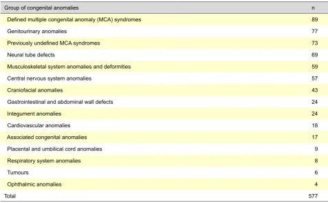
Maternal age ranged between 10 and 49, 26.9 on average. Among these mothers, approximately 14.5% were aged under 20 (adolescents), 69.9% were aged 20 to 34 and 15.6% were aged over 35. From our group of mothers, 577 (9.2%) had babies with congenital anomalies that were diagnosed at birth (Table 1). No statistically significant differences were found between mothers aged under or over 20 as regards the prevalence of babies with congenital anomalies (p = 0.43).
The distribution of babies with congenital anomalies according to maternal age is shown in Table 2. Neural tube defects were the leading anomaly in babies born to adolescent mothers, with higher prevalence rates compared to the remaining mothers (p = 0.027). No statistically significant differences were found between the groups regarding the remaining types of anomalies. Defined MCA syndromes (including chromosomal) were predominant in babies born to mothers aged over 20, whilst ophthalmic anomalies and tumours were those with the lowest prevalence in this group.
As regards the moment when anomalies were diagnosed, we found that 55.6% (321) of the anomalies were prenatally diagnosed and 44.4% (256) were diagnosed at the postnatal examination, with no statistically significant differences between these (p = 0.339). As regards prenatal diagnosis, we also found that it was obtained for 60% of babies born to adolescent mothers and for 54.6% of babies born to the remaining mothers (p = 0.483).
The distribution of types of anomaly according to the moment when these were diagnosed and to maternal age is shown in Table 3. We found that neural tube defects were
Table 1 – Classification of congenital anomalies
Group of congenital anomalies n
Defined multiple congenital anomaly (MCA) syndromes 89
Genitourinary anomalies 77
Previously undefined MCA syndromes 73
Neural tube defects 69
Musculoskeletal system anomalies and deformities 59
Central nervous system anomalies 57
Craniofacial anomalies 43
Gastrointestinal and abdominal wall defects 24
Integument anomalies 24
Cardiovascular anomalies 18
Associated congenital anomalies 17
Placental and umbilical cord anomalies 9
Respiratory system anomalies 8
Tumours 6
Ophthalmic anomalies 4
ARTIGO ORIGINAL
prenatally diagnosed in 100% of babies born to adolescent mothers and in 98.1% to mothers aged over 20. There were no MCA syndromes nor ophthalmic anomalies prenatally diagnosed.
DISCUSSION
Adolescent pregnancy is an important concern among public health authorities, mainly due to the rising incidence in this age group as well as to the socioeconomic factors involved. Nevertheless, few attention has been given to some complications that may relate to adolescent pregnancy, such as the study of congenital anomalies in their babies.7 An adequate prenatal monitoring of these women is crucial as, apart from maternal-foetal healthcare, all the involved psycho-social issues should be looked upon, including unplanned pregnancies, school dropout and recurrent pregnancy in this group.
Even though maternal age is well known as a risk factor for chromosomal abnormalities, the prevalence of birth defects is best known in the group of babies born to women aged under 20, as well as which are the most frequent types of anomaly and their outcomes.8,9 We should also mention that, when knowing the types and behaviour of congenital anomalies in babies born to adolescent mothers, preventive measures may be taken aimed to reduce their incidence. From a total of 6,257 pregnant mothers in our study, 14.5% (907 mothers) were adolescent (144.9 per 1,000 rate), which was in line with the average rate found in the state of São Paulo, although lower than the average in Brazil. According to data from the DATASUS,14 14.7% of deliveries occurred in 2011 at the state of São Paulo involved adolescent mothers vs. 19.2% (which was the national average). This rate of teenage deliveries is much higher than the rate found in developed countries such as Australia (1.98%), Canada (2.42%), Great Britain (2.84%), Germany (1.25%) and the USA (5.44%) (data regarding
mothers aged 15 to 19),15 showing that teenage pregnancy is an important public health issue in Brazil. In addition, the rate of low-birth-weight babies born to adolescent mothers is almost twice the rate in babies born to non-adolescent mothers.16 According to an American study, an adequate prenatal healthcare supply for adolescents that did not have any surveillance up to the sixth month of pregnancy would allow for a 50% reduction in low-birth-weight babies, with an average cost of only 95 USD.17
A 9.2% global prevalence of congenital anomalies diagnosed at birth was found in our population, which is above what is described in literature (between 3 to 5% prevalence).18 This high prevalence is due to the fact that the Hospital São Paulo is a referral public university hospital, attending to foetal anomalies clinically diagnosed or suspected in prenatal ultrasound from the whole metropolitan area of São Paulo (22 million people), as well as from other municipalities within the state of São Paulo as well as from other states in Brazil.
No statistically significant association was found between maternal age over or under 20 and the presence of congenital anomalies. In a study carried out in Atlanta, USA, between 1968 and 2000, involving 32,816 newborn babies born with non-chromosomal congenital anomalies, the number of babies born to adolescent mothers (aged 14 to 19) and to older mothers (aged 35 to 40) was similar, although showing different types of anomaly between the groups.10 Congenital cardiac anomalies, anencephaly, hydronephrosis, eardrum birth defects, cleft lip, omphalocele and gastroschisis were predominant in the group of babies born to adolescent mothers in this American study, whilst congenital cardiac anomalies, hypospadias and craniosynostosis were predominant in the older mother’s group. In line with our study, neural tube defects were the most frequent anomaly in the group of babies born to adolescent mothers.
Table 2 – Distribution of types of congenital anomalies according to maternal age
Types of congenital anomalies Maternal age (years) p
< 20
(n = 907) (n = 5 350)≥ 20
Neural tube defects 17 (1.76%) 52 (0.99%) 0.027*
Central nervous system anomalies 11 (1.21%) 46 (0.86%) 0.398
Craniofacial 6 (0.66%) 37 (0.69%) 0.908
Integument 4 (0.44%) 20 (0.37%) 0.990
Gastrointestinal and abdominal wall 3 (0.33%) 21 (0.39%) 0.990
Cardiovascular - 18 (0.34%) 0.157
Respiratory system - 8 (0.15%) 0.507
Genitourinary 14 (1.54%) 63 (1.18%) 0.446
Ophthalmic - 4 (0.07%) 0.910
Associated anomalies 2 (0.22%) 15 (0.28%) 0.980
Previously undefined MCA syndromes 14 (1.54%) 59 (1.10%) 0.327
Defined MCA syndromes 11 (1.21%) 78 (1.46%) 0.671
Tumours - 6 (0.11%) 0.668
Musculoskeletal and deformities 6 (0.77%) 53 (0.97%) 0.773
Placental and umbilical cord anomalies 2 (0.22%) 7 (0.13%) 0.853
ARTIGO ORIGINAL
Table 3 – Types of congenital anomalies according to the moment of diagnosis and to maternal age
Types of congenital anomalies Maternal age (years) Diagnosis
Prenatal Postnatal
Neural tube defects < 20 17 (100.0%)
-≥ 20 51 (98.1%) 1 (1.9%)
Central nervous system anomalies < 20 10 (90.9%) 1 (9.1%)
≥ 20 41 (89.1%) 5 (10.9%)
Craniofacial < 20≥ 20 1 (16.7%)2 (5.4%) 35 (94.6%)5 (83.3%)
Integument < 20 - 4 (100.0%)
≥ 20 1 (5.0%) 19 (95.0%)
Gastrointestinal < 20 1 (33.3%) 2 (66.7%)
≥ 20 15 (71.4%) 6 (28.6%)
Cardiovascular < 20 -
-≥ 20 9 (50.0%) 9 (50.0%)
Respiratory system < 20 -
-≥ 20 6 (75.0%) 2 (25.0%)
Genitourinary < 20 10 (71.4%) 4 (28.6%)
≥ 20 39 (61.9%) 24 (38.1%)
Ophthalmic < 20 -
-≥ 20 - 4 (100.0%)
Associated anomalies < 20 - 2 (100.0%)
≥ 20 - 15 (100.0%)
Previously undefined MCA syndromes < 20 11 (78.6%) 3 (21.4%)
≥ 20 44 (74.6%) 15 (25.4%)
Defined MCA syndromes < 20 5 (45.5%) 6 (54.5%)
≥ 20 49 (62.8%) 29 (37.2%)
Tumours < 20 -
-≥ 20 06 (100.0%)
Musculoskeletal and deformities < 20 - 6 (100.0%)
≥ 20 1 (1.9%) 52 (98.1%)
Placental and umbilical cord anomalies < 20 - 2 (100.0%)
≥ 20 1 (14.3%) 6 (85.7%)
However, there are studies with conflicting results, such as the study by Jacono et al.,19 in which a higher prevalence of congenital anomalies diagnosed at birth was found in babies born to adolescent mothers, when compared to babies born to mothers aged over 19. In a Chilean study (the Latin-American Collaborative study on Congenital Malformations), using the same classification as we adopted, a general 8.4% rate of malformations was found, similar to the 9.2% rate that we obtained in our population. In this Chilean study, the highest rates of foetal malformations were found in babies born to mothers aged under 20 and over 39, corresponding to 56% of all babies with malformations.20 Non-chromosomal congenital anomalies were the most prevalent anomaly in babies born to adolescent mothers (under 20 years of age), with a rate of 26.5 per 1,000 deliveries found in an European study involving 155 countries and 1.75 million deliveries. Teenage pregnancy was associated to gastroschisis, tricuspid atresia, anencephaly and gastrointestinal and nervous system malformations.21
ARTIGO ORIGINAL
When maternal age groups were analysed according to the presence of different types of congenital anomaly, we found that neural tube defects (1.76%) were the most prevalent in babies born to adolescent mothers whilst defined MCA syndromes (1.46%) were predominant in general population. This group included, for instance, babies with nevi (minor anomaly) and polydactyly (minor) or neural tube defect (major) and polydactyly (minor), diagnosed before hospital discharge.
In addition, teenage pregnancy (mothers aged 13-19) was also associated to an increased risk of central nervous system anomaly (odds ratio – OR: 1.08; 95% confidence interval – CI: 1.01-1.16), including neural tube defects such as anencephaly and spina bifida, in a retrospective study involving 5,542,861 single pregnancies in American women aged under 35.23
Central nervous system anomalies were also the most prevalent in a Brazilian study involving 335 babies with prenatally diagnosed foetal malformations born to mothers with an average of 27.1 years of age.24 Neural tube defects were the most frequent cause for termination of pregnancy due to congenital anomalies, in an Iranian study.25 The high percentage of prenatally diagnosed neural tube defects may relate to the high sensitivity - up to 92.8% - of ultrasound for the diagnosis of malformations of the central nervous system,22 whilst the high prevalence of this malformation in teenage pregnant mothers may be explained by the absence of folic acid preconception supplementation in unintended pregnancies, which is characteristic in adolescent women. A 16.1% frequency of neural tube defects (95% CI, 11.3-22.1) in the first group (175 pregnant mothers who had supplementation) and 47.1% in the second group (68 pregnant mothers with no supplementation; CI 95%, 35.6-58.7) was found in another Iranian study comparing two groups of pregnant mothers.26
The recent introduction of microarray-based comparative genomic hybridization techniques (array-CGH) offers higher resolution than standard karyotype for the diagnosis of unbalanced chromosomal abnormalities, allowing more frequently for an aetiological diagnosis of foetal or newborn morphological anomalies and consequently for a more accurate genetic counselling, namely regarding
the risk of recurrence.27 However, it should be mentioned that most congenital anomalies have a multifactorial origin such as neural tube defects and susceptibility of genetic variants remains unknown. In addition, this is an expensive technique and not available in most referral public hospitals in many developing countries, such as in Brazil.
CONCLUSION
In summary, the prevalence of congenital anomalies in newborn babies born to teenage mothers was in line with the one in newborn babies born to older mothers; in addition, neural tube defects were significantly more prevalent in babies born to adolescent mothers. These results show the need for a better preconception healthcare, with folic acid supplementation in this group of women and a personalized prenatal surveillance.
ACKNOWLEDGEMENTS
The authors wish to acknowledge the Coordenação de
Aperfeiçoamento de Pessoal de Nível Superior (CAPES)
for having assigned a Master’s grant to the student Leandro Valim dos Reis.
HUMAN AND ANIMAL PROTECTION
The study was approved by the Ethics Research Committee of the Federal University of São Paulo, file number 0387/04. The authors declare that the followed procedures were according to the Helsinki Declaration of the World Medical Association.
DATA CONFIDENTIALITY
The authors declare that they have followed the protocols of their work centre on the publication of patient data.
CONFLICTS OF INTEREST
The authors declare that there were no conflicts of interest in writing this manuscript.
FINANCIAL SUPPORT
The authors declare that there was no financial support in writing this manuscript.
REFERENCES
1. Colle M, Battin J. Physiologie de la puberte feminine et de la menstruation. Pediatrie. 1979;34:5-9.
2. World Health Organization. El embarazo y el aborto en la adolescencia. Ginebra: WHO; 1975.
3. Huang CC, Lin YC, Huang YT, Huang KH. Comparison of medical issues in antenatal and perinatal periods in early youth, adolescent, and young adult mothers in Taiwan: a 10-year nationwide study. BMC Pregnancy Childbirth. 2014;14:260.
4. Curtin SC, Abma JC, Ventura SJ, Henshaw SK. Pregnancy rates for U.S. women continue to drop. NCHS Data Brief. 2013;136:1-8. 5. Sokal R, Fleming KM, Tata LJ. Potential of general practice data for
congenital anomaly research: Comparison with registry data in the United Kingdom. Birth Defects Res A Clin Mol Teratol. 2013;97:546-53. 6. Centers for Disease Control and Prevention. Infant mortality-United
States. JAMA. 1994;271:15-7.
7. Croen LA, Shaw GM. Young maternal age and congenital malformations:
a population-based study. Am J Public Health. 1995;85:710-3. 8. Shin M, Besser LM, Kucik JE, Lu C, Siffel C, Correa A, et al. Prevalence
of Down syndrome among children and adolescents in 10 regions of the United States. Pediatrics. 2009;124:1565-71.
9. Egan JF, Smith K, Timms D, Bolnick JM, Campbell WA, Benn PA. Demographic differences in Down syndrome livebirths in the US from 1989 to 2006. Prenat Diagn. 2011;31:389-94.
10. Reefhuis J, Honein MA. Maternal age and non-chromosomal birth defects, Atlanta--1968-2000: teenager or thirty-something, who is at risk? Birth Defects Res A Clin Mol Teratol. 2004;70:572-9.
11. Shankaran S, Lester BM, Das A, Bauer CR, Bada HS, Lagasse L, et al. Impact of maternal substance use during pregnancy on childhood outcome.Semin Fetal Neonatal Med. 2007;12:143-50.
ARTIGO ORIGINAL
13. Sukchan P, Liabsuetrakul T, Chongsuvivatwong V, Songwathana P, Sornsrivichai V, Kuning M. Inadequacy of nutrients intake among pregnant women in the deep south of Thailand. BMC Public Health. 2010;10:572.
14. Brasil. Ministério da Saúde. DATASUS [homepage na Internet]. Proporção de nascidos vivos de mães adolescentes. Ministério da Saúde; 2011. [Consultado 2013 dez 23]. Disponível em: http://tabnet. datasus.gov.br/cgi/tabcgi.exe?idb2012/g15.def.
15. Singh S, Darroch JE. Adolescent pregnancy and childbearing: levels and trends in developed countries. Fam Plann Perspect. 2000;32:14-23.
16. Felice ME, Feinstein RA, Fisher MM, Kaplan DW, Olmedo LF, Rome ES, et al. Adolescent pregnancy current trends and issues: 1998. American Academy of Pediatrics Committee on Adolescence 1998– 1999. Pediatrics. 1999;103:516-20.
17. Hueston WJ, Quattlebaum RG, Benich JJ. How much money can early prenatal care for teen pregnancies save? a cost-benefit analysis. J Am Board Fam Med. 2008;21:184-90.
18. De Vigan C, Khoshnood B, Lhomme VV, Goujard J, Goffinet F. Prévalence et diagnostic prénatal des malformations em population parisienne: vingt ans de surveillance par le Registre des Malformations Congénitales de Paris. J Gynecol Obstet Biol Reprod. 2005;34:8-16. 19. Jacono JJ, Jacono BJ, St Onge M, Van Oosten S, Meininger E. Teenage
pregnancy: a reconsideration. Can J Public Health. 1992;83:196-9.
20. Nazer J, Cifuentes L, Águila A, Ureta P, Bello MP, Correa F, et al. Edad materna y malformaciones congenitas: un registro de 35 anos. 1970-2005. Rev Med Chile. 2007;135:1463-9.
21. Loane M, Dolk H, Morris JK; EUROCAT Working Group. Maternal age-specific risk of non-chromosomal anomalies. BJOG. 2009;116:1111-9. 22. Noronha Neto C, Souza AS, Moraes Filho OB, Noronha AM. Validação
do diagnostico ultrassonografico de anomalias fetais em centro de referencia. Rev Assoc Med Bras. 2009;55:541-6.
23. Chen XK, Wen SW, Fleming N, Yang Q, Walker MC. Teenage pregnancy and congenital anomalies: which system is vulnerable? Hum Reprod. 2007;22:1730-5.
24. Ramos JL, Carvalho MH, Zugaib M. Caracterização sociodemografica e resultados perinatais das gestacoes com diagnostico ultrassonografico de malformação fetal. Rev Assoc Med Bras. 2009;55:447-51. 25. Samadirad B, Khamnian Z, Hosseini MB, Dastgiri S. Congenital
anomalies and termination of pregnancy in Iran. J Pregnancy. 2012;2012:574513.
26. Hosseini MB, Khamnian Z, Dastgiri S, Samadi Raad B, Ravanshad Y. Folic acid and birth defects: a case study (Iran). J Pregnancy. 2011;2011:370458.