R E V I E W A R T I C L E
Radix entomolaris in children – A challenge to
pedodontist: A report of case series with literature review
N. B. Nagaveni, Gaurav Ram Chandani, Shilpa Kothari, P. PoornimaDepartment of Pedodontics and Preventive Dentistry, College of Dental Sciences, Davangere, Karnataka, India
Abstract
Permanent mandibular molars exhibit numerous variations pertaining to root or canal numbers. Vary rarely a supernumerary or extra root is found distolingually, which is given the term radix entomolaris in the literature. Thorough knowledge of this root variation is highly essential to provide ultimate success following root canal treatment. This paper
describes three case reports of permanent mandibular first molar with three roots (one mesial and two distal) and four canals (two in mesial and one in each distobuccal and
distolingual root).
Keywords: Anatomical variation, children, endodontic therapy, mandibular first molar, radix entomolaris
Correspondence
Dr. N. B. Nagaveni, Department of
Pedodontics and Preventive Dentistry, College of Dental Sciences, Davangere, Karnataka, India. E‑mail: [email protected]
Received 27 January 2015; Accepted 01 March 2015
doi: 10.15713/ins.ijcdmr. 53
How to cite the article:
N. B. Nagaveni, Gaurav Ram Chandani, Shilpa Kothari, P. Poornima, “Radix entomolaris in children – A challenge to pedodontist: A report of case series with literature review,” Int J Contemp Dent Med Rev, vol. 2015, Article ID: 360115, 2015. doi: 10.15713/ins.ijcdmr. 53
Introduction
The major intention of the root canal treatment is thorough debridement of root canals and their disinfection before obturation and sealing. One of the main reasons for failure of root canal treatment in molars is because the clinician has not removed all the pulp tissue and micro‑organisms from the entire root canal system.[1] Therefore, the knowledge of anatomy as
well as the variations in the number and shape of canals is of utmost importance for the clinician.
It is known that the permanent mandibular first molar can
display several anatomical variations and, like the number of root canals, the number of roots may also vary.[1,2] A major anatomical variant of the two‑rooted mandibular first molar is the third root known as radix entomolaris (RE), first mentioned in the
literature by Carrabelli.[3] This supernumerary root is located distolingually in mandibular molars, mainly the first molars and
most of the time it displays Vertucci Type I canal configuration. Sometimes an extra root is seen at the mesiobuccal side of main mesial root, and it is called radix paramolaris.[3]
The occurrence of RE associated with first mandibular molars
is found in some ethnic population of the world. The prevalence of its occurrence in Mongoloid populations including Chinese,
Eskimo and American Indians is 5‑30%.[4‑10] As the frequency
is high in these populations some authors consider it as normal
morphological entity or eumorphic root variation. However, in
Caucasians, as RE is not common, it is considered unusual or
dysmorphic root morphology. In dysmorphic supernumerary roots, their formation is related to external factors during odontogenesis, or due to penetration of an atavistic gene or polygenic system. In eumorphic roots, racial genetic factors
influence the most profound expression of a particular gene that
results in a more pronounced phenotypic manifestation.[6] Garg et al.[11] and Karale et al.[12] found a prevalence of 5% and 7.67%
in Indian population. However, Drennan[13] and Shaw[14] have investigated 0% prevalence of RE in South African Bushman and African Bantu races.
There are reports showing RE occurs more commonly on
the right side.[4,11] However, some reports also reported that RE
occurred on the left side in some individuals.[3,6‑8] With respect
to gender predilection Loh[15] and Colak et al.[16] did not found any significant difference for occurrence of RE with either sex.
Whereas, Tratman[4] has stated that it is more common on the
right side for males and bilateral for females. However, there are
reports showing prevalence of RE is similar in both sexes.[5‑8,11,12]
Midtbø and Halse[17] studied root length, crown height and
root morphology using an intra‑oral and panoramic radiographs
influences root formation. Curzon[18] suggested that certain
traits such as the “three‑rooted molar” was genetically dominant
as was reflected in the fact that pure Inuit and Inuit/Caucasian
mixes had similar prevalence of the trait. The prevalence of this extra root is equally manifested in males and females. However, the anomaly is more frequent on the left side and some studies
report a bilateral occurrence of RE from 50 to 67%.[19]
Carlsen and Alexandersen[20] have classified the RE into four
types according to its cervical part: Type A and Type B, distally located cervical part of RE with two normal mesial and one
normal distal component respectively; Type C is mesially located cervical part; and Type AC, central location, between distal and mesial root components. De Moor et al.[21] classified the RE into
three types according to buccolingual variations: Type I, straight
root/root canal; Type II, initially curvedentrance that continues as a straight root/root canal; Type III, initial curve in the coronal
third and a second curve beginning in the middle and continuing
to the apical third. Recently, Song et al.[22]categorized RE into five types such as Type I, II, III, small and conical type based on the curvature of RE. The purpose of this article is to report three
such cases who presented with this variation in morphology and its subsequent treatment.
Case Reports
Case 1
A 14‑year‑old male patient reported to the Department of Pedodontics and Preventive Dentistry, College of Dental Sciences, Davangere, India with a chief complaint of pain in his left lower back tooth region of mouth since 1 month. On clinical examination both permanent mandibular right and
left first molars (36, 46 ‑ FDI tooth notation) had deep caries on distoproximal surface. Both were tender on percussion and
showed no response to electric or thermal testing.
Radiographic evaluation with intra oral periapical radiograph showed radiolucency involving the pulp with widening of periodontal ligament space in 46, and periapical radiolucency in periapical region in relation to 36 [Figure 1a and 2a]. Based
on clinical and radiographic findings a diagnosis of chronic
periapical abscess with 36 and chronic reversible pulpitis with 46 was made treatment plan was decided as endodontic treatment for both the teeth followed by core build up, metal post and
finally with stainless steel crown.
An aesthesia was achieved for tooth number 36 by means of inferior alveolar nerve and buccal nerve block with 1.8 ml
of 2% lignocaine with 1:80,000 adrenaline. Conventional
root canal treatment was started, and all carious part was
removed and a trapezoidal access preparation was done with endo‑access bur and canal orifices were located with
DG 16 endodontic explorer. Initial negotiation of the root
canals was confirmed with K‑file 10. The fourth distolingual canal orifice was present far from distal root canal orifices.
The canal lengths were determined radiographically with K
file ISO 15 size [Figure 1b] and electronically with root ZX. They were cleaned with 2.5% sodium hypochlorite along
with saline. Calcium hydroxide powder mixed with saline was used as an intra‑canal medicament. At next appointment, patient was asymptomatic. Master cone radiograph revealed
proper fitting of cones [Figure 1c]. Canals were dried with paper point and obturation was done by lateral condensation technique using AH‑26 sealer [Figure 1d]. The access cavity was then temporarily sealed with intermediate restorative
material (IRM) and after 1 month post space was prepared
on the distobuccal canal space and metal post was cemented followed by core build‑up of the tooth using vitremer.
Similar treatment was done for mandibular right first
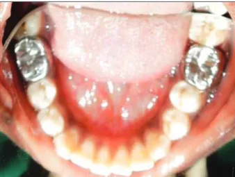
molar [Figure 2]. Finally, both teeth were restored with stainless
steel crown [Figure 3]. The patient remained asymptomatic after 1 year post treatment and is continued to be under active follow‑up.
Figure 1: (a-d) Endodontic treatment of permanent mandibular left molar with radix entomolaris (arrows)
Figure 2: (a-d) Permanent mandibular right first molar showing distolingual root or radix entomolaris (arrows)
a b
c d
a
c d
Case 2
A 15‑year‑old female patient reported to the same Department
with a chief complaint of pain in his left lower back tooth region
since 10 days. On clinical examination mandibular right first molar (46 ‑ FDI tooth notation) was deeply carious, tender
on percussion and radiographically there was widening in the periodontal ligament space. We also noticed the presence of a third root between the mesial and distal roots [Figure 4a]. Diagnosis was made as apical periodontitis with 46. Treatment plan was decided to do endodontic treatment for the same tooth followed by cast metal crown.
Conventional root canal treatment was started with 46.
After access opening the cavity was modified from triangular to trapezoidal in order to locate the RE. The root canal length determination was done with files [Figure 4b] and step back method was used for biomechanical preparation, teeth were
cleaned with 2.5% sodium hypochlorite along with saline
and closed dressing was given. Calcium hydroxide powder
mixed with saline was used as an intra‑canal medicament. At next appointment, patient was asymptomatic. Master cone radiograph revealed proper fitting of cones [Figure 4c]. Canals were dried with paper point and obturation was done by lateral condensation technique using AH‑26 sealer [Figure 4d]. The
access cavity was then temporarily sealed with IRM.
After root canal treatment, the tooth was prepared for receiving cast metal crown and subsequently the metal crown was delivered. The patient is asymptomatic and is under active follow up from past 1 year.
Case 3
A 10‑year‑old patient came with a chief complaint of pain in his lower left back tooth region since 30 days. On clinical
examination, deep occlusal caries was present in permanent left
mandibular first molar (36 ‑ FDI tooth notation) and was tender on percussion. Radiographic examination showed widening of periodontal ligament space [Figure 5a]. A diagnosis of apical
periodontitis was made, and treatment plan was decided to do a conventional root canal treatment. Access cavity was prepared,
and four distinct canal orifices were found. The canal lengths were
determined radiographically with K file ISO 15 size [Figure 5b] and electronically with root Z × . They were cleaned with 2.5%
sodium hypochlorite along with saline. Calcium hydroxide powder mixed with saline was used as an intra‑canal medicament. At next appointment patient was asymptomatic. Master cone radiograph revealed proper fitting of cones [Figure 5c]. Canals
were dried with paper points and obturation was done by using AH‑26 sealer [Figure 5d]. After 1 week, the tooth was prepared
and a stainless steel crown was delivered to the patient. The patient is asymptomatic and is under active follow‑up from past 6 months.
Discussion
Ingle[23] stated that the most frequent cause of endodontic failure is a canal that is left untreated because a clinician fails to recognize
Figure 3: Post-operative restoration of the teeth with stainless steel crowns
Figure 4: (a-d) Arrows showing radix entomolaris in permanent
mandibular right first molar Figure 5: (a-d) Radiographs showing complete root canal treatment of the radix entomolaris (arrows)
a b
d d
b
c a
it. Vertucci[24] studied the internal and external anatomy of teeth
and has shown that anatomical variations can occur within each group of teeth, within each person and, in general, within each racial group.
Few authors showed that RE was found in association
with an additional cusp on the buccal side that is usually called protostylid. Sometimes it is also seen with tuberculum paramolare or cervical prominence or convexity. Hence, it has been stated that the presence of extra root or canals is always associated with increased number of cusps.[3]
The presence of RE has clinical implications during endodontic
treatment. An accurate diagnosis of the supernumerary roots can avoid complications or a missed canal during root canal treatment. Apart from complicating the root canal procedures,
RE has been found to be a contributing factor to localized
periodontal destruction.[2] In addition, reports demonstrated significantly higher probing depths with attachment loss at the
distolingual aspect of three‑rooted molars.[2,25] During extraction
procedure, if rotational movements are used, there are chances
of RE root fracture due to its divergent morphology.[2,3] Presence of RE has orthodontic implications also. This root can make the orthodontic movement difficult.[17] However, few authors
also hypothesized that RE being an extra root gives additional
support and stability for molars by increasing the surface area of attachment to the alveolar bone.[26] Addition to this, RE has
forensic value in identifying the people of Mongoloid races.[5‑12,26]
The RE is usually situated in the same buccolingual plane
as the distobuccal root, so a superimposition of both roots can appear on the preoperative radiograph and result in an inaccurate diagnosis. The proper application of Clark’s rule or the buccal object rule facilitates locating additional canals or roots, and distinguishing between objects that have been superimposed.[3,27]
Hence, to reveal an RE, additional radiographs should be taken
from a more mesial or distal angle (20°). In 1985, Walker and
Quackenbush[10] concluded that panoramic radiographs reveal
90% accuracy in diagnosing RE. Along with this thorough
interpretation of some particular marks like an unclear view or
outline of the mesial or distal root/canal, contour can reveal the hidden RE.[3,27] Recently, the use of advanced techniques like
spiral computed tomography clearly shows the entire anatomy of the extra roots or canals.[28] The location of the additional canal orifice may be difficult because of overhanging dentine. If the orifice is not found, the root canal remains untreated and
infected, or necrotic tissue remnants may remain in the root canal, leading to treatment failure.
With the distolingually located orifice of RE, a modification of the classical triangular access cavity to a trapezoidal form,
so as to better locate and access the root canal is essential.[2,3,21] After access opening if the canal orifice of the RE root is not evident, clinician must carefully inspect the pulpal floor or wall at the distolingual region. As most of the RE root show severe inclination especially at the apical third part (like in Type III RE)
chances of formation of ledge in the root canal or loss of working length are possible.[21,28,29] Moreover, in these roots use of nickel
titanium rotary files are of more useful to provide a more centered
preparation and also to avoid procedural errors during root canal preparation.[3,21,28,29]
Conclusion
An accurate diagnosis of RE is essential to prevent procedural
complications during endodontic treatment. Correct radiographic techniques and interpretation of radiographs is
highly essential in identifying RE in mandibular molars.
References
1. Bonaccorso A, Tripi TR. Root canal treatment of a three-rooted mandibular first molar – A case report. ENDO 2008;2:211-8.
2. Nagaven NB, Umashankara KV. Radix entomolaris and paramolaris in children: A review of the literature. J Indian Soc Pedod Prev Dent 2012;30:94-102.
3. Calberson FL, De Moor RJ, Deroose CA. The radix entomolaris and paramolaris: Clinical approach in endodontics. J Endod 2007;33:58-63.
4. Tratman EK. Three-rooted lower molars in man and their racial distribution. Br Dent J 1938;64:264-74.
5. Pederson PO. The East Greenland Eskimo Dentition. Numerical Variations and Anatomy. A Contribution to Comparative Ethnic Odontography. Vol. 104. Copenhagen: Meddeleleser Om Gronland; 1949. p. 140-4.
6. Turner CG 2nd. Three-rooted mandibular first permanent molars and the question of American Indian origins. Am J Phys Anthropol 1971;34:229-41.
7. Curzon ME, Curzon JA. Three-rooted mandibular molars in the Keewatin Eskimo. J Can Dent Assoc (Tor) 1971;37:71-2. 8. Yew SC, Chan K. A retrospective study of endodontically treated
mandibular first molars in a Chinese population. J Endod 1993;19:471-3.
9. Reichart PA, Metah D. Three-rooted permanent mandibular first molars in the Thai. Community Dent Oral Epidemiol 1981;9:191-2.
10. Walker RT, Quackenbush LE. Three-rooted lower first permanent molars in Hong Kong Chinese. Br Dent J 1985;159:298-9. 11. Garg AK, Tewari RK, Kumar A, Hashmi SH, Agrawal N,
Mishra SK. Prevalence of three-rooted mandibular permanent first molars among the Indian Population. J Endod 2010;36:1302-6.
12. Karale R, Chikkamallaiah C, Hegde J, Aswathanarayana S, Santhosh L, Bashetty K, et al. The prevalence of bilateral three-rooted mandibular first molar in Indian population. Iran Endod J 2013;8:99-102.
13. Drennan MR. The dentition of the Bushmen tribe. Ann South Afr Mus 1929;24:61-87.
14. Shaw JC. The Teeth, the Bony Palate and the Mandible in Bantu Races of South Africa. London, UK: John Bale, Sons and Danielson; 1931.
15. Loh HS. Incidence and features of three-rooted permanent mandibular molars. Aust Dent J 1990;35:434-7.
mandibular permanent first molars among the Turkish population. Niger J Clin Pract 2012;15:306-10.
17. Midtbø M, Halse A. Root length, crown height, and root morphology in Turner syndrome. Acta Odontol Scand 1994;52:303-14.
18. Curzon ME. Miscegenation and the prevalence of three-rooted mandibular first molars in the Baffin Eskimo. Community Dent Oral Epidemiol 1974;2:130-1.
19. Segura-Egea JJ, Jiménez-Pinzón A, Ríos-Santos JV. Endodontic therapy in a 3-rooted mandibular first molar: Importance of a thorough radiographic examination. J Can Dent Assoc 2002;68:541-4.
20. Carlsen O, Alexandersen V. Radix entomolaris: Identification and morphology. Scand J Dent Res 1990;98:363-73.
21. Nagaveni NB, Umashankar KV, Radhika NB, Satisha TS. Third root (Radix Entomolaris) In Permanent Mandibular First Molars In Pediatric Patients – An Endodontic Challenge. J Oral Health Comm Dent 2011;5:49-51.
22. Song JS, Choi HJ, Jung IY, Jung HS, Kim SO. The prevalence and morphologic classification of distolingual roots in
the mandibular molars in a Korean population. J Endod 2010;36:653-7.
23. Nagaveni NB, Umashankar KV. Radix entomolaris in permanent mandibular first molars: Case reports and literature review. Gen Dent 2009;57:e25-9.
24. Vertucci FJ. Root canal morphology and its relationship to endodontic procedures. Endod Top 2005;10:3-29.
25. Huang RY, Lin CD, Lee MS, Yeh CL, Shen EC, Chiang CY, et al.
Mandibular disto-lingual root: A consideration in periodontal therapy. J Periodontol 2007;78:1485-90.
26. Mayhall JT. Three-rooted deciduous mandibular second molars. J Can Dent Assoc 1981;47:319-21.
27. White SC, Pharoh MJ. Oral Radiology: Principles and Interpretation. 4th ed. St. Louis: Mosby; 1999. p. 91.
28. Garg AK, Tewari RK, Agrawal N. Prevalence of three-rooted mandibular first molars among indians using SCT. Int J Dent 2013;2013:183869.