MECHANISMS THAT PROMOTE LIBERATION OF MITOTIC STRESS-INDUCED DEATH
Rebecca K. Sinnott
A dissertation submitted to the faculty of the University of North Carolina at Chapel Hill in partial fulfillment of the requirements for the degree of Doctorate of Philosophy in the Department of Pharmacology, School of Medicine.
Chapel Hill 2013
Approved by: Angelique W. Whitehurst
Robert A. Nicholas Stephen L. Rogers
ABSTRACT
REBECCA K SINNOTT: Mechanisms that promote liberation of mitotic stress-induced death
(Under the direction of Dr. Angelique Whitehurst)
Paclitaxel is an anti-mitotic drug that, due to its success in the clinic, has become a backbone of first-line chemotherapeutic regimens for many malignancies including non-small cell lung cancer (NSCLC). While paclitaxel-based regimens are efficacious for some NSCLC patients, response is often incomplete, rarely curative and unpredictable, indicating widespread intrinsic resistance in chemo-naïve tumors. Thus, there is an unmet need for new combinatorial treatment strategies to better target paclitaxel resistant tumor cells.
To study the molecular basis for this resistance, we first established a test bed of NSCLC-derived cell lines that evade cell death from high concentrations of paclitaxel due to an uncoupling of mitotic damage from cell death. We then employed a genome-wide loss-of-function cytotoxic screen to identify the molecular components that can re-engage paclitaxel-mediated cell death programs in an otherwise paclitaxel-resistant background. This screen was performed in the presence and absence of a mitotic damaging, yet sub-lethal, dose of paclitaxel. This approach revealed a cohort of proteins that support tumor cell viability in the presence of mitotic damage.
which is frequently co-amplified with KRAS, is essential for microtubule polymerization and mitotic spindle formation. We also identified TRIM69, an E3 ubiquitin ligase, that we find is recruited to the spindle poles during mitosis to support mitotic fidelity. Importantly, stable depletion of either CASC1, or TRIM69, attenuates tumor cell growth in vivo. Finally, we demonstrate that pharmacological inhibition of the APC collaborates
with an otherwise sublethal dose of paclitaxel.
ACKNOWLEDGEMENTS
First and foremost, I would like to thank my mentor, Dr. Angelique Whitehurst, for taking on a student with minimal experience, and providing countless hours, guidance and resources to support me throughout my dissertation research. I am grateful for her persistence in shaping me into a young scientist and demonstrating how to attack a problem and examine it from all angles.
I would like to thank Gary Johnson, Rob Nicholas and Ken Harden for shaping an excellent Pharmacology department in which to receive doctoral training and for their guidance when I needed support as a student. I would like to thank Charlene Ross in the UNC-Animal Core that assisted me with my xenograft studies, and Noah Sciaky for conversation and support in learning how to use the BD Pathway. I would also like to thank my committee, including Dr. Robert Nicholas, Dr. Stephen Rogers, Dr. Mohanish Deshmukh and Dr. Kimryn Rathmell for their insights.
TABLE OF CONTENTS
LIST OF TABLES ... viii
LIST OF FIGURES ... ix
LIST OF ABBREVIATIONS AND SYMBOLS ...x
Chapter I.Introduction ... Lung cancer treatment ... 1
Paclitaxel as a mainstay chemotherapeutic ... 2
Paclitaxels biological mechanism of action ... 3
The Spindle Assembly Checkpoint ... 5
Current challenges for paclitaxel based therapies ... 7
Project summary ... 10
Chapter II. Materials and Methods ... 14
Chapter III. Genome wide loss of function screen uncovers novel modulators of mitotic slippage... Defining screening platform ... 21
Genome wide loss of function screen ... 22
Secondary screening analysis ... 25
Prolonging a mitotic arrest restores paclitaxel sensitivity ... 26
Xenograft mouse models ... 29
Discussion ... 29
CASC1 Results ... 42
CASC1 Discussion ... 45
Chapter V TRIM69 is a centrosomal and microtubule associated protein that is essential for mitotic fidelity ... TRIM69 introduction ... 53
TRIM69 results ... 57
TRIM69 discussion ... 61
Chapter VI Summary ... 69
Future Directions ... 71
Final Conclusions ... 73
LIST OF TABLES
LIST OF FIGURES
Figure 1. The spindle assembly checkpoint ... 12
Figure 2. Variable fates following a prolonged mitotic delay ... 13
Figure 3 Defining a paclitaxel-resistant NSCLC screening platform ... 32
Figure 4 Pan-genomic loss of function screen in mitotic slippage prone HCC366 cells ... 34
Figure 5 Secondary screening analysis stratifies candidate chemosensitizers ... 36
Figure 6 Pan-genomic screen reveals conserved regulators of mitotic slippage ... 37
Figure 7 Prolonged engagament of the SAC recouples mitotic damage to cell death ... 38
Figure 8 Direct targeting of the APC/C collaborates with paclitaxel treatment ... 39
Figure 9 CASC1 and TRIM69 support tumor cell growth in vivo ... 40
Figure 10 CASC1 supports mitotic fidelity ... 48
Figure 11 CASC1 supports microtubule stability to satisfy the SAC ... 50
Figure 12 CASC1 is a tumor cell dependeny ... 52
Figure 13 TRIM69A is a testis enriched E3 ubiquitin ligase ... 61
Figure 14 TRIM69A supports mitotic fidelity ... 62
Figure 15 TRIM69A is a novel component of the MTOC ... 64
Figure 16 TRIM69 interacts with cancer testis antigen MAGEA4 ... 65
Figure 17 Slippage prone cell lines are sensitive to APC/C inhibition ... 72
LIST OF ABBREVIATIONS AND SYMBOLS ALK
ANAPC5 APC/C
Anaplastic lymphoma kinase Anaphase promoting complx 5
Anaphase promoting complex / Cyclosome APL
BSA BUB3
Acute promyelocytic leukemia Bovine serum albumin
Budding uninhibited by benzimidazoles 3 BUBR1 Budding uninhibited by benzimidazoles R1 CASC1
CDC20
Cancer susceptibility candidate 1 Cell division cycle 20
cDNA Complementary DNA
CTG CTRL
Cell titer glo Control
CTA(s) Cancer-testis antigen(s)
DAPI 4’-6-Diamidino-2-phenylindole DMSO
DNA
Dimethyl sulfoxide
Deoxyribonucleic acid
EGFR Epidermal growth factor receptor EGTA Ethylene glycol tetraacetic acid ELM4
FDA
Echinoderm microtubule-associated protein like-4 Food and Drug Administration
GFP-H2B Green fluorescent protein conjugated to histone 2B HAUS1
HBEC HECT
HAUS augmin-like complex, subunit 1 Human bronchial epithelial cell
Homologous to E6Ap carboxy terminus
HEPES 4-(2-hydroxyethyl)-1-piperazineethanesulfonic acid IACUC MAD2 MAGE MAGEA4 MAP(s) MCC MCL-1 MISP MTOC NCI NEXN Noc NSCLC
Institutional animal care and use committee Mitotic arrest deficient-like 1
Melanoma antigen
Melanoma antigen family A, 4 Microtubule associated protein(s) Mitotic checkpoint complex Myeloid cell leukemia sequence 1 Mitotic spindle positioning
Microtubule organizing center National Cancer Institute
Nexillin (F-actin binding protein) Nocodazole
Non-small cell lung cancer
NuMa Nuclear mitotic apparatus protein 1
Pac Paclitaxel
PLK1 Polo-like kinase 1 PML PP1 PPP1R18 qRT-PCR RARα Rb RhoA RING RNA Promyelocytic leukemia Protein phosphatase 1
Protein phosphatase 1, regulatory subunit 18, phostensin Quantitative Real-time polymerase chain reaction Retinoic acid receptor, alpha
Retinoblastoma protein
Ras homolog member, family A Really interesting new gene Ribonucleic acid
ROCK RPE1 SAC
Rho associated coiled-coiled containing kinase Retinal pigment epithelial cell line 1
Spindle assembly checkpoint SD Standard deviation
SDS Sodium dodecyl sulfate SEM Standard error of the mean shRNA Short hairpin RNA
Chapter I. Introduction Lung cancer treatment
Lung cancer is the leading cause of cancer-related death in the United States [1]. This high mortality rate is partially attributable to the often late stage of disease progression at the time of diagnosis, which leaves patients with limited therapeutic options beyond standard cytotoxic therapies [2]. Technological advancements over the past 10-15 years have allowed for the identification of patient subsets that carry specific genetic mutations that can be targeted to improve individual patient outcome. Currently, the epidermal growth factor receptor (EGFR) and the recently identified fusion protein, EML4-ALK, are examples of tumor vulnerabilities that can be targeted to improve non-small cell lung cancer (NSCLC) patient survival [3].
non-targetable oncogenic alterations, such as alterations to KRAS, or no known genetic lesion [5]. Thus, for a majority of patients, the current cytotoxic standard-of-care is the only available treatment option.
The current first-line, standard-of-care therapeutic regimen combines a DNA damaging reagent with the anti-mitotic paclitaxel [14, 15]. While this paclitaxel-based regimen has been shown to be efficacious, particularly in ovarian cancer, response in non-small-cell lung cancer has been notoriously poor with an approximate 30 % partial response rate at best [15-19]. Thus, there is an unmet need for new therapeutic approaches for treatment of NSCLC. Given the paucity of available targets for individualized therapy and the promise of taxane-based therapeutic regimens, novel therapeutic strategies that synergize with paclitaxel based treatment are in high demand.
Herein, I describe the undertaking of a genome-wide loss-of-function screen performed in a chemoresistant NSCLC-derived background in the presence of a damaging dose of paclitaxel. This screen was performed to uncover those molecular mechanisms that can disengage paclitaxel-mediated cell death programs. Therefore, a discussion on paclitaxel’s known mechanism of action and current therapeutic challenges is warranted.
Paclitaxel as a mainstay cytotoxic therapy
inhibitory specific activity [22]. Given paclitaxel’s initial identification as a tumor-inhibiting agent, clinical studies were performed in ovarian cancer. The standard-of-care for ovarian cancer at the time was a platinum-based cytotoxic reagent that met with limited success and intrinsic resistance [17, 21, 23]. Initial paclitaxel clinical trials reported an impressive therapeutic response with at least 30 % of patients demonstrating either a 50 % reduction in tumor volume or a complete clinical response [17]. Additionally, patients who had been resistant to the platinum-based therapy were responsive to paclitaxel treatment, setting the stage for combination therapy clinical trials [17, 24, 25]. In the late 1980s, paclitaxel was shown to be efficacious in the treatment of both advanced NSCLC and breast cancer and was approved by the Food and Drug Administration (FDA) for use in 1992 [21, 26, 27]. Since then, paclitaxel has become a mainstay of cytotoxic therapy and is broadly used as the first-line standard-of-care in ovarian, breast and non-small cell lung cancer.
Paclitaxel biological mechanism of action
Following the initial finding that paclitaxel was a potent anti-tumor agent, critical studies took place in order to understand it’s mechanism of action. Early studies led to the observation that paclitaxel can have profound impacts on microtubule dynamics and assembly [28, 29].
Microtubules exist as hollow tubes composed of 13 protofilaments, formed by α/β
The ability to rapidly alter microtubule kinetics is essential for many cellular processes including, but not limited to, protein and vesicle transportation, a cells motility and cell division. Paclitaxel alters microtubule dynamics by directly binding the β-subunit of the
α/β tubulin heterodimer. Binding of paclitaxel promotes a morphological change in the
α/β heterodimer that results in an increased affinity for a neighboring protofilament [32]. The resulting increased affinity makes microtubule depolymerization energetically unfavorable and thus supports overall microtubule stability [31]. The initial in vitro studies found that at saturating concentrations, paclitaxel binds β-tubulin in a 1:1 ratio and increases both microtubule stability and promotes microtubule polymerization [31, 32]. From these observations, paclitaxel is broadly classified as a microtubule stabilizing drug that at therapeutic doses functions by altering microtubule dynamics [31, 33, 34].
Shortly after the discovery of paclitaxel’s tumor inhibitory properties, paclitaxel was shown to induce damage to the mitotic spindle and inhibit appropriate mitotic division [29, 35]. Proper formation of the mitotic spindle for a high fidelity mitosis is critically reliant on rapid and stochastic remodeling of the microtubule network [31]. Given the paclitaxel-mediated impact on microtubule dynamics, it follows that the primary biological target of taxol treatment is mitosis.
microtubule growth and the ability to shrink if an attachment is not initially attained [31]. Further, inappropriate microtubule-kinetochore connections must depolymerize to avoid erroneous segregation [37]. Once a cell has aligned all chromosomes on the metaphase plate, the cell continues to rely on microtubule dynamicity to apply tension across the established mitotic spindle and physically pull aligned chromosomes apart [38, 39]. Thus, disruption of appropriate microtubule dynamics impairs formation of a bipolar mitotic spindle and creates mitotic stress. Mitotic spindle formation is monitored by the sentinel spindle assembly checkpoint (SAC) to prevent erroneous chromosomal segregation. Inhibition of bi-polar spindle formation prevents satisfaction of the SAC which will in turn delay mitotic exit and, ideally, the genomic damage that would result from incorrect chromosome segregation.
In summary, treatment with paclitaxel inhibits microtubule dynamics, disrupts mitotic spindle formation and results in prolonged engagement of the SAC, which delays mitotic division. A prolonged mitotic delay can lead to cell death signaling, imparting the therapeutic benefit of paclitaxel treatment. Although the SAC has been well studied, how a prolonged delay ultimately couples to cell death and why some tumors are more sensitive to SAC engagement than others is still largely unknown.
Spindle Assembly Checkpoint
assemble on the kinetochore in a dynamic mitotic checkpoint complex (MCC). The MCC inhibits APC/C activity by directly binding CDC20, an essential co-activator of the APC/C [40-42]. As kinetochore-microtubule attachments are established, the core MCC proteins, such as MAD2, are readily removed. Thus, the presence of MCC proteins at the kinetochore is interpreted as a lack of microtubule attachment [41, 43, 44]. Experiments inhibiting the APC/C and testing localization of MCC proteins at the kinetochore will be evaluated in chapters 4 and 5.
In a normal, non-transformed cell, the SAC remains engaged until microtubules establish connections with all kinetochores. Misalignment of a single unattached kinetochore has been shown to significantly delay cell cycle progression [45]. Once proper chromosome alignment has been achieved, the SAC is considered satisfied and CDC20 is released to activate the E3 ligase activity of the APC/C. The APC/C then polyubiquitylates key mitotic proteins, such as cyclin B1 and securin, marking them for degradation, which in turn allows for onset of anaphase (Figure 1) [41].
entry points to collaborate with paclitaxel treatment we must first understand the current therapeutic challenges.
Current challenges for paclitaxel based therapies
Paclitaxel has been a mainstay cytotoxic therapy for over twenty years. While treatment with paclitaxel can be highly efficacious, it is subject to the same challenges as most drugs, namely side-effects and drug resistance. Briefly, the dose-limiting side effects of paclitaxel treatment are neutropenia and peripheral neuropathy. These main side effects are accompanied by nausea, vomiting, alopecia and cardiac abnormalities [48]. Thus, even in paclitaxel-responsive patients, treatment regimens are limited due to the systemic effects on the patient. If mechanisms are identified that can synergize with paclitaxel treatment, one potential benefit could be reduction of the negative side-effects for the patient allowing for more aggressive treatment with lower doses of the cytotoxic drugs.
Resistance to paclitaxel treatment, both intrinsic and acquired, is common and can be achieved through several avenues [49, 50]. First, altering the propensity of a cell to induce apoptosis, thereby increasing general drug resistance, can dampen paclitaxel’s cytotoxic effects [51]. A second avenue to resistance is through over-expression of drug efflux pumps, which can limit the ability of paclitaxel to accumulate in the cell [52]. Mutations to tubulin, or the paclitaxel binding site on tubulin, are rare [53] however, alterations to microtubule dynamic instability, altering paclitaxel’s functional target, have been observed.
α-tubulin and seven β-tubulin isotypes that can form heterodimers in multiple combinations. β-tubulin isotypes have been demonstrated to impart different degrees of dynamic instability [54, 55]. An increased expression of βIII-tubulin, which may decrease microtubule dynamicity, has been reported in models of acquired resistance in NSCLC and is associated with resistance to microtubule-targeted drugs in a number of epithelial cancers [34, 54, 56]. Altering microtubule stability could impact the availability of paclitaxel’s biological target and diminish overall drug efficacy. Resistance to paclitaxel treatment can also be achieved through an uncoupling of paclitaxel-induced mitotic damage from cell death. While the mechanisms underlying mitotic stress-induced cell death are not well understood, the process of mitotic slippage allows cells to bypass the SAC and exit mitosis despite having a damaged mitosis or unaligned chromosomes. Thus, mitotic slippage may represent a fulcrum between mitotic cell survival and death. Mitotic slippage
undergo apoptosis in the following G1 due to their sustained genomic damage, exit the cell cycle, or continue to undergo additional rounds of division [57, 58] (Figure 2). It was originally thought that the ability of cells to aberrantly exit mitosis would be due to wide spread mutation or loss of core SAC proteins; however, this was found to be a fairly rare event [59]. While the mechanisms governing these various responses are not clearly defined, mitotic slippage has been suggested to be the result of competing kinetic mechanisms engaged upon entry into mitosis.
targeting cyclin B1.
In addition to slow degradation of cyclin B1, MCL-1, (myeloid cell leukemia sequence 1) a pro-survival protein, is targeted for degradation by both APC/CCDC20 dependent and independent mechanisms during a prolonged mitosis [65, 66]. It is posited that once a threshold of pro-apoptotic signals, such as the loss of MCL-1, accumulate, a cell will irreversibly commit to cell death programs. If sufficient cyclin B1 is degraded to allow for mitotic exit after the death threshold has been crossed, the cell may still undergo apoptosis in the resulting G1 [57, 67]. Thus, cell fate may be intimately tied to mitotic timing. However, the mechanisms ultimately coupling mitotic timing and mitotic damage to cell death have not been elucidated.
Currently the molecular mechanisms that govern the capacity to slip through a prolonged mitotic arrest are of high interest. Understanding those mechanisms that allow a tumor cell to uncouple mitotic stress from cell death, independent of other known resistance mechanisms, can lead to improved combinatorial strategies to improve taxane based therapies.
Project summary
tubulin mutations nor an increased expression of the multi-drug efflux pump MDR-1 (unpublished observation). Performing the screen both in the presence and absence of paclitaxel allowed for the identification of those molecular components that are specifically required to deflect mitotic stress-induced cell death. Secondary screening analysis coupled a medium through put cytotoxicity screen with a live-cell imaging platform. This strategy allowed stratification of high-interest chemosensitizers and revealed a cohort of proteins that support both tumor cell viability and mitotic slippage. We identify ANAPC5, a core component of the APC/C, whose genetic and pharmacological inhibition collaborates with paclitaxel treatment. Further we find that tumor cells are more sensitive to direct APC/C inhibition than normal cells suggesting that targeting mitotic timing may have a targetable therapeutic window.
Figure 1.
Figure 1. The spindle assembly checkpoint
Figure 2.
Figure 2. Variable fates following a prolonged mitotic delay.
Chapter II. Materials and Methods1
Cells and Reagents: HBEC and NSCLC cell lines, except A549, were a gift from John Minna, 293s, A549s and HeLas were a gift from Dr. Michael White at UTSW. HBEC cell lines were maintained in keratinocyte medium supplemented with supplied epidermal growth factor and bovine pituitary extract (Gibco). HeLa and 293 cells were maintained in DMEM (Gibco) supplemented with 10 % FBS. NSCLC cell lines were maintained in RPMI medium (Gibco) supplemented with 5 % fetal bovine serum (FBS). Paclitaxel (Sigma or Tocris), Nocodazole (Calbiochem) and ProTAME (Boston Biochem) were dissolved in Dimethyl Sulfoxide (DMSO). APO-ONE® and Cell-Titer Glo® (CTG) were obtained from Promega.
Paclitaxel Dose Curves: Cell lines were seeded in 96-well plates at densities such that they reached 50 % confluence 48 hours later. 48 hours post-plating, cells were treated with indicated concentrations of paclitaxel. 48 hours post drug exposure, cell viability was assessed by Cell-Titer Glo® assay (Promega).
Immunofluorescence: Immunofluorescence was performed as previously described [68, 69]. Briefly, cells were grown on coverslips in 24-well plates. Cells were 1
fixed in either 3.7 % formaldehyde, or methanol, permeabilized with 0.5 % Triton X-100 and blocked in a solution of PBTA: 1X PBS, 1 % Tween-20 and 5 % w/v Bovine Serum Albumin (BSA). For microtubule preservation (Figure 3D), cells were pre-extracted in
1
BRB80 (80 mM PIPES pH 6.8, 1 mM MgCl2, 5 mM EGTA) and 0.5 % Triton X-100 for 30 seconds, fixed in 0.5 % gluteraldehyde and quenched with 0.1 % sodium borohydride. Primary antibody incubations were performed in PBTA for 1 hour or overnight. Secondary antibodies were Invirogen Alexa Fluor (488, 546 and 648) conjugated anti-mouse or anti-rabbit and used at a dilution of 1:2000 for 30 minutes at 37°C. Cells were then washed and mounted using ProLong® Gold AntiFade with 4',6-diamidino-2-phenylindole (DAPI) reagent (Invitrogen). Mitotic index was scored as the % of cells in mitosis by manual inspection using phospho-H3B (Ser10) and/or DAPI stain for condensed chromatin. Slides were imaged on an Axioimager upright microscope (Zeiss) equipped with a charge-coupled device (CCD) camera.
Immunoblotting: Cells were lysed in boiling 2X Laemmli sample buffer as previously described [68, 69]. The primary antibodies used were from Santa Cruz (GAPDH, CASC1, PDE3B, Actin, Rabbit Myc-A14, Mouse Myc-9E10, GST), Epitomics (Cleaved Caspase-3), Covance (MAD2L1), Abgent (TRIM69, MAGE-A4), Sigma (β -tubulin), Abcam (Pericentrin), Roche (HA), and Millipore (phospho-histone-H3 (ser10)). Stable cell line production: Cell lines stably expressing myc-TRIM69A or green fluorescent protein-histone H2B (GFP-H2B) were generated by retroviral transduction. Retrovirus was produced by transfection of 293GP cells with vesicular stomatitis virus G protein (VSV-G) and either pLPCX-myc-TRIM69A or pCLNCX-H2B-GFP (gift from Dr. Gray Pearson at UTSW). Cells were infected with virus overnight and transduced cells were selected with puromycin (pLPCX-myc-TRIM69A) or geneticin (pCLNCX-GFP-H2B).
vectors using Lipofectamine 2000 (Invitrogen) or Fugene 6 (Promega) according to the manufacturer’s protocol. Plasmids used: Tomato-H2B, myc (Clontech), pCMV-myc-TRIM69A, pCMV-myc-TRIM69B and pCMV-myc-TRIM69A (C50S/C53S).
High-content live–cell imaging: Cell lines were transduced with green fluorescent protein-histone H2B (GFP-H2B) using retrovirus mediated gene delivery as previously described [68-70]. Cells were imaged on a BD Pathway 855 imager using a 20x high numerical-aperture objective [68, 69]. Single-cell lineage tracing was performed as previously described [68, 70]. Briefly, individual cells undergoing mitosis were monitored for mitotic transit time, which was calculated as the time between nuclear envelope breakdown and DNA decondensation. Mitotic fate was defined as either generation of two daughter cells, mitotic death or micronucleation. Nuclear blebbing and pyknotic nuclei that ceased to move were considered dying cells.
siRNA Transfection: Reverse transfections conditions were performed as previously described [68, 69]. Control siRNA transfections were performed with either (Dharmacon) a pool targeting DLNB14 or a non-targeting siRNA pool [68-71].
shRNA Infections: Lentiviral pLKO.1 vectors expressing short hairpin RNA (shRNA) were obtained from The RNAi Consortium TRC) (http://www.broadinstitute.org/rnai/public) through Open Biosystems. Virus was generated according to manufacturer protocol. 1 x 10⁶ HCC366 cells were infected for 12 hours and target mRNA knockdown was assessed 72 hours post infection by qPCR as indicated above.
72 hours post-infection. Cells were collected 96 hours post-infection and 2 x10⁶ cells were injected into the flank of female NSG (NOD.Cg-Prkdcscid Il2rgtm1Wjl/SzJ JAX®) mice. 3 x10⁶ non-transduced cells were used for taxol studies. All mice were housed in sterile conditions according to an approved IACUC protocol and abiding by all UNC Animal Welfare guidelines. Tumor growth was monitored by caliper measurement at indicated time points. Overall health of mice was monitored regularly according to IACUC regulations. When tumor burden met IACUC limits or earlier, mice were sacrificed and tumors fixed in formalin. In paclitaxel experiments, mice were treated with 20 mg/kg of paclitaxel 10 days post injection, at a frequency of two days per week for 4 weeks.
Hematoxylin and eosin staining (H&E): Tumors from xenograft mouse studies were excised, formalin fixed and paraffin embedded. Tissue was processed by routine microtomy into 5-6 micron sections for automated staining.
Quantitative PCR (qPCR): Cells were transfected with siRNAs for 72 hours. Total RNA was collected using the GenElute Mammalian Total RNA Miniprep Kit (Sigma). 2 µ g total RNA was used in subsequent reverse transcription using the High-Capacity cDNA reverse transcription kit (Applied Biosystems). Quantitative reverse transcription PCR (qRT-PCR) was performed with TaqMan gene expression assays and ribosomal protein L27 (RPL27) was used as the endogenous control. Probes spanned exon boundaries to avoid genomic contamination. The ddCT method was used to calculate relative amounts of mRNA.
Colony formation assays: 1x10⁵ HCC366 cells were reverse transfected with Dharmafect 2 in 24 well plates with either control or target siRNAs. 48 hours post-transfection, cells were treated with either vehicle control or 10 nM paclitaxel. 96 hours post transfection 2x10³ cells were trypsinized, counted in the presence of trypan blue, and replated in 6 well format. Cells were fed biweekly and monitored for up to 3 weeks. Cells were then fixed with 3.7 % paraformaldehyde and stained with Geimsa. Colonies were counted manually.
Microtubule Regrowth Assay – Cells were reverse transfected and seeded at 2x10⁴ (H1299) or 5x10⁴ (HBEC). Seventy-two hours post transfection cells were exposed to 11µM nocodazole for 1 hour to assure complete depolymerization. Cells were then washed once with PHEM buffer (60 mM PIPES, 25 mM HEPES, 10 mM EGTA, 2 mM, MgCl2, 1 M Paclitaxel) and allowed to regrow for indicated time. Cells were then permeabilized with 0.5 % Triton-X-100 for 1 min. Cells were subsequently fixed and immunostained as detailed above.
at 1.5 x 10⁵. 72 hours post transfection cells were rinsed in PBS and lysed in microtubule stabilizing buffer (100 mM PIPES, 2 M glycerol, 0.1 M MgCl2, 2 mM EGTA, 0.5 % TritonX-100, 5 µM Paclitaxel). Aliquots of whole cell lysates were collected and the remaining lysates were centrifuged for 30 minutes at 4°C and 16,000 RCF. The supernatant (monomeric tubulin) was collected and pellet (polymerized tublin) resuspended in microtubule stabilizing buffer. Lysates were then analyzed by immunoblot.
Analysis of Gene Expression Data Sets: Evaluation of CASC1 expression in The Cancer Genome Atlas datasets was performed through the CBioPortal [72]. Lung adenocarcinoma, breast and ovarian cases were provisional microarray data sets deposited by TCGA. Lung Squamous cases were previously reported [73]. Expression cutoff were based on z-score threshold of +/- 2. Odds ratios were also calculated by cBio Portal.
Immunoprecipitation: 293T cells were transfected in 3 µg of cDNAs using FuGENE®6. 24 hours post-transfection cells were lysed in a non-denaturing lysis buffer (20 mM TRIS pH 7.4, 50 mM KCl, 1 % NP40 and protease inhibitors (Sigma)). Following a 16,000 RCF spin for 30 minutes, soluble cell fractions were isolated and precleared with protein A/G beads (Invitrogen) and mouse or rabbit IgG for 1 hour. Lysates were then incubated with protein A/G beads for 3-5 hours with 1 µg antibody or control IgG. Beads were subsequently washed (3 x 5 min) in lysis buffer. Bound proteins were released from A/G beads into 2x Laemelli sample buffer and resolved via SDS-PAGE gel followed by immunoblotting.
E.coli (BL21) in the presence of ZnCl2. Bacteria were lysed with protein buffer (50 mM,
Tris pH 7.7, 150 mM KCl, 0.1 % Triton X-100, 1 mM DTT) supplemented with lysozyme. GST-TRIM69A was isolated using glutathione-agarose (Sigma) and eluted with protein buffer supplemented with glutathione.
In vitro auto ubiquitinylation assay: The Enzo® Auto-ubiquitinylation kit was
Chapter III. Genome wide loss of function screen uncovers novel modulators of mitotic slippage
Defining a screening platform for paclitaxel resistance.
Paclitaxel-based treatment has been fairly successful in ovarian patients; however response in NSCLC has been notoriously variable. The current standard of care, a combination paclitaxel and platinum-based cytotoxic regimen, results in a 30 % partial response at best with few to no patients exhibiting a complete response [15-19]. The molecular basis for this widespread intrinsic chemoresistance is not currently understood, however it can be recapitulated in NSCLC-derived cell lines. Exposing a panel of NSCLC-derived cell lines to escalating doses of paclitaxel reveals a cohort of chemoresistant cell lines, the HCC366, HCC1171, H2887 and HCC515s, that show minimal loss of viability in response to up to 1 µM paclitaxel (Figure 3A and B). While this cohort is able to evade apoptosis at high doses of paclitaxel, a low, clinically relevant dose of 10 nM is sufficient to produce abnormal mitotic spindles and the accumulation of micronucleated cells (Figure 3C). These observations indicate that although these cells are considered ‘resistant’, paclitaxel is hitting its biologically relevant target. This mitotic damage response was observed in all members of the resistant cohort as assessed by nuclear morphology (Figure 3D).
signaling remains intact and these resistant cells are capable of sensing and responding to mitotic stress. Although the SAC remains functional, micronucleated cells begin to accumulate at low doses of paclitaxel with a uniform mitotic slippage response at higher concentrations. In contrast, the paclitaxel-sensitive H1155 cell line undergoes a dramatic mitotic delay that resolves as either mitotic death or aberrant exit when treated with a damaging dose of paclitaxel (Figure 3E and F). Thus, paclitaxel-resistant HCC366 cells possess the capacity to bypass the SAC and may be considered a slippage-prone cell line. Further, live-cell imaging studies revealed that a majority of micronucleated HCC366 cells are able to survive for up to 40 hours, post-mitotic slippage. Approximately 8 % of the micronucleated cells underwent apoptosis and 8 % underwent an additional round of division (Figure 3G). Together, these data indicate that mitotic damage has become uncoupled from tumor cell death.
Finally, HCC366 cells were used to establish a xenograft mouse model to determine if the mitotic slippage event occurs in vivo. Indeed, micronucleated cells were found to accumulate in tumors following systemic treatment with 20 mg/kg paclitaxel (Figure 3H). Given the robust and uniform micronucleation response and resistance to cell death from high doses of paclitaxel, HCC366 cells represent an ideal screening platform to identify molecular components whose depletion will recouple mitotic stress to cell death.
Genome wide loss of function screen
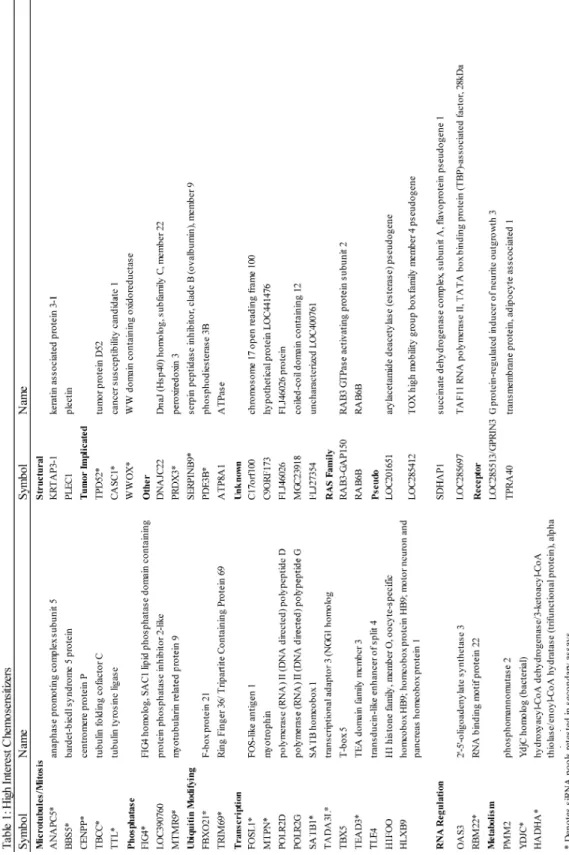
enrich for those genes that may have a therapeutic window, siRNA pools that impacted cell viability in a normal bronchial epithelial immortalized cell line, the HBEC3KT’s, were also removed from further analysis [74]. Ultimately, this analysis identified 49 candidate siRNA pools that correspond to annotated genes in the GENE database (Table 1). Depletion of the identified candidate chemosensitizers has minimal impact on cell viability on their own, but decreases cell viability by at least 15 % in the presence of sublethal dose of paclitaxel.
The identified chemosensitizers impact many diverse pathways, illustrating the global effect a drug or perturbagen can have on a cell system (Figure 4D and Table 1). The high interest chemosensitizers include a wide variety of proteins shown to regulate transcription. We identified MTPN, which regulates NF-kappa-B signaling [75], and SATB1, which is a transcriptional repressor that functions as a chromatin-remodeling factor and has been implicated in several tumorigenic settings [76-78]. Several genes that modify post translational modifications were also identified, including phosphatases, phosphatase inhibitors and components of the ubiquitin ligase cascade. These genes likely serve as nodes critical to regulating paclitaxel resistance. Further, we identified genes with more direct links to paclitaxel’s mechanism of action. We identified tubulin-binding cofactor C (TBCC) and tubulin tyrosine ligase (TTL), which are proteins integral to tubulin protein folding and posttranslational modification, respectively [79, 80]. Further, there was a small cohort of genes that play a defined role in mitosis, such as ANAPC5, a critical component of the anaphase promoting complex, and CENPP, a mitotic centromere protein [81].
unannotated function and several ‘pseudogenes’. Pseudogenes have been considered junk, or non-functional DNA, for a long time; however, since the ability to perform genome wide RNAi functional screens has been developed, it has become increasingly apparent that these “junk” genes can be critical to regulating numerous processes [82]. They have been found to regulate tumor suppressors and oncogenes and can be deregulated in cancer. Here, we suggest that due to the broad network engaged in paclitaxel resistance, there are likely novel therapeutic entry points with which to push taxane therapeutic strategies forward.
Secondary screening analysis
In order to identify the genes and processes most integral to paclitaxel resistance, we chose 23 candidates from a broad range of biology for additional secondary analysis. These candidate chemosensitizers were assessed for their ability to induce apoptosis, as measured by activation of caspase-3 and -7, and also their impact on mitotic progression and fate (Figure 5A and B). To monitor the impact of gene depletion on mitosis, a small scale, live-cell imaging screen was performed in HCC366 cells engineered to express GFP-H2B. Cells depleted of candidate chemosensitizers were evaluated for the amount of time spent in mitosis from nuclear envelope breakdown to cytokinesis, and the resulting mitotic fate was monitored. Further, to validate that the candidate siRNA pools were in fact knocking down their intended gene, target mRNA levels were also assessed (Figure 5C).
but less than a two fold increase in caspase induction. Genes identified in group III increased cell death, however they did not significantly impact mitotic timing. Group IV had a significant increase in both mitotic timing and cell death. Depletion of group IV genes, in addition to increasing mitotic delay by at least 20 %, exhibited the greatest induction of apoptosis. Importantly, all four of group IV siRNA pools met our criteria for off-target validation by having at least two of the four independent siRNAs, or a second non-redundant pool, recapitulate the loss of cell viability phenotype of the screen (Figure 5E). The four identified group IV chemosensitizing genes are ANAPC5, a core scaffolding component of the APC [81], CASC1, which had previously been implicated in lung tumorigenesis [83], PDE3B, a phosphodiesterase that has been recently identified to sensitize lung cancer cells to cisplatin [84], and TRIM69 [85], an uncharacterized E3 ubiquitin ligase.
Prolonged engagement of the SAC recouples mitotic damage to cell death
paclitaxel-resistant cell lines (Figure 6C). Together, these data indicate that ANAPC5, CASC1, PDE3B and TRIM69 are components that contribute to mitotic slippage in multiple genetic backgrounds.
To determine if the prolonged mitotic delay is required for post-mitotic cell death, cohort IV genes were co-depleted with siMAD2 in the presence and absence of paclitaxel and assessed at both the single cell and population level. At the single cell level, co-depletion with siMAD2 rescued the paclitaxel-mediated mitotic delay and nuclear blebbing, indicative of decreased cell death (Figure 7A). Co-depletion also prevented accumulation of cleaved caspase-3 at the population level as assessed by western blot analysis (Figure 7B). Taken together, these data indicate that engagement of the SAC and prolonged mitotic arrest is necessary for this cohort to re-couple mitotic damage to cell death.
Preliminary evidence suggests that co-depletion of HUWE1 and PDE3B rescues siPDE3B-induced cell death in the absence of paclitaxel (data not shown). This data suggests that PDE3B may target MCL-1 in a non-mitotic manner. Accordingly, depletion of ANAPC5, a core scaffolding component of the APC/C, stabilizes MCL-1. The accumulation of cleaved caspase-3 despite ANAPC5-mediated stabilization suggests that there are additional apoptotic signaling pathways engaged to couple mitotic slippage to cell death.
Based on these results, we hypothesize that mitotic slippage is a prominent pro-survival mechanism in NSCLC, despite the resulting damaged and micronucleated cells. Thus, targeting precocious mitotic exit either directly through core checkpoint proteins, or indirectly through proteins implicated herein, may be a viable therapeutic option.
cell death, above the use of either agent alone (Figure 8C). Thus, a collaborative interaction exists between APC/C inhibition and paclitaxel in an otherwise paclitaxel resistant setting.
Inhibiting mitotic slippage in vivo abrogates tumor growth
While ANAPC5 is a well documented component of the anaphase-promoting complex, CASC1, PDE3B and TRIM69 have no clearly defined roles in mitosis. Given PDE3B’s monogenic defects, we were particularly interested in further characterization of CASC1 and TRIM69. We next asked if inhibition of mitotic slippage, through stable depletion of CASC1 and TRIM69, would impact tumor cell growth in vivo. HCC366 cells were stably depleted of either CASC1 or TRIM69 and injected into the flank of immune-compromised mice to establish a xenograft model. By three weeks post-injection, tumors stably depleted of CASC1 and TRIM69 began to exhibit attenuation of tumor growth as evaluated by caliper measurement (Figure 9A and B). In an additional study, in which tumors from all mice were harvested at the same time, we observed a significant decrease in tumor volume in the shCASC1 and shTRIM69 tumors (Figure 9C). These findings suggest that CASC1 and TRIM69 support tumor cell growth in vivo. The remainder of this project has focused on elaborating the contributions of CASC1 and TRIM69 to mitotic slippage.
and II’s minimal induction of apoptosis suggests that the cell viability defect measured in the initial screen is likely due to other defects. For example, the observed viability defect may be due to an impact on proliferation. Thus, future investigation into additional genes that were not selected for further study herein is warranted.
Comparing groups II and IV, we see an un-coupling of mitotic delay and cell death. Depletion of all members of group II exhibit increased mitotic timing, yet minimal activation of caspase-3/-7 and apoptosis. In contrast, group IV recouples a minimum 20 % increase in mitotic delay to cell death. This begs the question, why do chemoresistance cells survive following depletion of group II genes. There may be many technical reasons for the observed uncoupling of mitotic delay and cell death. The uncoupling could be due to off target effects, insufficient depletion, or possibly cell death from depletion of group II targets may have been outside of the timing of our assays. However, if this is an on target effect, a mechanistic understanding of how mitotic timing has become uncoupled from cell death would advance the study of mitotic slippage. It is possible that a delayed mitosis only becomes re-coupled to cell death through discreet mechanisms and specific types of mitotic damage.
Figure 3. Defining a paclitaxel-resistant NSCLC screening platform
Figure 4.
Figure 4. Pan-genomic loss of function screen in mitotic slippage prone HCC366 cells.
Figure 5.
Figure 5. Secondary screening analysis stratifies candidate chemosensitizers
Figure 6.
Figure 7.
Figure 8.
Figure 9.
Figure 9. CASC1 and TRIM69 support tumor cell growth in vivo.
Chapter IV. CASC1 regulates microtubule stability to support mitotic slippage CASC1 Introduction:
Several lines of evidence suggest that there exists an inheritable genetic susceptibility to developing lung cancer [88-90]. To uncover these genetic predispositions free from environmental influence, Gariboldi et al took advantage of inbred mouse strains known to have varying degrees of susceptibility to urethane induced lung cancer, and performed a genome wide genetic linkage analysis [91, 92]. This analysis identified a region on mouse chromosome 6, later named the pulmonary susceptibility 1 (PAS1) locus, as responsible for approximately 50 % of the genetic variation between the two mouse models. Quantitative trait locus mapping by Zhang et al identified six candidate functional genes including the adjacent genes Casc1 and Kras [83, 93]. Casc1 has a single polymorphism at codon 60 resulting in a mutation from an aspargine to serine. This missense mutation is sufficient to alter tumor cell growth both by colony formation assays and in xenograft mouse models. While the mouse Casc1 and human CASC1 proteins are 67 % identical and 81 % similar, the key codon 60 is not conserved in the human homolog. Although there are no known genetic polymorphisms in human CASC1 that correlate with lung tumorigenesis or progression, ectopic expression of either mouse Casc1 allele significantly limited tumor cell growth in the NSCLC A549 cell line [94].
search of publically available protein interaction databases reveals no known interactors. One report finds that the mouse Casc1 protein co-sediments and co-immunoprecipitates with β-tubulin [95]. Additional evidence shows that overexpression of Casc1 results in an accumulation of binucleated cells suggesting an aberrant mitosis [95]. Given this previous association with lung cancer, and beginning characterization data, identification of CASC1 as a potential chemosensitizer in paclitaxel resistant NSCLC suggests CASC1 may influence lung cancer biology.
Results
CASC1 supports cell viability and mitotic spindle integrity.
paclitaxel, induced a dramatic mitotic delay which was frequently coupled to cell death in mitosis and aberrant mitotic figures. Together, these data suggest that CASC1 functions to buffer paclitaxel induced mitotic damage and support mitotic fidelity.
CASC1 stabilizes microtubule network.
Studies of the mouse homolog of CASC1 suggest an association with β-tubulin through a basic amino acid domain. This basic region is well conserved in the human protein suggesting that CASC1 may collaborate with paclitaxel treatment through a direct impact on microtubules [93, 95]. To date, no impact of CASC1 association with tubulin has been described.
To determine if CASC1 affects the microtubule cytoskeleton, we examined the microtubule network in H1299 cells where CASC1 impacts mitosis in the absence of paclitaxel. H1299 cells were depleted of CASC1 and assessed on the single cell level by immunofluorescence. This analysis revealed a diminished microtubule network in interphase (Figure 11A left). Differential centrifugation, which allows the separation of soluble and polymerized tubulin, confirmed that depletion of CASC1 results in a suppression of microtubule polymer formation (Figure 11A center). We also see a loss of acetylated tubulin, a general marker of microtubule stability, following CASC1 depletion (Figure 11A right).
cells have an impaired ability to regrow their microtubule network both in interphase (data not shown) and mitotic cells (Figure 11B). Together, these data suggests that CASC1 is a global regulator of microtubule stability.
To further investigate how CASC1’s impact on microtubules may alter mitotic slippage, we examined BUBR1 positive kinetochores during mitosis. BUBR1 is a key sentinel protein monitoring microtubule-kinetochore attachment (Figure 1). Following CASC1 depletion, we find an increased number of BUBR1 positive foci in mitotic cells, indicating fewer microtubule-kinetochore attachments are being made (Figure 11C). Together, these data suggest that CASC1’s impact on microtubule stability directly supports mitotic spindle formation.
CASC1 as an oncogenic dependency.
chromosome 12 (Figure 12C and D). It has previously been reported, in this progression model, that p53 alterations are sufficient to drive altered microtubule stability [97]. Together, these data suggest that CASC1’s functional support of microtubule stability may be necessary to buffer oncogenic stress.
Finally, we find that depletion of CASC1 induces damage to cells sufficient to drive stabilization of p53. In the normal HBEC3KT and HBEC30 cells this accumulation of p53 is sufficient to induce p21, suggesting a resulting delay in cell cycle progression. While CASC1 depletion in the tumor cells tested is able to stabilize p53, these cells are unable to induce p21 (Figure 12E). These data suggest a mechanism by which normal cells may survive CASC1 depletion as opposed to those tumor cells with a defective p53 signaling cascade.
CASC1 Discussion
functions to buffer mitotic defects induced by oncogenic changes.
Further, we find that depletion of CASC1 stabilizes p53 and induces p21 accumulation in normal but not tumorigenic backgrounds. The stabilization of p53 could be the result of several potential mechanisms. Several lines of evidence indicate that post mitotic failure, or an aberrant mitotic exit, cells may undergo a p53 and p21 dependent G1 arrest (Figure 2). This defect has been illustrated following treatment with paclitaxel [105-107]. As CASC1 supports mitotic spindle formation, it is possible that the accumulation of p53 is the result of a general, damaged mitotic exit. p53 has also been shown to accumulate in the nucleus post low dose paclitaxel treatment that interferes with microtubule dynamics [108]. Thus, it is possible that the p53 read out may be due to CASC1 interphase defects. Further, p53 accumulation could be due to CASC1-mediated genomic damage in the absence of paclitaxel.
Regardless, the accumulation of p21 in the normal bronchial epithelial backgrounds, and not the tumor cells, suggests that, normal cells may exit the cell cycle in response to CASC1 depletion, where as tumor cells with defective p53 signaling networks continue to proliferate. Thus, as the tumor cells continue to proliferate, they will be more susceptible to subsequent paclitaxel treatment then the normal cells that have exited the cell cycle. This hypothesis is supported by previous findings that the loss of normal p53 function can sensitize cells to paclitaxel [109]. This suggests a mechanism by which CASC1 is specifically required for cell viability following loss of p53 in the oncogenic progression model and suggests targeting CASC1 may have a therapeutic window.
Figure 10: CASC1 supports mitotic fidelity
Figure 11.
Figure 11: CASC1 supports microtubule stability to satisfy the SAC
Figure 12. CASC1 is a tumor cell dependency
Chapter V. TRIM69 is a centrosomal and microtubule associated protein that is essential for mitotic fidelity.
TRIM69 Introduction:
TRIM proteins as E3 ubiquitin ligases. TRIM69 is a member of the TRIpartite Motif containing (TRIM) family of proteins characterized by an ensemble of three types of domains, a RING E3 ligase domain, one or two B box domains and a coiled-coiled domain [115, 116]. This specific architecture, referred to as a RBCC domain structure, is highly conserved in combination, order, and spacing. If one domain is missing in a TRIM family member, the remaining domain structure is conserved. This suggests that TRIM proteins have evolved to carry out a specific basic function common to all TRIM family members [117]. RING finger domains are primarily associated with E3 ubiquitin ligase activity, and while the presence of a RING domain does not dictate ubiquitin ligase function, the TRIM family has been broadly classified as a group of “single protein RING finger E3 ubiquitin ligases” [117].
subsequent degradation is essential in order to respond to cellular signals such as satisfaction of the spindle assembly checkpoint [118].
The ubiquitin cascade is a complex and highly regulated, multistep mechanism achieved through sequential action of three enzymes, the ubiquitin activating (E1), ubiquitin-conjugating (E2) and ubiquitin-ligating (E3) enzymes. First, in an ATP-dependent manner, an E1 forms a thiol-ester bond with a free ubiquitin protein. The ubiquitin is then transferred to an E2 conjugating enzyme that then associates with an E3 ligase to transfer ubiquitin to the final substrate. Specificity increases going down the ubiquitin cascade. While there are two isoforms of the E1 activating protein, there are 35-40 potential E2 conjugating proteins and greater than 600 putative E3 ligases. E3 ligases are broadly classified based on their mechanism of transferring the activated ubiquitin from the E2 to the substrate. The primary E3 ligases families are the HECT (Homologous to E6Ap carboxy terminus) domain E3’s and the RING (really interesting new gene) finger-containing E3’s [119].
performed herein. RING family proteins can function as E3 ligases as part of large complexes, such as the APC/C and SCF (Skp1-Cullin-F-Box), or as in the case of TRIM proteins, as single protein ubiquitin ligases [119, 120].
TRIM protein function in pathological conditions. The TRIM family contains over 70 known members which have been implicated in a broad range of biological processes including development, differentiation, apoptosis and cell proliferation. A number of TRIM family proteins have previously been implicated in cancer and other pathological diseases [115]. TRIM18, or MID1, associates with microtubules throughout the cell cycle and targets the catalytic subunit of protein phosphatase 2 (PP2) for degradation [121]. Mutation of TRIM18 in the genetic syndrome, X-linked Opitz syndrome, results in decreased affinity for microtubules and altered PP2 activity which have been linked to characteristic defects in midline body structures, such as eye spacing, defects in the trachea or esophagus and cleft palate [122].
TRIM proteins have been implicated in either positively or negatively regulating oncogenesis in a context dependent manner [115]. TRIM19 or PML is subject to a chromosomal translocation which results in a fusion protein with the retinoic acid receptor-α (RARα). This PML-RARα fusion specifically occurs in acute promyelocytic leukemia (APL) [123]. In addition, recent work has identified TRIM proteins as forming functional complexes with members of the melanoma antigen (MAGE) family of cancer-testis antigens [124].
expression in tumors was identified to illicit an immune response in cancer patients. Until recently, CTAs have largely been studied from the field of tumor immunology with the possibility of using CTAs as cancer vaccines to stimulate an anti-tumor immune response. However the functions of CTAs in tumorigenesis have gone largely unstudied [125]. If CTAs are identified to participate in specific molecular pathways, this would identify pathways engaged to support tumorigenesis. TRIM-MAGE associations are found to functionally enhance the basal activity of TRIM family proteins through a yet undefined mechanism [124]. For example, it was demonstrated that TRIM28, which is over expressed in gastric cancer, can associate with up to four MAGE family proteins which enhance TRIM28 mediated degradation of p53 [124]. Together, this demonstrates that TRIM family proteins can be usurped by cancer specific mechanisms to support tumorigenesis. As discussed below, we find that, like TRIM28, TRIM69 associates with MAGE family proteins.
herein as TRIM69B, which does not contain the RING domain. While TRIM69’s cellular function has not been elucidated, it has been shown to contain a functional RING E3 ubiquitin ligase domain which we and others have demonstrated is sufficient to induce autoubiquitylation (Figure 13C) [85]. Identification of TRIM69 as a candidate chemosensitizer, whose depletion had minimal viability defects on its own made TRIM69 an intriguing gene to further characterize.
Results:
TRIM69 supports mitotic fidelity. To begin to determine the genetic penetrance of TRIM69 in supporting chemoresistance, depletion was evaluated in a panel of NSCLC patient derived cell lines of varying sensitivity to paclitaxel. TRIM69 was found to be required for cell viability in the presence or absence of paclitaxel in all but one tumor line tested and further, there is no observed induction of apoptosis in the normal HBEC3KT cells (Figure 14A). In addition to TRIM69 depletion causing a delayed mitosis, we find an accumulation of micronucleated cells in A549 and H1299 cells in the absence and presence of paclitaxel respectively, and an enhancement of paclitaxel-mediated multipolar spindles in HCC366 cells (Figure 14B). Depletion of TRIM69 in HCC366 cells also increased the number of BUBR1 foci in mitotic cells, indicating that without TRIM69, fewer stable kinetochore-microtubule attachments are made (Figure 14C). These data support the hypothesis that TRIM69 functionally supports mitotic fidelity.
TRIM69B and TRIM69A (C53S/C55S), a mutation of two conserved RING domain cysteines. TRIM69A was found to localize to microtubules in interphase and the spindle poles during mitosis. This localization is dependent, at least in part, on the RING domain as neither TRIM69B nor the RING domain mutant maintained microtubule or centrosomal accumulation (Figure 14D). TRIM69 association with microtubules induces microtubule bundling, a classic phenomenon observed with several microtubule associated proteins. The bundling resulting from overexpression of TRIM69A was sufficient to stabilize microtubules against nocodazole mediated depolymerization, indicating a more stable and likely crosslinked microtubule network (Figure 14E). Consistent with TRIM69A specifically impacting microtubule dynamics, exogenous expression of -A and not -B disrupts mitotic spindle formation and drives accumulation of micronucleated cells, indicating aberrant mitotic exit (Figure 14F). Finally, siRNAs targeting knockdown of only TRIM69A only, were assessed for their impact on mitotic progression. Depletion of TRIM69A, specifically, was sufficient to prolong a paclitaxel-mediated mitotic delay in both the HCC1171 and HCC366 cells (Figure 14G). Taken together, these data suggest that the ubiquitylation activity of TRIM69A supports mitotic fidelity and, like many integral mitotic proteins, either depletion or over expression can have detrimental impacts on mitosis.
remains at the spindle poles through cytokinesis (Figure 15A). Consistent with the observed localization, immunoprecipitation of endogenous pericentrin, a centrosome scaffolding protein, pulls down myc-TRIM69A (Figure 15B). Treatment with nocodazole was sufficient to disrupt myc-TRIM69A accumulation at the centrosome, indicating a microtubule-dependent recruitment (data not shown). Taken together, these data suggest that TRIM69A is recruited to the microtubule organizing centers (MTOCs) to support appropriate mitotic spindle formation.
TRIM69’s interaction network.
results in cytokinesis defects while its over expression results in a prolonged mitotic delay [127]. MYPT1, myosine phosphatase targeting subunit 1, forms a holoenzyme with the catalytic protein phosphatase 1 (PP1) and functions to target PP1 to substrates. In mitotic cells, MYPT1/PP1 has been found to be a negative regulator of PLK1 activity. Further, PP1 is integral to checkpoint silencing and removal of BUBR1 from kinetochores [128-131]. These results indicate a functional connection between TRIM69 and key mitotic proteins. Taken together, these studies have identified TRIM69A as a novel centrosomal component required for mitotic fidelity.
TRIM69 protein stability is regulated by a CT-antigen.
TRIM69 protein accumulation, accordingly, stabilizes microtubules against nocodazole depolymerization, indicating that the protein stabilization is physiologically relevant (Figure 16D and E). Further, MAGEA4 depletion in both HCC366s and H1299s results in accumulation of mitotic cells. Taken together, these data suggests that TRIM69 stability and thus, localization may be regulated by an aberrantly expressed cancer-testis antigen. Further, these data suggest that aberrant expression of cancer testis antigens may functionally support tumor cell mitosis.
TRIM69 discussion
Through the work presented here, we have found that TRIM69 is recruited to the mitotic spindle poles during mitosis to support mitotic fidelity. Localization to centrosomes requires an intact RING finger domain and further, depletion of the isoform that contains the RING domain is sufficient to drive mitotic defects. Together, these data suggest that TRIM69’s E3 ubiquitin ligase activity is required to maintain mitotic fidelity.
TRIM69 recruitment to the centrosome at the beginning of mitosis coincides with centrosome maturation. Centrosomes, as the microtubule organizing centers of the cell, undergo a process termed maturation at the onset of mitosis in order to expand microtubule nucleating capacity necessary for proper mitotic spindle formation [136]. Maturation is a dynamic process in which microtubule scaffolding proteins such as pericentrin accumulate at the centrosome and increase recruitment of microtubule nucleating proteins such as gamma-tubulin [136, 137]. Increased accumulation of gamma tubulin results in a dramatic increase in microtubule nucleation and thus, additional regulatory proteins are required to anchor microtubule ends to the centrosomes [138, 139]. The centrosomal maturation process is regulated by series of protein kinases and phosphatases including, but not limited to, PLK1 and PP1. TRIM69s cell cycle dependent recruitment to the centrosome, its demonstrated ability to bundle microtubules, and association with PP1 regulatory protein MYPT1 suggests TRIM69 may be required to support microtubule activities at the MTOCs during centrosomal maturation.
by tumors to alter TRIM69 expression and alter mitotic slippage. Further, we see that recruitment of TRIM69 to the spindle poles relies, at least in part, on the activity of MYPT1 and GNAI3.
Figure 13.
Figure 13. TRIM69A is a testes enriched E3 ubiquitin ligase.
Figure 14. TRIM69A supports mitotic fidelity
Figure 15.
Figure 15. TRIM69A is a novel component of the MTOC
Figure 16.
Figure 16. TRIM69 interacts with cancer testis antigen MAGEA4
Chapter VI. Summary
Understanding the mechanisms that promote escape from mitotic-stress induced tumor cell death is vital to improving the current standard-of-care cytotoxic regimens for multiple cancers, including breast, ovarian and non-small cell lung cancer. The work presented here undertook a global analysis to identify mechanisms of intrinsic paclitaxel resistance. Importantly, in Chapter 3 we find that the most potent method of re-coupling mitotic stress to cell death is to prolong a paclitaxel-mediated mitotic delay. Further, we find that even in the most resistant genetic backgrounds, prolonging a mitotic delay by as little as 20 % can be sufficient to allow loss of pro-survival proteins and engage cell death signaling.
delay, such as HeLa cells (Figure 17).
Identification of ANAPC5 as synthetic lethal with paclitaxel suggested that targeting the APC/C may have a therapeutic window. Previous studies have found that directly targeting mitotic exit, through depletion of CDC20, is highly lethal to cancer cells and “...killed all cells that entered mitosis..” [142]. Here, we were able to combine the APC/C inhibitor proTAME with paclitaxel and show a cooperative mitotic and cell death defect in the slippage prone HCC366 cells. We further expanded our analysis to additional APC/C subunits and found varying induction of cell death in the presence and absence of paclitaxel. Our studies suggest that APC/C inhibition may be ‘tunable’ in order to achieve a therapeutic effect.
In addition to directly targeting mitotic exit, we have identified novel mechanisms that indirectly support mitotic slippage. The work presented in chapter 4 identified CASC1 as a novel regulator of microtubule stability and suggested that separate but parallel insults to the microtubule network can collaborate therapeutically. Without CASC1, there is a loss of microtubule polymer that we propose leads to an insufficient ability to establish microtubule-kinetochore attachments. This defect ultimately leads to increased MCC protein BUBR1 at unattached kinetochores and an increased robustness of SAC signaling.
models of paclitaxel resistance, decreases over all network dynamicity leading, generally, to a more stable network. Thus, we suggest that tumor cells with a more stable microtubule network may be more resistant to anti-mitotic therapy.
In chapter 5 we find TRIM69 to be a novel E3 ubiquitin ligase that localizes to the spindle poles in a cell cycle dependent manner. TRIM69 ubiquitylation activity is required for proper localization and mitotic fidelity. Though we have been unable to define the direct function of TRIM69 at the centrosomes, we do find that toggling TRIM69 expression can have damaging impacts on mitosis. Further, we find TRIM69 expression can be altered by a cancer-testis antigen, MAGEA4. These data suggest that re-expression of testis specific proteins can be functionally employed to buffer or support tumor mitoses. Further, we find a functional connection between TRIM69 and the centrosomal proteins MYPT1 and GNAI3. Over expression co-immunoprecipitation confirms that MYPT1 and TRIM69 can physically associate (data not shown). Given MYPT1s functional impact on PLK1 and association with PP1, one intriguing hypothesis is that TRIM69 may be regulating SAC silencing through impacting PP1 function. Future Directions: