AN IN VITRO CHARACTERIZATION OF FUNCTIONAL INTERACTIONS BETWEEN PURIFIED TELOMERE REPEAT BINDING FACTORS 1 AND 2 AND RAD51 RECOMBINASE
Brian D. Bower
A dissertation submitted to the faculty of the University of North Carolina at Chapel Hill in partial fulfillment of the requirements for the degree of Doctor of Philosophy in the Curriculum of
Genetics and Molecular Biology.
Chapel Hill 2014
Approved By Jack D. Griffith
Shawn Ahmed
ABSTRACT
BRIAN D BOWER: AN IN VITRO CHARACTERIZATION OF FUNCTIONAL INTERACTIONS BETWEEN PURIFIED TELOMERE REPEAT BINDING FACTORS 1 AND 2 AND RAD51
RECOMBINASE
(Under the direction of Jack D. Griffith)
ACKNOWLEDGEMENTS I would like to thank:
My thesis advisor Jack Griffith for his unyielding support, generosity and guidance throughout the course of my doctoral research
My family and friends for their love and support
TABLE OF CONTENTS
LIST OF FIGURES ... ix
LIST OF TABLES ... xii
LIST OF ABBREVIATIONS AND SYMBOLS ... xiii
CHAPTER PAGE 1 INTRODUCTION ... 1
Telomeric DNA ... 1
Telomere proteins ... 2
Homologous recombination/repair proteins ... 2
Chromosome end capping ... 3
Higher order telomere structure ... 4
DNA repair at the telomeres ... 5
Telomeric proteins and DNA repair ... 6
Scope of dissertation ... 7
REFERENCES ... 9
2 TRF2 AND TRF1 DIFFERENTIALLY MODULATE RAD51-MEDIATED DISPLACEMENT LOOP FORMATION IN VITRO ... 13
INTRODUCTION ... 13
MATERIALS AND METHODS ... 14
RESULTS ... 18
Fluorescent D-Loop Assay ... 18
TRF2∆M Promotes Rad51-Mediated
Telomeric But Not Non-Telomeric D-Loop ... 26
TRF2∆B Inhibits Telomeric But Not Non-Telomeric Rad51-Mediated D-Loop Formation ... 28
TRF1 Promotes Rad51-Mediated Telomeric But Not Non-Telomeric D-Loop Formation ... 28
DISCUSSION ... 31
REFERENCES ... 35
3 BIOPHYSICAL AND ULTRASTRUCTURAL CHARACTERIZATION OF ADENO-ASSOCIATED VIRUS CAPSID UNCOATING AND GENOME RELEASE ... 38
INTRODUCTION ... 38
MATERIALS AND METHODS ... 39
RESULTS ... 46
Heat-induced DNA release depends on genome length ... 46
scAAV are more thermostable than ssAAV ... 50
Heat-induced exposure of VP1 is not dependent on genome size or complementarity ... 53
Tungsten-shadowing EM shows DNA secondary structure for dsDNA vectors ... 54
Molecular dynamics provides insight into intra-capsid genome organization ... 54
EM quantification of DNA release following thermal denaturation ... 58
Attempts at characterizing intra-capsid structure by EM ... 60
DISCUSSION ... 62
REFERENCES ... 66
4 A GUANOSINE-CENTRIC MECHANISM FOR RNA CHAPERONE FUNCTION ... 70
MATERIALS AND METHODS ... 70
RESULTS & DISCUSSION ... 75
Time-resolved SHAPE analysis of MuLV RNA dimerization ... 77
Model-free clustering of nt-resolution kinetic dimerization profiles ... 79
Initial Interactions between NC and UP1 with MuLV monomer ... 82
Role of guanosine in RNA structure and RNA chaperone mechanism ... 83
REFERENCES ... 90
5 AN INVESTIGATION OF HETEROGENOUS RIBONUCELOPROTEIN A1 AND UNWINDING PROTEIN 1 DNA BINDING CHARACTERISTICS ... 92
INTRODUCTION ... 92
MATERIALS AND METHODS ... 93
RESULTS ... 95
hnRNP A1 & UP1 bind to ssDNA ... 95
hnRNP A1 bind preferentially to telomeric ssDNA ... 99
Possible deleterious effects of N-terminal tagging ... 103
DISCUSSION ... 104
REFERENCES ... 106
6 CONCLUSIONS AND FINAL THOUGHTS ... 108
Homologous recombination/repair in telomere maintenance & protection ... 108
Intra-capsid AAV genome organization is unamenable to EM characterization ... 110
LIST OF FIGURES
FIGURE 1.1: Diagram of telomeric DNA ... 1
FIGURE 1.2: Telomere binding proteins ... 2
FIGURE 1.3: Representative telomere loops reported in the literature ... 5
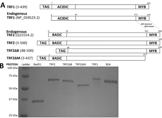
FIGURE 2.S1: Proteins Used ... 19
FIGURE 2.1: TRF-mediated telomeric D-loop formation ... 20
FIGURE 2.2: TRF2 inhibits Rad51-mediated telomeric but not non-telomeric D-loop formation ... 21
FIGURE 2.S5: Area under the curve (AUC) calculation procedures ... 22
FIGURE 2.S2: TRF2-mediated D-loop formation requires telomeric homology while Rad51 does not ... 23
FIGURE 2.S3: TRF2 inhibits Rad51-mediated telomeric D-loop formation only when added early in D-loop reactions ... 25
FIGURE 2.3: TRF2∆M promotes Rad51-mediated non-telomeric but not telomeric D-loop formation ... 27
FIGURE 2.4: TRF2∆B promotes Rad51-mediated telomeric but not non-telomeric D-loop formation ... 29
FIGURE 2.5: TRF1 promotes Rad51-mediated telomeric but not non-telomeric D-loop formation ... 30
FIGURE 2.6: TRF1 and TRF2 differentially modulate Rad51-mediated telomeric and non-telomeric D-loop formation ... 34
FIGURE 3.1: Ultrastructural characterization of AAV capsid uncoating ... 47
FIGURE 3.2: Effect of packaged genome length on AAV capsid uncoating. ssAAV ... 48
FIGURE 3.3: Quantitative analysis of TEM images ... 49
FIGURE 3.5: Fluorimetric analysis of AAV capsid uncoating
as a function of genome length and self-complementarity ... 52
FIGURE 3.6: Exposure of the VP1 N termini is not dependent on genome length or self-complementarity... 53
FIGURE 3.7: Tungsten-shadowing EM of released genomes from ssCMV-GFP ... 55
FIGURE 3.8: Computational modeling of internal capsid pressure and genome organization ... 57
FIGURE 3.9: Characteristics of capsid adherence and ________________genome release following thermal denaturation ... 59
FIGURE 3.10: EM analysis of end structure and genome length ... 61
FIGURE 3.11: Psoralen fails to crosslinks scAAV genomes in intact capsids ... 63
FIGURE 4.S1: Secondary structures of monomer and dimer states ... 76
FIGURE 4.1: Time-resolved SHAPE analysis of MuLV RNA dimerization... 77
FIGURE 4.2: Model-free clustering of nucleotide-resolution kinetic profiles for dimerization ... 79
FIGURE 4.S2: Time-resolved SHAPE of the native sequence MuLV RNA dimerization at 600nM and 200 nM ... 80
FIGURE 4.3: Initial interactions between NC and UP1 with the MuLV monomer ... 82
FIGURE 4.4: Role of guanosine in RNA structure and mechanism of chaperone-mediated RNA folding ... 83
FIGURE 4.S3: Native and inosine-substituted RNA secondary structures ... 84
FIGURE 4.S4: Visualization of MuLV genomic RNA dimerization by non-denaturing electrophoresis ... 87
FIGURE 4.S6: Interactions between NC and UP1 with
the MuLV monomer and dimer states ... 89 FIGURE 5.1: Electrophoretic mobility shift assay
demonstrating hnRNP A1 ssDNA binding ... 96 FIGURE 5.2: hnRNP A1 binds to M13 ssDNA ... 98 FIGURE 5.3: Dynamic light scattering reveals mostly homogenous hnRNP A1 ... 98 FIGURE 5.4: hnRNP A1 binds to strand displaced
telomeric but not non-telomeric DNA ... 99 FIGURE 5.5: hnRNP A1 binds preferentially to one end of a
template with telomeric and non-telomeric ssDNA
on opposite sides of a linear template ... 100 FIGURE 5.6: Comparative binding of E. coli and
LIST OF TABLES
LIST OF ABBREVIATIONS AND SYMBOLS
5-FAM 5-Carboxyfluorescein
6-JOE 6-Carboxy-4',5'-dichloro-2',7'-dimethoxyfluorescein Å Angstrom
θ Theta
κ Kappa
Σ Sigma
Δ Delta
α Alpha
AAV Adeno-associated virus
AFM Atomic force microscopy
AUC Area under the curve
ATM Ataxia telangiectasia mutated
bp Base pair
BSA Bovine serum albumin
BzCN benzoyl cyanide
CBA Chicken β Actin
CCD Charge coupled device
cDNA Coding DNA
CMV Cytomegalovirus
Cy3 Cyanine 3
∆ Delta
DNA Deoxyribonucleic acid
D-loop Displacement loop
DMSO Dimethyl sulfoxide
dsDNA Double-stranded deoxyribonucleic acid
EDTA Ethylenediamenetetraacetic acid
EF1a Elongation factor 1 α
EM Electron microscopy
EMSA Electrophoretic mobility shift assay FANCD2 Fanconi anemia group D2 protein
GAG Group specific antigen
GFP Green fluorescent protein
HEPES 4-(2-hydroxyethyl)-1-piperazineethanesulfonic acid
HJ Holliday junction
hnRNP Heterogenous ribonucleoprotein
HR Homologous recombination/repair
HRP Horseradish peroxidase
IRES Internal ribosome entry site
ITR Inverted terminal hairpin repeat
kbp Kilobase pair
MD Molecular dynamics
MRN Mre11/Rad50/Nbs1 complex
MuLV Moloney murine leukemia virus
MVM Minute virus of mice
NC Nucleoprotein capsid
NHEJ Non-homologous end joining
nt Nucleotide
PAL1 Palindromic sequence 1
PAL2 Palindromic sequence 2
PCR Polymerase chain reaction
PEG Polyethylene glycol
PTM Post-translational modification
RNA Ribonucleic acid
scAAV Self-complimentary adeno associated virus
SHAPE Selective 2’-hydroxyl acylation analyzed by primer extension
SL1 Stem loop 1
SL2 Stem loop 2
ssAAV Single-stranded adeno associated virus ssDNA Single-stranded deoxyribonucleic acid
ss/dsDNA Single-stranded to double-stranded deoxyribonucleic acid
SDS Sodium dodecyl sulfate
TBS Tris-buffered saline
TBS-T Tris-buffered saline with Tween 20
TE Tris-buffered EDTA solution
TEM Transmission electron microscopy
t-loop Telomere loop
Tm Melting temperature
Tris Tris(hydroxymethyl)aminomethane TRF1 Telomere repeat-binding factor 1
TRF2 Telomere repeat-binding factor 2 TIN2 TRF1 interacting nuclear protein 2
UP1 Unwinding protein 1
vg Viral genome
VP Viral protein
CHAPTER 1: INTRODUCTION
Telomeric DNA
Telomeres constitute a nucleoprotein structure which protects the termini of the linear chromosomes present in virtually all eukaryotes from aberrant recognition as DNA damage and aberrant repair. At the level of primary sequence, telomeres consist of a repetitive or quasi-repetitive dsDNA tract of varying length. These dsDNA tracks often also possess ssDNA overhangs, most often of 3’ character, which may derive from either the end-replication problem or from elongation of the telomeres by recombination mediated methods or retrotranscription via the telomerase ribonuceloprotein complex (TERT). In mammals telomeres consist of a tract of 5’-GGTTAG-3’ repeat base paired with complimentary 5’-CTAACC-3’ repeats. This track can vary in length from several kilobasepairs (kbp) in length to in excess of 100 kbp. This variability in telomere length is affected by species, organism age, and due to disease or genetic factors. In humans telomere length varies from 5-15 kbp, and the telomeres possesses 3’ ssDNA tails of between 50 and 500 nt2. The terminal sequence of the 3’ tail is weakly defined with the plurality of ends terminating in a ‘TAG’ sequence3. The ss-to-dsDNA junction is more strictly defined, with the majority of ends possessing a terminal; ‘ATC’3. The length of the overhang and the end sequences appear to be specified by post-replicative processing of the telomeres3.
Telomere Proteins
Telomeric dsDNA provides a binding site for two homologous human DNA binding proteins, telomere repeat-binding factors 1 and 24. While TRF2 is most often described as a telomeric dsDNA binding protein, considerable evidence exists which suggests the preferred binding site of TRF2 may be to the telomeric ss/dsDNA junction5-8. In addition to binding to telomeric dsDNA, TRF2 also interacts with an accessory protein, repressor-activator protein 1(RAP1) 9, 10. RAP1 plays poorly understood roles in mammalian telomere biology, but has been implicated in repressing repair processes at the telomere and perhaps to improving the binding specificity of TRF256. TRF1 and TRF2 also interact with a scaffolding protein, TRF2 interacting nuclear protein 1 (TIN2) 11. TIN2 in turn provides a binding site a bridging protein, TIN2 and POT1 Interacting Protein 1 (TPP1), 12 which stabilizes the binding of a telomere specific ssDNA bind protein, protection of telomeres 1 (POT1) 13.
Figure 1.2: Telomere binding proteins. Telomere repeat binding factors 1 and 2 (TRF1 and 2) bind to telomeric dsDNA, with TRF2 binding preferentially to the ss/dsDNA junction. TRF2 also interacts with an accessory protein, repressor activator protein 1 (RAP1). TRF1 and TRF2 provide a binding site for a scaffolding protein, TRF2 interacting nuclear protein 2 (TIN2). TIN2 likewise permits binding of a telomeric ssDNA binding protein, protection of telomeres 1 (POT1), via its interaction with TIN2 and POT1 interacting protein 1 (TPP1, alternately known as TINT1, PTOP and PIP1).
Homologous Recombination
promotes resection of the 5’ end of the DSB to generate a 3’ ssDNA tail vaguely similar in structure to the 3’ tail of a telomere14. This ssDNA is rapidly bound by replication protein A ( RPA)15, but is subsequently displaced by Rad51, the recruitment of which is promoted by breast cancer associated gene 2 (BRCA2) at the ss/dsDNA junction16, 17. Rad51 then facilitates a process of homology search, whereby the ssDNA substrate interrogates available dsDNA for complimentary sequence18. Rad51 first forms a protein-mediated complex between the substrate ssDNA and complimentary dsDNA and thereafter promotes protein-independent base pairing of the substrate and template in an ATP dependent manner generating a displacement loop (D-loop) within the template 18. Following displacement loop formation a variety of HR factors, including RecQ helicases such as Blooms (BLM) 19, 20 and Werner (WRN) 20 helicase, promote the migration and expansion of the D-loop and the eventual formation of a Holliday Junction (HJ) wherein both strands of the damaged substrate are paired with homologous sequences. These holiday junctions can then be cleaved or ‘resolved’ by HJ resolvases, such as the SLX1/4 complex, leaving behind two single-strand breaks (SSBs), which are then ligated21.
Chromosome End Capping
activation of MRNs exonucleolytic activities26. However, the exact mechanism or mechanisms that mediate telomere protection or ‘capping’ in vivo remain only poorly understood and despite these processes it appears that unperturbed telomeres are transiently recognized as DNA damage during the cell cycle in spite of these processes27.
Higher Order Telomere Structure
It has been discovered that telomeres possesses a variety of unusual structures both at the level of primary sequence and the tertiary structure of the telomere nucleoprotein complex. The G-rich ssDNA tail can form an unusual secondary structure wherein four triguanosine tracts can form a quadruplex (G4) stabilized by a number of monovalent ions28. This property is suspected to affect the replication of telomeric DNA as well29. The C-rich complement of the G-rich strand of the telomeres is replicated discontinuously. As such, the G-rich strand is left transiently single-stranded, which may permit formation of G4 structures. These structures are likely to inhibit DNA replication directly by interfering with nucleotide addition and indirectly by promoting fork slipping. Accordingly, telomeres have been observed to be particularly difficult to replicate, and are prone to defects consistent with fork stalling and slipping30. It has also been observed that telomeres from a variety of species often adopt a lariat or loop-like structure (T-loop)31-34, which appear to be stabilized by base pairing between the ssDNA tail and internal telomeric sequence. T-loops appear to be subject to cleavage by HJ resolvases, such as the SLX1/4 complex35, and resolution of T-loops is believed to result in the generation of extrachromosomal circular telomere DNAs (T-circles) 35. These T-circles are generated upon disruption of factors that may promote migration or dissociation of the D-loop or HJ that stabilizes the T-loop, such as RecQ helicases35, 36 or the regulator of telomere length 1 (RTEL1) helicase35. T-circles are also observed to be highly
Figure 1.3: Representative telomere loops reported in the literature. T-loops have been observed at the telomeres of a variety of only-distantly related species by a variety of groups31-34 using an assortment of techniques.
DNA Repair at the Telomeres
It has been paradoxically observed that telomere maintenance and protection require the activity of several DNA repair proteins. In contrast to HR, which is initiated by the MRN complex, NHEJ is initiated by binding of the Ku heterodimer (Ku 70 & Ku 80) and its associated DNA dependent protein kinase catalytic subunit (DNA-PKcs). While TRF2 and Rap1 inhibit DNA-PKcs activation at telomeres, it has been observed that both Ku and DNA-PKcs are required for proper telomere maintenance. Disrupting the expression of Ku or DNA-PKcs results in telomere shortening or aberrant telomere repair in a variety of mouse or human cell lines. This may be due to Ku’s interaction with TRF2 or WRN helicase, which may facilitate telomere protection. Alternately, the binding of Ku to telomeres may inhibit alternative
DSB repair pathways when activation of DNA-PKcs is inhibited by the TRF2/Rap1 complex. Likewise, it has been reported the BRCA2 recruits Rad51 to telomeres in a cell-cycle dependent
defect in telomere protection or capping. However, disrupting the expression of BRCA2 or Rad51 results in aberrant telomere repair even in quiescent and non-dividing cells, suggesting that the HR pathway is also required for telomere replication. It has been suggested that HR proteins including Rad51 may facilitate the formation of the T-loop structure. Immunodepletion of either TRF2 or Rad51 from nuclear extracts ablated the ability of those extracts to promote telomeric displacement loop formation; a requisite step in T-loop formation. This activity can be recovered upon addition of purified Rad51 or TRF2 to the immunodepleted extracts. However, there is evidence that TRF2 may also inhibit HR mediated processes at telomeres of humanized yeast strains56.
While these sundry DNA repair proteins are known to play some important role in telomere biology, the nature of that role remains poorly understood. Our data suggest that HR processes at the telomeres are differentially regulated by TRF1 and TRF2. Literature suggests that this differential regulation may also be influenced by a variety of post-translational modifications and are mediated by several different protein domains. Many of these domains remain incompletely characterized. As such our understanding the actual role or roles of DNA repair proteins in telomere maintenance and biology remains cursory at best.
Telomeric Proteins and DNA Repair
suggests that the basic domain of TRF2 facilitates non-telomeric D-loop formation, a necessary initiating step in HR.
TRF1 has also been observed to facilitate telomere replication in vivo30 despite previous reports that TRF1 actually inhibits replication of telomeric DNA in vitro42. It is plausible that in vitro TRF1 binding to telomeric DNA may pose an obstacle to DNA polymerase, which is alleviated by one or more factors in vivo. Supporting this hypothesis is has been observed that TRF1 is post-translationally modified (PTM) by replication complex associated proteins43-49. These PTMs reduce TRF1 binding affinity and may relieve the replication inhibition observed in an in vitro reconstituted replication reaction. Interesting these PTMs are inhibited in vivo by TIN243 and a component of the Fanconi Anemia pathway, FANCD246. This careful modulation of TRF1 binding may only transiently remove TRF1 from near the replication fork. The data we present herein suggests that retaining TRF1 near the replication fork may be advantageous. Telomeric DNA is prone to fork stalling and slipping. Fork stalling event can be mitigated or reversed in an HR dependent manner, and we report that TRF1 promoted Rad51-mediated telomeric D-loop formation. This is a critical step in some models of HR mediated replication fork restart.
Scope of Dissertation
Presented here in is an investigation of functional interactions between TRF1, TRF2 and Rad51 in vitro (Chapter 2). This characterization will be the main focus of this dissertation, as this project
constituted the bulk of my independent work in Dr. Griffith’s laboratory. Published data characterizing genome organization and capsid stability of adeno-associated virus (Chapter 3)50, and characterization of a guanosine centric mechanism of chaperone-mediated RNA folding (Chapter 4)51 are also described. Finally, unpublished data from a characterization of the DNA binding properties of hnRNP A1 and UP1 are described in Chapter 5. This projects are presented secondarily, as they constitute collaborative work on which I was not a first author, or work which did not yield data of sufficient quality for publication.
discovery that TRF2 inhibits Rad51-mediated telomeric D-loop formation. This finding appears to contradict previous nuclear extract work by the Karlseder lab27, 52, but may be supported by findings from Dr. Gilson’s lab that TRF2 inhibits HR-mediated processed at humanized yeast telomeres53. Our finding that TRF2ΔB promotes Rad51-mediated non-telomeric D-loop formation may explain how TRF2 can promote HR-mediated non-telomeric double-strand break repair41. Likewise, our finding that TRF1 promotes Rad51-mediated telomeric D-loop formation may suggest that TRF1 promotes telomeric replication fork restart, explaining how TRF1 may promote telomere replication in vivo30 despite findings that TRF1 and TRF2 actually inhibit telomeric DNA replication in vitro42.
Chapter 3 describes a collaborative project between the Griffith and Asokan laboratories wherein a multidisciplinary approach was used to investigate the relative thermal stability of scAAV and ssAAV when loaded with genomes of varying lengths50. I assisted with numerous EM examinations of AAV genomer release following thermal denaturation. However, most of this data was not used in the final publication. Portions of this relevant unpublished data will be described in Chapter 3.
Chapter 4 describes a collaboration with the laboratory of Dr Kevin Weeks and his collaborators. We provided purified UP1 protein, which was used as a key control to determine whether the guanosine-centric mechanism of chaperone-mediated RNA folding they identified when using the MuLV NC protein was a conserved feature of RNA chaperones.
REFERENCES
[1] Mehle, C., Ljungberg, B., and Roos, G. (1994) Telomere shortening in renal cell carcinoma, Cancer Res 54, 236-241.
[2] Miyake, Y., Nakamura, M., Nabetani, A., Shimamura, S., Tamura, M., Yonehara, S., Saito, M., and Ishikawa, F. (2009) RPA-like mammalian Ctc1-Stn1-Ten1 complex binds to single-stranded DNA and protects telomeres independently of the Pot1 pathway, Mol Cell 36, 193-206.
[3] Chow, T. T., Zhao, Y., Mak, S. S., Shay, J. W., and Wright, W. E. (2012) Early and late steps in telomere overhang processing in normal human cells: the position of the final RNA primer drives telomere shortening, Genes Dev 26, 1167-1178.
[4] Broccoli, D., Smogorzewska, A., Chong, L., and de Lange, T. (1997) Human telomeres contain two distinct Myb-related proteins, TRF1 and TRF2, Nat Genet 17, 231-235.
[5] Nora, G. J., Buncher, N. A., and Opresko, P. L. (2010) Telomeric protein TRF2 protects Holliday junctions with telomeric arms from displacement by the Werner syndrome helicase, Nucleic Acids Res 38, 3984-3998.
[6] Poulet, A., Buisson, R., Faivre-Moskalenko, C., Koelblen, M., Amiard, S., Montel, F., Cuesta-Lopez, S., Bornet, O., Guerlesquin, F., Godet, T., Moukhtar, J., Argoul, F., Declais, A. C., Lilley, D. M., Ip, S. C., West, S. C., Gilson, E., and Giraud-Panis, M. J. (2009) TRF2 promotes, remodels and protects telomeric Holliday junctions, EMBO J 28, 641-651.
[7] Khan, S. J., Yanez, G., Seldeen, K., Wang, H., Lindsay, S. M., and Fletcher, T. M. (2007) Interactions of TRF2 with model telomeric ends, Biochem Biophys Res Commun 363, 44-50.
[8] Fouche, N., Cesare, A. J., Willcox, S., Ozgur, S., Compton, S. A., and Griffith, J. D. (2006) The basic domain of TRF2 directs binding to DNA junctions irrespective of the presence of TTAGGG repeats, J Biol Chem 281, 37486-37495.
[9] Li, B., Oestreich, S., and de Lange, T. (2000) Identification of human Rap1: implications for telomere evolution, Cell 101, 471-483.
[10] Bae, N. S., and Baumann, P. (2007) A RAP1/TRF2 complex inhibits nonhomologous end-joining at human telomeric DNA ends, Mol Cell 26, 323-334.
[11] Ye, J. Z., Donigian, J. R., van Overbeek, M., Loayza, D., Luo, Y., Krutchinsky, A. N., Chait, B. T., and de Lange, T. (2004) TIN2 binds TRF1 and TRF2 simultaneously and stabilizes the TRF2 complex on telomeres, J Biol Chem 279, 47264-47271.
[12] Liu, D., Safari, A., O'Connor, M. S., Chan, D. W., Laegeler, A., Qin, J., and Songyang, Z. (2004) PTOP interacts with POT1 and regulates its localization to telomeres, Nat Cell Biol 6, 673-680. [13] Baumann, P., and Cech, T. R. (2001) Pot1, the putative telomere end-binding protein in fission yeast
and humans, Science 292, 1171-1175.
[15] Robison, J. G., Lu, L., Dixon, K., and Bissler, J. J. (2005) DNA lesion-specific co-localization of the Mre11/Rad50/Nbs1 (MRN) complex and replication protein A (RPA) to repair foci, J Biol Chem 280, 12927-12934.
[16] Liu, J., Doty, T., Gibson, B., and Heyer, W. D. (2010) Human BRCA2 protein promotes RAD51 filament formation on RPA-covered single-stranded DNA, Nat Struct Mol Biol 17, 1260-1262. [17] Jensen, R. B., Carreira, A., and Kowalczykowski, S. C. (2010) Purified human BRCA2 stimulates
RAD51-mediated recombination, Nature 467, 678-683.
[18] Chi, P., Van Komen, S., Sehorn, M. G., Sigurdsson, S., and Sung, P. (2006) Roles of ATP binding and ATP hydrolysis in human Rad51 recombinase function, DNA Repair (Amst) 5, 381-391. [19] Wu, L., Davies, S. L., Levitt, N. C., and Hickson, I. D. (2001) Potential role for the BLM helicase in
recombinational repair via a conserved interaction with RAD51, J Biol Chem 276, 19375-19381. [20] Sakamoto, S., Nishikawa, K., Heo, S. J., Goto, M., Furuichi, Y., and Shimamoto, A. (2001) Werner
helicase relocates into nuclear foci in response to DNA damaging agents and co-localizes with RPA and Rad51, Genes Cells 6, 421-430.
[21] Wyatt, H. D., Sarbajna, S., Matos, J., and West, S. C. (2013) Coordinated actions of SLX1-SLX4 and MUS81-EME1 for Holliday junction resolution in human cells, Mol Cell 52, 234-247.
[22] Dewar, J. M., and Lydall, D. (2012) Similarities and differences between "uncapped" telomeres and DNA double-strand breaks, Chromosoma 121, 117-130.
[23] van Steensel, B., Smogorzewska, A., and de Lange, T. (1998) TRF2 protects human telomeres from end-to-end fusions, Cell 92, 401-413.
[24] Bertoni, L., Attolini, C., Tessera, L., Mucciolo, E., and Giulotto, E. (1994) Telomeric and nontelomeric (TTAGGG)n sequences in gene amplification and chromosome stability, Genomics 24, 53-62.
[25] Bombarde, O., Boby, C., Gomez, D., Frit, P., Giraud-Panis, M. J., Gilson, E., Salles, B., and Calsou, P. (2010) TRF2/RAP1 and DNA-PK mediate a double protection against joining at telomeric ends, EMBO J 29, 1573-1584.
[26] Karlseder, J., Hoke, K., Mirzoeva, O. K., Bakkenist, C., Kastan, M. B., Petrini, J. H., and de Lange, T. (2004) The telomeric protein TRF2 binds the ATM kinase and can inhibit the ATM-dependent DNA damage response, PLoS Biol 2, E240.
[27] Verdun, R. E., Crabbe, L., Haggblom, C., and Karlseder, J. (2005) Functional human telomeres are recognized as DNA damage in G2 of the cell cycle, Mol Cell 20, 551-561.
[28] Dapic, V., Abdomerovic, V., Marrington, R., Peberdy, J., Rodger, A., Trent, J. O., and Bates, P. J. (2003) Biophysical and biological properties of quadruplex oligodeoxyribonucleotides, Nucleic Acids Res 31, 2097-2107.
[30] Sfeir, A., Kosiyatrakul, S. T., Hockemeyer, D., MacRae, S. L., Karlseder, J., Schildkraut, C. L., and de Lange, T. (2009) Mammalian telomeres resemble fragile sites and require TRF1 for efficient replication, Cell 138, 90-103.
[31] Griffith, J. D., Comeau, L., Rosenfield, S., Stansel, R. M., Bianchi, A., Moss, H., and de Lange, T. (1999) Mammalian telomeres end in a large duplex loop, Cell 97, 503-514.
[32] Murti, K. G., and Prescott, D. M. (1999) Telomeres of polytene chromosomes in a ciliated protozoan terminate in duplex DNA loops, Proc Natl Acad Sci U S A 96, 14436-14439.
[33] Munoz-Jordan, J. L., Cross, G. A., de Lange, T., and Griffith, J. D. (2001) t-loops at trypanosome telomeres, EMBO J 20, 579-588.
[34] Nikitina, T., and Woodcock, C. L. (2004) Closed chromatin loops at the ends of chromosomes, J Cell Biol 166, 161-165.
[35] Vannier, J. B., Pavicic-Kaltenbrunner, V., Petalcorin, M. I., Ding, H., and Boulton, S. J. (2012) RTEL1 dismantles T loops and counteracts telomeric G4-DNA to maintain telomere integrity, Cell 149, 795-806.
[36] Li, B., Jog, S. P., Reddy, S., and Comai, L. (2008) WRN controls formation of extrachromosomal telomeric circles and is required for TRF2DeltaB-mediated telomere shortening, Mol Cell Biol 28, 1892-1904.
[37] Li, B., Reddy, S., and Comai, L. (2011) Depletion of Ku70/80 reduces the levels of extrachromosomal telomeric circles and inhibits proliferation of ALT cells, Aging (Albany NY) 3, 395-406.
[38] Nosek, J., Rycovska, A., Makhov, A. M., Griffith, J. D., and Tomaska, L. (2005) Amplification of telomeric arrays via rolling-circle mechanism, J Biol Chem 280, 10840-10845.
[39] Tanaka, H., Mendonca, M. S., Bradshaw, P. S., Hoelz, D. J., Malkas, L. H., Meyn, M. S., and Gilley, D. (2005) DNA damage-induced phosphorylation of the human telomere-associated protein TRF2, Proc Natl Acad Sci U S A 102, 15539-15544.
[40] Huda, N., Tanaka, H., Mendonca, M. S., and Gilley, D. (2009) DNA damage-induced phosphorylation of TRF2 is required for the fast pathway of DNA double-strand break repair, Mol Cell Biol 29, 3597-3604.
[41] Mao, Z., Seluanov, A., Jiang, Y., and Gorbunova, V. (2007) TRF2 is required for repair of nontelomeric DNA double-strand breaks by homologous recombination, Proc Natl Acad Sci U S A 104, 13068-13073.
[42] Ohki, R., and Ishikawa, F. (2004) Telomere-bound TRF1 and TRF2 stall the replication fork at telomeric repeats, Nucleic Acids Res 32, 1627-1637.
[43] Smith, S., and de Lange, T. (2000) Tankyrase promotes telomere elongation in human cells, Curr Biol 10, 1299-1302.
[45] Beneke, S., Cohausz, O., Malanga, M., Boukamp, P., Althaus, F., and Burkle, A. (2008) Rapid regulation of telomere length is mediated by poly(ADP-ribose) polymerase-1, Nucleic Acids Res 36, 6309-6317.
[46] Lyakhovich, A., Ramirez, M. J., Castellanos, A., Castella, M., Simons, A. M., Parvin, J. D., and Surralles, J. (2011) Fanconi anemia protein FANCD2 inhibits TRF1 polyADP-ribosylation through tankyrase1-dependent manner, Genome Integr 2, 4.
[47] Dantzer, F., Giraud-Panis, M. J., Jaco, I., Ame, J. C., Schultz, I., Blasco, M., Koering, C. E., Gilson, E., Menissier-de Murcia, J., de Murcia, G., and Schreiber, V. (2004) Functional interaction between poly(ADP-Ribose) polymerase 2 (PARP-2) and TRF2: PARP activity negatively regulates TRF2, Mol Cell Biol 24, 1595-1607.
[48] Simbulan-Rosenthal, C. M., Rosenthal, D. S., Boulares, A. H., Hickey, R. J., Malkas, L. H., Coll, J. M., and Smulson, M. E. (1998) Regulation of the expression or recruitment of components of the DNA synthesome by poly(ADP-ribose) polymerase, Biochemistry 37, 9363-9370.
[49] Walker, J. R., and Zhu, X. D. (2012) Post-translational modifications of TRF1 and TRF2 and their roles in telomere maintenance, Mech Ageing Dev 133, 421-434.
[50] Horowitz, E. D., Rahman, K. S., Bower, B. D., Dismuke, D. J., Falvo, M. R., Griffith, J. D., Harvey, S. C., and Asokan, A. (2013) Biophysical and ultrastructural characterization of adeno-associated virus capsid uncoating and genome release, J Virol 87, 2994-3002.
[51] Grohman, J. K., Gorelick, R. J., Lickwar, C. R., Lieb, J. D., Bower, B. D., Znosko, B. M., and Weeks, K. M. (2013) A guanosine-centric mechanism for RNA chaperone function, Science 340, 190-195.
[52] Verdun, R. E., and Karlseder, J. (2006) The DNA damage machinery and homologous recombination pathway act consecutively to protect human telomeres, Cell 127, 709-720.
[53] Saint-Leger, A., Koelblen, M., Civitelli, L., Bah, A., Djerbi, N., Giraud-Panis, M. J., Londono-Vallejo, A., Ascenzioni, F., and Gilson, E. (2014) The basic N-terminal domain of TRF2 limits recombination endonuclease action at human telomeres, Cell Cycle 13.
[54] Kruger, A. C., Raarup, M. K., Nielsen, M. M., Kristensen, M., Besenbacher, F., Kjems, J., and Birkedal, V. (2010) Interaction of hnRNP A1 with telomere DNA G-quadruplex structures studied at the single molecule level, Eur Biophys J 39, 1343-1350.
[55] Flynn, R. L., Centore, R. C., O'Sullivan, R. J., Rai, R., Tse, A., Songyang, Z., Chang, S., Karlseder, J., and Zou, L. (2011) TERRA and hnRNPA1 orchestrate an RPA-to-POT1 switch on telomeric single-stranded DNA, Nature 471, 532-536.
[56] Saint-Leger, A., Koelblen, M., Civitelli, L., Bah, A., Djerbi, N., Giraud-Panis, M. J., Londono-Vallejo, A., Ascenzioni, F., and Gilson, E. (2014) The basic N-terminal domain of TRF2 limits recombination endonuclease action at human telomeres, Cell Cycle 13.
CHAPTER 2: TRF1 and TRF2 Differentially Modulate Rad51-Mediated Telomeric and Non-Telomeric Displacement Loop Formation In Vitro1
INTRODUCTION
Mammalian telomeres consist of 5-15 kilobase pairs (kbp) of TTAGGG repeats that terminate in a 50-200 nucleotide (nt) stranded DNA (ssDNA) 3’ tail. The telomere repeats and the single-stranded-to-double-stranded DNA (ss/dsDNA) junction provide a binding site for telomere-specific proteins that shelter telomeres from being recognized as DNA damage. While these shelterin proteins may directly inhibit DNA damage signaling,1, 2 the presence of a DNA loop at the end of the telomeres (t-loop) may also mediate telomere protection. One shelterin component, telomere repeat-binding factor 2 (TRF2), is required for t-loop formation in vivo,3 and can promote t-loop formation in vitro4 by facilitating a strand invasion reaction between the ssDNA tail and upstream dsDNA in a telomere. However, telomere protection also requires components of the homologous recombination/repair (HR) pathway, which may facilitate telomere replication or promote t-loop formation.
In vitro, telomeric replication forks are prone to slipping,5 and replication of telomeric DNA is inefficient6 and prone to defects consistent with fork stalling.7 In vivo fork stalling can be mitigated by proteins involved in the HR pathway.8 Accordingly, replication of telomeric DNA in vivo is sensitive to disruption of that pathway. The BRCA2 tumor suppressor recruits the Rad51-recombinase to telomeres during replication, and disrupting the expression of either of these proteins results in telomere shortening and fragility. These phenotypes are attenuated in cells possessing short telomeres and are exacerbated by chemical inhibition of DNA replication.9 As such it’s likely that these defects are due in part to a telomere replication defect.
___________________________
Disrupting the HR pathway in non-dividing cells results in aberrant telomere repair. Therefore, it is likely that the HR pathway also contributes to telomere protection in a replication-independent manner,9 possibly by promoting t-loop formation. Concordantly, both TRF2 and Rad51 are required for cell extracts to promote telomeric D-loop formation;10 a requisite step in t-loop formation. Interestingly, this relationship appears to be bi-directional. Overexpression of TRF2 promotes, while TRF2 knockdown inhibits HR in vivo.11 While these observations suggest that TRF2 and HR cooperate functionally in vivo, this hypothesis contradicts these proteins’ established in vitro activities. TRF2 induces positive supercoiling within telomeric dsDNA upon binding,12 but Rad51 most efficiently promotes D-loop formation when acting upon negatively supercoiled dsDNA templates.13
To investigate functional interactions between shelterin proteins and the HR pathway, we undertook an in vitro characterization of the combined activities of purified proteins from these pathways. While the use of purified proteins permits an examination of their isolated functional interactions in vitro, such interactions may be affected by other proteins in vivo. The absence of such other proteins likely explains why the results of our assay contradict previous cell-extract based characterizations.10 We report that TRF2 inhibits the ability of Rad51 to promote telomeric D-loop formation, suggesting that Rad51 does not promote t-loop formation and elucidating a novel mechanism by which TRF2 inhibits aberrant DNA repair at the telomeres. In contrast, we report that TRF1 promotes Rad51-mediated telomeric D-loop formation, possibly explaining why TRF1 is required for efficient telomere replication. Finally, we report that a TRF2 mutant lacking the dsDNA binding domain was able to promote Rad51-mediated D-loop formation, suggesting that one or more TRF2 domains can positively modulate Rad51 activity and possibly explaining how TRF2 can facilitate HR.
MATERIALS AND METHODS
DNA Substrates, Templates and Competitors
pRST15. A pBluescript derived plasmid containing a non-telomeric insert (pGL GAP) was generated as previously described.14 All plasmids were cultured in DH10B E. coli and purified using Qiagen Maxiprep kits. HPLC purified 5’ Cy3 labeled G-rich telomeric 90 mer oligonucleotide (T90:[Cy3] (GGTTAG)15),
D1 oligonucleotide
([Cy3]AAATCAATCTAAAGTATATATGAGTAAACTTGGTCTGACAGTTACCAATGCTTAATCA
GTGAGGCACCTATCTCAGCGATCTGTCTATTT) and T3 promoter primer ([Cy3]ATTAACCCTCACTAAAGGA) and HPSF purified unlabeled T7 promoter primer (TAATACGACTCACTATAGGG) were ordered from Eurofins MWG Operon. A 255 bp Cy3-labeled PCR product was amplified from pBB using the 5’ Cy3-labeled T3 and unlabeled T7 promoter primers and Q5 High Fidelity Polymerase (New England Biolabs) as per the manufacturer’s instructions and purified using a DNA Clean & Concentrator-25 column (Zymo Research).
Proteins
sonication following addition of 1 mg/ml egg white lysozyme and 20 μL of RQ1 DNAse (Promega) and 20 μL of RNAse A (Sigma). The crude lysate was then centrifuged in an SW-41 Ti rotor at 41,000 RPM for 1.5 h. The supernatant was collected and serially purified over 1 ml HisTrap HP, HiTrap Heparin HP and HiTrap Q FF columns using an ÄKTApurifier FPLC (GE Bioscience). Rad51, TRF2, TRF2∆B and TRF2∆M protein were recovered in 20 mM HEPES at pH 7.5, 150 mM NaCl, 10% Glycerol and 0.5 mM DTT, while TRF1 was recovered in 20 mM HEPES at pH 7.5, 300 mM NaCl, 10% Glycerol, 0.5 mM DTT. These proteins were aliquoted, flash frozen with liquid nitrogen and stored at -80°C until use. Protein concentration was determined using a Biorad Protein Assay calibrated against a Bovine Gamma Globulin standard set (Biorad). For all proteins homogeneity was assessed as >90% by Coomassie staining of SDS-PAGE gels. Immediately prior to use in experiments TRF2, TRF2∆B, TRF2∆M and TRF1 were diluted to a final concentration of 4.25 μM in buffer containing 19 mM HEPES-KOH, 203.8 mM NaCl, 1 mM CaCl, 1 mM ATP, 7% glycerol, and 0.7 mM DTT. All protein concentrations are reported as monomeric protein. Rad51 was purified to a concentration of 27.5 μM and was used un-diluted in all experiments. Fraction V Bovine Serum Albumin (Fisher) was un-diluted to 10 mg/ml in 20 mM potassium phosphate at pH 7.0, 50 mM NaCl, 5% Glycerol and 0.1 mM EDTA.
Displacement Loop Assay
For the displacement loop assay 2.4 μM in nucleotides (nt) of the 5’ Cy3 labeled telomeric 90 mer (26.67 nM Oligo) was incubated with no protein or 1,000-1,500 nM Rad51 at 37 °C for 10 min in a reaction buffer containing 5 mM HEPES-KOH at pH 7.5, 1 mM CaCl, 1 mM ATP, 0.8 mM DTT and 100
loading buffer (5% Glycerol, 1.67 mM Tris, 0.17 mM EDTA, 0.017% SDS) was then added to 1X and the samples were separated for 30 min in a small-format 1% 1/2X TBE agarose gel at 100 V (6.67 V/cm) in a light-protected box in a 4 °C cold room. All figures are labeled with the final respective protein concentrations.
Electrophoretic Mobility Shift and Binding Competition Assay
To demonstrate binding via an electrophoretic mobility shift assay, 2.55 μM in bp of the Cy3-labeled PCR product (10 nM product) was incubated with no protein or 100-500 nM TRF2, TRF2∆B, TRF2∆M or TRF1 at 37 °C for 10 min in reaction buffer supplemented with 100 μg/ml BSA. To demonstrate binding specificity via a competition assay, an additional set of 500 nM reactions were performed in buffer containing no competitor or between a 1:1 (2.55 μM bp) and 200:1 (510 μM bp) excess of pGL GAP and then incubated at 37 °C for 25 min. To demonstrate that the induced supershifts were protein-mediated a 500 nM reaction containing no competitor was incubated for 10 min then deproteinized with SDS and proteinase K for 15 min. Glycerol loading buffer containing no SDS was then added to 1X and the samples were separated for 30 min in a small-format 1/2X TBE agarose gel at 100 V (6.67 V/cm) in a light-protected box in a 4 °C cold room. All figures are labeled with the final respective protein concentrations.
Imaging
RESULTS
A Fluorescent TRF2 and Rad51-Mediated Displacement Loop Assay
To investigate functional interactions between Rad51 and TRF2 we developed a fluorescent displacement loop (D-loop) assay (Fig. 2.1A) adapted from previous TRF2 and Rad51 characterizations.12, 15 Untagged Rad51, and N-terminally hexahistidine tagged TRF1, TRF2 and TRF2 mutant proteins lacking either the N-terminal basic domain of TRF2 (TRF2∆B) or the C-terminal Myb domain of TRF2 (TRF2∆M) were purified from E. coli to >90% homogeneity (Supporting Information Fig. 2.S1). In this assay co-incubation of a Cy3-labeled telomeric ssDNA substrate (T90) with a dsDNA telomeric plasmid template (pBB) in the absence of any proteins resulted in low-to-undetectable levels (<0.5%) of spontaneous D-loop formation (Fig. 2.1B, C: Lane 1). In contrast, pre-incubation of the substrate with purified Rad51 protein prior to its addition to the template promoted D-loop formation in a Rad51-concentration dependent manner (Fig. 2.2A, B). Likewise, pre-incubation of the template with full length TRF2 protein prior to its addition to the substrate could promote D-loop formation across a discreet range of TRF2 concentrations (Fig. 2.1B). TRF2∆B exhibited only 47% of the activity of full-length TRF2 (Table 2.1), but this residual activity was similarly optimal across a narrow range of concentrations (Fig. 2.1B). In contrast, TRF2∆M and TRF1 respectively exhibited only 31% and 27% of the activity of full length TRF2 (Table 2.1), and were maximally active only at higher concentrations (Fig. 2.1C).
critically dependent upon telomeric homology. TRF2 could promote D-loop formation only between telomeric substrates and templates (Supporting Information Fig. 2.S2C, D).
TRF2 Inhibits Rad51-Mediated Telomeric But Not Non-Telomeric D-Loop Formation. To test for functional interactions between TRF2 and Rad51, D-loop assay reactions were
prepared in which the template was pre-incubated with either a fixed concentration of TRF2 or no protein, while the substrate was pre-incubated with one of several concentrations of Rad51 or no protein prior to the combination of the substrate and template reactions. Pre-incubation of a telomeric template with TRF2 reduced the ability of Rad51 to promote D-loop formation between the template and a homologous telomeric substrate by 52±5.1% (Table 2.1; Fig. 2.2A, B). In contrast, TRF2 did not significantly inhibit Rad51-mediated non-telomeric D-loop formation (Table 2.1; Fig. 2.2C, D). Taken together, these data suggested that TRF2 differentially modulates Rad51-mediated telomeric and non-telomeric D-loop formation.
Figure 2.1 TRF-mediated telomeric D-loop formation. (A) Diagram of D-loop assay. (B) TRF2 and TRF2∆B promote telomeric D-loop formation with an activity peak when included at a final concentration of between 100 nM (lane 3) and 150 nM (Lane 4) of protein. (C) TRF2∆M and TRF1 promote telomeric D-loop formation only at higher concentrations.
20
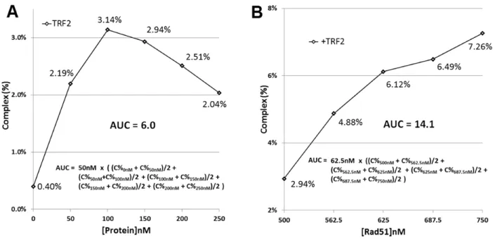
Figure 2.S5 Area under the curve (AUC) calculation procedures (C% = Complex %). (A) A representative activity trace from data shown in Figure 1C. AUC is calculated as (Complex% × [TRF2] nM) for all regions of the activity trace. (B) A representative activity trace from data shown in Figure 2A. AUC is calculated as (Complex% × [Rad51] nM) for 500-750 nM [Rad51].
Table 2.1 Properties of TRF2, TRF2∆B, TRF2∆M and TRF1
Proteins
TRF-Induced Telomeric
D-Loop Formation
AUC 0-250 nM % of TRF2
Rad51-Mediated Telomeric D-loop Formation
AUC 500-750 nM % Change From Buffer
Rad51-Mediated Non-Telomeric D-Loop Formation
AUC 500-750 nM % Change From Buffer
Telomeric DNA Binding C1/2, nM Telomeric Binding Specificity Migration in Agarose Gels
Buffer† N/A 30.3 ± 0.4 4.7 ± 0.4 N/A + -
0 ± 0.7% 0 ± 7.6%
TRF2
6.0 ± 0.15 13.3 ± 0.8 * 4.7 ± 0.0
111 ± 8 + -
100 ± 2.4% - 52 ± 5.1% * + 4 ± 2.2%
TRF2∆B
2.8 22.1 ± 0.7 * 5.1 ± 0.7
257 + -
47% - 31 ± 5.5% * + 5 ± 7.2%
TRF2∆M
1.8 29.0 ± 1.4 9.6 ± 0.4 *
319 - + / -
31% + 4 ± 4.0% + 112 ± 13.0% *
TRF1 27% 1.6 + 25 ± 1.0% * 38.5 ± 1.9 * + 9 ± 5.2% 5.4 ± 0.9 152 + +
†Buffer data are averaged. Proteins are statistically compared against matched buffer controls.
TRF-induced D-loop formation calculated as area under the curve (AUC: Complex% × [TRF] nM) from Figure 1. Rad51-mediated D-loop formation calculated as AUC (Complex% × [Rad51] nM) from Figures 2-5.
AUC calculation examples in Figure 2.S5.
C1/2 represents the concentration of TRF protein required to supershift 50% of template in EMSAs. Errors shown are 95% confidence intervals from three independent experiments.
Figure 2.S2 TRF2-mediated D-loop formation requires telomeric homology while Rad51 does not. (A) Rad51 promotes telomeric D-loop formation between pBB and T90 (top left), but not between pGL GAP and T90. Rad51 can promote non-telomeric D-loop formation between D1 and both pGL GAP and pBB. (B) Quantification of (A). (C) TRF2 promotes D-loop formation only between T90 and pBB. (C) Quantification of (A). (D) Quantification of (C).
23
Rad51-mediated D-loop formation is a multi-step process initiated by Rad51 binding to ssDNA to form a nucleoprotein filament, which subsequently interrogates dsDNA for matching antisense sequence in a process known as ‘homology search’. In this process a Rad51-coated substrate initially forms a protein-mediated complex with a homologous template. Subsequently Rad51 promotes D-loop formation between the substrate and template.20 To determine what step or steps of this process might be inhibited by TRF2 we performed several order of addition experiments.
We observed that the ability of TRF2 to inhibit Rad51-mediated telomeric D-loop formation was dependent upon addition of TRF2 early in the D-loop reaction (Fig. 2.S3). TRF2 could inhibit Rad51-mediated telomeric D-loop formation when pre-incubated with the telomeric template (T0) or when added to a combined reaction prior to D-loop formation (T0+10min). However, TRF2 could not inhibit Rad51-mediated D-loop formation if added after D-loop formation had already occurred (T0+3hrs). These observations suggested that TRF2 modulates Rad51-mediated D-loop formation via a passive mechanism, possibly by interfering with Rad51 filament formation, inhibiting homology search or by preventing subsequent D-loop formation.
Figure 2.S3 TRF2 inhibits Rad51-mediated telomeric D-loop formation only when added early in D-loop reactions. (A) Diagram of order of addition experiment. (B) TRF2 inhibits Rad51-mediated telomeric D-loop formation when added early in the reactions (T0 and T0+10min vs no TRF2) but not when added late in the reaction (T0+3hrs vs no TRF2). (C) Quantification of (B), error bars shown 95% confidence intervals from three independent experiments. (*) significant difference between indicated samples, paired samples t-test α=0.05. (NS) No significant difference.
25
To investigate whether the DNA binding activities of TRF2 mediate its ability to inhibit Rad51-mediated telomeric D-loop formation, we characterized the binding affinity and specificity of TRF2 using an electrophoretic mobility shift assay (EMSA) and a binding competition assay (Fig. 2.2E). Incubating a Cy3-labeled template containing a 103 base pair bp telomere tract with increasing concentrations of TRF2 resulted in a supershift of the template, consistent with stable TRF2 binding (Fig. 2.2F). The binding to TRF2 to the template was observed to be specific, and persisted even in the presence of high concentrations of non-telomeric competitor (Fig. 2.22F: lanes 8-11). Nearly all low-mobility species generated by TRF2 binding became trapped in the wells. This supershift was protein mediated, and could be disrupted by incubation with SDS and proteinase K (Fig. 2.2F: lane 12).
To further investigate possible mechanism by which TRF2 may inhibit Rad51-mediated telomeric D-loop formation we characterized the binding activity and the telomeric and non-telomeric Rad51-modulating activities of a variety of TRF2 mutant proteins and TRF1, a close homolog of TRF2.
TRF2∆M Promotes Rad51-Mediated Telomeric But Not Non-Telomeric D-Loop Formation.
TRF2∆B Inhibits Telomeric But Not Non-Telomeric Rad51-Mediated D-Loop Formation.
In addition to its Myb domain, TRF2 possesses an N-terminal domain rich in basic residues that has been implicated in directing the binding of TRF2 to ss/dsDNA junctions and unusual DNA structures.4, 21 This domain also promotes the annealing and migration of DNA joints in a manner not unlike that required during D-loop formation.22 To investigate whether the basic domain of TRF2 contributes to the ability of TRF2 to inhibit Rad51-mediated telomeric D-loop formation or the ability of TRF2∆M to promote Rad51-mediated non-telomeric D-loop formation we characterized the DNA binding affinity and specificity and Rad51-modulating activity of a TRF2 mutant protein lacking the basic domain of TRF2 (TRF2∆B).
Like TRF2 and in contrast to TRF2∆M, TRF2∆B was found to inhibit Rad51-mediated telomeric D-loop formation by 31±5.5% (Table 1; Fig. 2.4A, B), suggesting that the joint-binding activity of TRF2 is not required for TRF2 to inhibit Rad51-mediated telomeric D-loop formation. In contrast, TRF2∆B was not observed to affect Rad51-mediated non-telomeric D-loop formation (Table 2.1; Fig. 2.4C, D). Deletion of the basic domain resulted in an approximately 2.3-fold reduction in template binding affinity (Table 1) but did not reduce binding specificity (Fig. 2.4E: lanes 8-11) compared to full length TRF2. Like TRF2, TRF2∆B binding resulted in the template becoming trapped in the wells.
TRF1 Promotes Rad51-Mediated Telomeric But Not Non-Telomeric D-Loop Formation.
Figure 2.4 TRF2∆B promotes Rad51-mediated telomeric but not non-telomeric D-loop formation. (A) Rad51 promotes telomeric D-loop formation in a concentration dependent manner that is promoted by TRF2∆B. (B) Quantification of data in (A). (C) Rad51 promotes non-telomeric D-loop formation in a concentration dependent manner that is not affected by TRF2∆B. (D) Quantification of data in (C). (E) TRF2∆B binding supershifts the template into the wells. This binding is specific and persists in the presence of high concentrations of non-telomeric competitor and is protein-mediated. Error bars shown 95% confidence interval, significant difference between +Buffer and +TRF2∆B (*), paired samples t-test
be made with caution, as despite possessing comparable DNA binding affinity and telomeric sequence specificity (Table 2.1) their binding behavior is otherwise grossly different when examined in an EMSA. Whereas TRF2 binding shifts a telomeric template into the wells (Fig. 2.2F), TRF1 binding shifts the species into increasingly larger complexes as the TRF1 concentration is increased (Fig. 2.5E). This behavior is perhaps consistent with previous observations that while TRF2 binds to telomeric dsDNA as a large oligomeric structure, TRF1 binds as a smaller complex.24, 25 Likewise, this property may be consistent with observations that TRF2 can promote the formation of unusual DNA structures and induce topological changes within telomeric DNA to a greater degree than TRF1.12, 26
DISCUSSION
The results of this study suggest a model whereby TRF1 and TRF2 differentially regulate Rad51-mediated telomeric and non-telomeric D-loop formation. This would promote efficient telomeric DNA replication and non-telomeric HR while inhibiting aberrant HR at the telomeres. TRF1 promotes Rad51 mediated telomeric D-loop formation, which may facilitate replication fork restart and explain why TRF1 is required for efficient telomere replication. In contrast, TRF2 potently inhibits Rad51-mediated telomeric D-loop formation, providing yet another mechanism by which TRF2 can inhibit DNA repair at telomeres. Finally, TRF2∆M promotes Rad51-mediated D-loop formation, providing insight into how TRF2 may contribute to HR. Our findings are generally in good agreement with previous characterizations, and what contradictions exist are likely due to methodological differences.
possesses only a 103 bp telomeric tract. Finally, while the topology of telomeric DNA in vivo is unknown, the templates used in our assay were negatively supercoiled.
While we observed that TRF2 inhibits Rad51, it has previously been reported that TRF2 and Rad51 appear to exhibit functional cooperation. Immunodepletion of TRF2 or Rad51 from cell extracts ablates the ability of those extracts to promote telomeric D-loop formation.10 Moreover, supplementation of such immunodepleted extracts with purified Rad51 or TRF2 can restore telomeric D-loop formation.10, 27 However, the presence of factors in vivo that are absent from our in vitro characterization may affect
the activities of TRF2 and Rad51.
Although TRF1 and TRF2 can be found at telomeres throughout the cell cycle and TRF1 promotes efficient telomeric replication,7 TRF1 and TRF2 inhibit DNA replication in vitro.6 However, TRF1 and TRF2 binding are inhibited by post–translational modifications (PTMs), some of which are conferred by replication-complex associated proteins.28-34
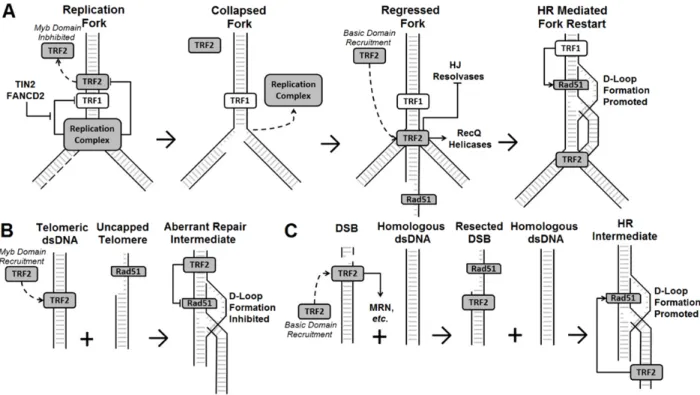
These proteins may facilitate replication by transiently removing TRF1 and TRF2 from telomeric DNA near the replication fork.30 Interestingly, PTMs that reduce TRF1 binding are inhibited in vivo by another shelterin protein, TIN2,28 and by FANCD2, a component of the Fanconi anemia pathway.31 Comparable PTMs of TRF2 are not likewise inhibited. However, these TRF2 PTMs likely disrupt TRF2 dimerization, which would be expected to abrogate Myb domain binding but that may not affect basic domain binding. As TRF1 can promote and TRF2 can inhibit Rad51-mediated telomeric D-loop formation, the depletion of TRF2 but not TRF1 from DNA near the replication fork may facilitate HR-mediated fork restart within the telomeres (Fig. 2.6A). This process may also be facilitated by basic-domain mediated recruitment of TRF2 to regressed forks, where it can both protect the nascent Holliday junction (HJ) from HJ resolvases22 and recruit RecQ helicases that can promote fork migration.35, 36 Likewise, the presence of TRF2 on telomeric dsDNA away from the fork may prevent HR-mediated strand invasion reactions and protect the telomeres from aberrant repair (Fig. 2.6B).
the basic domain of TRF2 but not its Myb-domain37 and can occur in an ATM deficient background. Additionally, TRF2 is phosphorylated by ATM38 in response to DNA damage,39 and mutations that disrupt TRF2 phosphorylation inhibit DNA repair.40 While it has been suggested that this DNA repair defect may be due to impaired non-homologous end joining (NHEJ),39 the defect may also be due to impaired HR. Overexpression of TRF2 and TRF2∆M promote HR in vivo.11 Likewise, knockdown of TRF2 inhibits HR but not NHEJ in vivo.11 Our finding that TRF2∆M can promote Rad51-mediated D-loop formation suggests a novel mechanism by which TRF2 can promote HR (Fig. 2.6C). Upon induction of a DSB, TRF2 may undergo basic-domain mediated recruitment to the site of damage. TRF2 may then help recruit proteins such as the Mre11/Rad50/Nbs1 (MRN) complex,41 which promotes end resection in preparation for HR. Following end resection, Rad51 binding and homology search, the basic domain of TRF2 may facilitate Rad51-mediated D-loop formation by promoting the opening of the template dsDNA in a manner similar to a Rad51 accessory protein, Rad54.13
This model of the interaction of TRF1, TRF2 and Rad51 provides insight into both telomere biology and the HR pathway. Previous characterizations suggested that TRF2 and Rad51 cooperate to promote telomeric D-loop and possibly t-loop formation in vivo, despite apparent incompatibilities in the in vitro activities of these proteins. Our finding that TRF2 inhibits Rad51-mediated telomeric D-loop
REFERENCES
[1] Bombarde, O., Boby, C., Gomez, D., Frit, P., Giraud-Panis, M. J., Gilson, E., Salles, B., and Calsou, P. (2010) TRF2/RAP1 and DNA-PK mediate a double protection against joining at telomeric ends, EMBO J 29, 1573-1584.
[2] Karlseder, J., Hoke, K., Mirzoeva, O. K., Bakkenist, C., Kastan, M. B., Petrini, J. H., and de Lange, T. (2004) The telomeric protein TRF2 binds the ATM kinase and can inhibit the ATM-dependent DNA damage response, PLoS Biol 2, E240.
[3] Doksani, Y., Wu, J. Y., de Lange, T., and Zhuang, X. (2013) Super-resolution fluorescence imaging of telomeres reveals TRF2-dependent T-loop formation, Cell 155, 345-356.
[4] Stansel, R. M., de Lange, T., and Griffith, J. D. (2001) T-loop assembly in vitro involves binding of TRF2 near the 3' telomeric overhang, EMBO J 20, 5532-5540.
[5] Fouche, N., Ozgur, S., Roy, D., and Griffith, J. D. (2006) Replication fork regression in repetitive DNAs, Nucleic Acids Res 34, 6044-6050.
[6] Ohki, R., and Ishikawa, F. (2004) Telomere-bound TRF1 and TRF2 stall the replication fork at telomeric repeats, Nucleic Acids Res 32, 1627-1637.
[7] Sfeir, A., Kosiyatrakul, S. T., Hockemeyer, D., MacRae, S. L., Karlseder, J., Schildkraut, C. L., and de Lange, T. (2009) Mammalian telomeres resemble fragile sites and require TRF1 for efficient replication, Cell 138, 90-103.
[8] North, J. A., Amunugama, R., Klajner, M., Bruns, A. N., Poirier, M. G., and Fishel, R. (2013) ATP-dependent nucleosome unwrapping catalyzed by human RAD51, Nucleic Acids Res 41, 7302-7312.
[9] Badie, S., Escandell, J. M., Bouwman, P., Carlos, A. R., Thanasoula, M., Gallardo, M. M., Suram, A., Jaco, I., Benitez, J., Herbig, U., Blasco, M. A., Jonkers, J., and Tarsounas, M. (2010) BRCA2 acts as a RAD51 loader to facilitate telomere replication and capping, Nat Struct Mol Biol 17, 1461-1469.
[10] Verdun, R. E., and Karlseder, J. (2006) The DNA damage machinery and homologous recombination pathway act consecutively to protect human telomeres, Cell 127, 709-720.
[11] Mao, Z., Seluanov, A., Jiang, Y., and Gorbunova, V. (2007) TRF2 is required for repair of nontelomeric DNA double-strand breaks by homologous recombination, Proc Natl Acad Sci U S A 104, 13068-13073.
[12] Amiard, S., Doudeau, M., Pinte, S., Poulet, A., Lenain, C., Faivre-Moskalenko, C., Angelov, D., Hug, N., Vindigni, A., Bouvet, P., Paoletti, J., Gilson, E., and Giraud-Panis, M. J. (2007) A topological mechanism for TRF2-enhanced strand invasion, Nat Struct Mol Biol 14, 147-154. [13] Sigurdsson, S., Van Komen, S., Petukhova, G., and Sung, P. (2002) Homologous DNA pairing by
human recombination factors Rad51 and Rad54, J Biol Chem 277, 42790-42794.
[15] Amunugama, R., He, Y., Willcox, S., Forties, R. A., Shim, K. S., Bundschuh, R., Luo, Y., Griffith, J., and Fishel, R. (2012) RAD51 protein ATP cap regulates nucleoprotein filament stability, J Biol Chem 287, 8724-8736.
[16] Smogorzewska, A., Karlseder, J., Holtgreve-Grez, H., Jauch, A., and de Lange, T. (2002) DNA ligase IV-dependent NHEJ of deprotected mammalian telomeres in G1 and G2, Curr Biol 12, 1635-1644.
[17] Broccoli, D., Smogorzewska, A., Chong, L., and de Lange, T. (1997) Human telomeres contain two distinct Myb-related proteins, TRF1 and TRF2, Nat Genet 17, 231-235.
[18] Biet, E., Sun, J., and Dutreix, M. (1999) Conserved sequence preference in DNA binding among recombination proteins: an effect of ssDNA secondary structure, Nucleic Acids Res 27, 596-600. [19] Tracy, R. B., Baumohl, J. K., and Kowalczykowski, S. C. (1997) The preference for GT-rich DNA
by the yeast Rad51 protein defines a set of universal pairing sequences, Genes Dev 11, 3423-3431.
[20] Forget, A. L., and Kowalczykowski, S. C. (2010) Single-molecule imaging brings Rad51 nucleoprotein filaments into focus, Trends Cell Biol 20, 269-276.
[21] Fouche, N., Cesare, A. J., Willcox, S., Ozgur, S., Compton, S. A., and Griffith, J. D. (2006) The basic domain of TRF2 directs binding to DNA junctions irrespective of the presence of TTAGGG repeats, J Biol Chem 281, 37486-37495.
[22] Poulet, A., Buisson, R., Faivre-Moskalenko, C., Koelblen, M., Amiard, S., Montel, F., Cuesta-Lopez, S., Bornet, O., Guerlesquin, F., Godet, T., Moukhtar, J., Argoul, F., Declais, A. C., Lilley, D. M., Ip, S. C., West, S. C., Gilson, E., and Giraud-Panis, M. J. (2009) TRF2 promotes, remodels and protects telomeric Holliday junctions, EMBO J 28, 641-651.
[23] Court, R., Chapman, L., Fairall, L., and Rhodes, D. (2005) How the human telomeric proteins TRF1 and TRF2 recognize telomeric DNA: a view from high-resolution crystal structures, EMBO Rep 6, 39-45.
[24] Poulet, A., Pisano, S., Faivre-Moskalenko, C., Pei, B., Tauran, Y., Haftek-Terreau, Z., Brunet, F., Le Bihan, Y. V., Ledu, M. H., Montel, F., Hugo, N., Amiard, S., Argoul, F., Chaboud, A., Gilson, E., and Giraud-Panis, M. J. (2012) The N-terminal domains of TRF1 and TRF2 regulate their ability to condense telomeric DNA, Nucleic Acids Res 40, 2566-2576.
[25] Khan, S. J., Yanez, G., Seldeen, K., Wang, H., Lindsay, S. M., and Fletcher, T. M. (2007) Interactions of TRF2 with model telomeric ends, Biochem Biophys Res Commun 363, 44-50. [26] Yoshimura, S. H., Maruyama, H., Ishikawa, F., Ohki, R., and Takeyasu, K. (2004) Molecular
mechanisms of DNA end-loop formation by TRF2, Genes Cells 9, 205-218.
[27] Verdun, R. E., Crabbe, L., Haggblom, C., and Karlseder, J. (2005) Functional human telomeres are recognized as DNA damage in G2 of the cell cycle, Mol Cell 20, 551-561.
[29] Ye, J. Z., and de Lange, T. (2004) TIN2 is a tankyrase 1 PARP modulator in the TRF1 telomere length control complex, Nat Genet 36, 618-623.
[30] Beneke, S., Cohausz, O., Malanga, M., Boukamp, P., Althaus, F., and Burkle, A. (2008) Rapid regulation of telomere length is mediated by poly(ADP-ribose) polymerase-1, Nucleic Acids Res 36, 6309-6317.
[31] Lyakhovich, A., Ramirez, M. J., Castellanos, A., Castella, M., Simons, A. M., Parvin, J. D., and Surralles, J. (2011) Fanconi anemia protein FANCD2 inhibits TRF1 polyADP-ribosylation through tankyrase1-dependent manner, Genome Integr 2, 4.
[32] Dantzer, F., Giraud-Panis, M. J., Jaco, I., Ame, J. C., Schultz, I., Blasco, M., Koering, C. E., Gilson, E., Menissier-de Murcia, J., de Murcia, G., and Schreiber, V. (2004) Functional interaction between poly(ADP-Ribose) polymerase 2 (PARP-2) and TRF2: PARP activity negatively regulates TRF2, Mol Cell Biol 24, 1595-1607.
[33] Simbulan-Rosenthal, C. M., Rosenthal, D. S., Boulares, A. H., Hickey, R. J., Malkas, L. H., Coll, J. M., and Smulson, M. E. (1998) Regulation of the expression or recruitment of components of the DNA synthesome by poly(ADP-ribose) polymerase, Biochemistry 37, 9363-9370.
[34] Walker, J. R., and Zhu, X. D. (2012) Post-translational modifications of TRF1 and TRF2 and their roles in telomere maintenance, Mech Ageing Dev 133, 421-434.
[35] Machwe, A., Xiao, L., and Orren, D. K. (2004) TRF2 recruits the Werner syndrome (WRN) exonuclease for processing of telomeric DNA, Oncogene 23, 149-156.
[36] Opresko, P. L., von Kobbe, C., Laine, J. P., Harrigan, J., Hickson, I. D., and Bohr, V. A. (2002) Telomere-binding protein TRF2 binds to and stimulates the Werner and Bloom syndrome helicases, J Biol Chem 277, 41110-41119.
[37] Bradshaw, P. S., Stavropoulos, D. J., and Meyn, M. S. (2005) Human telomeric protein TRF2 associates with genomic double-strand breaks as an early response to DNA damage, Nat Genet 37, 193-197.
[38] Tanaka, H., Mendonca, M. S., Bradshaw, P. S., Hoelz, D. J., Malkas, L. H., Meyn, M. S., and Gilley, D. (2005) DNA damage-induced phosphorylation of the human telomere-associated protein TRF2, Proc Natl Acad Sci U S A 102, 15539-15544.
[39] Huda, N., Abe, S., Gu, L., Mendonca, M. S., Mohanty, S., and Gilley, D. (2012) Recruitment of TRF2 to laser-induced DNA damage sites, Free Radic Biol Med 53, 1192-1197.
[40] Huda, N., Tanaka, H., Mendonca, M. S., and Gilley, D. (2009) DNA damage-induced phosphorylation of TRF2 is required for the fast pathway of DNA double-strand break repair, Mol Cell Biol 29, 3597-3604.
[41] Zhu, X. D., Kuster, B., Mann, M., Petrini, J. H., and de Lange, T. (2000) Cell-cycle-regulated association of RAD50/MRE11/NBS1 with TRF2 and human telomeres, Nat Genet 25, 347-352.
Chapter 3: Biophysical and Ultrastructural Characterization of Adeno-Associated Virus Capsid Uncoating and Genome Release1
INTRODUCTION
Adeno-associated virus (AAV) is a small (25 nm) nonenveloped virus belonging to the family Parvoviridae and genus Dependovirus. The AAV capsid packages a single-stranded (ssDNA) genome
approximately 4.7 kb in length1. The wild-type genome consists of two open reading frames flanked by two inverted terminal hairpin repeats (ITRs). The ITRs, which are 145 nucleotides each, are the only cis element in the AAV genome required for successful packaging2,3. The AAV capsid is composed of 60 (T = 1) viral protein subunits VP1, VP2, and VP3, in approximately the ratio 1:1:10. The three different subunits are generated from overlapping reading frames and interact within the capsid through the common VP3 subunit region. The largest VP1 subunit is known to possess a phospholipase A2 domain required for infectivity4. Because of its broad tropism, lack of pathogenicity, and flexibility in genome content, AAV has become a promising candidate for therapeutic gene transfer applications. In the past 2 decades, AAV has been utilized as a gene transfer vector in a number of phase I and phase II clinical trials treating various genetic diseases5.
Different AAV serotypes infect cells by engaging a variety of cell surface glycans and coreceptors, followed by endocytic uptake4,6,7. Viral particles are then thought to escape from the endosome and translocate to the nucleus, where the ssDNA genome is released and undergoes second-strand synthesis. Engineered AAV genomes containing a mutant 3′ ITR have been shown to package dimeric, self-complementary DNA (scDNA)8. Such scAAV vectors have the advantage of escaping ssDNA degradation9 and bypassing second-strand synthesis, which is a rate-limiting step preceding ________________________