MASTICATORY FUNCTION OF THE DENTULOUS VERSUS
PROSTHODONTICALLY TREATED EDENTULOUS MANDIBLE
Sorin Uram-Tuculescu
A thesis submitted to the faculty of the University of North Carolina at Chapel Hill in partial fulfillment of the requirements for the degree of Master of Science in the School of Dentistry (Prosthodontics).
Chapel Hill 2008
Approved by:
ABSTRACT
Sorin Uram-Tuculescu: Masticatory function of the dentulous versus prosthodontically treated edentulous mandible
(Under the direction of Dr. Greg Essick, DDS, PhD)
Background: There is general agreement that implant supported prostheses offer better masticatory function than traditional prostheses that are not supported by teeth or implants. However, solid evidence is lacking to fully support this, and contrary findings are mentioned in the literature.
Aim: To characterize differences in chewing and neuromuscular control characteristics of patients with upper and lower natural teeth, upper full dentures and lower natural teeth, upper and lower full dentures, upper full dentures and lower implant-supported overdentures, upper full dentures and lower implant supported fixed prostheses.
Materials and methods: Chewing function is evaluated subjectively (questionnaires) and objectively (electromyographic measurements), while subjects are chewing agar based model foods with 4 different consistencies.
Conclusions: Electromyographic methods using i) engineered test foods with controlled physical properties and ii) rigorous analytical techniques can detect and characterize meaningful differences among human subjects who differ in dental status and prosthodontic treatments.
To my wife Cora, who sacrificed a lot for me to continue my graduate education. To my son Dorin, who is the joy of my life.
AKNOWLEDGEMENTS
I would like to thank my mentor, Dr. Greg Essick for his outstanding support and scientific guidance throughout the course of thesis preparation. Also I would like to thank my other committee members, Dr. Lyndon Cooper, Dr. Allen Foegeding, and Dr. Chris Vinyard for their kind support and friendship. Special thanks are directed to my fellow residents, to Dr. Bongkosh Vardhanabhuti, Mr. Mike Young and Mr. Steven Smith for their help in the experimental and analytical part of the study.
This work was partially funded by a research grant from the Academy of Prosthodontics Foundation.
TABLE OF CONTENTS
LIST OF TABLES………..…x
LIST OF FIGURES………...xi
LIST OF ABBREVIATIONS AND SYMBOLS………...……….…xiv
CHAPTER 1. INTRODUCTION.………...1
1.1. METHODS USED FOR ASSESSMENT OF MASTICATORY FUNCTION.………….2
A. Subjective methods...………..2
B. Objective methods……….2
i) Food particle size reduction determination………4
ii) Masticatory muscle force evaluation………4
iii) Masticatory muscle EMG………5
iv) Jaw movements tracking...5
C. Test foods used in studies...6
D. Objective compared to subjective methods...7
1.2. MASTICATORY FUNCTION IN DENTATE INDIVIDUALS………...8
A . Subjective evaluation……….………8
B. Food particle size reduction...8
i) “Good” chewers and “bad” chewers...8
ii) Influence of age on particle size reduction………...…………9
C. Masticatory muscle force...9
i) Response to different textures. ………...……….10
ii) Influence of gender……….10
iii) Influence of age………...11
E. Jaw movements………...11
i) Influence of food texture………..11
ii) Influence of craniofacial type……….………12
iii) Influence of gender………. ..12
1.3. MASTICATORY FUNCTION IN CONVENTIONAL FULL DENTURE WEARERS12 A. Subjective evaluation...13
B. Food particle size reduction...13
C. Masticatory muscle EMG...13
i) Chewing duration, number of chews, and chewing frequency...14
ii) Peak amplitude...14
iii) Area under EMG curve...15
D. Jaw movements...16
E. Significance of the loss of periodontal receptors...16
1.4. MASTICATORY FUNCTION IN SUBJECTS WITH UPPER CONVENTIONAL FULL DENTURES AND LOWER IMPLANT-SUPPORTED OVERDENTURES……...17
A. Subjective evaluation...18
B. Food particle size reduction...18
C. Masticatory muscle EMG...19
D. Significance of the loss of periodontal receptors and addition of implants...19
1.5. MASTICATORY FUNCTION IN SUBJECTS WITH UPPER CONVENTIONAL FULL DENTURES AND LOWER IMPLANT-SUPPORTED FIXED PROSTHESES…....20
A. Subjective evaluation...20
1.6 RATIONALE OF OUR STUDY………...21
2. MATERIAL AND METHODS………..………...23
2.1 HUMAN SUBJECTS……….23
2.2. APPROVALS AND LEGAL DOCUMENTATION………24
2.3. TESTING PROCEDURES………...………25
A. Subjective evaluation………25
B. Objective evaluation.……….………25
i) Model foods preparation and storage.………..26
ii) Masticatory muscle electromyography. ………..27
iii) Processing of the EMG data. ……….29
2.4. DATA ANALYSIS ………...32
A. Characteristics of subject population. ………...32
B. Subjective evaluation of chewing. ………32
C. Objective evaluation of chewing. ………..32
3. RESULTS……….………..35
3.1. CHARACTERISTICS OF SUBJECT POPULATION ………35
3.2. SUBJECTIVE EVALUATION OF CHEWING ………..35
A. Evaluation of chewing ability. ………..35
B. Evaluation of reported parafunction. ………36
3.3. OBJECTIVE EVALUATION OF CHEWING ………37
A. Total duration of chewing ……….38
i) Dentate subjects only. ………..38
ii) Prosthodontically treated versus dentate subjects.………..38
iv) Effect of age. ………..39
B. Chewing Frequency………...39
i) Dentate subjects only. ………..39
ii) Prosthodontically treated versus dentate subjects. ……….40
iii) Prosthodontically treated subjects only. ………40
iv) Effect of age. ……….41
C. Duration of single chews ………..41
i) Dentate subjects only. ………..41
ii) Prosthodontically treated versus dentate subjects ………..41
iii) Prosthodontically treated subjects only. ………42
iv) Effect of age. ………..42
v) Chew to chew variation in cycle duration.………..43
D. Duty cycle of the jaw-closing muscles. ………43
i) Dentate subjects only. ………..43
ii) Dentate subjects versus prosthodontically treated individuals. ………..44
iii) Prosthodontically treated subjects only. ………44
iv) Effect of age. ………..45
E. EMG area-under-curve for the jaw-closing muscles ………45
i) Dentate subjects only. ………..46
ii) Prosthodontically treated versus dentate subjects.………..46
iii) Prosthodontically treated subjects only. ………48
iv) Effect of age. ………..49
F. Timing: Asynchrony in muscle recruitment ………..49
i) Dentate subjects only. ………..49
ii) Prosthodontically treated versus dentate subjects.………..50
iii) Prosthodontically treated subjects only. ………51
iv) Effect of age. ………..53
4. DISCUSSIONS….……….………...…..54
4.1. SUBJECTIVE ASSESSMENT OF MASTICATORY FUNCTION ………54
4.2. OBJECTIVE EVALUATION OF MASTICATORY FUNCTION ……….55
A. Total duration of chewing. ………55
B. Chewing frequency. ………..56
C. Duration of single chews. ……….56
D. Duty cycle of the jaw closing muscles . ………57
E. Relative muscle work: EMG area-under-curve for the jaw-closing muscles …………57
5. CONCLUSIONS………..………...58
LIST OF TABLES
Table
1.1 Masticatory function in dentate subjects………...59
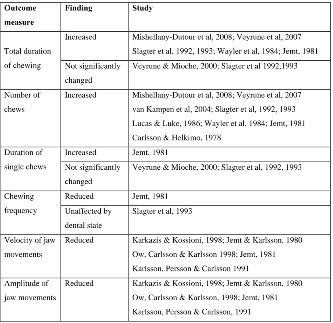
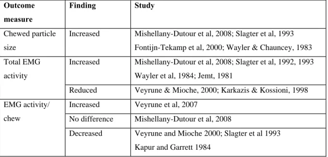
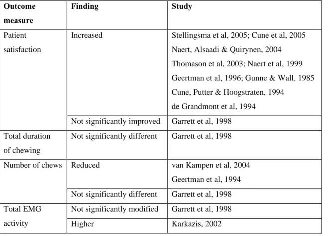
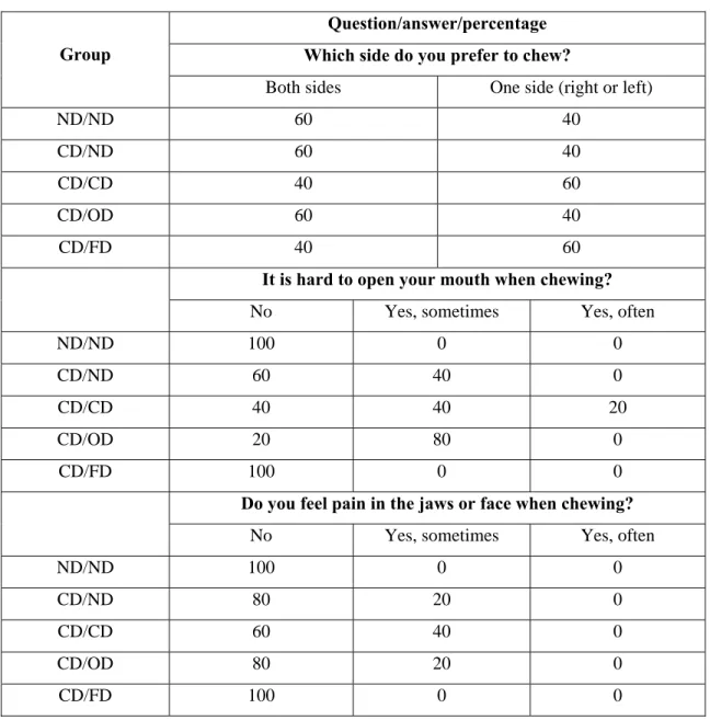
1.2 Masticatory function in conventional full denture wearers, compared to dentate individuals.………...………...………..60, 61 1.3 Masticatory function in patients with upper conventional dentures and lower implant-supported overdentures, compared to individuals with upper & lower conventional dentures………...………....62
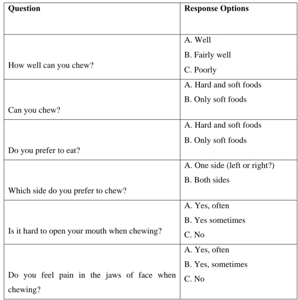
2.1 Reported chewing ability questionnaire (Ow et al 1998)……….63
2.2 Reported parafunction questionnaire (Miyake et al 2004)………64
3.1 Age and gender composition of the study population………..65
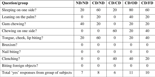
3.2 Reported chewing ability questionnaire results: percentage of subjects answers/question/group………...………..66, 67 3.3 Reported parafunction questionnaire results: percentage of “Yes” answers/group for each question………68
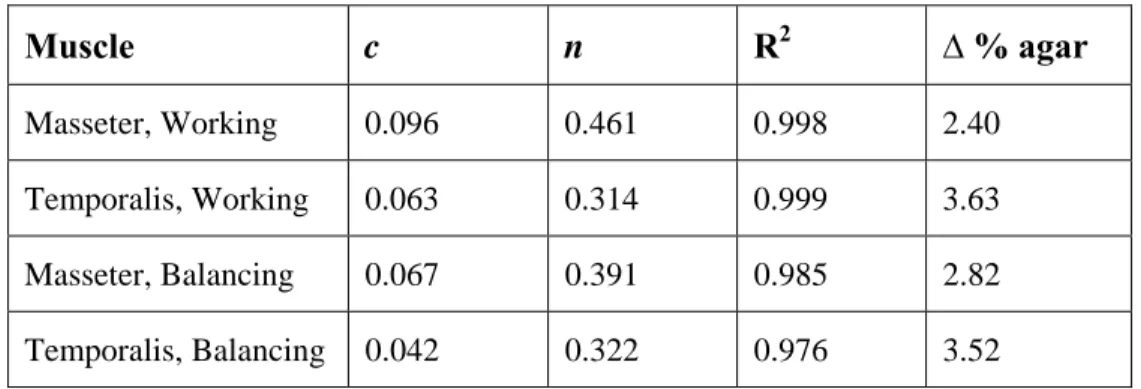
3.4 Power function analyses of the total duration of chewing versus model food firmness ……..69
3.5 Chewing frequency (chews/s) for the different subject groups………....70
3.6 Power function analyses of the muscle work, based on scaled EMG area-under-curve versus model food firmness (% agar) of all study subjects.……….71
3.7 Power function analyses of the muscle work, based on scaled EMG area-under-curve versus model food firmness (% agar) of 5 dentate subjects…….………72
3.8 Power function analyses of the muscle work, based on scaled EMG area-under-curve versus model food firmness (% agar) of 20 prosthodontically treated subjects……...….…...73
3.9 Time occurrence of peak EMG activity in stated muscle re: time of peak EMG activity in working side masseter muscle (time = 0)………..………...74
3.10 Time occurrence of peak EMG activity in stated muscle re: time of peak EMG activity in working side masseter muscle (time = 0)………..………...75
LIST OF FIGURES Figure
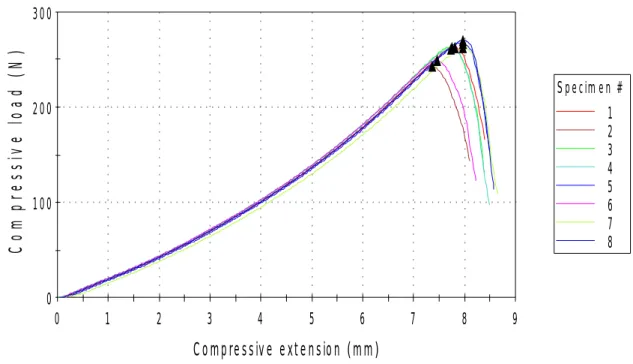
2.1 Testing results of 7% agar samples loaded to fracture, in order to evaluate the
consistency of mechanical properties……….……….76
2.2 Linear regression plot for stress fracture by agar concentration………...77 2.3 EMG electrodes positioning: parallel with the muscle fibers (schematically represented in
red), on the bulkiest part of the muscle belly, as determined by palpation, when subject is contracting the muscle……….…78 2.4 Raw EMG data for the six muscles investigated, as they appear on the computer screen
after recording a masticatory sequence (right side intentional chewing, 1.75% agar)……….79 2.5 Combined rms (root mean squares) data (all six muscles tested during one masticatory
sequence), processed in Labview software, and illustrated in Igor Pro software……….80 2.6 a: Expanded rms sequence allowing identification of different muscle waves, rough
estimation of peak timing, and delimitation of individual chewing cycles (data processed in Labview software, and illustrated in Igor Pro………..…81 2.6 b: Expanded rms sequence, with the first chewing cycle delimited between cursors, and
ready to be recorded……….………...82 2.6 c: Expanded rms sequence, with the second chewing cycle delimited between cursors, and
ready to be recorded……….83 3.1 Age composition of the study population………..………..84 3.2 Reported parafunction questionnaire results: percentage of “Yes” answers/group for each
question……….……...85 3.3 Total duration of chewing in the dentate group. Error bars denote ± 1 standard error of the
mean……….86 3.4 Total duration of chewing for the dentate group versus the prosthodontically treated
patients……….………87 3.5 Total duration of chewing for the prosthodontically treated subjects..……..………...88
3.8 Chewing frequency for the prosthodontically treated subjects………...………..91
3.9 Duration of single chew for dentate subjects…...……….92
3.10 Duration of single chew for dentate subjects.……….93
3.11 Duration of single chew by agar concentration for dentate subjects versus prosthodontically treated individuals.……….……….94
3.12 Duration of single chew by chew number for dentate subjects versus prosthodontically treated individuals………...………95
3.13 Duration of single chew by agar concentration for prosthodontically treated subjects…...96
3.14 Chew duration coefficient of variation by agar concentration…..………...97
3.15 Elevator duty by agar concentration for dentate subjects………...98
3.16 Elevator duty by chew number for dentate subjects…...………99
3.17 Elevator duty by agar concentration for dentate subjects versus prosthodontically treated individuals………...………...100
3.18 Elevator duty by chew number for dentate subjects versus prosthodontically treated individuals………...………...101
3.19 Elevator duty by agar concentration for prosthodontically treated individuals………102
3.20 Elevator duty by age for all groups………...103
3.21 Mean muscle work/chew, as measured by scaled area under the EMG curve in dentate subjects………...………...….104
3.22 Mean muscle work/chew, as measured by scaled area under the EMG curve for the four jaw elevators, in dentate subjects, and prosthodontically treated individuals……....……..105
3.23 Mean muscle work/chew, as measured by scaled area under the EMG curve for the four jaw elevators, in dentate subjects………...………...…106
3.24 Mean muscle work/chew, as measured by scaled area under the EMG curve in dentate subjects versus prosthodontically treated individuals………...………...107
3.25 Mean muscle work/chew, as measured by scaled area under the EMG curve in dentate subjects versus prosthodontically treated individuals..….……….108
3.26 Constant “c” and exponent “n” of the power functions for the relationship between muscle work and food firmness (% agar) by chew number....……….……….……...109
3.27 Mean muscle work/chew, as measured by scaled area under the EMG curve in
prosthodontically treated individuals………...…..110 3.28 Mean muscle work/chew, as measured by scaled area under the EMG curve for the four
jaw elevators, in prosthodontically treated individuals……….111
3.29 Mean muscle work/chew, as measured by scaled area under the EMG curve for the working side muscles versus the balancing side muscles in prosthodontically
treated subjects..…...112 3.30 Mean muscle work/chew, as measured by scaled area under the EMG curve in
prosthodontically treated individuals……….113
3.31 Timing of masticatory muscles, based on peak activation and referenced to the timing of working masseter (considered equal to 0 by convention), for dentate subjects versus
prosthodontically treated individuals……….114
3.32 Timing of masticatory muscles, based on peak activation and referenced to the timing of working masseter (considered equal to 0 by convention), for dentate subjects versus
prosthodontically treated individuals, by agar concentration in the samples chewed……....115 3.33 Timing of masticatory muscles, based on peak activation and referenced to the timing of
working masseter (considered equal to 0 by convention), for dentate subjects, by chew number………...……….116 3.34 Timing of masticatory muscles, based on peak activation and referenced to the timing of
working masseter (considered equal to 0 by convention), for prosthodontically treated
subjects………...117 3.35 Timing of jaw-opening masticatory muscles, based on peak activation and referenced
to the timing of working masseter (considered equal to 0 by convention),
LIST OF ABBREVIATIONS AND SYMBOLS
ACP – American College of Prosthodontists AUC – area under the electromyography curve BDA – balancing digastric anterior muscle BMM – balancing superficial masseter muscle BTA – balancing temporalis anterior muscle BS – balancing side
c – multiplicative constant
CD/CD – conventional full denture group
CD/FD – upper conventional full denture, lower implant-supported prosthesis group CD/ND – upper conventional full denture, lower natural dentition group
CD/OD – upper conventional full denture, lower implant supported overdenture group CD/XX – prosthodontically treated subjects
cv – coefficient of variation D – total duration of chewing DA-L – left anterior digastric muscle DA-R – right anterior digastric muscle EMG – electromyography
IRB - Institutional Review Board MM-L – left superficial masseter muscle MM-R – right superficial masseter muscle N – sample size
n - power function exponent ND/ND – natural dentition group
R2 – R-square goodness of fit of the data to a power function model
rms – root mean square
SAS –Statistical Analysis Software sd – standard deviation
TA-L – left anterior temporalis muscle TA-R – right anterior temporalis muscle UNC – University of North Carolina USP – United States Pharmacopeia W – relative muscle work
WDA – working digastric anterior muscle WMM – working superficial masseter muscle WS – working side
WTA – working temporalis anterior muscle w/w – weight percentage
∆ % - change in agar concentration
1. INTRODUCTION
Mastication is a highly coordinated neuromuscular function involving purposeful movements
of the jaw and continuous modulation of force (Karkazis and Kossioni 1998), representing a function
that matures with growth as the teeth erupt. It is characterized by complex movements of the
stomatognathic system structures, most importantly the mandible, which vary depending on the foods
ingested, resulting in manipulation of a food bolus, salivation, and associated oscillatory movements
of the head.
The commands underlying the basic rhythmical movements of mastication are generated
centrally, but those responsible for their adaptive control are regulated by afferent (sensory)
information, particularly related to oro-facial kinesthetic inputs. Sensory receptors, such as
periodontal mechanoreceptors and muscle spindle afferents, strongly influence the activity of motor
neurons and thus, control of the masticatory muscles. Much of the integration of sensory feedback
with the centrally generated drive occurs at the level of the protoneurons in the nucleus reticularis
parvocellularis and in the mesencephalic trigeminal nucleus and adjacent nuclei, neural substrates
associated with the central pattern generator for mastication (Lund 1991).
Mandibular kinematics and the masticatory muscle activity during chewing have been
described in numerous studies using various methods. Characteristic chewing patterns in children and
adults (Gibbs et al 1982; Neill and Howell 1988) seem to be established early in childhood with some
restricted adaptation to new conditions such as the locations of permanent teeth (Gibbs et al 1982) or
tooth loss. However, individual chewing patterns vary widely within (Plesh Bishop and McCall 1987)
The loss of teeth, and consequently, prosthodontic rehabilitation pose new challenges to the
stomatognathic system, which alter masticatory function. Relatively little is known about the extent to
which chewing differs between dentate subjects and completely or partially edentulous patients
treated in various ways. Moreover, the methods of assessment are relatively few and have often
provide conflicting data.
1.1. METHODS USED FOR ASSESSMENT OF MASTICATORY FUNCTION
Masticatory function can be evaluated subjectively (subject reported assessments) and
objectively (laboratory tests of function).
A. Subjective methods.
In subject reported assessments, subjects judge their masticatory function via questionnaires
or interviews (Agerberg and Carlsson 1981; Osterberg and Steen 1982; Uchida 1991). A patient’s
general satisfaction is determined by a number of factors directly related to treatment: comfort,
perceived chewing ability, oral health-related quality of life, dietary difficulties and overall nutritional
status enabled by the oral status. The subjective method is sensitive to differences in dental and
prosthetic state, while data gathering and analysis is easy. The method is inexpensive and the data is
easy to interpret.
B. Objective methods.
The most frequently used objective methods target the determination of the following
outcomes:
a) Total duration of mastication in subjects eating standard-sized pieces of food (Lindquist and
Carlsson 1985; Lucas et al 1986a, Jemt and Stalblad 1986, Peyron et al 2004). In addition,
several other associated measures can be extracted from observation of the masticatory
most commonly measured nowadays by electromyography (Garrett Kapur and Perez 1996,
Youssef et al 1997; Veyrune et al 2007; Mishellany-Dutour et al 2008) or jaw tracking
methods (Jemt 1981; Youssef et al 1997).
b) Size of particles of test foods after comminution (Slagter et al 1992a; Yurkstas Fridley and
Manly 1951; Feldman et al 1980; Helkimo Carlsson and Helkimo 1978; Kapur Soman and
Yurkstas 1964). Particle size reduction can be determined from sieving (Gunne et al 1982;
Gunne 1985; Slagter et al 1993; Fontijn-Tekamp et al 2000; Mishellany-Dutour et al 2008),
or computerized image analysis (Mahmood et al 1992; Nakano et al 1989), of chewed
particles of known initial size.
c) Masticatory forces (Haraldson et al 1988). Masticatory forces can be evaluated directly
between the upper and lower teeth using bite bars instrumented with strain gauges (Carlsson
and Lindquist 1994; Fontijn-Tekamp 2000; Hatch et al 2001), force transducers placed in
prostheses, or indirectly by sound transmitted between the teeth (Gibbs et al 1981) or EMG
activity (Youssef et al 1997) calibrated to known to force levels.
d) Electrical activity of jaw muscle. EMG analysis enables specific physiologically relevant
measures to be extracted, such as the peak amplitude, its time of occurrence, area under the
EMG curve (also referred to as ‘muscle work’ in some literature), and duty cycle of the jaw
closing muscles (Feine et al 1994a; Garrett et al 1996; Karkazis 2002; Kohyama Mioche and
Bourdiol 2003; Miralles et al 1989; Peyron et al 2004; Mishellany-Dutour et al 2008).
e) Jaw movement patterns during mastication (Jemt 1984; Jemt and Stalblad 1986; Feine and
Lund 2006), recorded using a jaw tracking system.
Probably the most commonly used objective methods in the assessment of masticatory function
are: i) determination of food particle reduction, ii) evaluation of masticatory muscle forces, iii)
evaluation of masticatory muscle EMG activity, and iv) analysis of jaw movements.
i) Food particle size reduction determination.
Particle size measurement is meant to assess the ability to pulverize food. Fractional sieving
as a technique of separating the food after chewing for a given time period has been used for a long
time (Gunne et al 1982; Gunne 1985), and is still considered to be a viable method (Fontijn-Tekamp
et al 2000; Mishellany-Dutour et al 2008). Some disadvantages of this method relate to the following:
data analysis is messy and time consuming, particularly if one tries to measure the distribution of
particle sizes. Additionally, the method is applicable only to brittle substances (Feine and Lund 2006).
Computer-assisted image processing can also be used to analyze the size of masticated test
particles (Mahmood et al 1992; Nakano et al 1989). Compared with sieving, image analysis of test
particles offers considerable advantages such as simplicity, speed, accuracy, reproducibility, and
hygiene. This method is also practical for measuring a large number of samples (Boretti Bickel and
Geering 1995).
ii) Masticatory muscle force evaluation.
Force measurements have been purported to measure masticatory efficiency. Since maximal
biting forces are not generated during normal chewing, it seems more relevant to measure the forces
generated during mastication (Feine and Lund 2006). This can be performed by placing force
transducers into prostheses, or using a sound transmission method to measure the mechanical
coupling between the upper and lower teeth when occluding (Gibbs et al 1981). Masticatory muscle
force can also be evaluated by calculations using EMG outputs (Youssef et al 1997).
The measurement of bite force is an indirect method for assessing masticatory efficiency, is
based on the assumption that function is correlated with bite force, and has been advocated by a
number of investigators (Helkimo Carlsson and Helkimo 1978). However, Jemt et al (Jemt Lindquist
and Hedegard 1985), and some other investigators, have found only a weak correlation between bite
iii) Masticatory muscle EMG.
EMG method provides a variety of measures, offering valuable information about the
amplitude of muscle activity during a chew and timing of masticatory muscle recruitment, with the
possibility to calculate multiple secondary behavioral outcomes, including total duration of the
chewing sequence and chewing frequency, number of chewing cycles, masticatory muscle activity
(e.g. area under the EMG curve), and duty cycle of the jaw closing muscles (e.g. the chewing cycle
when the jaw-closing muscles are active). EMG is considered a repeatable technique if done under
standardized conditions (Komi and Buskirk 1970; Nouri Rothwell and Duxbury 1976).
iv) Jaw movements tracking.
Jaw movement tracking represents a useful method to characterize masticatory function.
Mandibular movements while chewing can be quantified by a variety of measures, such as vertical
and mediolateral amplitude during a chewing cycle, opening and closing velocity (i.e. jaw speed),
closing acceleration (i.e. mean change in velocity during the closing portion of the chewing cycle),
and angle of approach (relative to the horizontal plane) to maximal intercuspation (Wilding and
Lewin 1994).
Jaw movements have demonstrated variability between individuals (related for example, to
dentofacial deformity, or gender) and also within the same individual, as it can be influenced by
dental state and/or various dental treatments as well as the mechanical properties of foods.
The usefulness of jaw movement tracking is well illusrrated in the orthodontic literature,
which has demonstrated a correlation between jaw function and altered chewing of individuals with
dentofacial deformities. As an example, the envelope of jaw motion and the chewing movements of
Class III (prognathic) individuals present characteristics, which when observed in Class I individuals
signify challenged jaw mobility and decreased chewing performance. The Class III phenotype
exhibits a restricted envelope of motion, i.e. protrusive and lateral jaw movements are limited in
extent compared to Class I or Class II (retrognathic) individuals (Zimmer et al 1991 1992; Ehmer and
Broll 1992). Class III subjects tend to chew more slowly and require additional chewing cycles to
masticate a standard food item (Ingervall et al 1979; Miyawaki et al 2001). Irregularity in jaw
movement (e.g. abrupt changes in velocity) is observed more frequently in Class III individuals than
in Class I individuals (Miyawaki et al 2001; Yashiro and Takada 2004). Finally, jaw closing
movement during chewing tends to occur vertically, rather than laterally, resulting in food substances
being chopped rather than ground and milled (Ehmer and Broll 1992). In contrast, smooth, flowing
movement and wide lateral closures are typical for Class I subjects, which are the two major
predictors of good chewing performance (Wilding and Lewin 1994) and are often lacking in Class III
individuals. One might hypothesize that many of the alterations in chewing movements observed for
Class III individuals also apply to individuals who wear conventional full dentures, although this has
never been tested.
C. Test foods used in studies.
The test foods used in objective studies of mastication can be either natural foods (e.g. bread,
cheese, carrots, nuts, apples) or artificial model foods (e.g. elastomers, gelatine, agar gels). The
natural foods present the undisputed advantage of relating to ecologically valid mastication, but their
size, texture, hardness, and rheological characteristics are difficult to standardize, and fabrication of
samples with reproducible properties is practically impossible. Use of model foods provides a means
to standardize (without seasonal variation) the dimensions, color, and taste/smell of test substances
with consistent, well characterized textural properties. Model foods can be repeatedly given to the
same subject from appointment to appointment, and to different subjects (Barrangou Daubert and
Foegeding 2006a; Barrangou Daubert and Foegeding 2006b; Brown et al 2003; Foegeding et al
2003).
Viscoelastic model products with progressively increasing hardness constitute a very useful
tool, as found by many studies (Peyron Lassauzay and Woda 2002; Lassauzay et al 2000; Peyron et al
specific study designs. Moreover, it has been shown that artificial test food substances, although not
swallowed, are chewed similarly as are natural foods with respect to the number of strokes and
particle size reduction (Fontijn-Tekamp et al 2004).
D. Objective compared to subjective methods.
A number of studies (Lindquist and Carlsson 1985; Slagter et al 1992a; Carlsson and
Lindquist 1994; Feine et al 1994a; Feine and Lund 2006; Gunne et al 1982; Gunne 1985; Slagter et al
1992b; Stellingsma et al 2005; Geertman et al 1999) report that subjects’ ratings of chewing ability
and the results of laboratory-based tests of chewing efficiency or performance are not significantly
related. For example, Garrett et al (1996) found that subjects reported improved chewing ability
when conventional full dentures were adjusted or replaced, but found no corresponding improvement
in laboratory measures of mastication. Other investigators have argued that patient-reported measures
are more sensitive than objective measures for detecting differences between prosthetic treatments (de
Grandmont et al 1994; Feine et al 1994a; Feine et al 1994b; Garrett Kapur and Perez 1996). The
discrepancy between patient’s perceptions and the outcomes of functional tests suggests that study
subjects’ and scientists’ concepts of masticatory function differ, or that subjective and objective data
assess very different aspects of masticatory behavior. Chewing duration or particle size measures are
used by scientists as indications of mechanical efficiency, but patient’s ratings of ease of chewing
reflect the experience of eating, which may more closely reflect stability and comfort (Garrett Kapur
and Perez 1996). This is supported by the observation that self-assessments of the ease of chewing are
consistent with those of general satisfaction, comfort, and stability (Tang et al 1997). Because
prosthodontic therapy is an art as well as a science, laboratory-based chewing tests cannot predict the
impact of treatment on patient’s quality of life, and questionnaires remain as valuable tools in the
assessment of chewing (Boretti Bickel and Geering 1995).
1.2. MASTICATORY FUNCTION IN DENTATE INDIVIDUALS
Masticatory function and physiology in dentate subjects have been extensively studied (Gibbs
et al 1982; Neill and Howell 1988; Plesh Bishop and McCall 1987; Lassauzay et al 2000) by various
means and methods, and the influence of different factors on the variation of chewing patterns has
been researched (Table 1.1). The origin of some variations is not clearly established and likely
depends in some extent on morphological features (Lassauzay et al 2000), related for example, to
cusp inclination (Jemt and Hedegard 1982), or malocclusion (Karlsson 1979).
A . Subjective evaluation.
Patient’s assessments of masticatory function, even though poorly correlated with objective
measurements, have proven able to detect differences related to the dental state, like quality of dental
treatments, number of remaining teeth (van der Bilt et al 1994), and dentofacial phenotype.
B. Food particle size reduction.
A prominent observation from these studies is the large variability between individuals in
terms of the particle size reduction of comminuted foods.
i) “Good” chewers and “bad” chewers.
Subjects who exhibit greater particle size reduction are regarded as “good” chewers, while
individuals who produce coarser boluses, are considered “bad” chewers. “Good” chewers and “bad”
chewers tend to preserve their chewing habits in terms of particle size reduction regardless of the
dental state, prosthetic treatment, or food characteristics (Fontijn-Tekamp et al 2004).
“Bad” chewers will not necessarily chew longer before swallowing than good chewers; as a
ii) Influence of age on particle size reduction.
If confounding factors such as missing teeth are controlled for, then ageing alone has little
impact on the ability of subjects to reduce food into small particles (Mishellany-Dutour et al 2008;
Wayler and Chauncey 1983; Feldman et al 1980; Fontijn-Tekamp et al 2000; Hatch et al 2001;
Carlsson 1984; Helkimo Carlsson and Helkimo 1978; Lucas and Luke 1986; Ship et al 1996). It was
demonstrated that aged dentate subject can exhibit increased particle size reduction, as compared to
their younger counterparts (Mishellany-Dutour et al 2008).
C. Masticatory muscle force.
Analysis of masticatory muscle force has identified differences with respect to gender and
age, which could be explained in part by variations in masticatory muscle mass (Newton et al 1987).
Masseter muscle cross-sectional area and thickness is related to craniofacial morphology (Weijs and
Hillen 1986; Bakke et al 1992; Raadsheer et al 1996, 1999), and body size (Raadsheer et al 1996;
Shiau et al 1999).
The masticatory muscle force appears to be decreased in women, as compared to men, and in
aged individuals, as compared to younger ones (Hatch et al 2001). Most studies suggest that
masticatory muscle force does not represent a strong predictor of masticatory function in dentate
individuals (Hatch et al 2001), since its influence on chewing function is relatively minor compared
to other factors, such as craniofacial phenotype, dental state, or prosthetic treatment.
D. Masticatory muscle EMG.
Electromyographic records in dentate individuals have demonstrated differences in
masticatory function related to food texture, as well as to subject factors. Lassauzay et al (2000)
detected large variation in EMG recordings between dentate individuals, despite the use of controlled
model foods and rigorous selection of the subjects by oral criteria. See also (Brown et al 1994).
Lassauzay et al (2000) also found differences between cycles during a masticatory sequence
with the largest difference observed between the first and second cycles), in accord with other studies
(Lucas et al 1986b; Schindler Stengel and Spiess 1998).
i) Response to different textures.
Analysis of EMG records have shown that with increased food toughness, dentate subjects
chew for a longer time with relatively unchanged masticatory force (Shiau Peng and Hsu 1999).
Plesh et al (Plesh Bishop and McCall 1986) found a slower chewing frequency (calculated by
dividing the number of chewing cycles by the chewing duration) when dentate subjects changed from
chewing a non-comminutable soft to a hard chewing gum. However, Horio and Kawamura (Horio
and Kawamura 1989) observed no differences in chewing frequency in dentate individuals chewing
various foods.
ii) Influence of gender.
Analyzing EMG records, no significant difference between males and females has been
established for the number of cycles in a masticatory sequence. However, a higher EMG activity per
cycle and per sequence was demonstrated in males versus females (Peyron et al 2004; Neill and
Howell 1988; Youssef et al 1997).
It was shown also that men have shorter chewing cycles (higher chewing frequency) than
women (Youssef et al 1997). The increased frequency observed in men as compared to women was
attributed to the greater muscle strength found in males. It may be explained by a larger
cross-sectional area of muscle and/or larger muscle fiber diameter. Moreover, for a given level of EMG
activity, it was suggested that the jaw muscles of men produce more masticatory force than do the jaw
iii) Influence of age.
Age is another factor which influences masticatory performance. It is documented that there
is a progressive decline in total body muscular mass (Porter Vandervoort and Lexell 1995) and
mechanical performance of muscles as age advances (Peyron et al 2004).
The adaptation of the masticatory function to ageing is seen as an increase of total EMG
activity, number of cycles, and sequence duration. Specifically, aged dentate subjects use
significantly more chewing strokes to reach swallowing threshold than younger dentate subjects, with
increased particle size reduction, longer chewing sequence duration, and greater total EMG activity
(Mishellany-Dutour et al 2008). As such, the formation of a food bolus ready to be swallowed uses
more energy in aged subjects (Mishellany-Dutour et al 2008; Peyron et al 2004). However, the EMG
per cycle was not significantly different between the young and old dentate groups
(Mishellany-Dutour et al 2008; Feldman et al 1980; Peyron et al 2004).
E. Jaw movements.
Mandibular kinematics recordings demonstrated significant differences between individuals,
(for example gender related, or specific to different dentofacial types) and also within the same
individual, depending on dental state, dental treatments. Variation between subjects eating the same
food was documented by Jemt (Jemt 1984). His data suggests that the chewing movements are more
related to individual behavior and type of test bolus, than to differences in oral status.
i) Influence of food texture.
Changes in food texture may affect the chewing frequency, as found by Plesh et al (Plesh
Bishop and McCall 1986). They found a slower chewing frequency when dentate subjects switched
from chewing a non-comminutable soft to a hard chewing gum. Other data (Horio and Kawamura
1989) revealed no differences in chewing frequency in dentate individuals chewing various foods.
ii) Influence of craniofacial type.
Class III individuals exhibit a low efficiency pattern, as compared to Class I and Class II
subjects, as documented by a number of studies (Zimmer et al 1991 1992; Ehmer and Broll 1992;
Ingervall et al 1979; Miyawaki et al 2001; Yashiro and Takada 2004; Wilding and Lewin 1994).
iii) Influence of gender.
It was shown that men have shorter chewing cycles than women, determining a higher
chewing frequency (Youssef et al 1997). However, another study found no significant gender related
difference in chewing frequency (Kiliaridis Karlsson and Kjellberg 1991).
A higher velocity of the chewing cycle was found in men, as compared to women (Youssef et
al 1997; Peyron et al 2004; Neill and Howell 1988). The vertical amplitude of the jaw movement
during chewing is also greater in men than in women (Peyron et al 2004; Neill and Howell 1988;
Youssef et al 1997).
1.3. MASTICATORY FUNCTION IN CONVENTIONAL FULL DENTURE WEARERS
Fully edentulous patients, even those with well-fitting dentures, can be regarded as
individuals having oral disabilities, with reduced capacity in various functions of the stomatognathic
system such as chewing force, chewing efficiency and oral perception of specific food characteristics
(Helkimo Carlsson and Helkimo 1978; Lucas and Luke 1986; Fontijn-Tekamp et al 2000; Akeel
Nilner and Nilner 1992; Jiffry 1983; Haraldson Karlsson and Carlsson 1979; Kelly 1975; Miralles et
al 1989; Slagter et al 1992a,b; Slagter et al 1993; Veyrune and Mioche 2000; Slagter Bosman and
Van der Bilt 1993; Kapur and Soman 1964). Table 1.2 presents findings of a number of studies
evaluating the masticatory function of conventional full denture wearers, as compared to dentate
In conventional full denture wearers, masticatory efficiency is considered to be decreased by
50 to 85 % compared with subjects with intact dentition (Carlsson 1984; Kapur Soman and Yurkstas
1964; Feldman 1983; Garcia Perlmuter and Chauncey 1989; Wayler et al 1984).
A. Subjective evaluation.
The altered masticatory function in conventional full denture wearers has been documented by
studies using questionnaires to rate chewing ability, food preference, and patient satisfaction
(Geertman et al 1996; Wayler and Chauncey 1983; Wayler et al 1984; Slagter et al 1992a).
B. Food particle size reduction.
It was established that denture wearers make a much coarser bolus than dentate subjects
(Mishellany-Dutour et al 2008; Wayler and Chauncey 1983; Slagter et al 1993; Fontijn-Tekamp et al
2000). The increased chewing activity in conventional full denture patients does not lead to a better
comminution of food. Therefore, the increased number of cycles does not compensate the impaired
masticatory performance in conventional full denture wearers (Wayler and Chauncey 1983; Hirai et al
1994; Mishellany-Dutour et al 2008).
C. Masticatory muscle EMG.
Mishellany-Dutour et al (2008) studied masticatory function by EMG methods in
conventional full denture wearers versus dentate individuals, while chewing groundnuts and carrots.
They showed that mastication in conventional full denture wearers reflects an effort to compensate
for their stomatognathic system deficiency by an increase in the number of chewing cycles, duration
of mastication sequence and EMG activity per sequence.
i) Chewing duration, number of chews and chewing frequency.
Veyrune et al (Veyrune et al 2007) compared the EMG recordings in dentate individuals
versus conventional full denture wearers, using gummy model foods. They found that preparing the
same food bolus for swallowing required a greater number of masticatory cycles and a longer
duration of mastication for conventional full denture wearers than for dentate subjects. The number of
masticatory cycles increased more with hardness in denture wearers than in dentate subjects (Veyrune
et al 2007). Another study (Veyrune and Mioche 2000), using meat as test food, found no differences
in chewing duration and individual chewing cycle duration between conventional full denture and
dentate groups, as previously mentioned by Slagter et al (Slagter et al 1993, 1992a), who used
artificial foods in their determinations.
The frequency of chews was found unaffected by food texture or dental state (Slagter et al
1993), and seems to be improved by stable dentures (Garrett et al 1996; Garrett Kapur and Perez
1996).
ii) Peak amplitude.
Differences between denture wearers and dentate subjects were also encountered for the
amplitude of muscular activity, particularly associated with the masseter muscles rather than the
temporalis muscles. Denture wearers present lower peak amplitudes, as compared to their dentate
counterparts. During a normal chewing cycle, the maximal crushing force is expected to occur during
the period of tooth contact. Denture wearers may limit this load due to pain from the oral mucosa if
this is pinched by the denture (Veyrune and Mioche 2000).
Slagter et al (1993) found that peak amplitudes of activity during mastication and maximal
voluntary clenching were more than twice as large in the dentate subjects as in the denture wearers. In
both groups, chewing softer food was associated with lower peaks of activity than with firmer food.
reduction in particle size (Slagter et al 1993), so the impact of chewing with low amplitude, and thus
low force, on chewing efficiency is not clear (Ingervall and Hedegard 1980).
iii) Area under EMG curve.
In general, conventional full denture wearers show lower EMG muscle activity during
chewing compared to dentate individuals (Karkazis and Kossioni 1998). Veyrune and Mioche
(Veyrune and Mioche 2000) performed EMG recordings from individuals chewing different samples
of beef, and also found that muscle work during chewing was reduced and poorly adapted to food
texture, in denture wearers, as compared to dentate subjects. It was also found that conventional full
denture wearers failed to increase EMG activity per cycle in response to hardness of food (Veyrune et
al 2007).
Another study found no significant difference in the EMG per cycle between dentate subjects
and denture wearers (Mishellany-Dutour et al 2008). Other studies have found an increase (Veyrune
et al 2007) or a decrease in EMG activity per cycle in denture wearers (Kapur and Garrett 1984;
Slagter et al 1993; Veyrune and Mioche 2000). When difficult to chew food is offered, such as meat,
full denture wearers may not accomplish mastication. They reject the food or display a markedly
decreased EMG activity (Veyrune and Mioche 2000), indicating that non-prepared pieces of food are
being swallowed. It was also been shown that, for a given food, only minimal variations occur as the
hardness of the same food is increased, regardless of whether natural or model foods were used
(Peyron Lassauzay and Woda 2002; Peyron et al 2004; Slagter et al 1993; Jemt and Hedegard 1982).
Garrett et al (1996) studied the effects of improvements of poorly fitting dentures and new
dentures on masseter EMG activity during chewing. The results revealed that new dentures or the
stabilization of poorly fitting dentures through occlusal correction and restoration of occlusal vertical
dimension permits patients to use less muscle while chewing; also the chewing rate is considerably
improved. In contrast, the loss of vertical dimension has little effect on temporalis activity; these
muscles are particularly responsible for guiding mandibular motion (Blanksma Van Eijden and Weijs
1992), and their contraction was found to be basically similar for both dentate and edentulous
subjects.
D. Jaw movements.
Jaw tracking methods found, in accord with EMG measurements, that conventional full
denture wearers exhibit a longer duration of mastication, and an increased number of chewing cycles
(Jemt 1981; Slagter et al 1993, 1992b; Veyrune et al 2007; Mishellany-Dutour et al 2008)
In denture wearers, both opening and closing velocity of masticatory cycles are diminished,
(Karkazis and Kossioni 1998; Jemt and Karlsson 1980; Jemt 1981; Ow Carlsson and Karlsson 1998;
Karlsson Persson and Carlsson 1991). Jemt (Jemt 1981) also showed that the longer chewing cycle in
denture wearers, as compared to dentate individuals, originated from a longer closing phase. The
closing phase appears to be the critical kinematic parameter differentiating denture wearers from
dentate subjects (Jemt 1981; Karlsson Persson and Carlsson 1991; Ow Carlsson and Karlsson 1998).
The shortened vertical displacement and the slowed velocity of the opening and closing
phases probably correspond to denture stabilization needs (Veyrune et al 2007). Moreover, the effect
of dental state on the movement and the velocity of the mandible appears to weaken with advancing
age. These differences between chewing performed with natural or artificial dentitions were less
accentuated with the elderly individuals (mean age 80 years), as found by Karlsson et al (1991).
E. Significance of the loss of periodontal receptors.
The extent in which the loss of teeth and elimination of periodontal afferent inputs affects the
neurophysiological mechanisms regulating mastication is not fully established. Karkazis and Kossioni
(Karkazis and Kossioni 1998) found that, despite the great individuality in the particular
characteristics of EMG patterns, there are similar general tendencies when chewing soft or hard foods
in dentate persons and denture wearers. This indicates that functional adjustments to food consistency
and Kossioni 1998). It can be speculated then that while in healthy dentate subjects periodontal
ligament mechanoreceptors have a considerable influence on mechanism regulating mastication, in
experienced denture wearers their role is successfully taken over by other receptors (Karkazis and
Kossioni 1998).
1.4. MASTICATORY FUNCTION IN SUBJECTS WITH UPPER CONVENTIONAL FULL DENTURES AND LOWER IMPLANT-SUPPORTED OVERDENTURES
Most studies on implant treatment and oral function demonstrated an improvement of
masticatory function in the mandible (Haraldson et al 1988; Geertman et al 1994, 1999; Pera et al
1998; Fontijn-Tekamp et al 2000; Bakke Holm and Gotfredsen 2002; Jemt Lindquist and Hedegard
1985; Jemt and Stalblad 1986; Schmitt and Zarb 1998; Naert et al 1999; Stellingsma et al 2005; van
Kampen et al 2004).
Mandibular implant-supported overdenture represents one of the successful treatment
alternatives in edentulous patients. It seems that most of the deficiencies encountered with lower
conventional full dentures can be efficiently alleviated.
Some data on masticatory function in subjects with upper conventional dentures and lower
implant-supported overdentures are presented in Table 1.3., as compared to individuals with upper
and lower conventional dentures.
A systematic review performed by Fueki et al (2007) concluded that the combination of a
mandibular implant-supported overdenture and maxillary conventional conventional full denture
provides significant improvement in masticatory performance compared to conventional full dentures
both in the mandible and maxilla for a limited population having persistent functional problems with
an existing mandibular conventional full denture due to severely resorbed mandible.
A. Subjective evaluation.
After implant treatment, patients report high levels of satisfaction regarding various aspects
of their denture function and they are more satisfied than patients with similar problems who receive
a conventional denture without implant support (Stellingsma et al 2005; Cune et al 2005; Cune de
Putter and Hoogstraten 1994; Naert et al 1999; Naert Alsaadi and Quirynen 2004; Thomason et al
2003; Geertman et al 1996; de Grandmont et al 1994; Gunne and Wall 1985). However, the data on
patient satisfaction reported by Garrett et al (1998) found no significant advantage of the
implant-supported overdentures, as compared to conventional dentures.
Tang et al (1999) compared two treatments for edentulous patients: the “hybrid” overdenture
(supported by 2 implants and mucosa/bone) and the long bar overdenture, which acts more like an
implant bridge, having more implant support (4), and very little mucosal support. All patients
preferred the long bar overdentures, despite the fact that objective functional assessments by EMG
showed no significant differences between the two alternatives (Tang et al 1999).
B. Food particle size reduction.
Objectively, subjects with mandibular implant-supported overdentures need 1.5 to 3.6 times
fewer chewing strokes than conventional full denture wearers to obtain a similar reduction in food
particle size (Geertman et al 1994). Van Kampen et al (van Kampen et al 2004) also reported that the
number of chewing cycles until swallowing slightly decreased after implant treatment, and a
significantly better masticatory performance was obtained. However, in another study, no significant
advantage in masticatory performance evaluated by the size of chewed particles was found for
implant-supported overdentures compared with conventional dentures (Garrett et al 1998).
The degree of mandibular overdenture support, 2 vs. 4 implants (Geertman et al 1994) did not
C. Masticatory muscle EMG.
Karkazis (Karkazis 2002) showed that implant-supported overdenture wearers presented
significantly higher EMG activity during chewing compared to denture wearers and in some instances
the recorded values were even higher than those of the young dentate persons.
In general when a conventional full denture is exchanged for a more stable and well
supported prosthesis, various chewing parameters are expected to improve. On the other hand,
individual analysis of the EMG tracings revealed an individual basic EMG pattern stable in repeated
registrations. This finding suggests the existence of a stable background mechanism obviously
capable of modifying itself with changes in the oral situation: teeth – dentures – implants (Karkazis
2002).
Van der Bilt et al (2006) evaluated the muscle activity in edentulous subjects who received 2
lower implants. They performed separate EMG recordings with the lower dentures unattached and
unsupported by the implants, and with the prostheses attached to the implants, while chewing natural
and artificial foods, as well (van der Bilt van Kampen and Cune 2006). Their findings show that when
subjects chewed with an unsupported denture, the masseter and temporalis muscle activities did not
differ significantly, whereas while chewing with an implant-supported denture the muscle activities of
the temporalis muscles were significantly larger than those of the masseter muscles.
D. Significance of the loss of periodontal receptors and addition of implants.
From a neurophysiological perspective, implant-supported overdentures present fundamental
differences. The lack of periodontal ligament mechanoreceptors with their specific functions results in
important alterations of the oral sensory perception skills (Karkazis 2002), with loss of inhibitory
reflexes. The reduced tactile function could lead to an impaired control of the maximum biting force
which is well reflected in the high EMG activity during chewing (Karkazis 2002).
However, activation of periosteal mechanoreceptors, or other intra-osseous neural endings
through bone deformation, might be an explanation for the capacity of most implant patients to
discriminate interocclusal thickness and perceive loads, a situation similar to the “osseoperception”
seen in patients with osseo-integrated amputation prostheses (Jacobs and van Steenberghe 1993).
1.5. MASTICATORY FUNCTION IN SUBJECTS WITH UPPER CONVENTIONAL FULL DENTURES AND LOWER IMPLANT-SUPPORTED FIXED PROSTHESES
The fully supported fixed prosthesis represents a further upgrade from the
implant-supported overdenture, with little to no tissue contact, and the feature of not being removable is
greatly appreciated by patients.
A. Subjective evaluation.
Lindquist & Carlsson (Carlsson and Lindquist 1994; Lindquist and Carlsson 1985) found that
treatment with implant-supported fixed prostheses, produced a marked improvement of the patients
assessment of their chewing ability, and of the results of chewing tests (particle size reduction and
masticatory force).
Feine et al (1994a) compared two different treatments for the lower jaw (long bar
implant-supported overdentures and implant-implant-supported fixed prostheses) for edentulous patients treated with
upper conventional dentures. They found that, despite the fact that the long bar implant-supported
overdenture treatment proved no less efficient in functional tests (e.g. EMG activity, chewing
duration), scoring even better than the implant-supported fixed prosthesis in some instances, patients
generally preferred the fixed prosthesis. It seems that patient’s perception plays an important role in
the acceptance of prosthesis and the overall treatment outcome. This is also supported by the findings
of Fueki et al (Fueki et al 2007), who established that mandibular fixed implant-supported prostheses
provide significant improvement in masticatory function compared to mandibular conventional full
1.6 RATIONALE OF OUR STUDY
Clinically, it is accepted that implant supported/retained prostheses offer better masticatory
function than traditional prostheses that are not supported or retained by teeth or implants (Geertman
et al 1996; de Grandmont et al 1994; Lindquist and Carlsson 1985). However, solid evidence is
lacking to fully support this, and contrary findings are found in the literature (van Waas Kalk and
Engels 1992; Kalk van Waas and Engels 1992; Haraldson et al 1988; Garrett et al 1998). For
example, Haraldson et al (1988) reported no significant improvement in chewing ability after
treatment with an implant-supported mandibular overdenture compared to a conventional full denture.
As a second example, Garrett et al (1998) performed a randomized clinical trial comparing the
efficacy of mandibular implant-supported overdentures and conventional dentures in diabetic patients,
in regard of masticatory performance. The mandibular implant-supported overdentures and
conventional dentures were considered functionally equivalent in terms of their ability to comminute
test foods.
There is insufficient data to explain how the loss of natural dental units and the replacement
with artificial dental units often results in poorer masticatory function. A possibility is that the
replacements, at least for traditional removable dentures, are not as retentive or stable as their natural
predecessors (Slagter Bosman and Van der Bilt 1993). Alternatively, the loss of periodontal feedback
might lead to impaired masticatory function from loss in the fine motor control of the jaw (Trulsson
and Johansson 1996; Trulsson and Gunne 1998).
Our pilot project aimed to develop and evaluate a novel method using model foods,
engineered to have specified physical properties, to study the masticatory sequence in human
subjects. Moreover, patients without and with periodontal sensory feedback were studied, and the
stability and retentiveness of the lower dentitions varied according to the patients’ dental status and
method of rehabilitation. Five groups of subjects were identified, albeit limited by small sample sizes
(N=5 for each subject group).
A group of subjects having natural dentition was used to evaluate the masticatory function
while chewing the model foods of different consistencies. The individuals in the four other study
groups were edentulous in the maxillary arch and accustomed to wearing conventional upper
dentures. One of the study groups consisted of patients with lower natural dentition, the other three
groups designated individuals treated in the lower arch with either a conventional full denture, an
implant-supported overdenture, or an implant-supported fixed prosthesis --- enabling a comparison of
different treatment modalities for the edentulous mandible, accepting the limitations of the small
sample size.
The objective evaluation of chewing function relied on a novel method to standardize the
dimensions, color, and taste/smell of test food substances, with consistent, well characterized
properties, for administration to different patients at different times (Barrangou Daubert and
Foegeding 2006a; Barrangou Daubert and Foegeding 2006b; Brown et al 2003; Foegeding et al
2003). The timing and symmetry measures in muscle activation during chewing and the level of EMG
activity were anticipated to bear directly on differences in the manner mastication is controlled by the
central nervous system to accommodate differences in the textures of the model foods as well as
putative differences related to the prosthodontic treatments. A long term goal of this study is to better
understand the functional impact of implant placement in the treatment of the edentulous deficient
2. MATERIAL AND METHODS
2.1 HUMAN SUBJECTS
Subjects were recruited for each of the five treatment groups from the University of North
Carolina School of Dentistry. The initial 20 subjects that were recruited and tested were all
edentulous in the maxilla, wearing an upper conventional full denture, and in the mandible had some
combination of natural teeth, crowns, and fixed partial dentures (5 subjects, group CD/ND), or wore a
conventional full denture (5 subjects, group CD/CD), an implant-supported overdenture (5 subjects,
group CD/OD), or an implant-supported fixed prosthesis (5 subjects, group CD/FP). An additional
group of 5 subjects with natural teeth in both arches was identified after the initial 20 subjects had
been tested. From the total pool of potential study subjects contacted (n = 35), seven declined to
participate, and three failed to qualify due to the inability to comply with the study protocol or to
comminute the model foods.
All study subjects were required to be 45 to 85 years old, to be able and willing to follow
study procedures and instructions, and to give written informed consent. Subjects in the dentate group
(ND/ND) were required to have at least 10 dental units (natural teeth, and/or tooth supported crowns
or fixed partial dentures) in each jaw. Subjects in all the other groups were required to have an upper
conventional full denture of satisfactory adaptation and function. Subjects in the CD/ND were
required to have at least 10 dental units (as defined above) in the mandible. Subjects in the other three
groups were required to have in the mandible one of the following: a conventional denture, with
satisfactory adaptation and function (group CD/CD); an implant-supported overdenture,
retained/partially supported by at least 2 implants (group CD/OD); or an implant-supported fixed
required to fall within the description of class II or III of the American College of Prosthodontists
(ACP) classification for complete edentulism.
Patients with uncontrolled diabetes, bruxism, class III ridge relationship, or prostheses older
than 5 years were excluded from the study.
2.2. APPROVALS AND LEGAL DOCUMENTATION
Biomedical IRB approval was obtained in the fall of 2006, then renewed in the fall of 2007 to
study the five additional subjects.
Potential subjects were selected from the UNC School of Dentistry patient’s pool via referrals
from colleagues and faculty members. Volunteers who responded to an e-mail notice addressed to the
UNC School of Dentistry faculty and staff were also considered. For those selected, a health history
questionnaire, comprehensive oral evaluation, and radiographic examination, which already were
available in patients charts, were consulted to confirm that potential study subjects met the inclusion
criteria. Subject contact was made by telephone, at which time the subject was appointed for testing.
The phone conversation did not deal with any sensitive information that could affect subject’s
privacy. A letter was also sent to potential study subjects, explaining in lay language the purpose of
the project, inclusion criteria, testing procedures, potential benefits and risks. Legal aspects, including
means of privacy protection were also dealt with in the letter.
At the initial appointment, the subject was briefly examined to confirm compliance with the
inclusion criteria, then was handed a hardcopy of the informed consent to read. Questions were
addressed by the principal investigator before being signed by the subject. Interviews and testing
procedures took place in a special designated office with the participation of the principal investigator
only.
Personal data collected from subjects included: names, telephone numbers, birth dates, street
address, medical record numbers as extracted from the School of Dentistry Electronic Patient Record
Subjects were identified with a unique number in all study documents that is linked to
dental/medical records via code known only by the principal investigator. The study records are
secured in locked areas to which only the study investigators have access. Access to electronic files is
protected by password, which was known only by the study investigators.
2.3. TESTING PROCEDURES
Any questions raised by the subjects were answered before testing, details about the model
foods, and study procedures were given.
Each subject was offered a calm atmosphere, so good relaxation and optimal cooperation was
obtained. The individual was seated in a dental chair, in comfortable position, as determined by
subject.
Chewing function was evaluated subjectively and objectively.
A. Subjective evaluation.
Chewing function was evaluated subjectively from the subject’s reported assessments. A
questionnaire, designed to rate chewing ability, chewing side preference, difficulty in mouth opening
and pain while chewing, as described by Ow et al (1998) – Table 2.1, was used. This questionnaire
has been used in the past to assess the masticatory performance in dentate, and edentulous subjects
(Ow et al 1998). The presence of parafunction was investigated also using the questionnaire of
Miyake et al (2004) – Table 2.2. This questionnaire has been used in the past to assess whether oral
parafunction is associated with symptoms of temporomandibular disorders (Miyake et al 2004).
B. Objective evaluation.
Chewing function was evaluated objectively using physiological measures extracted from
electromyographic activity (EMG) from the masticatory muscles. The EMG was recorded while the
subject chewed each of four foods on both the right and left sides of the mouth until the bolus was
ready to swallow – providing a total of eight masticatory sequences per subject for analysis. The four
foods were prepared in a highly standardized manner to differ systematically in compressive load to
fracture (i.e., ‘firmness’).
i) Model foods preparation and storage.
The model foods were cylindrical samples of agar gels. They were prepared from food-grade
agar powder (TIC Gums, Belcamp, MD) and food-grade glycerol (Star Glycerine, USP, Procter &
Gamble Chemicals, New Milford, CT) in the Food Science Department at the North Carolina State
University under the supervision of Dr. Allen Foegeding. The requisite quality of the samples was
established by Dr. Foegeding and the model foods were prepared following a strict protocol.
Gels contained 1.75 to 7 % w/w agar, 60% w/w glycerol and deionized water as the
remaining mass. Also 0.02 grams of strawberry flavor (Mother Murphy’s, Greensboro, NC) was
added to each sample to make the model food more palatable. Dispersions were made by slowly
sprinkling agar powder into the water/glycerol liquid while stirring to prevent clumping. Dispersions
were rapidly heated in a microwave to 90 to 95°C, then held in a water bath at 85°C for 30 min to
assure complete molecule unfolding. Hot solutions were poured into cylindrical glass tubes (19 mm
inner diameter and 150 mm long) having rubber stoppers at one end, held at room temperature (25 ±
2°C) for at least 2 hrs, and refrigerated (4°C) 16-24 hrs for gel formation.
Initially, several gel samples were prepared with the different agar concentrations and were
tested to fracture using an Instron 5565 Universal Testing Machine (Instron Corporation, MA). The
aim was to confirm the consistency in the value of fracture stress and strain for a given % agar
(Figure 2.1). Briefly summarized, the test samples were removed from the preparation tubes, cut in to
19-mm height pieces, and placed between the 50-mm diameter upper plate and the 150-mm lower
plate of the testing machine. The samples were compressed to 80% of their original height at a rate of
15 mm/min using a load cell of 500 N. The data were processed using Bluehill2® software (Instron
Based on data available in Dr. Foegeding’s lab, fracture stress is linearly related to % agar, as
shown in Figure 2.2:
Fracture stress = -5.54 + 51.9 x Agar Concentration [1]
The four different concentrations of agar (1.75, 3.5, 5.25, and 7%) used in the present study resulted
in gels with fracture stress values ranging roughly from 85 to 357 KPa.
Gels containing 60% glycerol have a water activity of <0.8 and are therefore considered
shelf-stable. Nonetheless, gels can be held or shipped such that the temperature remains below 10°C.
However, gels were made in advance only after an appointment with a certain study subject was
made, in order to assure a fairly consistent period of refrigeration between gel formation and subject
testing.
Samples used to evaluate mastication were cut into 1.9 cm long cylinders before subject
testing, and placed into sealed bags, in order to avoid dehydration during the short storage period
between cutting and testing. After refrigeration (4oC), the samples need to be equilibrated at room
temperature (25±2oC) for 1 hr before testing.
ii) Masticatory muscle electromyography.
Surface muscle activity was recorded from the right (R) and left (L) side superficial masseter
(MM), anterior temporalis (TA), and anterior digastric (DA) muscles using a BioEMG II
electromyograph (Bioresearch Inc., 9275 North 69th Street Suite 150, Milwaukee, WI 53223, tel:
800-251-2315).
The study subject was comfortably seated. After cleaning the skin with an alcohol pad, the
subject was asked to clench the teeth together to identify the borders of each muscle. Particular
attention was paid to female subjects, make-up (if present) was carefully removed in order to assure a
proper contact between the electrode and the skin. Also, prescription glasses were removed for the