New
Technologies
for
Elucidating
Opioid
Receptor
Function
Michael
R.
Bruchas
1,*
and
Bryan
L.
Roth
2,*
Recentadvances intechnology,includinghigh resolutioncrystalstructuresof
opioidreceptors,novelchemicaltools,andnewgeneticapproacheshave
pro-videdanunparalleledpaletteoftoolsfordeconstructingopioidreceptoractionsin
vitroandinvivo.Hereweprovideabriefdescriptionofourunderstandingofopioid
receptorfunctionfrombothmolecularandatomicperspectives,aswellastheir
roleinneuralcircuitsinvivo.Wethenshowhowinsightsintothemoleculardetails
ofopioidactionscanfacilitatethecreationoffunctionallyselective(biased)and
photoswitchable opioid ligands. Finally, we describe how newly engineered
opioid receptor-based chemogenetic and optogenetic tools, and new mouse
lines,areexpandingandtransformingourunderstandingofopioidfunctionand,
perhaps,pavingthewayfornewtherapeutics.
NewInsightsintotheStructure andFunctionof OpioidReceptorsFacilitate SmallMoleculeandChemogeneticTechnologies
Althoughthehistoricalaspectsofopioidreceptorsciencehavebeenextensivelysummarized [1,2],itishelpfultoconsiderthatthreemajorclassesofclassicalopioidreceptors–m,d,k–were originallyidentifiedviabothpharmacologicalandradioligandbindingapproaches,withoutany insightsintotheir molecularstructure(see,forexample,[3–5]).Parenthetically,it isusefulto considerthat,priortothemolecularcloningofthefourknownmajoropioidreceptorsubtypes [6–9], some had even suggested that opioid receptors might not be proteins but rather cerebrosidesulfate(see[10]forexample).
Itwaswithsomeexcitementthenthattheinactivestatestructuresofallfourknownmammalian opioidreceptorswerereportedin2012.Thus,thestructuresofthemousem[11],humank[12], moused[13],andhumannociceptin(NOP)[14]receptorsappearedinthesameissueofNature. Boththe authorsofthe structuralelucidationstudies (see[11–14]) andothers[15,16]have predictedthatthesenewstructureswillacceleratestructure-guideddrugdiscovery.Todate, modestsuccesseshavebeenreportedforstructure-guideddrugdiscoveryofnewNOP[17] andk-opioidreceptor(KOR)[18] ligands,providing newchemotypes withmodest potency. Additionally,nMpotency,selectivem-opioidreceptor(MOR)G-protein-biasedagonistsofnovel chemotypes from structure-based screens in silico have been reported (A. Manglik et al., unpublished).Giventheseinitialsuccesses,continuingandexpandingthesestructure-guided approachescouldprovidemanynewopioidreceptorligandswithgreatertherapeuticpotential andreducedsideeffects(A.Mangliketal.,unpublished).
Structuralelucidationofopioidreceptors–asmightbeexpected–hasalsobeenusefulfor identifyingpotentialmodesbywhichligandsbindtomultiplereceptors.Thus,forinstance, site-directedmutagenesisandstructure-guideddockingstudieshaveprovidednovelinsightsinto KORbindingforbothconventionalandnovelagonistsandantagonists[19].Thesestudieshave
Trends
Crystalstructuresoftheinactivestates for all four receptors (m, d, k, and nociceptin)andtheactivestateofmhave beenelucidatedandthesestructures are acceleratingthe structure-guided designofnovelopioidligands.
Functionallyselective,orbiased,opioid ligandsforseveralopioidreceptorsexist andholdpromiseasimproved thera-peuticswithfewerliabilities.
New chemogenetic and optogenetic opioid receptors hold promise for transforming basic and translational opioidreceptorresearch.
Genetically engineered mice and photocaged opioid ligands allow unprecedentedspatiotemporalcontrol of opioid receptors, opioid peptide release,andopioidligandexpression.
1
DepartmentsofAnesthesiologyand Neuroscience,WashingtonUniversity, SchoolofMedicine,StLouis,MO, USA
2
DepartmentofPharmacology, UniversityofNorthCarolinaSchoolof Medicine,ChapelHill,NC,USA
*Correspondence:
[email protected](M.R.Bruchas)
Glossary
Chemogenetics:thetermhasbeen usedtodescribetheprocessesby whichmacromolecules(proteinssuch asreceptors)canbeengineeredto interactwithpreviouslyunrecognized smallmolecules.DesignerReceptors ExclusivelyActivatedbyDesigner Drugs(DREADDs)areacommonly usedexampleinwhichGPCRshave beenengineeredtorespondtoinert ligandsCNOorsalvinorinB. Cre-recombinase:isanenzyme derivedfromtheP1bacteriophage. Theenzymeisamemberofthe integrasefamilyofsite-specific recombinasesanditisknownto catalyzethesite-specific recombinationeventbetweentwo DNArecognitionsites(loxPsites).This 34basepair(bp)loxPrecognitionsite consistsoftwo13bppalindromic sequencesflankingan8bpspacer region.TheproductsofCre-mediated recombinationatloxPsitesare dependentuponthelocationand relativeorientationoftheloxPsites. DREADD:DesignerReceptors ExclusivelyActivatedbyDesigner Drugsrepresentatypical GPCR-basedchemogenetictool. FLP-recombinase:similartocre,is asite-directedrecombination technology,tomanipulatean organism'sDNAundercontrolled conditionsinvivo.Itisanalogousto Cre-loxrecombination,butinvolves therecombinationofsequences betweenshortflippaserecognition target(FRT)sitesbytherecombinase (Flp)-derivedfromthe2mmplasmid ofbaker'syeast.
Optogenetics:atechniquethat involvestheuseoflighttocontrol cellsinlivingtissue,typicallyneurons, whichhavebeengeneticallymodified toexpresslight-sensitiveproteins. Opto-XR:basedonthewordingof HarGobindKhoranaandothers, chimericGPCRshavebeen developedthatreplacethe
intracellularloopsofbovinerhodopsin withspecificintracellularcomponents ofGPCRs,includingopioid, adrenergic,adenosine,and serotonergicversions. Photostimulation:involvestwo methodstoengagebiological processes.Oneutilizesanuncaging processtomakeacompound biologicallyactiveinresponsetolight; theotheruseslight-sensitiveproteins suchasrhodopsinthatcanexcite, inhibit,orengageaparticularcelltype. U69593
Balanced agonist
Salvinorin A Balanced agonist
ICI199441
β-Arresn biased
RB-64 G-Protein biased Minimal sedaon Minimal impaired Coordinaon Minimal anhedonia Sedaon Impaired coordinaon Anhedonia Analgesia Analgesia Analgesia Minimal impaired
Coordinaon No effect
β-ARR2 KO mice WT mice WT mice KOR KO mice
(A)
(B)
(C)
Figure1.IdentificationofG-Protein-andb-Arrestin-Biasedk-OpioidAgonists.(A)Molecularmodelofdocking poseofU69593tok-opioidreceptor(KOR)andstructuresofU69593andICI199441.ICI199441wasidentifiedasab -arrestin-biasedagonist.(B)DockingposeofsalvinorinAtoKOR,structureofsalvinorinA,andRB-64.RB-64wasidentified asaG-protein-biasedKORagonist.(C)SalvinorinAshowsalloftheprototypicalactionsofKORagonistsinwild-type(WT) micealthoughcertainsideeffects(sedation,impairedcoordination,andanhedonia)arereducedinb-arrestin2knockout (KO)mice.RB-64hasanalgesicactionsandmildlyimpairscoordination,butisapparentlydevoidofanhedoniaandsedation inWTmice.RB-64hasnoeffectinKORKOmice,indicatingitseffectsarelikelyduetoKORagonists.
revealedthat differentchemotypes likely adoptdifferent poses intheKOR bindingpocket. ArylacetamidessuchasU69593(Figure1A)andditerpenessuchassalvinorinA(Figure1B)are predictedtoadoptbothdistinctandoverlappingbindingmodesinKOR[19].Indeed,itisclear thatsalvinorin A,for instance, differs from all otherKORagonists inthat its bindingis not dependentuponastrongionicinteractionwiththehighlyconservedasparticacidin transmem-branedomainIII(TMIII;Figure1B)[19].
FunctionallySelectiveOpioidLigands
GiventhatpreviousstudiesrevealedthatRB-64isactiveinvivo[23],theauthors comprehen-sivelystudiedtheactionsofRB-64comparedwithreferenceKORagoniststoclarifytherole(s)of G proteinversus b-arrestin-ergic signaling inmice. Initial studies indicated thatRB-64 has psychotomimetic-likeactivity[23]inthatitdisruptedtheprepulseinhibitionofstartleresponse, whichiswidelyusedtopredictpsychotomimeticactionsofdrugs[24].Next,studiesinwild-type (WT)andb-arrestin2(bARR2)knockout(KO) micerevealedthattheanalgesiceffectsofthe balancedKORagonistsU69593andsalvinorinAaswellastheG-protein-biasedagonistRB-64 wereunaffectedbyb-arrestin2genedisruption[25,26],suggestingthatKORanalgesiawasdue atleastinparttoGproteinsignaling.SimilarresultswererecentlyreportedforKOR-mediated inhibitionof pruritus [27].Thus, aG-protein-biased agonist ofa differentchemotype – iso-quinolinone2.1–wasaseffectiveasthebalancedagonistU50488Hfortheinhibitionofpruritus [27].ThesefindingsarebroadlysupportiveofpreviousstudiesperformedwithbalancedKOR agonists [28], suggesting that analgesic actions of KOR agonists might be mediated by canonicalGproteinsignaling(Figure1C).
KORagonists,inaddition to their analgesic [29] andpsychotomimeticactions[29–32],are sedative[3],aversive[33],impaircoordination[3],andinducedysphoria[31]andanhedonia [28,34].Significantly,theG-protein-biasedagonistRB-64displayedasloweronsetanddecline ofanalgesiawhencomparedwithsalvinorinA–asmightbepredictedbasedonitsweakactivity atarrestin-ergicsignalingasarrestin‘arrests’orinhibitsGproteinsignalingofG-protein-coupled receptors(GPCRs)[35,36].Thus,30minfollowingadministration,micetreatedwithRB-64still displayedan analgesicresponse,whilemice treatedwith U69593 andsalvinorinA didnot. RB-64hadnoeffectinamodelofanhedonia,althoughitdidinduceconditionedplaceaversion inbothWTandbARR2KOmice[25].RB-64hadlittleeffectonlocomotionintheopenfieldand treatedmiceshowedalowerdegreeofmotoriccoordination–similartoresultsobtainedfor salvinorinAinbArrestin2KOmice.Takentogether,theseresults[25,27]supportthenotionthat G-protein-biasedKORagonistsmightrepresentnovelanalgesicagentswithareducedside effectprofilewhen comparedwith balanced,centrallyactive KORagonists.Additionally, as differencesinsignalingbiasarebutoneexplanationforthesefindings,furtherstudieswithmore highly biased compounds having good drug-like properties are needed to fully test this hypothesis.
HighResolutionStructuresofOpioidReceptorsandRelevanceforChemogenetics
Recently,ahighresolutionstructureofananobodystabilizedstateofthemouseMORwas reported,alongwithbiochemicalandmolecular dynamicssimulationsoftheMORactivation process[37,38].NanobodiesaresinglechainantibodiesthatareincreasinglyusedinGPCR structural biology to stabilize various active states [39].Additionally, the highest resolution structuretodateforanyopioidreceptor(1.8Å)wasreportedfortheinactivestateofd-opioid receptor(DOR)[40].Further,thefirstx-raycrystallographic[41]andNMR-based[42]structures ofpeptidesincomplexwith opioidreceptors havebeenrecently reported. Notsurprisingly, globalconformationalchangesareevidentwhencomparingthenanobodystabilized confor-mationofMORwiththeinactivestate[37,38].Thesearesimilartothosethathavebeenseen previouslywhen comparingnanobodystabilized activeand inactivestates ofb2-adrenergic [43,44]andM2muscarinic[45]receptors.Ofnote,thehighlyconservedsodiumionsite,which stabilizestheinactivestateofmanyGPCRs(Figure2A,B)[46–49]hasdisappearedintheactive stateofMOR(Figure2D),althoughitispredictedtooccurintheinactiveMORstate(Figure2C). Sodiumionshavebeendemonstratedasnegativeallostericmodulatorsforopioidreceptorsin situ[50,51],aswellasclonedandpurifiedopioidreceptorsinvitro[40].
(A)
(B)
(C)
(D)
(E) (F)
Sodium site Naltrindole
W6.48
S7.46 D2.50
N1.50
N3.35
D3,32 N
3,32
Salvinorin B Salvinorin B
D2.50 D2.50
S7.46
S7.46
N1.50 N1.50
N3.35 N3.35
Na+
Na+
Inacve δ-opioid receptor Inacve δ-opioid receptor
Inacve μ-opioid receptor Acve μ-opioid receptor
κ-opioid receptor κ-opioid receptor DREADD
Figure2.MolecularInsightsintoOpioidReceptorActionsYieldStructure-BasedDesignofNewDREADD.(A, B)Overviewandclose-upviewofthesodium(Na+
)siteinthed-opioidreceptor[40].In(B)residuesarenumberedaccording totheBallesteros–Weinsteinconvention[95].Panel(C)showstheputativelocationoftheNa+
siteintheinactivestateofthe
m-opioidreceptor[11],while(D)showsstructuralrearrangementsleadingtothelossofthissiteintheactivatedand presumablyG-protein-coupledstate[37].Panel(E)showsdockingresultsforsalvinorinB–aninactivemetaboliteof salvinorinAtothewild-typek-opioidreceptor[52].Panel(F)showshowsalvinorinBispredictedtointeractwiththeD3.32N mutantopioidreceptor,thek-opioidDREADD(KORD).Abbreviation:DREADD,DesignerReceptorsExclusivelyActivated byDesignerDrugs.
giventheubiquitousnatureoftheinteractionofD138withbasicnitrogenseeninallendogenous opioidpeptides–andaspredictedbystructuralstudies[41,42]–theauthorsanticipatedthatthe D138Nmutationwouldalsobeinsensitivetoopioidpeptidesaswellasnon-peptideKOR agonists.
was also insensitive to all tested opioid peptides and nitrogen-containing non-peptide agonists[52].
GiventhattheD138NmutantcouldbeactivatedbytheinactiveKORligandsalvinorinB,itwas dubbedk-opioidreceptorDREADD(seeGlossary)(DesignerReceptorExclusivelyActivatedby DesignerDrug[53])orKORD[52].SeveralreportshavenowdemonstratedthatKORDsilences neuronsinvivoandthatthissilencingaffectsbehaviorsinamannerconsistentwithneuronal silencing [52,54,55]. Additionally, as KORD is activated by salvinorin B, it can beused in combinationwith theclozapine-N-oxide(CNO)-based DREADDs[53,56]forthe multiplexed chemogeneticmodulationofsignalingandbehavior[52,57].Thus,basedonhighresolution structuresofKOR,anewDREADD-basedchemogenetictoolhasbeendevelopedthatshould bebroadlyusefulforinterrogatingneuralcircuitsandsignaling.
OptogeneticToolsforSimulatingOpioid SignalingInVitroandInVivo
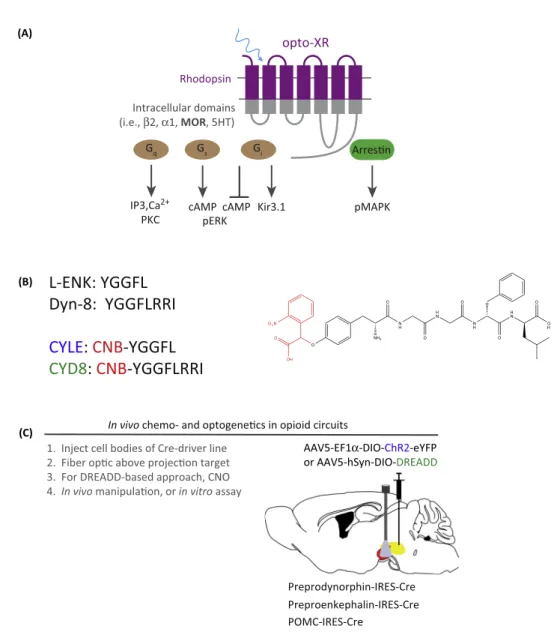
Thefieldofoptogeneticinnovationhasbeengrowingrapidly,withmostoftheeffortsbyprotein engineersfocusedondevelopingnovelchannelopsins,withshiftedkinetics,on/offrates,orion filters[58–60].However,afewgroups,includingourown,havebeenworkingtodevelopand characterizeGPCRversionsofoptogenetictools,whichwouldallowforspatiotemporal engage-mentofopioid signalinginvitro andinvivo[61–64].Capitalizingonthese recenteffortsvia molecular modeling in iTASSER (http://zhanglab.ccmb.med.umich.edu/I-TASSER/), the authorsgeneratedachimericreceptorthatcontainstheintracellularcomponents(loopsand Cterminus)oftheratMORfusedtothehydrophobicandextracellularcomponentsoftherat rhodopsinreceptor[62].Our goalwas todesign andimplementa photosensitiveMOR-like receptorthatrespondstolight-basedstimulationbysignalingtointracellularpathwayswiththe sameproperties ofits WTcounterpart.In this report[62],the authors demonstratedthat photostimulationresultsincanonicalMORsignalingasmeasuredbyinhibitionofcyclicAMP production,receptordesensitization,couplingtoG-protein-coupledinwardlyrectifying potas-sium(GIRK)channels,activationofmitogen-activatedproteinkinasecascades,andreceptor internalization. Furthermore, using this approach combined with cre-loxP mouse genetic approaches,theauthorsexpressedanAAV5–opto-MOR–YFPreceptorinGABAergicneurons ofthe ventral tegmentalarea (VTA) and found that photostimulation ofthis pathway was rewarding,duetoanopioid-likedisinhibitionofGABAergictone[65,66].Otherrecentreports haveusedexpressionofbothadrenergicandserotoninergicrhodopsin-likeopto-GPCRsin structuresincludingthedorsalrapheandbasolateralamygdalatoregulateanxietybehavior [61,64,67].Takentogether,theseresultssuggestnewversionsoftheopto-MOR,orother opioidreceptor rhodopsinchimeric approaches, could facilitatethe millisecondcontrol of opioidsignaling,andtherebyelucidateitsrelevancefortemporallyprecisebehaviorsindefined circuits.
Additionally,theresultsobtainedwithopto-MOR[62],alongwiththoseobtainedwiththeKOR– DREADD[52],highlightthepotentialtoengagemultipleopioidreceptorsignalingpathwaysin thesamecelltype.Giventhatmanyofthesereceptorsarecoexpressed,thesenewtoolsmay provideamultiplexedmethodfordissectingreceptorinteractions,signaling,andcircuitlevel effectsinvitro andinvivo.Future studies arewarrantedto determine howopto-opioid-like receptorsfunctioninperipheralcircuits,andwhethertheycanbefurthermutatedorenhancedto bettermimicendogenousopioidreceptorfunction,aswellastobetterdissecttheroleofbiased opioidreceptorsignalinginvivowithspatiotemporalcontrol[28,68,69].
OptopharmacologyforEngagingOpioid ReceptorSignalingwith SpatiotemporalPrecision
opioidreceptorsandtheirendogenousneuropeptidesare‘how,where,andwhen’endogenous peptidesactwithinintactneuralcircuits.Tobegintodissectthis,theresearchteamsofBernardo Sabatini and John T. Williams, led by efforts of Matthew Banghart and others [70], have developedtwoopioidagonistpeptideanalogs:[Leu5]-enkephalin(CYLE)andtheeightamino acidformofDynorphinA(CYDyn-8).TheseanalogscontainamodifiedN-terminal carboxyni-trobenzyl(CNB)chromophore,whichisreleasedatahighquantumefficiencyuponphotolysis. Importantly, these modified peptides are inert and functionally inactive in the absence of photolysis.However, whenexposedtoapulseofUVlight(405nm),theybecomefunctional andmaythenactivateendogenousopioidreceptors.BanghartandSabatini[70]haveshown robustinvitrodata,demonstratingm-opioidreceptor-coupledGIRKchannelcoupling, suggest-ingthatthesecompoundscanbeusedreliablyfordissectingMORfunction.
Furthermore,smallmoleculephotoswitchableopioidligandshavealsobeendevelopedthatcan acttoantagonizeendogenousorexogenousopioidreceptoragonistsinvitro[71]. Carboxyni-troveratryl-naloxone(CNV-NLX)wasgeneratedas acagedanalogofthecompetitiveopioid receptorantagonistnaloxone(NLX).Theauthors[71]investigateditsutilityinbothHEKcelland slicepreparations.TheyreportedthatCNV-NLX, withdermorphinastheagonist,canblock opioidreceptor-mediated GIRKchannel coupling after photo-uncaging. Interestingly, inthis reporttheauthorswereabletoutilizethisnoveltooltodemonstratethatsomeMORagonists havealternatedeactivationrates,whicharegovernedbytheirGproteinsignaling,yetothersare determinedbyagonistdissociationrate.Inanelegantcomplementarystudy,usingCYLEwith CNV-NLX,twodifferentalterationsinopioidsignalingweredeterminedinMORdesensitization withinlocuscoeruleusneurons[72].Theauthorconcludesthatopioidreceptordesensitizationis bothareductionin‘active’receptornumberaswellasadecreaseinagonistreceptoraffinityof theremainingreceptorpool.Therapidspatiotemporalcontrolofopioidligandsusing photo-uncagingaffordstheinvestigatortheabilitytoassesskineticsofassociationanddissociation. Further,photo-uncagingcouldrevealhowquicklyreceptor-inducedsignaling activation/deac-tivationfollowsfollowingreceptoroccupancybyligand.Theseapproachesarepowerful addi-tionstotheopioidreceptortoolboxinvitro,andperhapscouldeventuallybeusedforbehavioral and systems level experiments in vivo. In vivo photopharmacology has been a significant challengebecausedeliveryofUVlight todeepbrain structures,alongwith pharmacological infusionistechnicallychallenging,althoughnewwirelessdevicesthatcanco-deliverlightand drug simultaneouslymaybepromising inthisrespect [73].Inthis recentreport,delivery of opioids(DAMGO)wasdemonstratedusingamicrofluidicprobe.Extensionsofthistechnology usingUVLEDsorotherphotoswitchable ligands could transformour understandingofthe relationshipsbetweenopioidligandsandreceptoractivitywithinthespatiotemporalframework ofintactneuralcircuits[73].
RodentGeneticToolsforDissectingOpioid ReceptorFunctionInVivo
Additionalintersectional(e.g.,usingFLP-recombinase)viralandknock-inapproachesmay further provide a powerful tool set for dissecting the functions of subsets of neurons expressing opioid receptors and their ligands [83–85]. A recent example showed that selectivedeletionofKOR fromdopamine neurons (DATcre+) hasanxiolytic-likeproperties andalterscocaine-inducedplasticity[81].Inacomplementarystudy,Ehrichandcolleagues showedtherescueofKORinKORKOmice(usinganewCre-dependentKORvirus)onlyin DATcre+ cells restores KOR-mediated conditioned place aversion [86]. These types of experiments highlight both the genetic specificity and defined neural circuit contributions ofdistinctopioidreceptors.
RodentGeneticToolsforDissectingtheRolesofOpioidPeptidesInVivo
Extendingtheseconditionalapproachesforexaminingreceptorfunctionwithindiscretecelltypes havebeeneffortstodevelopconditionalKOmiceforeachendogenousopioidpeptide,alongwith Cre-driver micefor each, sothat neuronscontaining these peptidescan betargeted using chemogeneticandoptogeneticapproaches[56](Figure3).ConditionalKOmicefor proopiome-lanocortin,proenkephalin, andprodynorphin haveeachbeendevelopedandareintheinitial testingphasesforviabilityandphenotyping[87–89].Sinceeachoftheseprepropeptidesgenerate amultiplicityofactivepeptidespecies,deletingtheprecursorswillresultinthedeletionofmany activepeptides.Viralcre-recombinaseorINTERSECT[85]approachesarelikelytohaveimportant implicationswhenisolatingthecontributionsoftheseneuropeptidestobehavioralsystemslevel questions.Inparallel,cre-drivermiceofeachopioidpeptidewillbecomemorewidelyavailable. Thesewillallowforexpressionofopto-andchemogeneticactuatorsinneuronsexpressingopioid peptides.SuchCre-drivermicewouldbeusefulfordissectingtheroleofendogenousopioidtone, circuit-basedopioidreceptorfunction,howopioidsarereleased,andwhetherthereare promis-cuousopioidpeptide–receptorinteractions,ashavebeenhypothesizedandsuggestedoverthe pastseveralyears.Finally,severallaboratoriesarealsodevelopingreceptor-basedcre-drivermice (NOP-cre,MOR-cre,KOR-cre,DOR-cre) forisolatingpopulationsofcellsthatexpress opioid receptors.
Recenteffortshavebeguntousedynorphin-credrivermice[87,88]inbothchemogeneticand optogeneticexperimentstodefinetheendogenousnatureofdynorphinergictoneonfeeding behavior, reward, and aversion. The surprising findings thusfar include the observation of optogeneticallyevokedopioidneuropeptidereleaseafterrelativelymildstimulation,aswellas noncanonicalrolesfork-opioid-mediatedbehavioraleffects[87](Figure3).Bycontrast, proo-piomelanocortin-cre driver animals have beenuseful for targeting the arcuate nucleus and hippocampus[90],yetlittleisknownaboutwhethertheseneuronscanbeevokedtorelease proendorphin,theendogenousMORagonist,inanactivity-dependentmanner.Enkephalin-cre driver mice have recently beengenerated by the Allen Brain Institute, and are likelyto be widely used in studies of basal ganglia function, as well as in reward neurobiology and neuropharmacology.
Preprodynorphin-IRES-Cre
AAV5-EF1α-DIO-ChR2-eYFP
or AAV5-hSyn-DIO-DREADD
Preproenkephalin-IRES-Cre (A)
(B)
Rhodopsin
cAMP
Arresn
pMAPK pERK
opto-XR
Intracellular domains (i.e., β2, α1, MOR, 5HT)
cAMP G
i G
s G
q
Kir3.1 IP3,Ca2+
PKC
L-ENK: YGGFL
Dyn-8: YGGFLRRI
CYLE:
CNB
-YGGFL
CYD8
:
CNB
-YGGFLRRI
(C)
1. Inject cell bodies of Cre-driver line 2. Fiber opc above projecon target 3. For DREADD-based approach, CNO 4. In vivo manipulaon, or in vitro assay
In vivo chemo- and optogenecs in opioid circuits
POMC-IRES-Cre
Figure 3.Summary ofModern OptogeneticApproaches for Dissecting Opioid Peptide and Receptor FunctionInVitroandInVivo.(A)Cartoondepictingchimeric‘Opto-XR’approachinwhichrhodopsincDNAisfused withwild-typeG-protein-coupledreceptor(GPCR)cDNAintracellularloopsandtailtogenerateaphotosensitivereceptor systemcapableofspatiotemporalengagementofcanonicalGPCRsignalingpathwayssuchasGq,Gs,andGiorarrestin recruitmentinselectedcelltypeswhencombinedwithviralandgeneticapproachesinvivo.Opto-MORreceptors[62]take advantageofsimilaritiesbetweenRO4Gicoupledopsinsandm-opioidreceptors.(B)Left,One-letteraminoacidsequences ofLE,Dyn-8,andtheircorrespondingphotoswitchableCNB-modifiedanalogsCYLEandCYD8.Right,Thechemical structureofCYLE.TheCNBmoiety(photoswitch)ishighlightedinred.Adaptedfrom[70].(C)Summaryofcircuit-based chemogeneticandoptogenetictargetingapproachandavailableendogenousopioidcre-drivermice.Strategyisprovided usingadouble-invertedopenreadingframe(DIO)construct,andfiberopticsforoptogeneticmanipulation.For DREADD-basedapproaches,injectionandthenCNOmanipulationswouldoccurinvivoorinvitro.Extensionsofthisapproachare alsopossible,wherebyotheropsinsorDREADDscanbeusedformultiplexingorinhibitionexperiments.Abbreviations: CNB,carboxynitrobenzyl;CNO,clozapine-N-oxide;CYLE,[Leu5
]-enkephalin;DREADD,DesignerReceptorsExclusively ActivatedbyDesignerDrugs;Dyn-8,eightaminoacidformofDynorphinA;MOR,m-opioidreceptor.
ConcludingRemarks
Insummary,wehaveoutlinedsomeoftherecentadvancesintechnologythathaveallowedfora deeperandmorerigorousunderstandingofopioidreceptorneurobiologyandpharmacology. Wehavebynomeansprovidedacomprehensivesurveyofthisrapidlyprogressingfield,but insteadwehavefocusedonsomeoftheemergingtechniques,tools,andapproaches,which havethepotentialtounravelhistoricallycriticalmysteriesinthefield(seeOutstandingQuestions).
Clearly, improvements in high resolution structural determination of opioid receptors and complexesviacrystallographyalongwithenhancedGPCRmodelinganddockingapproaches [94]willprovidepowerfultemplatesforthestructure-guideddiscoveryofnovelopioidreceptor ligands.Additionalrefinementsofbothchemogeneticsandoptogeneticsaswellasviraldelivery platformswillprovidethetechnologiesforresolvinglong-standingissuesrelatedtocell type-specificactionsofopioidligandsandreceptors.Finally,developmentofsuitablydrug-likeand highlyG-protein-andb-arrestin-biasedligandsforallfouropioidreceptorswillbeextraordinarily valuableforelucidatingtherelativerolesofcanonicalversusnoncanonicalsignalingforthemany actionsmediatedbyexogenousandendogenousopioids.Althoughthefieldofopioidreceptor pharmacologywillalwaysrestonpharmacologicalmethodsandconceptsforitsfoundation, theseemergingtechnologiesprovideanunparalleledopportunityforaddressingkeyenigmasin opioidreceptorstructureandfunction.
References
1. Waldhoer,M.etal.(2004)Opioidreceptors.Annu.Rev.Biochem. 73,953–990
2. Pasternak,G.W.and Pan,Y.X.(2013)Muopioidsandtheir receptors:evolutionofaconcept.Pharmacol.Rev.65,1257– 1317
3. Martin,W.R.etal.(1976)Theeffectsofmorphineand morphine-like drugs in the non-dependent and morphine-dependent chronic spinal dog. J. Pharmacol. Exp. Ther. 197, 517–532 4. Robson, L.E. and Kosterlitz, H.W. (1979) Specific protection of the
binding sites of D-Ala2-D-Leu5-enkephalin (delta-receptors) and dihydromorphine (mu-receptors). Proc. R. Soc. Lond. B Biol. Sci. 205, 425–432
5. Kosterlitz, H.W. and Leslie, F.M. (1978) Comparison of the recep-tor binding characteristics of opiate agonists interacting with mu-or kappa-receptors. Br. J. Pharmacol. 64, 607–614 6. Evans, C.J. et al. (1992) Cloning of a delta opioid receptor by
functional expression. Science 258, 1952–1955
7. Chen, Y. et al. (1993) Molecular cloning and functional expression of a mu-opioid receptor from rat brain. Mol. Pharmacol. 44, 8–12 8. Li, S. et al. (1993) Molecular cloning and expression of a rat kappa
opioid receptor. Biochem. J. 295, 629–633
9. Mollereau, C. et al. (1994) ORL1, a novel member of the opioid receptorfamily.Cloning,functionalexpressionandlocalization. FEBSLett.341,33–38
10.Loh,H.H.etal.(1975)Opiatebindingtocerebrosidesulfate:amodel system for opiate–receptor interaction. Life Sci. 16, 1811–1817 11.Manglik, A. et al. (2012) Crystal structure of the micro-opioid
receptor bound to a morphinan antagonist. Nature 485, 321–326 12.Wu, H. et al. (2012) Structure of the human kappa-opioid receptor
in complex with JDTic. Nature 485, 327–332
13.Granier, S. et al. (2012) Structure of the delta-opioid receptor bound to naltrindole. Nature 485, 400–404
14.Thompson, A.A. et al. (2012) Structure of the nociceptin/orphanin FQ receptor in complex with a peptide mimetic. Nature 485, 395–399
15.Shoichet, B.K. and Kobilka, B.K. (2012) Structure-based drug screening for G-protein-coupled receptors. Trends Pharmacol. Sci. 33, 268–272
16.Filizola, M. and Devi, L.A. (2013) Grand opening of structure-guided design for novel opioids. Trends Pharmacol. Sci. 34, 6–12 17.Daga, P.R. et al. (2014) Structure-based virtual screening of the nociceptin receptor: hybrid docking and shape-based
approachesforimprovedhitidentification.J.Chem.Inf.Model. 54,2732–2743
18.Negri,A.etal.(2013)Discoveryofanovelselectivekappa-opioid receptoragonistusingcrystalstructure-basedvirtualscreening.J. Chem.Inf.Model.53,521–526
19.Vardy,E.etal.(2013)Chemotype-selectivemodesofactionof kappa-opioid receptor agonists. J. Biol. Chem. 288, 34470–34483 20.Urban, J.D. et al. (2007) Functional selectivity and classical con-cepts of quantitative pharmacology. J. Pharmacol. Exp. Ther. 320, 1–13
21.Kenakin, T. and Christopoulos, A. (2013) Signalling bias in new drug discovery: detection, quantification and therapeutic impact. Nat. Rev. Drug Discov. 12, 205–216
22.White, K.L. et al. (2014) Identification of novel functionally selective kappa-opioid receptor scaffolds. Mol. Pharmacol. 85, 83–90 23.Yan, F. et al. (2009) Structure-based design, synthesis, and
bio-chemical and pharmacological characterization of novel salvinorin A analogues as active state probes of the kappa-opioid receptor. Biochemistry 48, 6898–6908
24.Geyer, M.A. and Braff, D.L. (1987) Startle habituation and senso-rimotor gating in schizophrenia and related animal models. Schiz-ophr. Bull. 13, 643–668
25.White,K.L.etal.(2015)TheGprotein-biasedkappa-opioid recep-toragonistRB-64isanalgesicwithauniquespectrumofactivities invivo.J.Pharmacol.Exp.Ther.352,98–109
26.Kroeze, W.K. et al. (2015) PRESTO-Tango as an open-source resource for interrogation of the druggable human GPCRome. Nat. Struct. Mol. Biol. 22, 362–369
27.Morgenweck, J. et al. (2015) Investigation of the role of barrestin2 in kappa opioid receptor modulation in a mouse model of pruritus. Neuropharmacology 99, 600–609
28.Bruchas, M.R. and Chavkin, C. (2010) Kinase cascades and ligand-directed signaling at the kappa opioid receptor. Psycho-pharmacology (Berl.) 210, 137–147
29.Martin, W.R. (1979) History and development of mixed opioid agonists, partial agonists and antagonists. Br. J. Clin. Pharmacol. 7, 273S–279S
30.Pfeiffer, A. et al. (1986) Psychotomimesis mediated by kappa opiate receptors. Science 233, 774–776
31.Kumor, K.M. et al. (1986) Human psychopharmacology of keto-cyclazocine as compared with cyclazocine, morphine and pla-cebo.J.Pharmacol.Exp.Ther.238,960–968
OutstandingQuestions
Whatarethemoleculardetailsofopioid receptoractivation?
Dotheactivationstatesdiffer depend-inguponbindingbypeptideorsmall moleculeligands?
How do different ligands engage diverse intracellular signaling path-ways,andwhatisthestructuralbasis forfunctionalselectivity?
How,where,andwhatpeptidespecies arereleaseduponneuronalstimulation andaredifferentspeciesdifferentially released?
32.Roth,B.L.etal.(2002)SalvinorinA:apotentnaturallyoccurring nonnitrogenouskappaopioidselectiveagonist.Proc.Natl.Acad. Sci.U.S.A.99,11934–11939
33.Iwamoto, E.T. (1985) Place-conditioning properties of mu, kappa, and sigma opioid agonists. Alcohol Drug Res. 6, 327–339 34.Todtenkopf, M.S. et al. (2004) Effects of kappa-opioid receptor
ligands on intracranial self-stimulation in rats. Psychopharmacol-ogy (Berl.) 172, 463–470
35.Lohse, M.J. et al. (1990) b-Arrestin: a protein that regulates beta-adrenergic receptor function. Science 248, 1547–1550 36.Lohse, M.J. et al. (1992) Receptor-specific desensitization with
purified proteins. Kinase dependence and receptor specificity of b -arrestin and arrestin in the b2-adrenergic receptor and rhodopsin systems. J. Biol. Chem. 267, 8558–8564
37.Huang, W. et al. (2015) Structural insights into micro-opioid recep-tor activation. Nature 524, 315–321
38.Sounier, R. et al. (2015) Propagation of conformational changes during m-opioid receptor activation. Nature 524, 375–378 39.Steyaert, J. and Kobilka, B.K. (2011) Nanobody stabilization of G
protein-coupled receptor conformational states. Curr. Opin. Struct.Biol.21,567–572
40.Fenalti,G.etal.(2014)Molecularcontrolofdelta-opioidreceptor signalling.Nature506,191–196
41.Fenalti, G. et al. (2015) Structural basis for bifunctional peptide recognition at human delta-opioid receptor. Nat. Struct. Mol. Biol. 22, 265–268
42.O’Connor, C. et al. (2015) NMR structure and dynamics of the agonist dynorphin peptide bound to the human kappa opioid receptor. Proc. Natl. Acad. Sci. U.S.A. 112, 11852–11857 43.Rasmussen, S.G. et al. (2011) Crystal structure of the
b2adrener-gic receptor–Gs protein complex. Nature 477, 549–555 44.Vardy, E. and Roth, B.L. (2013) Conformational ensembles in
GPCR activation. Cell 152, 385–386
45.Kruse, A.C. et al. (2013) Activation and allosteric modulation of a muscarinic acetylcholine receptor. Nature 504, 101–106 46.Katritch, V. et al. (2014) Allosteric sodium in class A GPCR
signal-ing. Trends Biochem. Sci. 39, 233–244
47.Gutierrez-de-Teran, H. et al. (2013) The role of a sodium ion binding site in the allosteric modulation of the A(2A) adenosine G protein-coupled receptor. Structure 21, 2175–2185 48.Zhang,C.etal.(2012)High-resolutioncrystalstructureofhuman
protease-activatedreceptor1.Nature492,387–392 49.Vardy,E.etal.(2015)Singleaminoacidvariationunderlies
spe-cies-specific sensitivity to amphibian skin-derived opioid-like pep-tides. Chem. Biol. 22, 764–775
50.Pert, C.B. et al. (1973) Opiate agonists and antagonists discrimi-nated by receptor binding in brain. Science 182, 1359–1361 51.Simon, E.J. and Groth, J. (1975) Kinetics of opiate receptor
inactivation by sulfhydryl reagents: evidence for conformational change in presence of sodium ions. Proc. Natl. Acad. Sci. U.S.A. 72, 2404–2407
52.Vardy, E. et al. (2015) A new DREADD facilitates the multiplexed chemogenetic interrogation of behavior. Neuron 86, 936–946 53.Armbruster, B.N. et al. (2007) Evolving the lock to fit the key to
create a family of G protein-coupled receptors potently acti-vated by an inert ligand. Proc. Natl. Acad. Sci. U.S.A. 104, 5163–5168
54.Marchant, N.J. et al. (2016) Behavioral and physiological effects of a novel kappa-opioid receptor-based DREADD in rats. Neuro-psychopharmacology 41, 402–409
55.Denis,R.G.etal.(2015)Palatabilitycandrivefeedingindependent ofAgRPneurons.CellMetab.22,646–657
56.Urban,D.J.andRoth,B.L.(2015)DREADDs(DesignerReceptors ExclusivelyActivatedbyDesignerDrugs):chemogenetictoolswith therapeuticutility.Annu.Rev.Pharmacol.Toxicol.55,399–417 57.English, J.G. and Roth, B.L. (2015) Chemogenetics –a
transfor-mational and translational platform. JAMA Neurol. 72, 1361–1366 58.Deisseroth, K. (2011) Optogenetics. Nat. Methods 8, 26–29 59.Berndt, A. and Deisseroth, K. (2015) OPTOGENETICS. Expanding
the optogenetics toolkit. Science 349, 590–591
60.Berndt,A.etal.(2014)Structure-guidedtransformationofchannel rhodopsinintoalight-activatedchloridechannel.Science344, 420–424
61.Siuda, E.R. et al. (2015) Optodynamic simulation of beta-adrener-gic receptor signalling. Nat. Commun. 6, 8480
62.Siuda, E.R. et al. (2015) Spatiotemporal control of opioid signaling and behavior. Neuron 86, 923–935
63.Airan, R.D. et al. (2009) Temporally precise in vivo control of intracellular signalling. Nature 458, 1025–1029
64.Masseck, O.A. et al. (2014) Vertebrate cone opsins enable sus-tained and highly sensitive rapid control of Gi/o signaling in anxiety circuitry. Neuron 81, 1263–1273
65.Tan, K.R. et al. (2012) GABA neurons of the VTA drive conditioned place aversion. Neuron 73, 1173–1183
66.van Zessen, R. et al. (2012) Activation of VTA GABA neurons disrupts reward consumption. Neuron 73, 1184–1194 67.McCall, J.G. et al. (2015) CRH engagement of the locus coeruleus
noradrenergic system mediates stress-induced anxiety. Neuron 87, 605–620
68.Bohn, L.M. et al. (2015) Exploring the biology of G protein-coupledreceptorsfrominvitrotoinvivo.Mol.Pharmacol.88, 534–535
69.Roth,B.L.andKroeze,W.K.(2015)Integratedapproachesfor genome-wide interrogation of the druggable non-olfactory G pro-tein-coupled receptor superfamily. J. Biol. Chem. 290, 19471–
19477
70.Banghart, M.R. and Sabatini, B.L. (2012) Photoactivatable neuro-peptides for spatiotemporally precise delivery of opioids in neural tissue. Neuron 73, 249–259
71.Banghart, M.R. et al. (2013) Caged naloxone reveals opioid sig-naling deactivation kinetics. Mol. Pharmacol. 84, 687–695 72.Williams, J.T. (2014) Desensitization of functional micro-opioid
receptors increases agonist off-rate. Mol. Pharmacol. 86, 52–61 73.Jeong, J.W. et al. (2015) Wireless optofluidic systems for pro-grammable in vivo pharmacology and optogenetics. Cell 162, 662–674
74.Ansonoff, M.A. et al. (2006) Antinociceptive and hypothermic effects of Salvinorin A are abolished in a novel strain of kappa-opioid receptor-1 knockout mice. J. Pharmacol. Exp. Ther. 318, 641–648
75.Kas,M.J.etal.(2004)Mu-opioidreceptorknockoutmiceshow diminishedfood-anticipatoryactivity.Eur.J.Neurosci.20,1624–
1632
76.Contet,C.etal.(2006)Dissociationofanalgesicandhormonal responsestoforcedswimstressusingopioidreceptorknockout mice. Neuropsychopharmacology 31, 1733–1744
77.Sharifi, N. et al. (2001) Generation of dynorphin knockout mice. Brain Res. Mol. Brain Res. 86, 70–75
78.Koster, A. et al. (1999) Targeted disruption of the orphanin FQ/ nociceptin gene increases stress susceptibility and impairs stress adaptation in mice. Proc. Natl. Acad. Sci. U.S.A. 96, 10444–
10449
79.Brady, L.S. et al. (1999) Region-specific up-regulation of opioid receptor binding in enkephalin knockout mice. Brain Res. Mol. Brain Res. 68, 193–197
80.Gaveriaux-Ruff, C. et al. (2011) Genetic ablation of delta opioid receptors in nociceptive sensory neurons increases chronic pain and abolishes opioid analgesia. Pain 152, 1238–1248 81.Van’t Veer, A. et al. (2013) Ablation of kappa-opioid receptors from
brain dopamine neurons has anxiolytic-like effects and enhances cocaine-induced plasticity. Neuropsychopharmacology 38, 1585–1597
82.Weibel,R.etal.(2013)Muopioidreceptorsonprimaryafferent nav1.8neuronscontributetoopiate-inducedanalgesia:insight from conditional knockout mice. PLoS ONE 8, e74706
83.Ray, R.S. et al. (2011) Impaired respiratory and body temperature control upon acute serotonergic neuron inhibition. Science 333, 637–642
85.Fenno,L.E.etal.(2014)Targetingcellswithsinglevectorsusing multiple-featureBooleanlogic.Nat.Methods11,763–772 86.Ehrich,J.M.etal.(2015)Kappaopioidreceptor-inducedaversion
requires p38 MAPK activation in VTA dopamine neurons. J. Neu-rosci. 35, 12917–12931
87.Al-Hasani, R. et al. (2015) Distinct subpopulations of nucleus accumbens dynorphin neurons drive aversion and reward. Neuron 87, 1063–1077
88.Krashes, M.J. et al. (2014) An excitatory paraventricular nucleus to AgRP neuron circuit that drives hunger. Nature 507, 238–242 89.McHugh, T.J. et al. (2007) Dentate gyrus NMDA receptors mediate
rapid pattern separation in the hippocampal network. Science 317, 94–99
90.Garfield, A.S. et al. (2015) A neural basis for melanocortin-4 receptor-regulated appetite. Nat. Neurosci. 18, 863–871
91.Ozawa,A.etal.(2015)Knock-inmicewithNOP-eGFPreceptors identifyreceptorcellularandregionallocalization.J.Neurosci.35, 11682–11693
92.Scherrer, G. et al. (2009) Dissociation of the opioid receptor mechanisms that control mechanical and heat pain. Cell 137, 1148–1159
93.Bardoni, R. et al. (2014) Delta opioid receptors presynaptically regulate cutaneous mechanosensory neuron input to the spinal cord dorsal horn. Neuron 81, 1312–1327
94.Huang, X.P. et al. (2015) Allosteric ligands for the pharmacologi-cally dark receptors GPR68 and GPR65. Nature 527, 477–483 95.Ballesteros, J.A. and Weinstein, H. (1995) Integrated methods for