Outcome prediction models in AQP4-IgG
positive neuromyelitis optica spectrum
disorders
Jacqueline Palace,
1,* Dan-Yu Lin,
2Donglin Zeng,
2Masoud Majed,
3,4Liene Elsone,
5Shahd Hamid,
5Silvia Messina,
1Tatsuro Misu,
6Jessica Sagen,
7Daniel Whittam,
5Yoshiki Takai,
6Maria Isabel Leite,
1Brian Weinshenker,
3Philippe Cabre,
8Anu Jacob,
5Ichiro Nakashima,
6Kazuo Fujihara
6,9and Sean J. Pittock
3,4,*
*These authors contributed equally to this work.
Pathogenic antibodies targeting the aquaporin-4 water channel on astrocytes are associated with relapsing inflammatory neuro-myelitis optica spectrum disorders. The clinical phenotype is characterized by recurrent episodes of optic neuritis, longitudinally extensive transverse myelitis, area postrema attacks and less common brainstem and cerebral events. Patients often develop major residual disability from these attacks, so early diagnosis and initiation of attackpreventing medications is important. Accurate prediction of relapse would assist physicians in counselling patients, planning treatment and designing clinical trials. We used a large multicentre dataset of 441 patients from the UK, USA, Japan and Martinique who collectively experienced 1976 attacks, and applied sophisticated mathematical modelling to predict likelihood of relapse and disability at different time points. We found that Japanese patients had a lower risk of subsequent attacks except for brainstem and cerebral events, with an overall relative relapse
risk of 0.681 (P= 0.001) compared to Caucasians and African patients, who had a higher likelihood of cerebral attacks, with a
relative relapse risk of 3.309 (P= 0.009) compared to Caucasians. Female patients had a higher chance of relapse than male
patients (P= 0.009), and patients with younger age of onset were more likely to have optic neuritis relapses (P50.001).
Immunosuppressant drugs reduced and multiple sclerosis disease-modifying agents increased the likelihood of relapse
(P50.001). Patients with optic neuritis at onset were more likely to develop blindness (P50.001), and those with older age
of onset were more likely to develop ambulatory disability. Only 25% of long-term disability was related to initial onset attack, indicating the importance of early attack prevention. With respect to selection of patients for clinical trial design, there would be no gain in power by selecting recent onset patients and only a small gain by selecting patients with recent high disease activity. We provide risk estimates of relapse and disability for patients diagnosed and treated with immunosuppressive treatments over the subsequent 2, 3, 5 and 10 years according to type of attack at onset or the first 2-year course, ethnicity, sex and onset age. This study supports significant effects of onset age, onset phenotype and ethnicity on neuromyelitis optica spectrum disorders outcomes. Our results suggest that powering clinical treatment trials based upon relapse activity in the preceding 2 years may offer little benefit in the way of attack risk yet severely hamper clinical trial success.
1 Nuffield Department of Clinical Neurosciences, Oxford, UK
2 Department of Biostatistics, University of North Carolina, Chapel Hill, NC 27599–7420, USA
3 Department of Neurology, Mayo Clinic College of Medicine, 200 First Street S.W., Rochester, Minnesota 55905, USA
4 Laboratory Medicine and Pathology, Mayo Clinic College of Medicine, 200 First Street S.W., Rochester, Minnesota 55905, USA 5 The Walton Centre, NHS Foundation Trust, Liverpool, UK
6 Department of Neurology, Tohoku University Graduate School of Medicine, Sendai, Japan
7 Clinical Research Unit, Mayo Clinic College of Medicine, 200 First Street S.W., Rochester, Minnesota 55905, USA
Received September 21, 2018. Revised December 23, 2018. Accepted January 13, 2019. Advance Access publication April 1, 2019
ßThe Author(s) (2019). Published by Oxford University Press on behalf of the Guarantors of Brain.
8 Department of Neurology, Fort-de-France University Hospital Center, Pierre Zobda Quitman Hospital, Fort-de-France, Martinique, France
9 Department of Multiple Sclerosis Therapeutics, Fukushima Medical University School of Medicine and Multiple Sclerosis and Neuromyelitis Optica Center, Southern TOHOKU Research Institute for NeuroScience, Koriyama, Japan
Correspondence to: Sean J. Pittock, MD
Mayo Clinic, Department of Neurology, 200 First Street S.W., Rochester, MN 55905, USA E-mail: [email protected]
Correspondence may also be addressed to: Jacqueline Palace, FRCP Nuffield Department of Clinical Neurosciences, Oxford, UK E-mail: [email protected]
Keywords:neuromyelitis optica; aquaporin-4; outcome prediction; disability
Abbreviations:EDSS = Expanded Disability Status Scale; IST = immunosuppressive treatment; NMOSD = neuromyelitis optica
spectrum disorders
Introduction
The neuromyelitis optica spectrum disorders (NMOSD) are autoimmune, inflammatory disorders of the CNS with a predilection for the optic nerves and spinal cord and are distinct from multiple sclerosis (Wingerchuk et al., 2007). The majority of patients have antibodies to aquaporin-4 (AQP4) water channels, which are situated predominantly on astrocyte foot processes; hence AQP4-IgG-positive NMOSD is now recognized as an autoimmune astrocyto-pathy with secondary demyelination (Lennon et al., 2004, 2005). The AQP4-IgG seronegative group (considered sero-negative NMOSD) likely represents a heterogeneous group of both monophasic and relapsing inflammatory CNS dis-orders that include post-infectious inflammation and condi-tions caused by unidentified antibodies (Wingerchuket al., 2015). Recently some patients in this group have been re-ported to be positive for antibodies targeting myelin oligo-dendrocyte glycoprotein (MOG) and these patients have a primary demyelinating disorder (Waterset al., 2015; Jarius
et al., 2016; Peschl et al., 2017).
There are important differences between those who are seropositive for AQP4-IgG and MOG-IgG. AQP4-IgG-posi-tive NMOSD in contrast to MOG-antibody-associated disor-ders (MOGAD) is far more common in females (Queket al., 2012), has a non-Caucasian ethnic bias, commonly co-asso-ciates with other autoantibodies and diseases, is relapsing if untreated and is associated with significant morbidity and mortality from relapse-related disability (Kitleyet al., 2014b; Flanagan et al., 2016; Jurynczyk et al., 2017; Cobo-Calvo
et al., 2018; Jitprapaikulsan et al., 2018a, b). Additionally, current immunosuppressive treatments (ISTs), such as pred-nisolone, azathioprine, mycophenolate mofetil and rituximab, may not suppress the disease adequately. Thus there is a growing interest in developing new treatments and currently there are three immune-modulatory drugs being tested in international multicentre phase 4 randomized controlled trials: inebilizumab (anti-CD19 monoclonal antibody target-ing B cells), eculizumab (anti-C5 monoclonal antibody
targeting complement) and two utilizing satralizumab (anti-IL-6R monoclonal antibody targeting T and B cell acti-vation, Th17 differentiation, and plasmablast survival). These studies enrol exclusively or primarily patients with AQP4 antibodies.
Because the outcomes across patients with AQP4-IgG-posi-tive NMOSD are heterogeneous and because data that permit power calculations for clinical trials are sparse (Weinshenker
et al., 2015), we combined datasets from five centres with detailed prospective data collection systems, across four coun-tries with varied ethnicities. We developed a joint modelling framework to understand the factors that influence relapses and disability and predict future attacks and disability events.
Materials and methods
Patient cohort
An international database was created by merging prospectively collected datasets from five neuromyelitis optica (NMO) specia-lized centres: Oxford and Liverpool (UK), Mayo Clinic (USA), Sendai (Japan) and Martinique. Information collected included sex, ethnicity, onset attack type (optic neuritis, transverse myelitis, brainstem attack, cerebral, and mixed), age at onset, Expanded Disability Status Scale (EDSS) scores and visual acuity and chronic immunomodulatory treatment. All data were anon-ymized and satisfied the local ethics requirements.
Analysis
the random effects vary among patients, representing the pa-tient characteristics that are not captured by the measured covariates. The mean of the random effect in the population is set to zero. A patient with a positive value of the random effect tends to have more attacks than an average patient, whereas a patient with a negative value of the random effect tends to have fewer attacks than an average patient. The vari-ance of the random effect reflects the degree of heterogeneity, with a larger value indicating greater heterogeneity.
We formulated the effects of baseline covariates and treat-ment histories on the hazard function of a disability event through a proportional hazards model (Cox, 1972) in which the shared random effect from the model for recurrent attacks enters as an additional covariate. The regression coefficient for the shared random effect captures the dependence of disability on recurrent attacks. This joint modelling approach allows us to assess the effects of baseline covariates and time-dependent covariates (e.g. treatments) on the rate of recurrence for each type of NMO attack and on the risk of occurrence for each type of disability while accounting for the patient heterogeneity that is not accounted for by the measured covariates. It also allows us to predict future attacks and disability events using not only the baseline characteristics and treatment histories but also the event histories. This model uses a time-dependent ana-lysis which allows for any variation in follow-up times across the different covariate subgroups. The mathematical formula-tion, estimation procedure, and prediction algorithm are de-tailed in the Supplementary material.
For the baseline covariates we included ethnicity, sex, age at disease onset, and baseline attack type. We classified patients according to five major ethnicity groups: Caucasian, African (includes all those of African descent), Hispanic, Japanese, and non-Japanese Asian. We combined Hispanic and unknown ethnicity with Caucasian, which serves as the reference. We divided patients into three age groups according to the tertiles:
435 years, 35–48 years, and448 years, with the last tertile as the reference. Such tertiles were used as a balanced distri-bution yields more stable estimates. The clinical and demo-graphic characteristics of each tertile are shown in Supplementary Table 1. For the baseline attack, we combined brainstem and cerebral and set transverse myelitis as the reference.
There were two time-dependent covariates: ISTs were com-bined into one group (chronic prednisolone/prednisone, azathioprine, mycophenolate, rituximab, methotrexate or any combination) and were allocated the value of 0 before initi-ation of the IST treatment and the value of 1 afterwards; licensed multiple sclerosis disease-modifying treatments were combined as one group (multiple sclerosis disease-modifying treatment: mainly interferon beta and glatiramer acetate; anti-CD20 monoclonals were not included in this group) and were allocated the value of 1 between the starting and stop-ping dates and the value of 0 otherwise. No patients were treated with natalizumab or ocrelizumab. The treatment com-parisons pertain to IST versus no treatment and multiple scler-osis disease-modifying treatments versus no treatment.
We combined some types of events in order to increase power and stability. Specifically, we combined unilateral and bilateral optic neuritis, classified unknown attack types (13 of 1976 events, 0.7%) as transverse myelitis attacks, which were the most common type, used the composite endpoint of
one-eye blindness and two-one-eye blindness and the composite end-point of EDSS 8.0 and death.
The results are based on the joint model with four types of NMO attacks (i.e. optic neuritis, transverse myelitis, brain-stem, and cerebral) and the disability events of blindness and EDSS 6.0, except for the results on EDSS 8.0/death, which are based on a second joint model with EDSS 6.0 replaced by EDSS 8.0/death, and for the results on all relapses, which com-bine the four types of NMO attacks into a single sequence of recurrent events. If a patient had a mix of optic neuritis and transverse myelitis, then he/she would contribute to both types of events in the analysis.
Data availability
The data that support the findings of this study are available from the corresponding authors, upon request.
Results
Clinical and demographic
characteristics of the international
NMOSD attack database
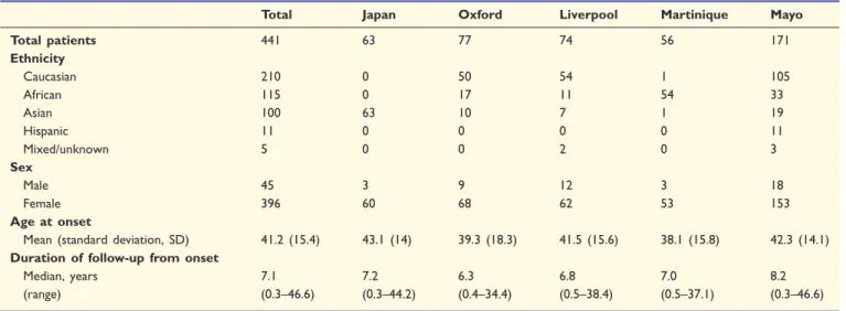
A total of 441 AQP4-IgG-positive NMOSD patients from the five sites were included (Table 1). There were 396 fe-males and 45 fe-males. The age of onset ranged from 2.7 to 82.7 years, with a median of 40.8 years. Over a median disease duration of 7.1 years (range: 0.3–46.6), 1976 at-tacks were documented. Supplementary Table 2 shows the frequencies and types of attacks and disability outcomes during the disease course according to baseline characteris-tics, such as attack type, site and ethnicity.
Effects of age, sex, ethnicity and
treatment on likelihood of relapse
(P50.001), with greater effects on optic neuritis, trans-verse myelitis and cerebral attacks (Fig. 1), whereas mul-tiple sclerosis disease-modifying treatments increased the risk of relapse. Supplementary Fig. 1 shows that the risk of relapse decreases over time, most dramatically after 10 years.
Effects of age, sex and ethnicity on
likelihood of developing disability or
blindness
The estimation results for the effects of covariates on the likelihood of developing disability (EDSS56) or blindness
Table 1 Demographics of AQP4-IgG-positive NMOSD patients
Total Japan Oxford Liverpool Martinique Mayo
Total patients 441 63 77 74 56 171
Ethnicity
Caucasian 210 0 50 54 1 105
African 115 0 17 11 54 33
Asian 100 63 10 7 1 19
Hispanic 11 0 0 0 0 11
Mixed/unknown 5 0 0 2 0 3
Sex
Male 45 3 9 12 3 18
Female 396 60 68 62 53 153
Age at onset
Mean (standard deviation, SD) 41.2 (15.4) 43.1 (14) 39.3 (18.3) 41.5 (15.6) 38.1 (15.8) 42.3 (14.1) Duration of follow-up from onset
Median, years 7.1 7.2 6.3 6.8 7.0 8.2
(range) (0.3–46.6) (0.3–44.2) (0.4–34.4) (0.5–38.4) (0.5–37.1) (0.3–46.6)
Table 2 Estimation of the effects of covariates on the rates of recurrence for attacks
Optic neuritis Transverse myelitis Brainstem Cerebral All
Covariate Rate ratio P-value Rate ratio P-value Rate ratio P-value Rate ratio P-value Rate ratio P-value Site ethnicity
African 0.968 (0.188) 0.865 0.963 (0.120) 0.761 0.813 (0.310) 0.588 3.309 (1.517) 0.009 1.003 (0.099) 0.974 Japanese 0.587 (0.141) 0.026 0.588 (0.092) 0.001 1.651 (0.575) 0.150 1.719 (0.921) 0.312 0.681 (0.076) 0.001 Non-Japanese Asian 1.124 (0.292) 0.653 0.921 (0.178) 0.669 0.617 (0.402) 0.458 0.717 (0.631) 0.706 1.010 (0.138) 0.940 USA Caucasian
and others
1 – 1 – 1 – 1 – 1 –
Sex
Female 0.903 (0.145) 0.524 1.501 (0.167) 50.001 0.860 (0.316) 0.681 1.596 (0.711) 0.294 1.209 (0.087) 0.009
Male 1 – 1 – 1 – 1 – 1 –
Age, years
435 2.078 (0.393) 50.001 0.872 (0.114) 0.295 1.767 (0.643) 0.118 0.823 (0.414) 0.698 1.090 (0.104) 0.365 35–48 1.468 (0.296) 0.057 0.790 (0.105) 0.075 1.942 (0.762) 0.091 1.507 (0.765) 0.419 0.930 (0.090) 0.456
448 1 – 1 – 1 – 1 – 1 –
Baseline attack
Optic neuritis 1.608 (0.270) 0.005 0.836 (0.103) 0.146 0.838 (0.306) 0.627 1.145 (0.509) 0.761 1.026 (0.096) 0.783 Brainstem/cerebral 1.686 (0.498) 0.077 1.084 (0.216) 0.685 3.903 (1.636) 0.001 2.929 (1.518) 0.038 1.287 (0.197) 0.098 Mixed 0.992 (0.293) 0.978 0.893 (0.139) 0.470 1.719 (0.750) 0.215 2.284 (1.180) 0.110 0.936 (0.114) 0.584
Transverse myelitis 1 – 1 – 1 – 1 – 1 –
Treatment
IST 0.662 (0.064) 50.001 0.611 (0.042) 50.001 0.900 (0.220) 0.665 0.486 (0.172) 0.042 0.668 (0.028) 50.001 MS-DMT 1.325 (0.242) 0.124 1.382 (0.142) 0.002 0.672 (0.377) 0.479 1.941 (0.852) 0.131 1.383 (0.107) 50.001
No treatment 1 – 1 – 1 – 1 – 1 –
Standard errors are shown in parentheses.
(in terms of hazard ratio) are summarized in Table 3. African patients were more likely to develop blindness and Japanese patients least likely to reach EDSS 8.0/ death; both differences were significant compared to the reference Caucasian group. Females had higher risk for all types of disability events. Patients with a younger age of onset were not only more likely to develop recurrent optic neuritis but also had a higher likelihood of developing blindness. They had a lower risk of developing EDSS 6.0 and EDSS 8.0 or death. In contrast, patients in the oldest age of onset group had a significantly higher likelihood of having ambulatory disability (need for cane or wheelchair) compared with those in the two younger age groups.
Patients with optic neuritis or mixed onset attacks were more likely to develop blindness (P50.001). Although ISTs were associated with a lower likelihood and multiple sclerosis disease-modifying agents were associated with a higher likelihood of blindness and EDSS 6.0, neither reached statistical significance. In contrast, both ISTs and multiple sclerosis disease-modifying agents were associated with a higher likelihood of reaching EDSS58 although there were fewer events.
The estimation results for the random effects are pre-sented in Supplementary Table 3. The variances of the shared and type-specific random effects are all quite large, indicating strong patient heterogeneity (due to unobserved
confounders) in recurrent NMO attacks, especially for optic neuritis and transverse myelitis attacks. In addition, recurrent NMO attacks substantially increase the risks for all types of disability. Specifically, the coefficients of the shared random effect for blindness, EDSS 6.0, and EDSS 8.0/death are all estimated at 1.5, indicating that a half unit change in the shared random effect for recurrent at-tacks would double the risk of each type of disability event.
Relationship between onset attack
and significant long-term disability
Only 25% of patients who experienced EDSS56 reached that disability milestone due to the onset attack. Only 17% of patients experiencing EDSS 8.0 reached that disability milestone due to the onset attack. For patients developing blindness in one or both eyes, 41% and 21%, respectively, reached that level of disability due to the onset attack. Thus, most patients require multiple attacks in order to acquire significant disability.
Predicting risk of relapse and disability
based on age, sex and historical attack
frequency and phenotype
Our model can be used to predict the outcomes of individual patients according to their characteristics. For example,
Supplementary Fig. 2 displays the estimated cumulative inci-dence functions of any relapse and an optic neuritis attack after Year 2 for a Japanese female patient who was diagnosed with a transverse myelitis attack at age 40 and did not receive mul-tiple sclerosis disease-modifying treatment. The IST treatment reduces the incidence of any relapse and optic neuritis attack, while having two optic neuritis attacks in the first 2 years substantially increases the incidence of future NMO attacks, especially optic neuritis attacks. As a second example, Supplementary Fig. 3 displays the estimated cumulative inci-dence functions of blindness and EDSS 6.0 after Year 2 for a Caucasian male patient who was diagnosed at age 30, started IST at disease onset, and did not receive multiple sclerosis dis-ease-modifying agents. If such a patient was diagnosed with an optic neuritis attack as opposed to a transverse myelitis attack at disease onset, then his risk of blindness is increased by nearly 3-fold and his risk of EDSS 6.0 is reduced by nearly 40%. Having two new optic neuritis attacks in the first 2 years increases both the risks of blindness and EDSS 6.0 considerably.
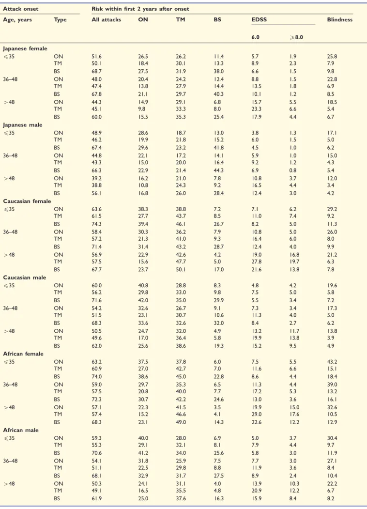
Outcome prediction tables: a helpful
tool for clinicians and patients.
Tables 4–6 provide estimates for the risks of recurrent at-tacks and disability events over time for patients who were treated (i.e. with immunosuppressants from onset and
Table 3 Estimation of the effects of covariates on the occurrence of disability events
Blindness EDSS 6.0 EDSS 8.0/death
Covariate Hazard ratio P-value Hazard ratio P-value Hazard ratio P-value
Site ethnicity
African 1.724 (0.248) 50.001 1.059 (0.280) 0.828 0.876 (0.306) 0.704 Japanese 0.853 (0.160) 0.395 0.799 (0.274) 0.514 0.297 (0.154) 0.020 Non-Japanese Asian 1.375 (0.270) 0.105 0.829 (0.316) 0.622 0.628 (0.347) 0.400
USA Caucasian and others 1 – 1 – 1 –
Sex
Female 1.633 (0.258) 0.002 1.522 (0.290) 0.027 1.508 (0.349) 0.076
Male 1 – 1 – 1 –
Age, years
435 1.490 (0.229) 0.009 0.332 (0.090) 50.001 0.339 (0.119) 0.002
35–48 1.284 (0.211) 0.128 0.522 (0.135) 0.012 0.270 (0.109) 0.001
448 1 – 1 – 1 –
Baseline attack
Optic neuritis 3.810 (0.561) 50.001 0.622 (0.162) 0.068 0.832 (0.275) 0.578 Brainstem/cerebral 1.253 (0.446) 0.527 0.726 (0.270) 0.389 0.663 (0.359) 0.448
Mixed 2.153 (0.420) 50.001 0.829 (0.246) 0.526 0.801 (0.321) 0.580
Transverse myelitis 1 – 1 – 1 –
Treatment
IST 0.735 (0.126) 0.072 0.708 (0.141) 0.082 1.647 (0.409) 0.045
MS-DMT 1.498 (0.399) 0.129 1.511 (0.502) 0.214 1.865 (0.862) 0.177
No treatment 1 – 1 – 1 –
Standard errors are shown in parentheses.
not multiple sclerosis disease-modifying treatments). We considered 54 combinations of baseline characteristics: ethnicity (Japanese, Caucasian, African), sex, age group (535, 35–48, 448), and onset attack type (optic neuritis, transverse myelitis, brainstem). Table 4 provides risk esti-mates over the first 2 years. Table 5 and Supplementary Table 4 provide risk estimates at Year 2 and over the sub-sequent 2, 3, 5 and 10 years for the patients who have not reached the disability endpoints at Year 2. Table 5 and Supplementary Table 4 pertain to patients without any re-lapse within 2 years after the onset attack. Table 6 and Supplementary Table 5 provide risk estimates at Year 2 over the subsequent 2, 3, 5 and 10 years for the patients who have not reached the disability endpoints at Year 2. Table 6 and Supplementary Table 5 pertain to patients who had one relapse in the first 2 years after the onset attack. These prediction tables can be used in the clinic setting to inform physicians and patients on the choice of initial treatment.
Optimization of patient recruitment
for clinical trials
Our data can be used to more accurately power future clinical trials. It is noteworthy that because disability is solely relapse-related in NMO and because such disability may be severe (and in contrast to multiple sclerosis), pri-mary outcome measures in phase 3 NMO clinical trials to date, have been time to first relapse only (for ethical rea-sons) as a surrogate for disability. Thus we estimated the risk of relapsing over a 1- and 2-year period to aid trial design. Because there are limited restrictions that can eth-ically be applied to clinical trial eligibility (e.g. sex and ethnicity criteria would not be acceptable inclusion cri-teria), we selected baseline characteristics such as prior re-lapse activity and disease duration to calculate risk of relapse over the subsequent 1 and 2 years. Table 7 shows the estimated proportion of patients who relapse over a 1-year and 2-1-year period, dependent on the disease duration category and the number of NMO attacks in the preceding 2 years for those on IST because we assumed all diagnosed AQP4-IgG-positive patients will be on IST. A patient with disease duration of55 years and with at least three attacks in the past 2 years has 73% chance of relapse within the next 2 years (representing 2.7% of the NMO population), whereas any patient (no restriction) has a 54% chance of relapse in 2 years (representing 100% of the population). Such information is useful for powering future clinical trials. The percentage of total AQP4-IgG-positive NMOSD patients who would satisfy these criteria is also included in the table because there will be a trade-off be-tween selecting a rare group of highly active patients versus a broader range of less active patients; the former is favour-able for statistical powering purposes whereas the latter favours ease of recruitment and a broader licensing indica-tion. The loss of study eligible patients would be more
deleterious to an effective study design than loading the study with highly active patients. For example, there ap-pears no overall advantage in selecting recent onset patients or patients with higher disease activity in the preceding 2 years (used as a criteria for the currently ongoing three randomized control trials) because the increase in disease activity is modest and the loss of eligible patients large so that the entry criteria could be broadened.
Discussion
This study has highlighted several important issues, includ-ing the effects of ethnicity, sex, onset age, treatment and onset attack phenotype on relapse and disability risks. Using these risk factors we have been able to produce a new useful prognostic tool allowing prediction of the likely outcome in individual patients according to their baseline features and at 2 years depending on their early disease course. Second, we have produced data on the relapse risk over 1 and 2 years based upon disease activity in the prior 2 years as a tool to power future clinical treatment trials, and we have shown that the activity in the prior 2 years has only a modest effect on the subsequent 2 year activity. Third, we have used a model that removes the usual before and after treatment biases, and our results still support the effectiveness of IST and negative effect of multiple sclerosis drugs in AQP4-IgG-positive NMOSD patients. Finally, we have shown that the disability is often due to relapses and not the onset attack, highlighting the importance of starting treatment soon after the index clinical event.
Several previous studies have reported risk factors for relapse and disability using univariate and multivariate re-gression analysis (Weinshenker et al., 2006; Collongues
et al., 2010a, b, 2014; Jiao et al., 2013; Kim et al., 2013). The strength of the analysis model in this study is that it takes into account the timing of the event and allows time-dependent treatment effects to be used. This will for example remove the positive bias due to onset attacks (which occur off treatment) being more severe, independent of treatment. Additionally we can combine the different factors in individual patients to produce individual patient predictions of risk. Importantly in contrast to other models, we can account for the correlation of recurrent attacks and disability events accounting for their dependence explicitly and use the histories of certain events to predict the devel-opments of other events.
Table 4 Likelihood (%) of developing attacks and disability by Year 2 for patients on IST from onset
Attack onset Risk within first 2 years after onset
Age, years Type All attacks ON TM BS EDSS Blindness
6.0 58.0
Japanese female
435 ON 51.6 26.5 26.2 11.4 5.7 1.9 25.8
TM 50.1 18.4 30.1 13.3 8.9 2.3 7.9
BS 68.7 27.5 31.9 38.0 6.6 1.5 9.8
36–48 ON 48.0 20.4 24.2 12.4 8.8 1.5 22.8
TM 47.4 13.8 27.9 14.4 13.5 1.8 6.9
BS 67.8 21.1 29.7 40.3 10.1 1.2 8.5
448 ON 44.3 14.9 29.1 6.8 15.7 5.5 18.5
TM 45.1 9.8 33.3 8.0 23.3 6.6 5.4
BS 60.0 15.5 35.3 25.4 17.9 4.4 6.7
Japanese male
435 ON 48.9 28.6 18.7 13.0 3.8 1.3 17.1
TM 46.2 19.9 21.8 15.2 6.0 1.5 5.0
BS 67.4 29.6 23.2 41.8 4.5 1.0 6.2
36–48 ON 44.8 22.1 17.2 14.1 5.9 1.0 15.0
TM 43.3 15.0 20.0 16.4 9.2 1.2 4.3
BS 66.3 22.9 21.4 44.3 6.9 0.8 5.4
448 ON 39.2 16.2 21.0 7.8 10.8 3.7 12.0
TM 38.8 10.8 24.3 9.2 16.5 4.4 3.4
BS 56.1 16.8 26.0 28.4 12.4 3.0 4.2
Caucasian female
435 ON 63.6 38.3 38.8 7.2 7.1 6.2 29.2
TM 61.5 27.7 43.7 8.5 11.0 7.4 9.2
BS 74.3 39.4 46.1 26.7 8.2 5.0 11.3
36–48 ON 58.4 30.3 36.2 7.9 10.8 5.0 26.0
TM 57.2 21.3 41.0 9.3 16.4 6.0 8.0
BS 71.4 31.4 43.2 28.7 12.4 4.0 9.9
448 ON 56.9 22.9 42.6 4.2 19.0 16.8 21.2
TM 57.5 15.6 47.7 5.0 27.8 19.7 6.3
BS 67.7 23.7 50.1 17.0 21.6 13.8 7.8
Caucasian male
435 ON 60.0 40.8 28.8 8.3 4.8 4.2 19.6
TM 56.2 29.8 33.0 9.8 7.5 5.0 5.8
BS 71.6 42.0 35.0 29.9 5.5 3.4 7.2
36–48 ON 54.2 32.6 26.7 9.1 7.3 3.4 17.3
TM 51.5 23.1 30.7 10.6 11.3 4.0 5.0
BS 68.3 33.6 32.6 32.0 8.4 2.7 6.2
448 ON 50.5 24.7 32.0 4.9 13.2 11.7 13.8
TM 49.6 17.0 36.4 5.8 19.9 13.8 3.9
BS 62.0 25.6 38.6 19.3 15.2 9.5 4.9
African female
435 ON 63.2 37.5 37.8 6.0 7.5 5.5 43.2
TM 60.9 27.0 42.7 7.0 11.6 6.6 15.1
BS 74.0 38.6 45.0 22.8 8.6 4.4 18.4
36–48 ON 59.0 29.7 35.3 6.5 11.3 4.4 39.0
TM 57.5 20.8 40.0 7.7 17.2 5.3 13.2
BS 72.3 30.7 42.2 24.6 13.0 3.6 16.1
448 ON 57.1 22.3 41.5 3.5 19.9 15.0 32.6
TM 57.4 15.2 46.6 4.1 29.0 17.6 10.5
BS 68.3 23.1 49.0 14.3 22.6 12.2 12.9
African male
435 ON 59.3 40.0 28.0 6.9 5.0 3.7 30.4
TM 55.3 29.1 32.1 8.1 7.9 4.4 9.7
BS 70.6 41.2 34.0 25.6 5.8 3.0 11.9
36–48 ON 54.1 31.8 25.9 7.5 7.7 3.0 27.1
TM 51.1 22.5 29.8 8.8 11.9 3.6 8.4
BS 68.1 32.9 31.7 27.5 8.9 2.4 10.4
448 ON 50.3 24.1 31.1 4.0 13.9 10.3 22.2
TM 49.1 16.5 35.5 4.8 20.9 12.2 6.7
BS 61.9 25.0 37.6 16.3 15.9 8.4 8.2
disability than the other groups. There were fewer brain attacks in Caucasians than other groups. Additionally, young onset patients were more likely to develop visual disability and older onset patients to develop motor disabil-ity and those with optic neuritis onset attacks were more likely to develop visual disability. Initiating IST before the first relapse was associated with longer time to relapse. However, the study had less power and did not adjust for interactions between race, age and onset phenotype, did not factor in time-dependent treatment effects nor in-corporate the effects of relapses and disability over time.
Long et al. (2017) reported in a cohort of 292 Chinese AQP4-IgG-positive patients an earlier time to relapse in those presenting with non-optic neuritis non-transverse myelitis attacks although the relapse rates were eventually similar to those presenting with optic neuritis or transverse myelitis (Long et al., 2017). This optic neuritis non-transverse myelitis onset group had lower EDSS scores at follow-up. However, these outcomes were not adjusted for other baseline differences such as the younger age of onset and varied follow-up times. Table 2 from our study shows that patients with cerebral or brainstem onset attacks had the highest relapse risk, and Table 3 shows that this group had non-significant lower risks of visual disability, a similar risk to optic neuritis but lower risk than transverse myelitis to reaching EDSS 6.0, and lower risk of EDSS58.0.
Seok et al. (2017) noted in Korean AQP4-IgG-positive patients that those with late onset compared to those with early onset disease had a lower risk of relapse (al-though not time to relapse) and subsequent risk of non-transverse myelitis attacks, a lower risk of visual disability and a trend to a higher risk of EDSS 6.0; however, differ-ences between the onset phenotypes, follow-up times, and use of multiple sclerosis drugs were not adjusted for (Seok
et al., 2017). The authors noted their older onset patients appeared to have lower EDSS scores than the Caucasian patients from Kitley et al. (2012).
One important advantage of our analysis model is its ability to predict the risk of future outcomes in individual patients at any time point and account for the number of events (relapses and disability events) already experienced. This model requires a large dataset and the confidence of the prediction will depend not only on the size of the data-set but also on the diversity of the population (age, sex, ethnicity etc.). We have included two illustrative scenarios: one from disease onset, which requires the diagnosis of AQP4-IgG-positive NMOSD to have been made and as-sumes long-term IST since disease onset, and one at 2 years depending on whether the patients have had no re-lapse or one rere-lapse over this period and have not reached the disability endpoint of interest (because 0–1 relapse is the most common relapse frequency over the first 2 years). Ideally this model should be set up as an online tool and continuously updated with more patient data, and individ-ual risks for all clinical scenarios could be estimated. A similar tool for multiple sclerosis was developed but not established because of lack of long-term resources (Daumer et al., 2007).
We also provided useful information to power future clinical trials. In contrast to multiple sclerosis, NMO dis-ability is primarily relapse acquired and because the re-lapses can be severely disabling, the primary outcome in NMO trials has been time to relapse. The current phase 3 clinical trials have focused on recruiting active patients with recent and often multiple relapses. There have been challenges in recruiting partly due to the rarity of the con-dition and the smaller subpopulation that meet these cri-teria. However, it has been assumed that activity in the last 2 years has a large effect on the relapse risk going forward. Our data suggest that randomizing all patients (assuming on IST) would produce a reasonable number of relapses within 1 and 2 years and allow a much greater pool of patients to recruit from. Restricting recruitment to very active disease in order to optimize the likelihood of
Table 7 Identification of NMOSD patients for drug trials: risk of relapse at 1 and 2 years based on numbers of attacks in preceding 2 years
No. of attacks in the past 2 years
Disease duration
% of patients that relapse in 1 year
% of patients that relapse in 2 years
Average % patients fulfilling criteria over timea
At least 3 Any 46.60% 67.90% 6.6%
55 years 52.00% 73.30% 2.7%
55 years 44.90% 66.30% 3.9%
At least 2 Any 42.60% 63.70% 17.6%
55 years 46.90% 68.00% 5.0%
55 years 40.20% 62.30% 12.6%
At least 1 Any 38.70% 59.20% 40.7%
55 years 41.10% 62.50% 7.7%
55 years 36.6% 58.20% 33.0%
Any Any 34.50% 54.20% 100%
55 years 37.90% 57.50% 14.8%
55 years 33.40% 53.10% 85.2%
a
on-study attacks significantly reduces the pool of eligible patients prolonging the recruitment period or increasing the number of centres or both. Our data indicate that such patient selection criteria only moderately increase re-lapse risk. Additionally, the drug license may be limited to patients who meet the study entry criteria and thus expand-ing the eligibility could broaden access to treatment. Thirty-four per cent of the total cohort of IST-treated patients relapsed within 1 year from a single time point and this appears surprisingly high. A previous letter (Kitley et al., 2014a, b) noted 25% of all patients from a single time point (on and off IST) relapsed within 7 months and 50% within 19 months (Kitley et al., 2014a). From onset of IST, 50% relapsed within 23 months when early relapses from initiation were included. Neither of these outcomes is directly comparable to our category of patients but these data support our figures although we have used a more practically relevant outcome for clinical trial recruitment i.e. taking all already on IST.
The lack of randomized controlled trials to support the use of IST has been used to advocate the use of pla-cebo-controlled trials in neuromyelitis optica. Cree (2015) noted the biases of using before (historical) and after (post-initiation of IST) comparisons of relapse rates, such as re-gression towards the mean (Cree, 2015). Additionally the natural history of reduction of relapses over time we have demonstrated would add to this bias. Although not rando-mized controlled data, our analysis removes these biases and shows a positive effect of IST in all relapse and dis-ability outcomes except for EDSS 8.0/death. There ap-peared a negative effect of IST on this latter outcome and this is out of keeping with the other IST effects in our cohort and would be at odds with the literature, thus we think this is likely to be a random effect due to the smaller numbers for this outcome.
The purpose of this study was not to compare efficacy of different immunosuppressive medications as attack prevent-ive therapies in NMOSD. Most but not all previous obser-vational studies suggest that rituximab is more effective than azathioprine (Mealy et al., 2014; Jeong et al., 2016; Stellmann et al., 2017). Furthermore, a recent randomized controlled trial also showed superiority of rituximab over azathioprine at relapse prevention (Nikoo et al., 2017). Our non-randomized allocation of treatments would not have added better evidence to the literature and would have reduced our ability to see the influences of other factors on outcome. Lumping all ISTs together does not include a bias because it is standard practice in all centres to advise all AQP4-IgG-positive patients to take ISTs. Additionally if we split the groups into more subgroups it reduces the power to see the effects of relevant and unbiased covariates. For ex-ample, there were only 20 patients who started with ritux-imab as the first line treatment, and among them, only six experienced disability events. We, therefore, did not analyse separately all the different IST or multiple sclerosis disease-modifying treatments for relative efficacy.
We have also demonstrated a negative time-dependent effect of multiple sclerosis disease-modifying treatments, which supports previous reports of increase in relapses and case reports/series of clinical worsening (Papeix et al., 2007; Shimizu et al., 2010; Uzawa et al., 2010; Barnett
et al., 2012; Kleiter et al., 2012; Min et al., 2012). We have also demonstrated that 75% of EDSS 6.0 outcomes and 79% of bilateral visual disability outcomes occur sub-sequent to the onset attack demonstrating the potential for reducing long-term disability. Thus, our study strengthens the evidence for early IST in NMO. On the other hand, given that 41% were blind in one eye after incident optic neuritis and 17% remained wheelchair-bound or worse after incident transverse myelitis, the development of regen-erative and reparative strategies in the future warrants emphasis.
Our study has several weaknesses and strengths. Firstly, although all centres have prospectively collected databases, the analysis was not preplanned and some data points were occasionally missing. Some retrospectively collected relapses were included particularly from the early phases of the dis-ease before the diagnosis was made and this relied on pa-tient reporting. The cases in this study may not be representative of disease course and disability in the com-munity (population-based). All centres except Matinique (which is closest to population-based cohort with 31 preva-lent cases from 2011) receive referrals from other centres so the referral bias is likely to be similar among centres. It would not be possible to perform a population-based cohort study as the numbers of NMOSD patients in such populations are too small to allow such a mathematical analysis. There are only five patients in Olmsted County with AQP4-IgG positive NMOSD. It is possible than some may have been included in the Mayo cohort, but given the small number, we doubt this would have any significant effect.
Additionally, biases will exist due to patients lost to follow-up although most centres will follow-up patients be-cause they are on long-term immunosuppression. This bias may lead to loss of some patients with milder disease and may explain the variability in mortality rates depending on the completeness of data obtained. However, these data represent the outcomes within NMO specialist centres across different countries that allow for different treatment and follow-up practices making it more relevant to hetero-geneous populations of NMO.
Acknowledgements
We would like to thank/acknowledge Amy Pace of Alexion Pharmaceuticals for statistical input/review. Alexion Pharmaceuticals provided a courtesy medical review of the manuscript. We thank Mary Curtis, Valerie Peterson, and Sara Vinje for technical assistance.
Funding
This study was funded by Alexion Pharmaceuticals and Mayo Clinic’s Centre MS and Autoimmune Neurology.
Competing interests
The authors report no competing interests.
Supplementary material
Supplementary material is available at Brain online.
Web resources
https://clinicaltrials.gov/ct2/show/NCT01892345 https://clinicaltrials.gov/ct2/show/NCT02003144 https://clinicaltrials.gov/ct2/show/NCT02028884 https://clinicaltrials.gov/ct2/show/NCT02073279 https://clinicaltrials.gov/ct2/show/NCT02200770
References
Barnett MH, Prineas JW, Buckland ME, Parratt JD, Pollard JD. Massive astrocyte destruction in neuromyelitis optica despite natali-zumab therapy. Mult Scler 2012; 18: 108–12.
Cobo-Calvo A, Ruiz A, Maillart E, Audoin B, Zephir H, Bourre B,
et al. Clinical spectrum and prognostic value of CNS MOG auto-immunity in adults: the MOGADOR study. Neurology 2018; 90: e1858–69.
Collongues N, Marignier R, Jacob A, Leite MI, Siva A, Paul F,et al.
Characterization of neuromyelitis optica and neuromyelitis optica spectrum disorder patients with a late onset. Mult Scler 2014; 20: 1086–94.
Collongues N, Marignier R, Zephir H, Papeix C, Blanc F, Ritleng C,
et al. Neuromyelitis optica in France: a multicenter study of 125 patients. Neurology 2010a; 74: 736–42.
Collongues N, Marignier R, Zephir H, Papeix C, Fontaine B, Blanc F,
et al. Long-term follow-up of neuromyelitis optica with a pediatric onset. Neurology 2010b; 75: 1084–8.
Cree BA. Placebo controlled trials in neuromyelitis optica are needed and ethical. Mult Scler Relat Disord 2015; 4: 536–45.
Daumer M, Neuhaus A, Lederer C, Scholz M, Wolinsky JS, Heiderhoff M. Prognosis of the individual course of disease–steps in developing a decision support tool for Multiple Sclerosis. BMC Med Inform Decis Making 2007; 7: 11.
Flanagan EP, Cabre P, Weinshenker BG, St Sauver J, Jacobson DJ,
Majed M, et al. Epidemiology of aquaporin-4 autoimmunity and
neuromyelitis optica spectrum. Ann Neurol 2016; 79: 775–83.
Jarius S, Kleiter I, Ruprecht K, Asgari N, Pitarokoili K, Borisow N,
et al. MOG-IgG in NMO and related disorders: a multicenter study of 50 patients. Part 3: Brainstem involvement - frequency, presenta-tion and outcome. J Neuroinflamm 2016; 13: 281.
Jeong IH, Park B, Kim SH, Hyun JW, Joo J, Kim HJ. Comparative analysis of treatment outcomes in patients with neuromyelitis optica spectrum disorder using multifaceted endpoints. Mult Scler 2016; 22: 329–39.
Jiao Y, Fryer JP, Lennon VA, Jenkins SM, Quek AM, Smith CY,et al.
Updated estimate of AQP4-IgG serostatus and disability outcome in neuromyelitis optica. Neurology 2013; 81: 1197–204.
Jitprapaikulsan J, Chen JJ, Flanagan EP, Tobin WO, Fryer JP,
Weinshenker BG, et al. Aquaporin-4 and myelin oligodendrocyte
glycoprotein autoantibody status predict outcome of recurrent optic neuritis. Ophthalmology 2018a; 125: 1628–37.
Jitprapaikulsan J, Chiriboga ASL, Flanagan EP, Fryer JP, McKeon A,
Weinshenker BG,et al. Novel glial targets and recurrent
longitudin-ally extensive transverse myelitis. JAMA Neurol 2018b; 75: 892–5. Jurynczyk M, Messina S, Woodhall MR, Raza N, Everett R,
Roca-Fernandez A, et al. Clinical presentation and prognosis in
MOG-antibody disease: a UK study. Brain 2017; 140: 3128–38.
Kim SM, Park J, Kim SH, Park SY, Kim JY, Sung JJ,et al. Factors
associated with the time to next attack in neuromyelitis optica: accelerated failure time models with random effects. PLoS One 2013; 8: e82325.
Kitley J, Leite MI, Elsone L, Jacob A, Palace J. Time to next relapse as a primary endpoint in neuromyelitis optica clinical trials. J Neurol Neurosurg Psychiatry 2014a; 85: 589–90.
Kitley J, Leite MI, Nakashima I, Waters P, McNeillis B, Brown R,
et al. Prognostic factors and disease course in aquaporin-4 antibody-positive patients with neuromyelitis optica spectrum disorder from the United Kingdom and Japan. Brain 2012; 135 (Pt 6): 1834–49.
Kitley J, Waters P, Woodhall M, Leite MI, Murchison A, George J,
et al. Neuromyelitis optica spectrum disorders with aquaporin-4 and myelin-oligodendrocyte glycoprotein antibodies: a comparative study. JAMA Neurol 2014b; 71: 276–83.
Kleiter I, Hellwig K, Berthele A, Kumpfel T, Linker RA, Hartung HP,
et al. Failure of natalizumab to prevent relapses in neuromyelitis optica. Arch Neurol 2012; 69: 239–45.
Lennon VA, Kryzer TJ, Pittock SJ, Verkman AS, Hinson SR. IgG marker of optic-spinal multiple sclerosis binds to the aquaporin-4 water channel. J Exp Med 2005; 202: 473–7.
Lennon VA, Wingerchuk DM, Kryzer TJ, Pittock SJ, Lucchinetti CF,
Fujihara K, et al. A serum autoantibody marker of neuromyelitis
optica: distinction from multiple sclerosis. Lancet 2004; 364: 2106–12.
Long Y, Liang J, Wu L, Lin S, Gao C, Chen X, et al. Different
Phenotypes at Onset in Neuromyelitis Optica Spectrum Disorder Patients with Aquaporin-4 Autoimmunity. Front Neurol 2017; 8: 62. Mealy MA, Wingerchuk DM, Palace J, Greenberg BM, Levy M. Comparison of relapse and treatment failure rates among patients with neuromyelitis optica: multicenter study of treatment efficacy. JAMA Neurol 2014; 71: 324–30.
Min JH, Kim BJ, Lee KH. Development of extensive brain lesions following fingolimod (FTY720) treatment in a patient with neuro-myelitis optica spectrum disorder. Mult Scler 2012; 18: 113–5. Nikoo Z, Badihian S, Shaygannejad V, Asgari N, Ashtari F.
Comparison of the efficacy of azathioprine and rituximab in neuro-myelitis optica spectrum disorder: a randomized clinical trial. J Neurol 2017; 264: 2003–9.
Papeix C, Vidal JS, de Seze J, Pierrot-Deseilligny C, Tourbah A,
Stankoff B, et al. Immunosuppressive therapy is more effective
than interferon in neuromyelitis optica. Mult Scler 2007; 13: 256–9.
Peschl P, Schanda K, Zeka B, Given K, Bohm D, Ruprecht K,et al.
Quek AM, McKeon A, Lennon VA, Mandrekar JN, Iorio R, Jiao Y,
et al. Effects of age and sex on aquaporin-4 autoimmunity. Arch Neurol 2012; 69: 1039–43.
Seok JM, Cho HJ, Ahn SW, Cho EB, Park MS, Joo IS,et al. Clinical
characteristics of late-onset neuromyelitis optica spectrum disorder: A multicenter retrospective study in Korea. Mult Scler 2017; 23: 1748–56.
Shimizu J, Hatanaka Y, Hasegawa M, Iwata A, Sugimoto I, Date H,
et al. IFNbeta-1b may severely exacerbate Japanese optic-spinal MS in neuromyelitis optica spectrum. Neurology 2010; 75: 1423–7. Stellmann JP, Krumbholz M, Friede T, Gahlen A, Borisow N, Fischer
K,et al. Immunotherapies in neuromyelitis optica spectrum disorder:
efficacy and predictors of response. J Neurol Neurosurg Psychiatry 2017; 88: 639–47.
Uzawa A, Mori M, Hayakawa S, Masuda S, Kuwabara S. Different responses to interferon beta-1b treatment in patients with neuromye-litis optica and multiple sclerosis. Eur J Neurol 2010; 17: 672–6.
Waters P, Woodhall M, O’Connor KC, Reindl M, Lang B, Sato DK,
et al. MOG cell-based assay detects non-MS patients with inflam-matory neurologic disease. Neurol Neuroimmunol Neuroinflamm 2015; 2: e89.
Weinshenker BG, Barron G, Behne JM, Bennett JL, Chin PS, Cree BA,
et al. Challenges and opportunities in designing clinical trials for neuromyelitis optica. Neurology 2015; 84: 1805–15.
Weinshenker BG, Wingerchuk DM, Vukusic S, Linbo L, Pittock SJ,
Lucchinetti CF,et al. Neuromyelitis optica IgG predicts relapse after
longitudinally extensive transverse myelitis. Ann Neurol 2006; 59: 566–9.
Wingerchuk DM, Banwell B, Bennett JL, Cabre P, Carroll W, Chitnis
T,et al. International consensus diagnostic criteria for neuromyelitis
optica spectrum disorders. Neurology 2015; 85: 177–89.
Wingerchuk DM, Lennon VA, Lucchinetti CF, Pittock SJ,