ASSESSMENT OF THE 3D DEVIATION IN IMPLANT PLACEMENT USING COMPUTER-GUIDED SURGERY
Syeda S. Zafrin
A thesis submitted to the faculty at the University of North Carolina at Chapel Hill in partial fulfillment of the requirements for the degree of Master of Science in the Division of
Comprehensive Oral Health- Periodontology in the Adams School of Dentistry.
Chapel Hill 2020
ABSTRACT
Syeda S. Zafrin: Assessment of the 3D deviation in implant placement using computer-guided surgery
(Under the direction of Thiago Morelli)
This study describes a clinical trial involving twenty-four subjects recruited at the Adams School of Dentistry, University of North Carolina at Chapel Hill. The aim of this study was to evaluate the clinical outcomes of implant placement using stereolithographic guide. Additionally, to report the mean platform and apical 3D global deviations between virtually planned and clinically achieved implant positions using the CoDiagnostix system. The deviations were subdivided into BL (buccolingual), MD (mesiodistal) and vertical (corono-apical) direction for the dental implants placed using surgical guides. A standardized CBCT scan was obtained for implant planning and fabrication of static stereolithographic guide. Dental implants were placed using the tooth-supported static guides. A second CBCT (small) field was obtained at week-2 (14 days) post-surgery to evaluate implant position and to compare with the initial CBCT planning. The postoperative positions of the dental implants were superimposed with preoperative
planning completed on coDiagnostiX software to calculate implant deviations.
TABLE OF CONTENTS
LIST OF TABLES ... vi
LIST OF FIGURES ... vii
LITERATURE REVIEW ... 1
1. Dental Implant Planning ... 1
1.1 Computer-Assisted Implant Surgery... 2
2. Computer-Guided Static Surgery ... 4
2.1 Advantage of Static-Guided Surgery ... 6
2.2 Disadvantage of Static Guided Surgery ... 6
3. Computer-Navigated Dynamic Surgery ... 7
3.1 Advantages of Dynamic Navigation Surgery ... 9
3.2 Disadvantages of Dynamic Navigation Surgery ... 9
REFERENCES ... 11
MANUSCRIPT ... 14
1. Introduction ... 14
2. Materials and Methods ... 16
2.1 Implant Planning ... 17
2.2 Radiographic Analysis ... 18
2.3 Statistical Analysis ... 19
3.1 Implant Deviation Analysis ... 20
4. Discussion ... 21
4.1 Comparison of Static vs. Dynamic Navigation System ... 25
4.2 Computer-Assisted Implant Surgery vs. Freehand Surgery ... 26
5. Conclusion ... 27
REFERENCES ... 29
APPENDIX 1: INCLUSION CRITERIA ... 32
LIST OF TABLES Table
1. Subject demographics ... 35
2. Deviations in guided implant placement ... 36
3. Descriptive statistics of the outcome ... 37
4. Vertical deviations ... 38
LIST OF FIGURES
Figure 1 - Three dimensions of direction ... 40
Figure 2 - Reference regions ... 41
Figure 3 - Cluster column chart for platform points ... 42
LITERATURE REVIEW 1. Dental Implant Planning
Dental implants have become a widespread method for tooth replacement by achieving
functional and esthetic satisfaction. Clinical success of implant placement is largely influenced
by precise preoperative planning. Preoperative planning involves study casts, wax-ups, and
computed tomography (CT) or cone beam computed tomography (CBCT) scans (Wagner et al.
2003). The American Academy of Oral and Maxillofacial Radiology recommends CBCT
imaging as the method of choice for evaluation and assessment of proposed implant sites due the high diagnostic yield and low radiation dose risk. Thus, CBCT volumetric data sets have become standard practice in implant treatment planning (Tyndall 2012). Three-dimensional (3D) based preoperative scans allows detailed evaluation of the alveolar bone and facilitates in planning
optimal number and position of dental implants.
Specialized radiographic techniques have become more available for preoperative
planning for dental implants, as the use of implant therapy for treatment of partially as well as
fully edentulous patients has increased in the past two decades (BouSerhal et al. 2002). Based on
results from literature review, it can be stated that many clinical situations demand the use of
cross-sectional imaging techniques for optimal preoperative planning of implant placement
(Widmann & Bale 2006). Thorough preoperative planning is an important requirement for successful dental implant placement and restoration. In the past, the traditional surgical protocol
included pre-surgical planning including radiographic assessment of accessible bone volume,
radiograph for evaluation of implant positioning (Hultin et al. 2012). In the last decade, the
development of radiographic 3D digital information with respect to anatomical and prosthetic
parameters using CBCT and 3D implant planning software have become standard in implant
therapy (Van Assche et al. 2012).
Proper dental implant position is an essential prerequisite for ensuring successful treatment outcome. Poor treatment planning and deficient surgical procedures may lead to compromised implant positon and outcome, and short or long-term complications (Buser et al. 2012). The optimal 3D implant placement is essential for single or multiple dental implant restorations and appropriately designed prosthesis for proper esthetics, function, and peri-implant health (Widmann and Bale, 2006). Conventional methods for implant placement, such as
freehand placement or use of prosthetic surgical guide made on the study model, do not provide reliable reproduction of the optimal planned implant position in the surgical site (Kaewsiri et al. 2019). 3D implant planning is becoming more popular in dentistry with increased availability, reduced radiation and lower costs (Schneider et al. 2009). With the current trend in implant dentistry to focus on fast and simplified use, many systems are now available where computer-guided implant placement can be implemented in a complete sequence, from flapless implant placement to immediate loading with prefabricated fixed prosthesis (Fortin et al. 2004). 1.1 Computer-Assisted Implant Surgery
imaging at low dose and relatively low cost has increased the applicability and strengthened the justification for 3D based pre-surgical planning (Guerrero et al. 2006).
The principle of CAIS is to use CT combined with an implant planning software for virtual implant placement in 3D position (Fortin et al. 1995). As the optimal 3D position is planned, the virtual planning can be transferred to surgical site using two different systems- either static or dynamic (Widmann and Bale, 2006). The static navigation can further be divided into full-guided and half-guided implant surgery. Static navigation can also be classified based on the type of the surgical guide: mucosa, bone and tooth-supported guide. The accuracy of transferring the virtual implant position to the patient using static and dynamic navigation system has been shown to be superior compared to conventional implant placement methods (Block et al. 2017). The advancement of digital technology is now assisting to accurately transfer the digitally planned optimal 3D implant position to the surgical site.
Static navigation refers to the use of static surgical templates for the bone implant drilling sequence and the implant placement. With the advancement of Computer-Aided
Design/Computer-Assisted Manufacturing (CAD/CAM) technique and digital data from surgical
plan, it can now transfer to clinical settings using computer-milled templates or
stereolithographic guides (Koop et al. 2013). Clinicians are now using software for virtual
implant placement using the acquired digital data from the CT scan and then transferring that
implant position into the patient’s mouth using guides that are fabricated manually in a dental
described for freehand implant placement that involves a wide variety of tools, devices and technological advancements. Although, freehand technique and the static navigation method remain the most widely used methods, transfer to the surgical field need to achieve clinical and medico-legally acceptable accuracy (Van Steenberghe et al. 2003). More clinicians are now utilizing computer-guided (static) surgery, or computer-navigated (dynamic) surgery for placing dental implants in the most optimal position.
2. Computer-Guided Static Surgery
Computer-guided static surgery uses static surgical templates during the bone implant drilling sequence and implant placement. The surgical templates transmit the information from the pre-surgical prosthetic and surgical planning to the patient (Sethi and Sochor, 1995). Via a scan prosthesis, the bone volume and ideal implant position is visualized, so that the anatomic and prosthetic aspects can be taken into account. The scan prosthesis can be prepared using a radiopaque resin or radiographic markers that patients can wear during the scan (Bou Serhal et al.2002). Another method is the double-scan procedure that was developed in mid-1990s by research team at University of Leuven (Verstreken et al. 1996). This requires double scans: patient with the prosthesis, and prosthesis with radiographic markers alone. It then integrates the scan prosthesis or radiological template using the visible radiographic markers (D’souza and Aras, 2012). Regardless of the method uses, the position of the scan prosthesis and stabilization of the template in patient’s mouth during scan is essential. An optimal fit of patient’s soft tissue with the scan prosthesis is crucial, as that will minimize air or gap that can introduce error in fit.
implant surgery planning (Jacobs et al. 1999). The specific software transform the original data set in a Digital Imaging and Communication in Medicine (DICOM) format. The DICOM images are imported in a software program, fusion of the scan prosthesis via the markers is
accomplished (Bou Serhal et al.2002). These programs allow different size of implants to bevirtually placed or imported into the jawbone image. The position of the implants in this virtual planning can be performed in different planes and visualize the cortex and trabecular bone. For computer-guided surgery, a static surgical guide is needed to transfer the virtual implant planning from CT scan to surgical site. Once the planning is complete, the guides can also be fabricated by computer-aided design (CAD)/computer-assisted manufacture technology (CAM), such as stereolithography, or manually in a dental laboratory or using a 3D printer (Van Assche et al. 2012). The static system uses CAD/CAM generated surgical guide with an
embedded ‘sleeve’ that precisely guide implant drilling and placement using the instructed drilling sequence (Somogyi‐Ganss et al. 2015). Before the surgery, the surgical guide is seated in the patient mouth to check for proper fit and correct positioning. The drilling sequence involves the use of drill keys inserted in the sleeves within the guide, which guides the drills of different diameters in the correct position and angulation (Rungcharassaeng et al 2015). Since the guide can be full-guided, half-guided, and can be tooth-supported, bone-supported or
mucosa-supported, the decision for which guide to use is made by the number of remaining teeth for support of the guide and the desire to take a flapless approach. A half-guided or partial-guided implant surgery can involve prosthetic and pre-surgical planning with CBCT exploration and cast-model planning (Jacobs et al. 1999).
In planning full-guided or guided surgery with static guides, it requires CBCT, 3D
The computer-guided template then guides the entire surgical procedure from drilling bone preparation to implant placement. Full-guided flapless surgery depends on the 3D planning and computer-guided template that can provide adequate information. A flapless surgery requires sufficient bone volume and keratinized tissue to avoid grafting at the time of surgery. In
situations with insufficient bone or keratinized tissue, flap surgery along with full-guided implant placement may be indicated for bone grafting and preservation of keratinized tissue (Gargallo-Albiol et al. 2019). Regardless, full-guided flapless surgery is most accurate and can benefit from a shorter-chair-time compared to freehand method or half-guided surgery (Arisan et al. 2013). 2.1 Advantage of Static-Guided Surgery
One of the major advantages of a full-guided implant surgery is accuracy. Full-guided surgery offers high accuracy in transmission of the implant positioning from pre-surgical planning to patient’s surgical site (Vercruyssen et al. 2015). Additionally, full-guided implant placement was found to have significantly less interproximal emerging error than freehand technique. Full-guided flapless surgery can significantly reduce the postoperative discomfort, pain, swelling, and analgesic consumption, as well, reduced hemorrhages during and after surgery (Pozzi et al. 2014). Lastly, full-guided implant surgery allows a more predictable temporary restoration over dental implants, and reduced time in immediate loading temporary crowns (Amorfini et al. 2017).
2.2 Disadvantage of Static Guided Surgery
pre-surgical planning will result in inaccurate implant positioning (Gargallo-Albiol et al. 2019). Proper pre-surgical planning and surgical experience with full-guided template is recommended to avoid inaccuracy and inconvenience. The cost for full-guided surgery is higher compared to freehand surgery. The actual clinical usage of static guide should be thoroughly evaluated to avoid problems associated with patient mouth opening range, surgical guide shape, length of metal sleeve and surgical drill, template supporting problem (Block et al. 2017). The most common surgical complication was fracture of the surgical guide and the most common prosthetic complication was misfit of the prosthesis (Vercruyssen et al. 2015)3. Computer-Navigated Dynamic Surgery.
3. Computer-Navigated Dynamic Surgery
Dynamic navigation system utilizes motion-tracking technology to track implant drilling instruments and patient’s jaw position (Kaewsiri et al. 2018). The navigation systems can track a surgical tool and to dynamically show the position of the surgical tooth within the patient’s pre-surgical CT scan that is updated in real time (Ewers et al. 2004). It guides the surgeon to plan the surgery and placement of a virtual implant in terms of angulation, depth, size, a location, thus allowing for a well-planned surgery (Block et al. 2017). Radiopaque markers attached to the patient’s jaw while taking the CT scan are used during the surgery to provide a synergic
attached to patient’s jaw and handpiece, continuously track sensors during the surgical procedure (Kaewsiri et al. 2019). Therefore, any deviation of the drill and implant from the virtual plan can be seen in real-time and adjustment of the drilling depth and angle or implant position can be performed at any time. In additional to oral implant surgery, dynamic guidance has proven to be a valuable tool in various surgical procedures, such as zygoma implant surgery, removal of tumors and foreign bodies, orthognathic and reconstructive surgery, temporomandibular joint surgery, skull base surgery, and for education and training purposes (Vercruyssen et al. 2014).
Before navigation is possible, the patient’s physical space coordinates have be to linked to the patient’s image coordinates; a process called registration (Eggers et al. 2006). After registration, the navigation system is ready for surgery with the tracked surgical drill and the dynamic reference frame that is continuously recorded by the stereoscopic camera. As viewed on head-mounted devices or computer screen, special guidance helps find the location of the
3.1 Advantages of Dynamic Navigation Surgery
The advantages of dynamic navigation includes precision of implant placement compared to freehand method and pilot-drill half-guided surgery (Block et al. 2017). Dynamic navigation improves the ability to directly observe and irrigate the surgical site without interference of the surgical guide. It is helpful in placing implants in molar locations with difficult direct
visualization (Widmann & Bale 2006). Based on a multicenter prospective clinical study, Block and colleagues (2017) observed that the improved accuracy in terms of implant angulation is the main feature of dynamic navigation when compared to freehand or half-guided implant surgery. The dynamic navigation allows correct implant placement with parallelism and provide feedback that can allow clinicians to modify planned surgical approach during the surgery (Brief et al. 2005). Dynamic navigation can allow implant placement in patients with limited mouth opening, difficult access areas, when direct vision is difficult, where static guide can interfere in tight interdental spacing, and implant placement on the same day of the CBCT scan (Widmann & Bale 2006). It is flexible, and allows clinicians to change surgical plan at the time of surgery. It does not require any laboratory work, which allows surgeon to scan, plan and guide the implant placement accordingly.
3.2 Disadvantages of Dynamic Navigation Surgery
REFERENCES
Amorfini, L., Migliorati, M., Drago, S., Silvestrini-Biavati, A., 2017. Immediatelyloaded implants in rehabilitation of the Maxilla: a two-year randomized clinicaltrial of guided surgery versus standard procedure. Clin. Implant Dent. Relat. Res.19 (2): 280–295. Arisan, V., Karabuda, C.Z., Mumcu, E., Ozdemir, T. (2013). Implant positioning errorsin
freehand and computer-aided placement methods: a single-blind clinical comparative study. Int. J. Oral Maxillofac. Implants. 28: 190–204.
Block, M. S., Emery, R. W., Lank, K., & Ryan, J. (2017). Implant Placement Accuracy Using Dynamic Navigation. International Journal of Oral and Maxillofacial Implants. 32(1), 92–99.
Bou Serhal C, Jacobs R, Quirynen M, van Steenberghe D. (2002). Imaging technique selection for the preoperative planning of oral implants: a review of the literature. Clin Implant Dent Relat Res. 4: 156–172.
Brief J, Edinger D, Hassfeld S, Eggers G. (2005). Accuracy of image-guided implantology. Clin Oral Implants Res. 16: 495–501.
Buser, D., Janner, S. F., Wittneben, J. G., Bragger, U., Ramseier, C. A., & Salvi, G. E. (2012). 10‐year survival and success rates of 511 titanium stereolithographic implants with a sandblasted and acid‐etched surface: A retrospective study in 303 partially edentulous patients. Clinical Implant Dentistry Related Research. 14(6): 839–85.
D’souza KM, Aras MA. (2012). Applications of computer-aided design/computer-assisted manufacturing technology in dental implant planning. J Dent Implant. 2:37–41. Eggers G, Muhling J, Marmulla R. (2006). Image-to-patient registration techniques in head
surgery. Int J Oral Maxillofac Surgery. 35: 1081–1095.
Ewers R, Schicho K, Truppe M, Seemann R, Reichwein A, Figl M, Wagner A. (2004).
Computer-aided navigation in dental implantology: 7 years of clinical experience. J Oral Maxillofac Surg. 62: 329–334.
Fortin T, Isidori M, Blanchet E, Perriat M, Bouchet H, Coudert JL. (2004). An image-guided system-drilled surgical template and trephine guide pin to make treatment of completely edentulous patients easier: a clinical report on immediate loading. Clin Implant Dent Relat Res. 6: 111–119.
Guerrero ME, Jacobs R, Loubele M, Schutyser F, Suetens P, van Steenberghe D. (2006). State-of-the-art on cone beam CT imaging for preoperative planning of implant placement. Clin Oral Investig. 10: 1–7.
Hultin, M., K. G. Svensson and M. Trulsson (2012). "Clinical advantages of computer-guided implant placement: a systematic review." Clin Oral Implants Res 23 Suppl 6: 124-135.
Jacobs R, Adriansens A, Naert I, Quirynen M, Hermans R, Van Steenberghe D. (1999). Predictability of reformatted computed tomography for pre-operative planning of endosseous implants Dentomaxillofac Radiol. 28: 37–41.
Kaewsiri D, Panmekiate S, Subbalekha K, Mattheos N, Pimkhaokham A. (2019). The accuracy of static vs. dynamic computer-assited implant surgery in single tooth space: A
randomized controlled trial. Clinical oral implants research. 30: 505-514.
Koop R. Vercruyssen M, Vermeulen K, Quirynen M. Tolerance within the sleeve inserts of different surgical guides for guided implant surgery. Clin Oral Implants Res 2013: 24: 630–634.
Mischkowski RA, Zinser MJ, Neugebauer J, Kubler AC, Zoller JE. (2006). Comparison of static and dynamic computer-assisted guidance methods in implantology. Int J Comput Dent. 9: 23–35.
Pozzi, A., Tallarico, M., Marchetti, M., Scarfo, B., Esposito, M., (2014). Computer-guided versus free-hand placement of immediately loaded dental implants: 1-yearpost-loading results of a multicentre randomised controlled trial. Eur. J. Oral Implantol. 7 (3): 229– 242.
Rungcharassaeng, K., Caruso, J. M., Kan, J. Y., Schutyser, F., & Boumans, T. (2015). Accuracy of computer‐guided surgery: A comparison of operator experience. Journal of Prosthetic Dentistry. 114(3): 407–413.
Schneider D., P. Marquardt, M. Zwahlen, R. Jung (2009). “A systemic review on the accuracy and the clinical outcome of computer-guided template-based implant dentistry.” Clinical Oral Implant Research. 20(4):19-22.
Sethi A, Sochor P. (1995). Predicting esthetics in implant dentistry using multiplanar angulation: a technical note. Int J Oral Maxillofac Implants. 10: 485–490.
Somogyi‐Ganss, E., Holmes, H. I., & Jokstad, A. (2015). Accuracy of a novel prototype dynamic computer‐assisted surgery system. Clinical Oral Implants Research. 26(8), 882–890.
beam computed tomography. Oral surgery, oral medicine, oral pathology and oral radiology, 113(6), 817-826.
Van Assche N, Vercruyssen M, Coucke W, Teughels W, Jacobs R, Quirynen M. (2012). Accuracy of computer aided implant placement. Clin Oral Implants Res. 23: 112–123. Van Steenberghe D, Malevez C, Van Cleynenbreugel J, Bou SC, Dhoore E, Schutyser F, Suetens
P, Jacobs R. (2003). Accuracy of drilling guides for transfer from three-dimensional CT-based planning to placement of zygoma implants in human cadavers. Clin Oral Implants Res. 14: 131–136.
Vercruyssen, M., De Laat, A., Coucke, W., Quirynen, M., (2014). An RCT comparing patient-centred outcome variables of guided surgery (bone or mucosasupported with
conventional implant placement. J. Clin. Periodontol. 41: 724–732.
Verstreken K, Van Cleynenbreugel J, Marchal G, van Steenberghe D, Suetens P. (1996). Computer-assisted planning of oral implant surgery. An approach using virtual reality. Stud Health Technol Inform. 29: 423–434.
Vercruyssen, M., Coucke, W., Naert, I., Jacobs, R., Teughels, W., Quirynen, M., (2015).Depth and lateral deviations in guided implant surgery: an RCT comparingguided surgery with mental navigation or the use of a pilot-drill template. Clin.Oral Implants Res. 26 (11): 1315–1320,
Wagner A., F. Wanschitz., W. Birkfellner, K. Zauza, C. Klug, K. Schicho, F. Kainberger, C. Czerny, H. Bergmann, R. Ewers. “Computer-aided placement of endosseous oral implants in patients after ablative tumour surgery: assessment of accuracy.” Clin Oral Implants Res. 2003 Jun; 14(3):340-8.
Widmann, G., & Bale, R. J. (2006). Accuracy in computer‐aided implant surgery–a review. International Journal of Oral and Maxillofacial Implants. 21(2): 305–313.
MANUSCRIPT 1. Introduction
The guided implant placement is primarily aimed at improving diagnostic, surgical and prosthetic precision (Deeb et al. 2017). In theory, the computer-guided implant placement may offer several clinical advantages. It allows a better positioning of the implants and distance from
anatomical structures such as the mandibular canal, mental foramen, maxillary sinus, and allows
the potential to avoid bone augmentation by optimizing implant positioning (Ersoy et al. 2008).
Clinicians with accurate information of the bony anatomy as well as information of the
restorative prosthesis can achieve an ideal implant placement. Therefore, an optimal implant
position may positively affect the final prosthesis and aid in function, speech and aesthetics
(Nickenig et al. 2007).
Guided implant placement makes it possible to transfer the planned 3D position of the
dental implant from the computer planning to the surgical site. It also allows the restoration to be
fabricated prior to implant placement and possibly allow temporary replacement into the
patient’s mouth immediately after surgery (Whitley et al. 2017). Studies performed both ex vivo and in vivo described the accuracy of the transfer from the implant planning to the implant
placement using surgical guides (Koop et al. 2013). In the rapid development of advanced
computer technology, which has influenced the traditional dental implant planning and
implementation of different treatment concepts, the clinical benefit of computer-guided implant
placement has to be constantly assessed (Schneider et al. 2009). Computer-guided implant
placement, thus its performance has to be critically evaluated for sensible clinical practice. Static stereolithographic guides for guided implant surgery can be fabricated using implant treatment software that combines patient’s anatomical data from CBCT in DICOM format. As the implant size is planned by determining the depth and angulation, the software is directed to send the information to design and fabricate the guide to a dental laboratory or implant manufacturer. While guided surgery has long been proven as the most accurate way to place a dental implant, its use in clinical practice is limited largely due to the cost of fabrication (Whitley et al. 2017). Development of low cost 3D printers with high precision and accessibility, now enables surgical guides to be conveniently designed and cost-effectively printed in a practitioner’s facility
(D’souza and Aras, 2012).
The 3D printing technology has made it possible to use data from CT scan to plan an implant rehabilitation, and to transfer this information to the surgery. This techniques uses stereolithography, a laser-driven polymerization process that fabricates an anatomic model and surgical templates (Lal et al. 2006). Preoperative CBCT is utilized for virtual implant planning and referencing for the creation of static stereolithographic guide, and then a ‘sleeve’ is
embedded to precisely guide the implant drilling and placement.
Dental implant planning and positioning with accurate transfer to surgical site can be
considered one of the most important factors in successful implant-supported restoration
(Westendorf et al. 2010). Previously reported interpretation of data on accuracy suggests that the
direction of the deviation with guided implant placement is not being reported consistently
for comparison, as there has not been many studies on deviation of implant placement using mucosa-supported or bone-supported guided implant surgery. Consequently, more clinical studies are needed as implant surgery requires careful planning and placement with continuous verification and assessments.
The accuracy of the implant placement procedure is defined as the deviation between the
position of the placed and the planned implant. It is a quantitative evaluation, which calculates
the 3D deviation between virtually planned and clinically achieved implant positions. The
measurements are performed on superimposed pre- and post-operative CBCT images. The aim of this study was to evaluate the clinical outcomes of implant placement using stereolithographic guide. Additionally, to report the mean platform and apical 3D global deviations between
virtually planned and clinically achieved implant positions using the CoDiagnostix system. The deviations were subdivided into BL (buccolingual), MD (mesiodistal) and vertical (corono-apical) direction for the dental implants placed using surgical guides.
2. Materials and Methods
The study was reviewed in accordance with federal regulations governing human
research and obtained IRB approval by full board review, thus approved by the UNC Biomedical
IRB#16-0832. All study participants read and signed a consent form that detailed the risks,
benefits, and requirements for participation in the study. Twenty-four subjects (10 males, 14
females) were recruited by means of advertisements and flyers from the patient, student, and
staff population at the University of North Carolina at Chapel Hill. All subject visits occurred at
the Go-Health Clinic at the University of North Carolina School of Dentistry (Chapel Hill, North
Carolina, United States). All subjects were required to be at least 18-years-old, did not suffer
incisor with a restorative or periodontal hopeless prognosis. Individuals with dehisced,
fenestrated, or fractured labial/buccal alveolar bone plates as determined after baseline CBCT
were excluded from the study. Complete inclusion and exclusion criteria are listed in Appendix 1
and 2.
At the initial examination, all subjects completed a medical health history questionnaire,
and full mouth clinical measurements, including probing pocket depth, clinical attachment level,
bleeding on probing, and gingival index on all teeth. A standardized CBCT scan was obtained
prior to implant planning. Tooth-supported stereolithographic guides were printed for the guided implant surgery at the General & Oral Health Center at UNC. A second CBCT (small field) was obtained at the 2-week (14 days) to evaluate implant position. The superimposed pre- and post
CBCTs were compared using coDiagnostiX software, which is a digital implantology solution
that allows dental implant planning and design of surgical guide.
2.1 Implant Planning
Within two weeks of the initial examination, the implant planning and fabrication of the
surgical guides were performed. The implant surgery were completed according to the approved
IRB# 16-0832 study protocol. All surgeries were planned and performed by the same surgeon.
Before each surgery, the teeth-supported surgical guide was seated in the patient mouth to check for proper fit and correct positioning. Per standard of care, the surgeon prepared the osteotomy with sequential drills as dictated by the Straumann guided surgical protocol and placed a bone
level, threaded titanium alloy implant with an internal connection using a stereolithographic
3-4 mm below the cemento-enamel junction of adjacent teeth. The implant insertion torque
values were recorded at implant placement. Immediately after the completion of the surgery, all
subjects received an immediate implant provisional crown. A screw retained single-unit
provisional were inserted and hand tightened into place. The provisional crown was fabricated
using polymethyl methacrylate (PMMA), which are polymeric resins that consist of acrylic and composite resins (Frazer et al. 2005). Occlusion on this provisional crown were adjusted to ensure that no centric occlusion or lateral contacts were present.
Subjects returned at week-2 for postoperative implant surgery evaluation and to take a
small field CBCT to evaluate the final implant position. Then, subjects returned at month-3 for
final clinical evaluation of implant stability and to be referred back to their restorative provider
for final implant restoration.
2.2 Radiographic Analysis
The baseline CBCT scans were performed at pre-surgical treatment planning. Using
coDiagnostiX software, virtual surgical templates were designed. A small field CBCT was
performed following week-2 post-implant. The postoperative position were matched to the
preoperative planning using CoDiagnostix that generated a computer-assisted superimposition by selecting areas of the data set where no changes had taken place from prior to post treatment for implant deviation analysis.
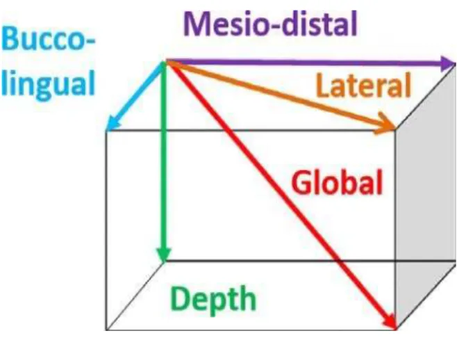
The global deviation is defined as the 3D distance between the platform and apical points
of the planned and placed implants. Depth deviation is the distance between platform or apical
points of the longitudinal axis (corono-apical) of the planned and placed implants. Moreover, a
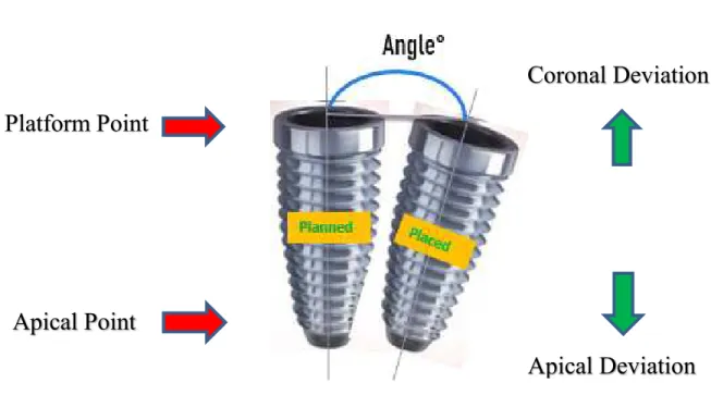
deviation could be calculated (Figure 1). The angular deviations and reference regions are used
to assess the accuracy of guided dental implant placement on all subjects (Figure 2).
2.3 Statistical Analysis
The pooled data set, the sample distributions of all variables were described univariately
by showing means, medians, standard deviations, and extreme values. All statistical estimates
will be calculated with corresponding confidence intervals. There is no statistical analysis for this
proposed study. The goal of this exploratory study was to generate results that will lend support
to hypotheses that can be studied in later, larger-scale randomized clinical studies. Adverse events (including onset, duration, treatment, outcome and suspected causality), negative factors
and reasons for withdrawals are summarized.
3. Results
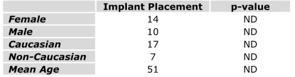
A total of 24 subjects were admitted and completed the study. Ten males between the
ages of 38 and 69 years (mean age 50.6±12.29) and fourteen females between the ages of 31 and
68 years (mean age 51.0±11.42) were enrolled. Patient demographics are listed in Table 1. Seven
subject’s data sets could not be used for the final analysis due to either limited mouth opening,
surgical guide shape, length of metal sleeve and surgical drill, or fracture or misfit of the guide. Five subject’s data sets were excluded from the final analysis due to lack of reproducibility and
accuracy of baseline and 2-week’s CBCT superimposition using DICOM files for comparison.
Hence, twelve subject’s data sets were omitted when calculating the final results. A total of
twelve subjects were included in the pre- and post-CBCT evaluation and were used for implant
3.1 Implant Deviation Analysis
The results of the implant position deviations are calculated based on implant placed
using static stereolithographic guide compared with preoperative CBCT planning to
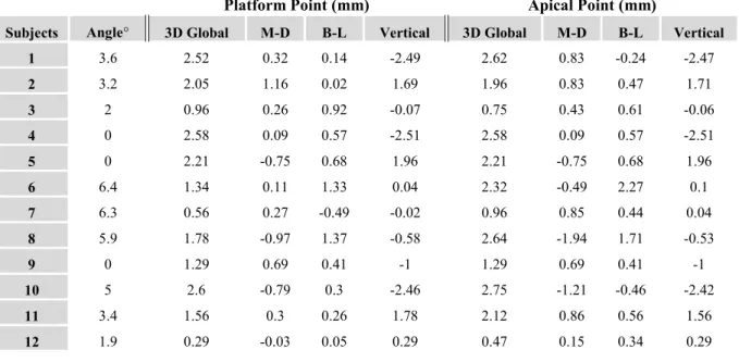
postoperative CBCT using coDiagnostiX. Table 2 lists all implant deviations in 3D global,
mesiodistal (mesial; positive values, distal; negative values), buccolingual (buccal; positive
values, lingual; negative values) and vertical (coronal; positive values, apical; negative values)
directions at the platform and apical points for all twelve subjects.
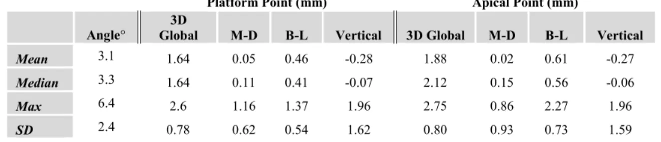
The mean 3D global deviations between planned and postoperative implant positions is 1.6 mm at the platform point and, 1.8 mm at the apical point, and 3.1° in angulation (Table 3).
Table 3 shows the calculated mean, median, maximum, and standard deviation in guided implant placement at the platform and apical point for all subjects. For the platform point, the mean deviations in the mesiodistal and buccolingual directions are 0.05 mm and 0.46 mm. For the apical point, mean deviation in the mesiodistal and buccolingual direction is 0.02 mm and 0.61 mm. This finding confirms mean deviation in the mesiodistal direction is less than the buccolingual direction.
The median 3D global deviation for platform point is less than the apical point (1.6 mm < 2.1 mm). Therefore, more deviation is expected at the apical point of guided implants.
Nonetheless, the standard deviation for 3D global at platform and apical points were similar in both groups (0.78 and 0.8) as given in Table 3. The extreme values for 3D global deviation at platform point and apical points were similar to each other (2.6 mm and 2.75 mm) as the maximum deviations. The extreme value for maximum angle deviation is 6.4° (Table 3).
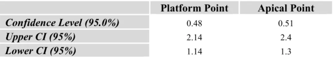
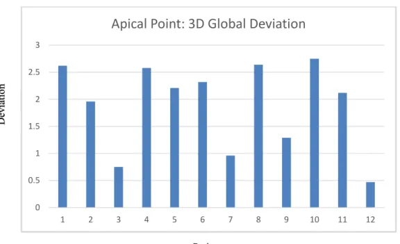
with maximum coronal deviation of 1.37 mm and maximum apical deviation of -2.51 mm. The average vertical deviation at the apical point is -0.28 mm with maximum coronal deviation of 1.37 mm and maximum apical deviation of -2.51 mm as shown in Table 4. As a results, more deviation is expected in the apical direction with an average apical deviation of 2.5 mm. Provided in Table 5, implant deviation at the platform point appeared to be smaller (95% confidence interval: 2.1 – 1.1) in comparison to apical point (95% confidence interval: 2.4 - 1.3). Cluster column chart displays the calculated mean 3D global deviation of platform and apical points in all patients (Figure 3 & 4).
4. Discussion
The results of this study demonstrates that it is possible to accurately transfer a virtual
implant position based on computer planning to the surgical site using stereolithographic guide
for implant placement. Based on the implant deviation analysis, the mean 3D global deviations found in implant positions at platform and apex was 1.6 mm and 1.8 mm, respectively, and mean angular deviation of 3.1°. The present study exhibits similarity with a retrospective study in which they reported 1.12 mm and 1.53 mm deviation at the implant platform and apex
(Tahmaseb et al. 2014). The results of this study also reports similar results with a recent meta-analysis in regards to mean deviation at the entry point of 1.25 mm and apex of 1.57 mm, but with a higher mean angular deviation of 4.1° (Zhou et al. 2018). Additionally, similarities were found in an in vivo study that reported mean global deviations between planned and placed implant positions at the coronal and apex to be 1.47 mm and 1.83 mm, but with a higher mean angular deviation of 5.09° as well (Cassetta et al. 2012).
at the implant platform and apex in static group was 0.97 mm and 1.28 mm with angular deviation of 2.84°, while the dynamic group was 1.05 mm and 1.29 mm with angular deviation of 3.06° with no statistical significant difference between the groups. Since both the static and dynamic groups had similar implant placement accuracy, it can be considered that our findings are comparable to deviations found with dynamic navigation.
The reported median 3D global deviation of platform point (1.6 mm) being less than apical point (2.1 mm) is similar to findings by Smitkarn et al. (2019) in static group that reported the deviation in platform (0.9 mm) was also less than apical (1.2 mm), although their values were smaller. Yet, the median angular deviation was similar to our result (3.1° vs. 2.8°). In this
randomized controlled trial comparing implant position accuracy between static guided and freehand surgery, they reported a statistically significant difference between the two groups with freehand median angular deviation of 7.0°. Therefore, our findings also suggest a statistically significantly less deviation compared with freehand implant placement.
The present clinical trial indicates implant deviation existence between preoperative
planning data and postoperative results using static full-guided implant surgery. The deviations
in implant position observed in this study requires detailed analysis. The mean 3D global deviations in the platform point (1.6 mm) and the apical point (1.8 mm) were calculated to be less than 2 mm. This result confirms the empirical requirement of a minimum distance of 2 mm from important anatomical structures (inferior alveolar nerve, sinus, adjacent tooth). The
confidence intervals and deviations found at the coronal positions to be smaller compared to apical positions with possible reasoning of coronal site being close to the drill sleeves.
Data from seven subjects were excluded in the final analysis of this study, as the guide was not used for implant placement due to either limited mouth opening, surgical guide shape, length of metal sleeve and surgical drills, or fracture or misfit of the guide. Regarding these clinical findings, several studies reported similar technology-related problems. The most
common reported complication was limited inter-occlusal distance in posterior segments, which
can make drill insertion through stereolithographic guide inadequate (Arisan et al. 2013).
Fractures of stereolithographic guides was also reported, which emphasizes the need for resistant
and rigid materials for guide fabrication (Schneider et al. 2009). Another limitation could be due
to the increase in sleeve height to the bone or in osteotomy length, which can produce larger
apical and coronal deviations as well as angulations for hand held sleeve inserts (Van Assche et
al. 2012). Consequently, improvement is needed in materials and fabrication of
stereolithographic guide to minimize technology related issues.
Additionally, five subjects were excluded from radiographic analysis due to lack of reproducibility and superimposition accuracy of baseline and 2-week DICOM files. This was likely due to CBCT scatter created by pre-existing metal and ceramic restorations on subject’s teeth. The scatter may make it difficult to create an accurate superimposition of images, and increases the error measurement of the final superimposition. The under- or overestimation of bone volume during CT-data analysis and virtual implant planning can reduce the predictability
of implant positioning, which can result in insufficient implant stability and or misjudgment in
contribute to more precise guided implant placement (Ersoy et al. 2008). It is essential to have
appropriate estimation of bone volume for proper CT analysis and implant planning. For future studies, it is recommended to exclude potential subjects who have a heavily restored dentition in the arch of interest.
A study by Deeb et al. (2017) demonstrated that in-office 3D printer fabricated stereolithographic implant surgical guides presents similar accuracy to laboratory or
manufacturer prepared guides. Data comparison on guide supported by bone, teeth or implants does not confirm superior accuracy when compared to mucosa-supported guide (Nazari et al.
2016). A previous precision analysis study for sleeves positions for surgical guide templates provided reliable results breakdown, with 3D deviation of 0.22 mm in the center of the sleeve top, 0.24 mm in the center of the sleeve bases and a mean angular deviation of 1.5° compared with the virtual position (Kuhl et al. 2015). Thus, the results reported in this present study using tooth-supported stereolithographic 3D printed guide demonstrates similar accuracy to
manufacturer prepared guide, as well as bone or mucosa-supported guide with the expected deviations in sleeves position as reported in above studies.
Several possible factors can affect the diagnostic and therapeutic procedures that may
allow deviations in implant placement using stereolithographic guide. For precision in implant
placement, the stability of the surgical guide is crucial. The results of this study supports that
improper seating of guide has a negative influence on the precision of implant positioning.
Therefore, the complete seating of the guide and adequate mouth opening to insert drills must be
carefully evaluated in order to avoid extreme deviations and protect important anatomical
structures. Overall, stereolithographic guides has shown to improve the accuracy in implant
4.1 Comparison of Static vs. Dynamic Navigation System
The different guided implant navigation surgery approaches rely on how the information from the pre-surgical planning to the surgical site is transferred during the implant placement, and has different characteristics that offer advantages or setbacks during the implant placement. In contrast to dynamic guidance, the static guidance via surgical templates does not allow
changes to be made to the surgical plan at the time of surgery. However, the template bur sleeves in static guides allow rigidly and highly controllable drillings, which may be an advantage in areas of irregular bone (Block et al. 2017). In addition, the accuracy of implant placement using tooth-supported static system was not significantly different between experienced and
inexperienced operators (Rungcharassaeng et al 2015). Cassetta and Bellardini (2017)
demonstrated in full edentulous patients, the accuracy of implant placement using static system was not significantly different between experienced and inexperienced surgeons.
The dynamic navigation surgery can be performed using conventional instruments and drills of several implant systems included in the database. Whereas, static system requires specific drilling system, guided instruments and possibly specific implant fixtures, which can incurs costs and time as well as laboratory fees (Kaewsiri et al. 2019). Additionally, static system typically requires less surgical time compared with dynamic system as the surgical template provides mechanical guidance of the drill and implant position. Whereas, dynamic system requires clear line of sight between tracking cameras and sensors, a registration prior to surgery that can take couple minutes, and the cost of the navigation machine also play an important factor. (Ruppin et al. 2008).
Despite the excellent results obtained with the full-guided navigation surgery,
have been attributed that render the use of full-guided static or dynamic navigation. Challenges can include reduction of accuracy in fully-edentulous arches compared to partially edentulous ridge, reduced accuracy in bone-supported templates when compared with mucosa-supported or tooth-supported templates, inaccurate adjustment of temporary prosthesis prepared in advance for immediate loading protocols, and mouth-opening limitations, especially in posterior region that may prevent the use of static surgical guides (Vercruyssen et al. 2015). Additionally, bone augmentation procedures require flap reflection, which further limit their use in the flapless approach. Nonetheless, full-guided implant placement can still be utilized in cases that may require flap reflection, though it may cause reduction in accuracy.
4.2 Computer-Assisted Implant Surgery vs. Freehand Surgery
The surgical guide allows a highly significant improvement in drilling accuracy compared with freehand drilling (Hoffmann et al. 2005). Freehand implant placement may contribute to reduced time in clinical preparation, and offer more surgical view during treatment without interfering direct vision to the implant site. It allows greatest contact of cooling fluids to the drill, resulting in better bone temperature control (Liu et al. 2018). Several randomized clinical trials showed no additional benefits with the full-or half-guided techniques compared to the freehand method in terms of peri-implant parameters commonly used such as marginal bone loss, bleeding on probing, plaque index and gingival index (Pozzi et al., 2014). Freehand implant surgery is least costly, as it does not involve any surgical template fabrication.
Disadvantage of freehand implant surgery includes least amount of accuracy in
which accompanies more patient morbidity, more post-operative pain and swelling reducing patient satisfaction, longer chair-time and higher risk of intra- and post-operative hemorrhages (Artisan et al. 2010). Furthermore, bacterial contamination in freehand implant surgery with flap elevation has shown to be three times greater than when performing flapless surgery and full-guided implant placement (Pozzi et al. 2014).
5. Conclusion
Stereolithographic guides were previously shown to improve the accuracy in implant
placement (Vercruyssen et al. 2008). Therefore, assessing the accuracy of guided implant surgery can play an important role in reducing errors. While precision transfer of the virtual planned implant position is desired, an universal ‘acceptable’ deviation cannot be defined since
in some clinical situations the malposition implant does not pose a problem, while in other
situations the malposition implant can be detrimental, such as nerve injury (Ersoy et al. 2008).
This study highlights deviations between the postoperative position and the preoperative planning at the platform and apical points of implants placed using a guide. The results of this clinical study indicates acceptable transfer accuracy when using static stereolithographic guide for implant placement after 3D implant planning, by taking into account all sources of
inaccuracies. The analysis confirms slight deviations between planned and placed implants that can occur using static stereolithographic guides. Clinicians should be aware not to overestimate advocated surgical safety by using static navigation tools. Computer-aided planning and
for better esthetics and functional outcome. Nonetheless, additional clinical trials are required to
REFERENCES
Arisan, V., Karabuda, C.Z., Mumcu, E., Ozdemir, T. (2013). Implant positioning errorsin freehand and computer-aided placement methods: a single-blind clinical comparative study. Int. J. Oral Maxillofac. Implants. 28: 190–204.
Block, M. S., Emery, R. W., Lank, K., & Ryan, J. (2017). Implant Placement Accuracy Using Dynamic Navigation. International Journal of Oral and Maxillofacial Implants. 32(1), 92–99.
BouSerhal C., R. Jacobs, M. Quirynen and D. van Steenberghe (2002). "Imaging technique selection for the preoperative planning of oral implants: a review of the literature." Clin Implant Dent Relat Res 4(3): 156-172.
Cassetta, M., & Bellardini, M. (2017). How much does experience in guided implant surgery play a role in accuracy? A randomized controlled pilot study. International Journal of Oral and Maxillofacial Surgery. 46(7): 922–930.
Cassetta M., Stefanelli L., Giansanti M., Calasso S. (2012). Accuracy of implant placement with a stereolithographic surgical template. International Journal of Oral and Maxillofacial Implants. 27(3):655-63.
Deeb G., Allen R., Hall P, Whitley R. (2017). How accurate are implant surgical guides
produced with desktop stereolithographic 3D-printers? Journal of Oral and Maxillofacial Surgery. 75:12.
D’souza KM, Aras MA. (2012). Applications of computer-aided design/computer-assisted manufacturing technology in dental implant planning. J Dent Implant. 2:37–41.
Ersoy, A., I Turkyilmaz, O. Ozan, E. McGlumphy (2008). Reliability of implant placement with stereolithography guides generated from computed tomography: clinical data from 94 implants. J Perio. 79(8): 1339-1345.
Frazer R., R. Byron, P. Osborne, K. West (2005). PMMA: an essential material in medicine and dentistry. J Long Term Eff Med Implants, 15: 629-639.
Gargallo-Albiol J, Barootchi S, Salomo-coll O, Wang H. (2019). Advantages and disadvantages of implant navigation surgery. A systemic review. Elsevier. 225:1-10.
Hoffmann J, Westendorff C, Gomez-Roman G, Reinert S. (2005). Accuracy of navigation-guided socket drilling before implant installation compared to the conventional free-hand method in a synthetic edentulous lower jaw model. Clin Oral Implants Res. 16: 609–614.
Kaewsiri D, Panmekiate S, Subbalekha K, Mattheos N, Pimkhaokham A. (2019). The accuracy of static vs. dynamic computer-assited implant surgery in single tooth space: A
randomized controlled trial. Clinical oral implants research. 30: 505-514.
Khadem R, Yeh CC, Sadeghi-Tehrani M, Bax MR, Johnson JA, Welch JN, Wilkinson EP, Shahidi R. (2000). Comparative tracking error analysis of five different optical tracking systems. Comput Aided Surgery. 5: 98–107.
Koop, R., M. Vercruyssen, K. Vermeulen and M. Quirynen (2013). "Tolerance within the sleeve inserts of different surgical guides for guided implant surgery." Clin Oral Implants Res 24(6): 630-634.
Kuhl S., Payer M., Zitzmann N., Lambrecht J., Filippi A. (2015). Technical accuracy of printed surgical templates for guided implant surgery with the coDiagnostiX software. Clin Implant Dent Relat Res. 1:177-82.
Lal K, White GS, Morea DN, Wright RF. Use of stereolithographic templates for surgical and prosthodonticimplant planning and placement. Part I. The concept. J Prosthodont. 2006;15:51–58.
Liu, Y.F., Wu, J.L., Zhang, J.X., Peng, W., Liao, W.Q., (2018). Numerical and experimental analyses on the temperature distribution in the dental implant preparation area when using a surgical guide. J. Prosthodontics. 27 (1): 42–51.
Naziri E., A. Schramm, F. Wilde (2016). “Accuracy of computer-assisted implant planning with insertion template.” GMS Interdiscip Reconstr Surg DGPW. 5: Doc15.
Nickenig HJ., S. Eitner. “Reliability of implant placement after virtual planning of implant positions using cone beam CT data and surgical (guide) templates.” Journal
Craniomaxillofac Surg. 2007 Jun-Jul; 35(4-5):207-11.
Pozzi, A., Tallarico, M., Marchetti, M., Scarfo, B., Esposito, M., (2014). Computer-guidedversus free-hand placement of immediately loaded dental implants: 1-yearpost-loading results of a multicentre randomised controlled trial. Eur. J. Oral Implantol. 7 (3): 229–242.
Ruppin J, Popovic A, Strauss M, Spuntrup E, Steiner A, Stoll C. (2008). Evaluation of the accuracy of three different computer-aided surgery systems in dental implantology: optical tracking vs. stereolithographic splint systems. Clin Oral Implants Res. 19: 709– 716.
Smitkarn P., Subbalekha K., Mattheos N., Pimkhaokham A. (2019). The accuracy of single-tooth implants placed using fully digital-guided surgery and freehand implant surgery. J.
Clinical Perio. 46(9):949-057.
Schneider D., P. Marquardt, M. Zwahlen, R. Jung (2009). “A systemic review on the accuracy and the clinical outcome of computer-guided template-based implant dentistry.” Clinical Oral Implant Research 20(s4):19-22.
Stubinger S., Buitrago-Tellez C., Cantelmi G. (2012). Deviations between placed and planned implant positions: an accuracy pilot study of skeletally supported stereolithographic surgical templates. Clinical Implant dentistry and Related Research. 16(4): 540-551.
Tahmaseb, A., Wismeijer, D., Coucke, W., & Derksen, W. (2014). Computer technology applications in surgical implant dentistry: A systematic review. International Journal of Oral and Maxillofacial Implants. 29: 25–42.
Van Assche, N., M. Vercruyssen, W. Coucke, W. Teughels, R. Jacobs and M. Quirynen (2012). "Accuracy of computer-aided implant placement." Clin Oral Implants Res 23 Suppl 6: 112-123.
Vercruyssen, M., R. Jacobs, N. Van Assche and D. van Steenberghe (2008). "The use of CT scan based planning for oral rehabilitation by means of implants and its transfer to the surgical field: a critical review on accuracy." J Oral Rehabil 35(6): 454-474.
Vercruyssen, M., Laleman, I., Jacobs, R., Quirynen, M. (2015). Computer-supported implant planning and guided surgery: a narrative review. Clin. Oral Implants Res. 26: 69–76. Verstreken K, Van Cleynenbreugel J, Marchal G, van Steenberghe D, Suetens P. (1996).
Computer-assisted planning of oral implant surgery. An approach using virtual reality. Stud Health Technol Inform. 29: 423–434.
Wagner A., F. Wanschitz., W. Birkfellner, K. Zauza, C. Klug, K. Schicho, F. Kainberger, C. Czerny, H. Bergmann, R. Ewers. “Computer-aided placement of endosseous oral implants in patients after ablative tumour surgery: assessment of accuracy.” Clin Oral Implants Res. 2003 Jun; 14(3):340-8.
Westendorf S., E. Naziri, H. Eisenmann, RG. Luthardt, A. Schramm. “3D techniques in dental implantology.” Int J Digital Dent News. 2010;4:32–45.
Whitley D, 3rd, Eidson RS, Rudek I, Bencharit S. (2017). In-office fabrication of dental implant surgical guides using desktop stereolithographic printing and implant treatment planning software: A clinical report. The Journal of prosthetic dentistry. 2017.
APPENIX 1: INCLUSION CRITERIA
• Subjects must be adult males or females age 18 to 80 years (inclusive).
• Subjects must be able and willing to follow study procedures and instructions in English.
• Subjects must have read, understood and signed an informed consent form in English.
• Subjects must have a maxillary premolar, canine, lateral incisor, or central incisor with a restorative or periodontal hopeless prognosis in which an implant is indicated without any sinus lift required.
APPENDIX 2: EXCLUSION CRITERIA
• Individuals who have a chronic disease with oral manifestations. • Individuals who exhibit gross oral pathology.
• The use of either antibiotics or chronic use (more than 7 days) of NSAIDs within 1 month prior to screening examination.
• Individuals that require antibiotic prophylaxis prior to dental treatment.
• Chronic treatment (i.e. two weeks or more) with any medication known to affect
periodontal status (e.g. phenytoin, calcium antagonists, cyclosporine, Coumadin) within 1 month prior to screening examination.
• Uncontrolled diabetes mellitus (HbA1c >7) within 3 months prior to screening examination.
• Individual with uncontrolled parafunctional habits, such as clenching and bruxing on objects, that could adversely impact implant survival.
• Individuals with a history of intravenous bisphosphonates.
• Individuals with active infectious diseases such as hepatitis, HIV or tuberculosis.
• Current cigarette smokers.
• Individuals who are known to be pregnant, breastfeeding or planning to become pregnant within 6 months.
• Individuals with blood disorders (hemophilia) and /or currently taking anticoagulant medications, such as heparin, warfarin, or clopidrogel.
• Individuals receiving any therapy known to affect healing, such as high dose corticosteroids, radiation therapy or chemotherapy.
• Individuals who require maxillary sinus augmentation prior to dental implant therapy.
TABLE 1: Subject demographics
Implant Placement p-value
Female 14 ND
Male 10 ND
Caucasian 17 ND
Non-Caucasian 7 ND
TABLE 2: Deviations in guided implant placement
M-D, mesial-distal; B-L; buccal-lingual
Table 2: Deviations in guided implant placement. The 3D global, M-D, B-L and vertical deviations at the platform and apical points for all subjects.
Platform Point (mm) Apical Point (mm)
Subjects Angle° 3D Global M-D B-L Vertical 3D Global M-D B-L Vertical
1 3.6 2.52 0.32 0.14 -2.49 2.62 0.83 -0.24 -2.47
2 3.2 2.05 1.16 0.02 1.69 1.96 0.83 0.47 1.71
3 2 0.96 0.26 0.92 -0.07 0.75 0.43 0.61 -0.06
4 0 2.58 0.09 0.57 -2.51 2.58 0.09 0.57 -2.51
5 0 2.21 -0.75 0.68 1.96 2.21 -0.75 0.68 1.96
6 6.4 1.34 0.11 1.33 0.04 2.32 -0.49 2.27 0.1
7 6.3 0.56 0.27 -0.49 -0.02 0.96 0.85 0.44 0.04
8 5.9 1.78 -0.97 1.37 -0.58 2.64 -1.94 1.71 -0.53
9 0 1.29 0.69 0.41 -1 1.29 0.69 0.41 -1
10 5 2.6 -0.79 0.3 -2.46 2.75 -1.21 -0.46 -2.42
11 3.4 1.56 0.3 0.26 1.78 2.12 0.86 0.56 1.56
TABLE 3: Statistical analysis
M-D, mesial-distal; B-L; buccal-lingual; Max, Maximum; SD, standard deviation;
Table 3: Statistical analysis. The calculated average, median, maximum, and standard deviation in guided implant placement at the platform and apical points.
Platform Point (mm) Apical Point (mm)
Angle°
3D
Global M-D B-L Vertical 3D Global M-D B-L Vertical
Mean 3.1 1.64 0.05 0.46 -0.28 1.88 0.02 0.61 -0.27
Median 3.3 1.64 0.11 0.41 -0.07 2.12 0.15 0.56 -0.06
Max 6.4 2.6 1.16 1.37 1.96 2.75 0.86 2.27 1.96
TABLE 4: Vertical deviations
Table 4: Vertical deviations. The average and maximum vertical deviations in coronal and apical directions for platform and apical points with guided implant placement.
Platform Point (mm) Apical Point (mm)
Average Vertical Deviation -0.28 -0.27
Maximum Coronal Deviation 1.37 1.96
TABLE 5: Confidence intervals
Platform Point Apical Point
Confidence Level (95.0%) 0.48 0.51
Upper CI (95%) 2.14 2.4
Figure 1: Three dimensions of deviations
Fig. 1: Three dimensions of deviations. Red: global coronal deviation, orange: lateral
deviation, green: depth deviation, blue: buco-lingual deviation, purple: mesio-distal deviation.
Figure 2: Reference regions
Figure 2: Reference regions. Illustration demonstrates the angle, platform and apical points, as well as coronal and apical direction as reference regions to assess the deviation of planned and inserted dental implants.
Apical Point
Platform Point
Coronal Deviation
Figure 3: Cluster column chart for platform points
Figure 3: Cluster column chart for platform points. Displaying 3D global deviation at
platform point for each implant insertion.
0 0.5 1 1.5 2 2.5 3
1 2 3 4 5 6 7 8 9 10 11 12
Platform Point: 3D Global Deviation
Patients
D
ev
ia
ti
o
n
Figure 4: Cluster column chart of apical points
Figure 4: Cluster column chart of apical points. Displaying 3D global deviation in apical point of each implant insertion
0 0.5 1 1.5 2 2.5 3
1 2 3 4 5 6 7 8 9 10 11 12
Apical Point: 3D Global Deviation
Patients
D
ev
ia
ti
o
n