R E S E A R C H
Open Access
Split luciferase complementation assay to detect
regulated protein-protein interactions in rice
protoplasts in a large-scale format
Yukichi Fujikawa
1, Takahiro Nakanishi
1, Hiroko Kawakami
1, Kanako Yamasaki
2, Masa H Sato
2, Hiroyuki Tsuji
3,
Makoto Matsuoka
4and Naohiro Kato
5*Abstract
Background:The rice interactome, in which a network of protein-protein interactions has been elucidated in rice, is a useful resource to identify functional modules of rice signal transduction pathways. Protein-protein interactions occur in cells in two ways, constitutive and regulative. While a yeast-based high-throughput method has been widely used to identify the constitutive interactions, a method to detect the regulated interactions is rarely developed for a large-scale analysis.
Results:A split luciferase complementation assay was applied to detect the regulated interactions in rice. A transformation method of rice protoplasts in a 96-well plate was first established for a large-scale analysis. In addition, an antibody that specifically recognizes a carboxyl-terminal fragment ofRenilla luciferase was newly developed. A pair of antibodies that recognize amino- and carboxyl- terminal fragments ofRenillaluciferase, respectively, was then used to monitor quality and quantity of interacting recombinant-proteins accumulated in the cells. For a proof-of-concept, the method was applied to detect the gibberellin-dependent interaction between GIBBERELLIN INSENSITIVE DWARF1 and SLENDER RICE 1.
Conclusions:A method to detect regulated protein-protein interactions was developed towards establishment of the rice interactome.
Keywords:Interactome; Split luciferase complementation; Regulated protein-protein interactions
Background
Interactome analysis collects data of proteprotein in-teractions occurring in cells. A useful application of the analysis is to identify functional modules of signal trans-duction pathways in which a network of protein-protein interactions mediate the signals (Barabasi et al. 2011; Pawson and Nash 2000). Protein-protein interactions occur in cells in two ways, constitutive and regulative. Constitutive protein interactions occur all the time in cells. In many cases, even when interaction is examined
in vitro, a constitutive interaction is detectable. On the other hand, regulated protein interactions occur when cells are exposed to selected environments such as stress
and environmental cue. When cells are exposed to selected environments, some of proteins change their subcellular localization to interact with other proteins. Alternatively, cells may produce small compound (i.e., hormone) that me-diates a protein-protein interaction (Ritter and Hall 2009), or a protein in the cells be modified (i.e., phosphorylation) to attract other proteins (Pawson 2004). Importantly, regulated protein-protein interactions are often at pivotal positions within the interaction network in a signal trans-duction pathway (Wehr et al. 2008).
Because plants highly adaptive to different environments, signal transduction pathways in plants are thought to be complicated (Vanstraelen and Benkova 2012; Santner and Estelle 2009). Genomes ofOryzagenus have been modified to adapt diverse environments and increase the value as a crop (Vaughan et al. 2003). Hence, genetic analysis ofOryza sativa, rice, is recently spurred considerable interest to
* Correspondence:[email protected] 5
Department of Biological Sciences, Louisiana State University, 226 Life Sciences Building, Baton Rouge, LA 70803, USA
Full list of author information is available at the end of the article
understand the genomic evolution. However, rice interac-tome analysis, which would reveal the molecular mechan-ism of the rice adaptation, has been limited to constitutive protein-protein interactions that are determined by the yeast two-hybrid assay (Gu et al. 2011; Seo et al. 2011). Although the assay would detect protein-protein interac-tions in absent or present of certain regulators, it is diffi-cult to detect regulated protein-protein interactions that may occur in cellular conditions specific to rice.
The split luciferase complementation (SLC) assay is one of methods that detect regulated proteprotein in-teractionsin situ(Ozawa et al. 2001; Kaihara et al. 2003; Luker et al. 2004). In the assay, N- and C-terminal frag-ments of luciferase are genetically fused to a protein pair of interest. The luciferase activity, which emits light through oxidation of a substrate, is complemented when the protein pair interacts with each other, but ceases when the protein pair does not interact. Hence, one can determine a protein interaction by a flash of light. Because the assay is capable of identifying not only interaction but also dissociation of protein pairs, it is suitable for analyzing kinetics of protein interactionsin situ (Luker et al. 2004). Moreover, the result of the SLC assay is reproducible with high signal-to-background ratios, which allows for conducting interactome analysis, at least, in animal cells (Cassonnet et al. 2011).
In plants, the assay was first applied to detect a histone-histone interaction in protoplasts of Arabidopsis leaves (Fujikawa and Kato 2007). Since then, the assay has been applied to detect interactions between membrane proteins (Kato et al. 2009), bacterial effector proteins and their protein targets (Chen et al. 2008), auxin response factors (Li et al. 2011), 14-3-3 regulator proteins (Gehl et al. 2011), coiled-coil–nucleotide-binding site–leucine-rich repeat (CC-NB-LRR) protein (Inoue et al. 2013), and kinase (Schmidt et al. 2013). We previously showed with the SLC assay that protein-protein interactions in Arabidopsis protoplasts are sensitive to environmental conditions (Kato and Bai 2010), indicating that the SLC assay would be well-suited for analyzing regulated protein-protein interactions in plant cells.
Currently, however, the use of the SLC in plants is limited to detecting constitutive protein-protein interac-tions and only in dicot model plants such as Arabdidopsis and tobacco, but not in monocot plants such as rice. Comparative transcriptional analyses revealed that rice and Arabidopsis differently transduce a stress signal (Yazaki et al. 2004; Narsai and Whelan 2013). Moreover, gene annotation and transcriptome analyses revealed that rice and Arabidopsis differently regulate the flow of potassium in the plasma membrane as a result of stress and environmental cue (Very et al. 2014; Ma et al. 2012). Potassium is the major inorganic cation in the cytoplasm, and known to regulate the activities of many enzymes
including ones involved in signal transductions in plant cells (Wang and Wu 2013). These analyses support the idea that rice may possess a unique network in regu-lated interactions, which could not be identified when a protein-interaction assay is conducted in different organ-isms (i.e. Arabidopsis and yeast).
Here we present aRenilla luciferase-based technology breakthrough that allows the identification of regulated protein-protein interactions in rice in near-real time, which can also be expanded to a large-scale analysis.
Results and discussion
A transformation method for rice protoplasts in a 96-well plate was established
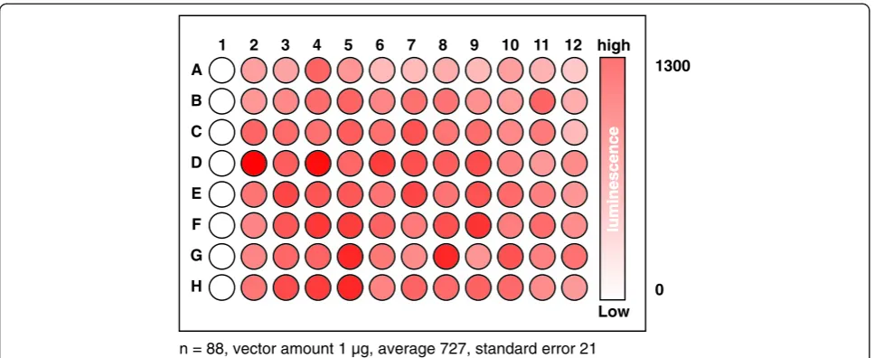
Simultaneously transforming an organism of interest with a large number of independent vectors is one of the key technologies for an interactome assay. Although methods to transform rice protoplasts in individual micro-centrifuge tubes have been published elsewhere (Bart et al. 2006; Zhang et al. 2011; Datta and Datta 1999), a method simultaneously to transform rice cells in a 96 well-plate hat allows transforming the cells with a large number of independent vectors has not been estab-lished for rice. To this end, we estabestab-lished a method in which rice protoplasts are transformed with polyethylene glycol (PEG) in a 96-well plate, based on the method previously developed for Arabidopsis leaf protoplasts (Kato and Jones 2010). We used a vector expressing full-length Renillaluciferase as a transgene so that the transformation method was most optimized for the assay. We found that 0.5–1.0 g of 1–2 week old rice shoots that are vertically cut into 2 mm wide strips toward the leaf sheath produced enough high quality protoplasts to conduct 96 independent transformations in the plate. We also found that transforming the rice protoplasts in a 96-well plate did not generate a bias toward column and row in the plate with respect to transformation efficiency (Figure 1). In other words, transformation efficiency is slightly deviated in each well but not by columns and rows.
An antibody that recognizes the C- terminal fragment of Renillaluciferase was generated
It is important to understand quality and quantity of recombinant proteins accumulated in transformed proto-plasts. This ensures interaction kinetics is correctly inter-preted from luminescence emitted from the protoplasts. We previously found that a commercially available
properly, rice protoplasts were transformed with vectors expressing recombinant proteins (Figure 2). The
recom-binant proteins expressed were Arabidopsis HISTONE
2A (H2A, AT4G27230), Arabidopsis HISTONE 2B (H2B,
AT5G22880), rice GIBBERELLIN INSENSITIVE DWARF1 (GID1, Os05g0407500), or rice SLENDER RICE 1 (SLR1, Os03g0707600). The cDNAs encoding these proteins were inserted in pDuEx vectors (Fujikawa and Kato 2007). These vectors contain NRLuc or CRLuc, and express the recom-bined gene from the cauliflower mosaic virus 35S promoter (Fujikawa and Kato 2007; Kato and Jones 2010). Recombin-ant proteins were expressed in rice protoplasts in various combinations (Figure 2). To conduct immunoblot assays for evaluating theRenillaluciferase antibodies, proteins in the transformed rice protoplasts were extracted by lysing the cells 16 h after the transformation. The proteins were then electrophoresed and transferred to a membrane in order to blot with the newly generated anti-CRLuc antibody, or the commercially available antibody that recognizes NRLuc. The results revealed that both anti-bodies detect recombinant proteins accumulated in rice protoplasts (Figure 2A). Also, the recombinant proteins did not degrade much because signal bands in the immunoblot were not smeared. The anti-CRLuc antibody detected multiple proteins whose sizes are between 40 and 75 kDa, which do not correspond to the CRLuc-fused proteins. This indicated that careful analysis would be required when recombinant proteins whose sizes are similar to those unknown proteins are examined. It also suggested that levels of protein accumula-tions would depend on a protein expressed. For Instance,
the accumulation level of the NRLuc-GID1 protein is lower than that of the NRLuc-H2A protein (Figure 2A).
Luminescence emitted from protoplasts depends on not only the affinity (dissociation constant) of a protein pair that is fused to NRLuc and CRLuc, respectively, but also the amounts the recombinant proteins accumulated in the protoplasts (Kato et al. 2009). Because lumines-cence units determined in the assay are relative but not absolute, it remains a challenge to deduce an absolute value of the dissociation constant. However, the anti-CRLuc antibody developed in this study will allows addressing the question whether low luminescence in the assay is caused by a low affinity of the protein pair or low levels of the protein accumulation in one or both of the protein pair as discussed in the following sections.
Histone2A-histone2B interaction was detected with a high signal-to-background ratio
To examine whether a protein interaction was detected by bioluminescence in rice protoplasts, relative lumines-cence units (RLU) were measured soon after adding the luciferase substrate to the culture solution in each well of the 96-well plate but before lysing the protoplasts for the immunoblot assay. Because H2A interacts with H2B (Fujikawa and Kato 2007), we expected rice protoplasts expressing NRLuc-H2A and CRLuc-H2B to emit lumi-nescence light at high levels upon addition of viviRen®, the substrate for Renilla luciferase. On the other hand, we expected rice protoplasts expressing NRLuc-H2A and CRLuc-SLR1 or NRLuc-GID1 and CRLuc-H2B to emit luminescence light at very low levels, similar to
high
Low 1
1300
0
2 3 4 5 6 7 8 9 10 11 12
A
B
C
D
E
F
G
H
n = 88, vector amount 1 µg, average 727, standard error 21
mock-transformed protoplasts, because these protein pairs would not interact with each other. The rice proto-plasts expressing NRLuc-H2A and CRLuc-H2B showed 21-fold higher RLU, compared to the mock transformed protoplasts (Figure 2B). On the other hand, the proto-plasts expressing NRLuc-H2A and CRLuc-SLR1 or those expressing NRLuc-GID1 and CRLuc-SRL1 showed RLU at low levels relative to the mock transformed protoplasts. This suggested that rice protoplasts are suitable to deter-mine protein-protein interactions by the SLC assay in a large-scale format as we previously found inArabidopsis
protoplasts (Fujikawa and Kato 2007).
Gibberellin-dependent interaction of GID1 and SLR1 was detected in near-real time
Adaptation of plant growth and development to the environment is largely achieved through hormone pro-duction and distribution in the plant (Vanstraelen and Benkova 2012; Santner and Estelle 2009). Hence, signal transduction pathways and a protein interaction net-work in plant cells are expected to be modulated by hormones. To test whether our system could detect hormone-dependent protein interactions, we took ad-vantage of the previously characterized interaction be-tween two components of gibberellin signaling, GID1 and SLR1 (Ueguchi-Tanaka et al. 2005).
GAs are a large family of plant hormones that widely play roles in plant development, such as seed germination, stem elongation, leaf expansion, flowering, and pollen maturation (Thomas and Sun 2004). A breakthrough in the GA signaling study is the discovery of a soluble GA receptor, GID1 (Ueguchi-Tanaka et al. 2005). The gid1
mutant rice shows dwarf and insensitive to the exposure of exogenous GA. Structural analyses of GID1 proteins of rice and Arabidopsis revealed that an N-terminal por-tion of the protein funcpor-tions as a lid to bind and cover GA (Murase et al. 2008; Shimada et al. 2008). The N-terminal lid is also found to involve in the GA-dependent interaction with SLR1, the repressor in GA signaling (Murase et al. 2008; Shimada et al. 2008). Interestingly for us, while the rice (Oryza sativa) genome encodes one GID1 protein OsGID1, the Arabidopsis (Arabidopsis thaliana) genome encodes the OsGID1 orthologous protein AtGID1a and the paralogous proteins AtGID1b (Nakajima et al. 2006). AtGID1b constitutively interacts with SLR1 without GA (Yamamoto et al. 2010). This supports the idea that rice and Arabidopsis cells may have different networks in the GA-regulated protein-interactions.
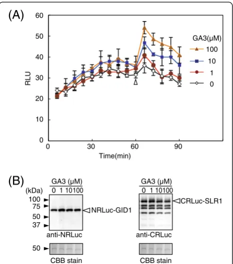
Because the interaction of GID1 and SLR1 occurs in the nucleus in a gibberellin-dependent manner in rice (Ueguchi-Tanaka et al. 2005; Ueguchi-Tanaka et al. 2007b), we tracked changes of RLU in rice protoplasts transformed with vectors expressing NRLuc-GID1 and CRLuc-SLR1 (Figure 3). In the experiment, different concentrations NRLuc-H2A
NRLuc-GID1
RLU
NRLuc fusion
Signal ratio CRLuc fusion 200
150
100
50
0
H2A
1.9 1
H2A
23
H2B H2B
GID1
1.1 SLR1
CRLuc-H2B CRLuc-SLR1 anti-NRLuc
anti-CRLuc
CBB stain
NRLuc-H2A
and CRLuc-SLR1
Mock NRLuc-GID1 and CRLuc-H2BNRLuc-H2A and CRLuc-H2B
100 75
50 37
25 (kDa)
100 75
50 37
25
50
(A)
(B)
of gibberellin 3 (GA3), an active form of gibberellin, were added to the culture solution 60 min after adding the luciferase substrate. The protoplasts showed low RLU (about 20 RLU) soon after adding the substrate, suggesting interaction did not occur at this point. Al-though the RLU gradually increased with a function of time, most likely due to passive transport of the sub-strate from the culture solution into the cells, the RLU remained at low levels for 90 min without GA3. On the other hand, the RLU increased soon after adding GA3, and the RLU remained at high levels even 30 min after adding GA3 (90 min after adding the substrate). Fold changes of the RLU after adding GA3 (up to 1.5-fold, compared to the mock treated protoplasts) was not as high as the H2A-H2B interaction that also occurs in the nucleus (10-fold, compared to a non-interaction protein pair) (Figure 2). This suggested that the dis-sociation constant of GID1-SLR1 might be lower than
that of H2A-H2B. An additional or alternative possibility is that topologies of NRLuc-GID1 and CRLuc-SLR1 do not allow for complementation of the luciferase activity to the extent of that with NRLuc-H2A and CRLuc-H2B. In other words, three-dimensional positions of NRLuc and CRLuc may not be optimized to reconstitute the lu-ciferase activity when NRLuc-GID1 and CRLuc-SLR1 interact. Nevertheless, the increased RLU was statisti-cally significant (P < 0.01) and depended on the concen-tration of GA3. Moreover, GA3 did not increase RLU in the protoplasts that expressed NRLuc-H2A and CRLuc-H2B (Additional file 1: Figure S1). We also confirmed by immunoblot assay that the changes of the RLU were not due to changes of the amounts of the recombinant proteins accumulated in the transformed protoplasts (Figure 3B). Together the results indicated that the in-creased RLU in the protoplasts expressing NRLuc-GID1 and CRLuc-SLR1 was due to increased association of the recombinant proteins by GA3.
Interaction of GID1 and SLR1 was not affected by other plant hormones in rice protoplasts
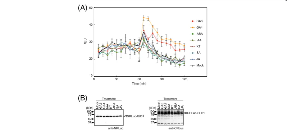
Gibberellin signaling pathways exhibit crosstalk with path-ways that are regulated by other plant hormones such as abscisic acid and auxin (Weiss and Ori 2007). To investi-gate whether other plant hormones besides gibberellin trigger the GID1-SLR1 interaction in rice cells, we tracked changes of RLU in the rice protoplasts transformed with vectors expressing NRLuc-GID1 and CRLuc-SLR1 (Figure 4). In the experiment, GA3 and GA4, another ac-tive form of gibberellin, as well as other plant hormones, including abscisic acid (ABA), indoleacetic acid (IAA), kin-etin (KT), salicylic acid (SA), and jasmonic acid (JA) added to the culture solution 60 min after addition of the lucifer-ase substrate. RLU increlucifer-ased soon after adding GA3 and GA4, and the RLU remained at high levels even 30 min after adding GA3 and GA4 (90 min after adding the sub-strate). Fold changes of the RLU, compared to the mock transformed protoplasts, after adding GA4 was significantly (P < 0.01) higher than that of GA3. This suggested that GID1 and SLR1 might interact more firmly with GA4 than GA3. This agrees with a previous report that GA4 gives higher promoter activity in a GID1-SLR1 yeast two-hybrid assay compared to GA3 (Ueguchi-Tanaka et al. 2007a).
Although other hormones increased RLU, the increases were not significant (P > 0.10) compared to the mock transformed protoplasts. We also confirmed by immuno-blot assays that amounts of the recombinant proteins accu-mulated in the transformed protoplasts were not affected by different plant hormones (Figure 4B). This suggested that the GID1-SLR1 interaction was mediated by gibberel-lin specifically in the rice cells. This also showed that the SLC assay is capable of detecting a protein-protein inter-action that is regulated by a hormone.
0 10 20 30 40 50 60
0 30 60 90
CRLuc-SLR1 100
75 50 37 (kDa)
GA3 (µM)
0 1 10100 0 1 10100
NRLuc-GID1
GA3 (µM) Time(min)
RLU
50
anti-NRLuc anti-CRLuc
CBB stain CBB stain
(A)
(B)
100
10
1
0 GA3(µM)
Conclusions
We previously discussed about advantage and disadvantage of SLC by comparing the results of SLC in Arabidopsis protoplasts with that of other interaction assays such as FRET (Fluorescence resonance energy transfer) and Co-IP (Co-immunoprecipitaiton) assay (Fujikawa and Kato 2007; Kato et al. 2009). We, furthermore, used the SLC assay to detect a light-dependent interaction in Arabidopsis protoplasts (Kato et al. 2009). Here we showed that the SLC is capable of detecting a protein-protein interaction that is regulated by a hormone in rice protoplasts in near-real time. Because pDuEx vectors used in this SLC assay contain the Gateway® recombinant sites (Fujikawa and Kato 2007), one can relatively easily construct a large number of expression vectors. Moreover, four differ-ent types of pDuEx vectors that allow fusing NRLuc or CRLuc to N- or C-terminal ends of a target protein, were constructed (GenBank: EF565883, EF56588, GU370778 and GU370779). All together, these provide a suite of vectors for a large-scale SLC assay in rice protoplasts. The suite would aid in revealing regulated protein-protein interactions in rice and other monocot crop cells in a large-scale format.
Methods
Plasmid constructs
The coding sequences in the cDNAs including a stop codon (GID1; GIBBERELLIN INSENSITIVE DWARF1,
Genbank Acc. No. AB211399, SLR1; SLENDER RICE1 Genbank Acc. No. AB262980) were amplified by PCR using pGBKT7-GID1 and pGADT7-SLR1 (Ueguchi-Tanaka et al. 2005) as the templates, respectively. The products of PCR were then cloned into a pDONR/Zeo vector (Invitrogen, CA). The resultant plasmids were subse-quently used to clone into pDuExAn6 or D7 expression vectors (Fujikawa and Kato 2007) with the Gateway® cloning system (Invitrogen, CA).
Rice cultivation and treatments
Mature seeds of rice (Oryza sativa L. Japonica) were soaked in water for 3 days at 28°C in the dark after being sterilized with 50% (v/v) bleach for 10 min. The seeds were then placed on soil in a pot (8 cm wide by 7.5 cm high) with Kimura B solution [365 μM (NH4)2SO4,
547 μM MgSO4, 183 μM KNO3, 365 μM Ca(NO3)2,
182μM KH2PO4, 19μM Fe-EDTA, 48.7μM H3BO3, 9μM
MnSO4, 0.3 μM CuSO4, 0.7 μM ZnSO4, 0.099 μM
Na2MoO4] containing 1 nM uniconazole. The seedlings
were incubated at 28°C in 16 h of light (light intensity; 100μmol m−2sec−1) and 8 h of dark for 1 to 2 weeks.
Protoplast preparation and transformation
Rice shoots (0.5 - 1.0 g, 1–2 week old) were cut into 2 mm-wide sections toward the leaf sheath by using a surgical knife (No.21 FETHER, Osaka, Japan). Protoplasts were isolated by enzymatic hydrolysis using cellularase RS
10 20 30 40 50
0 30 60 90 120
RLU
Time (min)
100 75 50 37 (kDa)
100 75 50 37 (kDa)
Treatment Treatment
NRLuc-GID1
CRLuc-SLR1
anti-NRLuc anti-CRLuc
Mock GA3 GA4 IAA KT ABA SA JA Mock GA3 GA4 IAA KT ABA SA JA
(A)
(B)
GA3 GA4 ABA IAA KT SA JA Mock
(Yakult pharmaceutical industry. Tokyo, Japan) and macer-ozyme R10 (Yakult pharmaceutical industry). The pieces of the rice shoots were first incubated with the enzyme solution (4% cellulase RS and 2% macerozyme R10, 0.6 M mannitol, 20 mM MES pH 5.7, 20 mM KCl, and 10 mM CaCl2) for 4 h on a rotary platform shaker at
35 rpm/min after vacuuming the samples for 30 min. The samples were then filtered through a 40μm nylon mesh to remove clumps. The isolated protoplasts were washed two times with 10 ml of W5 buffer (0.125 M CaCl2, 5 mM KCl, and 2 mM MES pH 5.7) by
centrifu-gation at 200 × g for 3 min. The protoplasts were fur-ther suspended in 10 ml of W5 buffer and placed at 4°C for 30 min. The protoplasts were collected by cen-trifugation at 200 × g for 3 min, and re-suspended in
MMg solution (0.4 M mannitol, 15 mM MgCl2, and
4 mM MES pH 5.7) so that the concentration of the protoplasts in the solution would be 4 × 10⁵cells/ml. Protoplast transformation was conducted using a 96-well plate (U-bottom plate; U96 MicroWell Plates, Nalge Nunc International, NY) based on the method previously published (Kato and Jones 2010). Briefly, the expression vectors dissolved in 10 μl of distilled water were used to transform 1.6 × 10⁴protoplasts in 40 μl of the MMg buffer. After adding 60 μl of poly-ethylene glycol solution containing 40% (w/v) PEG4000 (Sigma-Fluka, MO), 0.2 M mannitol and 0.1 M CaCl2 to
the wells, the plate was vortexed with a digital vortex mixer (GENIE2, Scientific industries) at 900 rpm for 15 sec. The transformed protoplasts in each were then washed with 200 μl of W5 buffer. To replace the solution in each well, the plate was centrifuged at 200× g for 3 min at room temperature and 200 μl of the supernatant was removed from each well. The remaining protoplasts were washed four-times in 200μl of W5 buffer. The transformed proto-plasts (1.6 × 10⁴cells) were suspended in 100μl of W5 buf-fer. Finally, the plate was shaken for 5 sec and incubated in the dark at 28°C overnight.
Measuring luciferase luminescence
Luminescence was measured by a microplate luminometer (ARVOx4 2030 Multilabel Reader, Perkin Elmer, MA)
immediately after adding 10 μl of 0.12 mM ViviRen®
(Promega) in each well where the transformed protoplasts were incubated in the 100μl solution. The luminescence signals in each well were integrated for 0.5 sec in one measurement every 1.5 min. When hormones were added, luminescence signals in a 96-well plate were measured about every 6 min for 90 min or 120 min before and after adding plant hormones. GA3 and GA4 were purchased from Sigma-Fluka. Abscisic acid (ABA) and jasmonic acid (JA) were from Wako Pure Chemical (Osaka, Japan). Indoleacetic acid (IAA), kinetin (KT) and salicylic acid (SA) were from Nakarai tesque (Kyoto Japan).
Generation of antibody
cDNA encoding the C-terminal fragment ofRenilla lucifer-ase was amplified from pDUExD7 (Fujikawa and Kato 2007) by PCR using CRLuc-F (5’-CCGGAATTCAAGCCCGACG TCCAGATT-3’) and CRLuc-R (5’-CCGCTCGAGCTGCTC GTTCTTCAGCACGCG-3’) primers. The resulted PCR product was subcloned into pGEX5X-1 (GE Healthcare, WI). The resultant plasmid was introduced into BL21, and the GST-fused CRLuc protein fragment was induced by adding 0.1 mM IPTG for 12 hr at 30°C. The fusion pro-tein was purified by glutathione Sepharose 4B according to the manufacture’s instruction (GE Helthcare). The purified protein was used to obtain a polyclonal antibody using rabbit.
Immunoblot analysis
Rice protoplasts (3.2 × 10⁴ cells) suspended in 10 μl of W5 buffer were homogenized with 10μl of 2x SDS-PAGE sample buffer containing 0.2 M Tris–HCl (pH 6.8), 20% (v/v) glycerol, 4% (w/v) SDS, 0.15 M sodium chloride, 2% (v/v) Triton X-100, 0.005% (w/v) brome phenol blue, 4% (v/v) 2-mercaptoethanol, and 6 M urea. The samples were boiled for 5 min and loaded on a 5-20% gradient polyacrylamide gel (E-R520L, Atto Corp., Tokyo, Japan). Proteins in the gel were then transferred to a polyvinylidene difluoride membrane (Immobilon-P transfer membrane, Millipore Corp., MA) using a semidry electrotransfer (15 V constant). The membrane was incubated for 90 min in a blocking solution [4% (w/v) nonfat dry milk and 0.1% (v/v) Triton X-100 in 10 mM Tris–HCl, pH 7.4, 150 mM sodium chloride), and further incubated overnight with mouse monoclonal anti NRLuc (1:20,000, MAB4400, Millipore Corp) or anti-CRLuc rabbit serum (1:10,000) in the blocking solution. The binding of these antibodies was detected with an ECL Western blotting system (Amersham Bioscience/GE Healthcare, WI).
Additional file
Additional file 1: figure S1.Gibberellin does not affect the H2A-H2B interaction.
Competing interests
The authors declare that they have no competing interests.
Authors' contributions
FY, NT and KH carried out the experiments. FY also drafted the manuscript. MHS and KY participated generated an antibody. HT and MM participated in constructing expression vectors for rice genes. NK conceived of the study, and participated in its design and coordination, and drafted the manuscript. All authors read and approved the final manuscript.
Authors' information
Acknowledgements
The study was supported by the USDA Cooperative State Research, Education and Extension Service–National Research Initiative–Plant Genome Program, award no. 2006-35604-16627 for NK. We thank Dr. Aaron Smith for his comments and suggestions on the manuscript.
Author details
1
Graduate School of Biosphere Science, Hiroshima University, 1-4-4 Kagamiyama, Higashi-Hiroshima, Hiroshima 739-8528, Japan.2Faculty of
Human Environmental Sciences, Kyoto Prefectural University, Kyoto 606-8522, Japan.3Department of Plant Biology, Graduate School of Biological Sciences,
Nara Institute of Science and Technology, 8916-5 Takayama, Ikoma, Nara 630-0192, Japan.4Bioscience and Biotechnology Center, Nagoya University,
Nagoya, Aichi 464-8601, Japan.5Department of Biological Sciences, Louisiana State University, 226 Life Sciences Building, Baton Rouge, LA 70803, USA.
Received: 9 September 2013 Accepted: 27 May 2014
References
Barabasi AL, Gulbahce N, Loscalzo J (2011) Network medicine: a network-based approach to human disease. Nat Rev Genet 12(1):56–68, doi:10.1038/nrg2918 Bart R, Chern M, Park C-J, Bartley L, Ronald P (2006) A novel system for gene
silencing using siRNAs in rice leaf and stem-derived protoplasts. Plant Methods 2(1):13
Cassonnet P, Rolloy C, Neveu G, Vidalain PO, Chantier T, Pellet J, Jones L, Muller M, Demeret C, Gaud G, Vuillier F, Lotteau V, Tangy F, Favre M, Jacob Y (2011) Benchmarking a luciferase complementation assay for detecting protein complexes. Nat Methods 8(12):990–992, doi:10.1038/nmeth.1773 Chen H, Zou Y, Shang Y, Lin H, Wang Y, Cai R, Tang X, Zhou JM (2008) Firefly
luciferase complementation imaging assay for protein-protein interactions in plants. Plant physiology 146(2):368–376, doi:10.1104/pp.107.111740 Datta K, Datta S (1999) Transformation of Rice via PEG-Mediated DNA Uptake into
Protoplasts. In: Hall R (ed) Plant Cell Culture Protocols, vol 111. Methods In Molecular Biology‚Ñ¢. Humana Press, Humana Press, pp 335–347, doi:10.1385/1-59259-583-9:335
Fujikawa Y, Kato N (2007) Split luciferase complementation assay to study protein-protein interactions in Arabidopsis protoplasts. Plant J 52(1):185–195 Gehl C, Kaufholdt D, Hamisch D, Bikker R, Kudla J, Mendel RR, Hansch R (2011)
Quantitative analysis of dynamic protein-protein interactions in planta by a floated-leaf luciferase complementation imaging (FLuCI) assay using binary Gateway vectors. The Plant journal 67(3):542–553, doi:10.1111/ j.1365-313X.2011.04607.x
Gu H, Zhu P, Jiao Y, Meng Y, Chen M (2011) PRIN: a predicted rice interactome network. BMC Bioinformatics 12:161, doi:10.1186/1471-2105-12-161 Inoue H, Hayashi N, Matsushita A, Xinqiong L, Nakayama A, Sugano S, Jiang CJ,
Takatsuji H (2013) Blast resistance of CC-NB-LRR protein Pb1 is mediated by WRKY45 through protein-protein interaction. Proc Natl Acad Sci U S A 110 (23):9577–9582, doi:10.1073/pnas.1222155110
Kaihara A, Kawai Y, Sato M, Ozawa T, Umezawa Y (2003) Locating a protein-protein interaction in living cells via split Renilla luciferase complementation. Anal Chem 75(16):4176–4181
Kato N, Bai H (2010) Expression, localization, and interaction of SNARE proteins in Arabidopsis are selectively altered by the dark. Plant Signaling & Behavior 5(11) Kato N, Jones J (2010) Split luciferase complementation assay. In: Henning L,
Kohler C (eds) Methods in Plant Development. Methods in Molecular Biology. Humana Press Inc, NY, pp 359–376
Kato N, Fujikawa Y, Fuselier T, Adamou-Dodo R, Nishimura A, Sato MH (2009) Luminescence detection of SNARE-SNARE interaction in Arabidopsis protoplasts. Plant Mol Biol:Publsihed online 12(December 2009)
Li JF, Bush J, Xiong Y, Li L, McCormack M (2011) Large-scale protein-protein interaction analysis in Arabidopsis mesophyll protoplasts by split firefly luciferase complementation. PLoS One 6(11):e27364, doi:10.1371/journal.pone.0027364 Luker KE, Smith MC, Luker GD, Gammon ST, Piwnica-Worms H, Piwnica-Worms D
(2004) Kinetics of regulated protein-protein interactions revealed with firefly luciferase complementation imaging in cells and living animals. Proc Natl Acad Sci U S A 101(33):12288–12293, doi:10.1073/pnas.0404041101 0404041101 [pii]
Ma TL, Wu WH, Wang Y (2012) Transcriptome analysis of rice root responses to potassium deficiency. BMC Plant Biol 12:161, doi:10.1186/1471-2229-12-161
Murase K, Hirano Y, Sun TP, Hakoshima T (2008) Gibberellin-induced DELLA recognition by the gibberellin receptor GID1. Nature 456(7221):459–463, doi:10.1038/nature07519
Nakajima M, Shimada A, Takashi Y, Kim YC, Park SH, Ueguchi-Tanaka M, Suzuki H, Katoh E, Iuchi S, Kobayashi M, Maeda T, Matsuoka M, Yamaguchi I (2006) Identification and characterization of Arabidopsis gibberellin receptors. Plant J 46(5):880–889
Narsai R, Whelan J (2013) How unique is the low oxygen response? An analysis of the anaerobic response during germination and comparison with abiotic stress in rice and Arabidopsis. Front Plant Sci 4:349, doi:10.3389/fpls.2013.00349 Ozawa T, Kaihara A, Sato M, Tachihara K, Umezawa Y (2001) Split luciferase as an
optical probe for detecting protein-protein interactions in mammalian cells based on protein splicing. Anal Chem 73(11):2516–2521
Pawson T (2004) Specificity in signal transduction: from phosphotyrosine-SH2 domain interactions to complex cellular systems. Cell 116(2):191–203 Pawson T, Nash P (2000) Protein-protein interactions define specificity in signal
transduction. Genes & development 14(9):1027–1047
Ritter SL, Hall RA (2009) Fine-tuning of GPCR activity by receptor-interacting proteins. Nat Rev Mol Cell Biol 10(12):819–830, doi:10.1038/nrm2803 Santner A, Estelle M (2009) Recent advances and emerging trends in plant
hormone signalling. Nature 459(7250):1071–1078, doi:10.1038/nature08122 Schmidt R, Mieulet D, Hubberten HM, Obata T, Hoefgen R, Fernie AR, Fisahn J,
San Segundo B, Guiderdoni E, Schippers JH, Mueller-Roeber B (2013) Salt-responsive ERF1 Regulates Reactive Oxygen Species-Dependent Signaling during the Initial Response to Salt Stress in Rice. The Plant cell, doi:10.1105/tpc.113.113068
Seo YS, Chern M, Bartley LE, Han M, Jung KH, Lee I, Walia H, Richter T, Xu X, Cao P, Bai W, Ramanan R, Amonpant F, Arul L, Canlas PE, Ruan R, Park CJ, Chen X, Hwang S, Jeon JS, Ronald PC (2011) Towards establishment of a rice stress response interactome. PLoS Genet 7(4):e1002020, doi:10.1371/journal. pgen.1002020
Shimada A, Ueguchi-Tanaka M, Nakatsu T, Nakajima M, Naoe Y, Ohmiya H, Kato H, Matsuoka M (2008) Structural basis for gibberellin recognition by its receptor GID1. Nature 456(7221):520–523, doi:10.1038/nature07546 Thomas SG, Sun TP (2004) Update on gibberellin signaling. A tale of the tall and
the short. Plant physiology 135(2):668–676, doi:10.1104/pp.104.040279 Ueguchi-Tanaka M, Ashikari M, Nakajima M, Itoh H, Katoh E, Kobayashi M, Chow
TY, Hsing YI, Kitano H, Yamaguchi I, Matsuoka M (2005) Gibberellin insensitive DWARF1 encodes a soluble receptor for gibberellin. Nature 437(7059):693–698, doi:10.1038/nature04028
Ueguchi-Tanaka M, Nakajima M, Katoh E, Ohmiya H, Asano K, Saji S, Hongyu X, Ashikari M, Kitano H, Yamaguchi I, Matsuoka M (2007a) Molecular interactions of a soluble gibberellin receptor, GID1, with a rice DELLA protein, SLR1, and gibberellin. Plant Cell 19(7):2140–2155
Ueguchi-Tanaka M, Nakajima M, Motoyuki A, Matsuoka M (2007b) Gibberellin receptor and its role in gibberellin signaling in plants. Annu Rev Plant Biol 58:183–198
Vanstraelen M, Benkova E (2012) Hormonal interactions in the regulation of plant development. Annu Rev Cell Dev Biol 28:463–487, doi:10.1146/ annurev-cellbio-101011-155741
Vaughan DA, Morishima H, Kadowaki K (2003) Diversity in the Oryza genus. Curr Opin Plant Biol 6(2):139–146
Very AA, Nieves-Cordones M, Daly M, Khan I, Fizames C, Sentenac H (2014) Molecular biology of K transport across the plant cell membrane: What do we learn from comparison between plant species? J Plant Physiol, doi:10.1016/j.jplph.2014.01.011
Wang Y, Wu WH (2013) Potassium transport and signaling in higher plants. Annu Rev Plant Biol 64:451–476, doi:10.1146/annurev-arplant-050312-120153 Wehr MC, Reinecke L, Botvinnik A, Rossner MJ (2008) Analysis of transient
phosphorylation-dependent protein-protein interactions in living mammalian cells using split-TEV. BMC Biotechnol 8:55, doi:10.1186/1472-6750-8-55 Weiss D, Ori N (2007) Mechanisms of cross talk between gibberellin and other
hormones. Plant physiology 144(3):1240–1246, doi:10.1104/pp.107.100370 Yamamoto Y, Hirai T, Yamamoto E, Kawamura M, Sato T, Kitano H, Matsuoka M,
Ueguchi-Tanaka M (2010) A rice gid1 suppressor mutant reveals that gibberellin is not always required for interaction between its receptor, GID1, and DELLA proteins. The Plant cell 22(11):3589–3602, doi:10.1105/tpc.110.074542 Yazaki J, Shimatani Z, Hashimoto A, Nagata Y, Fujii F, Kojima K, Suzuki K, Taya T,
comparative analysis between rice and Arabidopsis. Physiol Genomics 17 (2):87–100, doi:10.1152/physiolgenomics.00201.2003
Zhang Y, Su J, Duan S, Ao Y, Dai J, Liu J, Wang P, Li Y, Liu B, Feng D, Wang J, Wang H (2011) A highly efficient rice green tissue protoplast system for transient gene expression and studying light/chloroplast-related processes. Plant Methods 7(1):30
doi:10.1186/s12284-014-0011-8
Cite this article as:Fujikawaet al.:Split luciferase complementation assay to detect regulated protein-protein interactions in rice protoplasts in a large-scale format.Rice20147:11.
Submit your manuscript to a
journal and benefi t from:
7Convenient online submission
7Rigorous peer review
7Immediate publication on acceptance
7Open access: articles freely available online
7High visibility within the fi eld
7Retaining the copyright to your article