Volume 3 Issue 3 Sept 2018 29
Leukaemia Detection using Image Processing
Techniques
Ms. Neha Patil, Ms. Soniya Bastawade
[email protected], [email protected]
Department of Computer Engineering DYPCOE Akurdi
ABSTRACT
Leukaemia happens when a lot of abnormal white blood cells produced by the bone marrow. Medical specialist makes use of the microscopic study of human blood that ends up in a necessity of methods with microscopic colour imaging, segmentation, classification and agglomeration which will permit identification of patients who have leukaemia. The microscopic pictures can be inspected visually by haematologists and therefore the method is long and exhausting. The automated image process system is requirement of time and may overcome connected constraints in visual inspection. The planned system will be based on microscopic pictures or images to detect leukaemia.
Keywords
: Image Processing, Segmentation, classification, SVMINTRODUCTION
THE most necessary part of any physical body is blood as it keeps one alive. It performs several necessary functions like to transfer oxygen, CO2, mineral and etc. to the entire body in order to take care of metabolism. Blood consists of 3 main elements that red blood cell, White Blood Cells and Platelets [I]. Insufficient quantity of the blood might have an effect on the metabolism greatly that can be terribly dangerous if early treatment isn't taken. One amongst the common blood disorders is Leukaemia. Leukaemia is that
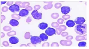
common sort of cancer in children. All cancers begin in body cells, and leukaemia may be a cancer that begins in blood cells. Generally, cells grow and multiply to create new cells because the body desires them. When body cells grow old day by day, they die and new cells take their place. Sometimes, this cycle doesn't work properly. In cancer, new cells area unit shaped once the body doesn't would like them, and old cells don't die once they should[l]. The bone marrow is that the web site wherever lymphocytes and alternative blood cells are created. It’s a spongy tissue found within several large bones of the body. The bone marrow produces 3 types of blood cells: RBCs contain haemoprotein and carry oxygen and alternative materials to the tissues throughout the body. Platelets facilitate to create clots; WBCs facilitate rebuff infections in the body.
Figure 1 Microscopic Image of Blood
Volume 3 Issue 3 Sept 2018 30 created, the blood won't clot properly, and also
the patient might bleed simply. Once WBCs are not plentiful enough, the body cannot rebuff germs and also the person might develop a frequent infection. Leukaemia will be either acute or chronic in type[l].
The first and quick identification of leukaemia greatly aids in providing the acceptable treatment. Initial segmentation is completed victimization applied mathematics parameters like mean, a typical deviation that segregates white blood cells from different blood parts i.e. erythrocytes and platelets. Geometrical features like space, perimeter of the white vegetative cell nucleus investigated for diagnostic prediction of leukaemia. The planned method is with success applied to an oversized variety of pictures, showing promising results for varied image quality. Different image process algorithms like Image sweetening, Thresholding.
LITERATURE SURVEY
S.Jagadeesh, Dr.E.Nagabhooshanam, Dr.S.Venkatachalam proposed segmentation of the image of bone marrow aspirate mistreatment watershed formula,[1] the extraction of individual cells from the image, automatic generation of different options of the cell, assessment of the feature quality of the cells mistreatment analysis of distribution, correlation and principal part analysis, application of support vector machine for final recognition and classification of cells.
Jamali Firmat Banzi, Xue Zhaojun projected a framework.[1] the primary step is to accumulate the photographs then they performed median filter to get rid of the noise while not blurring the image then next step distinction between the cytoplasm, and nuclei and extracellular elements were enhanced mistreatment Associate in Nursing unsharp filter is finished. Then thresholding is finished
and that they performed fourier remodel and then log remodel is finished. Then mean filter is applied and in ending line plot is finished. MATLAB is employed to perform this work. Ms. Sneha Dhakne, Ms. Kumudini K. Borkute, Ms. Priyanka Ikhar conferred 2 techniques like Watershed formula and mix agglomeration besides filtering for determination of cell [2]. Monika Mogra , Vivek Srivastava., projected watershed transform and morphological image process technique[3] to spot affected White vegetative cell and applied this method in MATLAB.
Subhan, Ms. Parminder Kaur., mentioned KNN and Hough Transform algorithms to observe abnormal cells that cause leukaemia [4]. Khot S.T, Sneha Bhalekar, Divya Jaggi and Dolly aristocrat used Support Vector Machine classifier to observe leukemic cells.[5] The options are extracted from the image and applied to classifier.
Ms. Minal D. Joshi, Prof. Atul H. Karode, Prof. S.R.Suralkar projected a technique to observe cancer of the blood. By bar chart effort methodology, [6] enhance distinction of the grayscale image. Applied international thresholding Otsu method so feature extraction is finished. Supported the feature extracted, classifier classifies the affected cells or normal cells and with the accuracy of ninety three. kNN classification is employed.
Emad A. Mohammed, Mostafa M. A. Mohammed, Christopher Naugler, Behrouz H. way projected a way for leucaemia cell segmentation.[7] within the proposed approach for segmenting a nucleus initial an optimal threshold worth is obtained mistreatment Otsu’s methodology. Canny edge detector is applied followed by erosion and dilation. Isolated pixels ar removed and a metameric nucleus mask is obtained.
Volume 3 Issue 3 Sept 2018 31 Myelogenous Leukemia) in microscopic
images of blood.[8] 3 clusters ar known that represent nucleus, background and alternative cells like erythrocytes, leukocytes etc. each pel of the image is assigned to 1 of those clusters supported the cluster property. The technique performs some preprocessing followed by k-means agglomeration for segmentation purpose. The segmentation is performed to extract the leukocytes’ nuclei mistreatment color based mostly agglomeration.
Subrajeet Mohapatra, Dipti Patra and Sanghamitra Satpathy projected Color based mostly agglomeration for blood cell nucleus segmentation of stained blood smear pictures followed by relevant feature extraction for cancer of the blood detection.[9] Few normal agglomeration techniques, viz., KMeans, KMedoid, Fuzzy C-Means (FCM), Gustafson Kessel (GK) and Fuzzy Possibilistic C suggests that (FPCM) were used for color based mostly segmentation and their performance were compared. Nucleus boundary irregularities are measured mistreatment hausdroff dimension and contour signature. Results were obtained by mistreatment SVM classifier with projected options.
PROPOSED METHOD
Figure 2 System Architecture
a) Pre-processing
The images generated by digital microscopes are sometimes in RGB colour space. Usually the blood cells and image background vary greatly with relation to colour and intensity. This is often caused by multiple reasons like camera settings, varying enlightenment, and aging blemish. Cell segmentation is different with relation to these variations, therefore a method is used to convert RGB input image into the CIELAB colour house [1].The a and b elements is employed to create correct color balance corrections. The L*a*b*color space with dimension L represents the lightness of the colour, element a, that represents its position between red/magenta and inexperienced, and part b*that represents its position between yellow and blue.
b) Segmentation
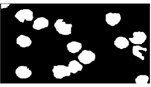
Volume 3 Issue 3 Sept 2018 32 different cell. The cluster that contains the blue
nucleus are measured, that is important for the feature extraction.
Figure 3Pre-processing Image
Figure 4 Clustering on Image
Transformation of the input file into the set of options is termed feature extraction. Feature choice influences the classifier performance in order that an accurate alternative of options should be known. I even have thought of the subsequent feature for the economical work.
c) Feature Extraction
i) Hausdorff dimension
HD is employed to observe the sting of the nucleus and it's thought of a necessary feature. Box investigation technique is implemented to observe the sting of nucleus. The perimeters ar detected to achieve the perimeter roughness of the nucleus. When the grid becomes finer and finer the perimeter roughness of the nucleus will increase.
ii) Native Binary Pattern
The native Binary Pattern (LBP) is employed for texture classification [2]. LBP quality options have the following characteristics: LBP is strong against illumination variations; quick to compute; don't need several parameters; a neighbourhood feature; LBP is invariant to
monotonic grey scale transformations and scaling it's performed all right in abundant computer vision application. The LBP technique has tested to vanquish in, Linear Discriminated Analysis (LDA) and the Principal element Analysis (PCA). To covenant with textures at completely different scales, the LBP operator is stretched to use regions of various dimensions.
d) Classification
Support Vector Machine (SVM) is employed for constructing a choice surface within the feature house that bisects the 2 categories. Here the classification relies upon 2 categories i.e., cancerous and noncancerous. A 2 category classifier is used to categorise the categories. The feature values are premeditated on the choice surface and most hyper-plane are drawn separating the 2 categories. The support vector is drawn by creating a large margin. To evade misclassification the boundary are wider from the hyper plane. The technique is affordable and doesn't would like kernel trick. It’s same to be performed sensible.
RESULTS AND DISCUSSION
The work is developed for automatically detecting and classifying the AML in Blood microscopic pictures. a hundred and twenty blood microscopic pictures are classified and therefore the analysis is additionally measured consistent with the performance analysis technique. The pre-processing methodology looks to be effective for strong segmentation. The segmentation technique suits for color pictures in order that straightforward to spot the blue nucleus. The feature extracted is helpful for classification of the info. The performance assessment shows the classifier performance.
CONCLUSION
Volume 3 Issue 3 Sept 2018 33 which may know through examination of blood
and bone marrow smears by trained consultants. This Manual review method is long and error prone, therefore a computer-based system for automatic detection of Acute Lymphocytic Leukaemia could offer an assistive diagnostic tool for pathologists. The automated segregation and identification algorithmic program aims to reduce the phase involved in treatment, that is typically life threatening. The planned automatic system is tested with Nearest Neighbour (kNN) and SVM Classifier on the dataset of one hundred twenty pretested samples, the accuracy achieved is 90.8%. The results show that algorithm planned achieves an appropriate performance for the designation of Acute Lymphocytic Leukaemia; additional the devised methodology conjointly addresses the segmentation of overlapping cells.
REFERENCES
[1] S.Jagadeesh 1, Dr.E.Nagabhooshanam 2, Dr.S.Venkatachalam., “Image processing based approach to cancer cell prediction in blood samples” International Journal of Technology and Engineering Sciences Vol.1 (1), ISSN: 2320-8007
[2] Jamali Firmat Banzi, Xue Zhaojun., “Detecting Morphological Nature of Cancerous Cell Using Image Processing Algorithms” International Journal of Scientific and Research Publications, Volume 3, Issue 12, December 2013 1 ISSN 2250-3153
[3] Ms. Sneha Dhakne, Ms. Kumudini K. Borkute, Ms. Priyanka Ikhar., “Detection of cancer cell in blood samples using an Effective algorithm” (IJCESR) ISSN (PRINT): 2393-8374, (ONLINE): 2394-0697, VOLUME-2, ISSUE-6, 2015
[4] Monika Mogra , 2,Vivek Srivastava., “ A Comprehensive Review of Analysis of Counting Blood Cells Using Different Image Processing Algorithms” International Journal of Engineering Science Invention ISSN (Online): 2319 – 6734, ISSN (Print): 2319 – 6726 www.ijesi.org Volume 3 Issue 6ǁ June 2014 ǁ PP.29-31
[5] Subhan, Ms. Parminder Kaur., “ Significant Analysis of Leukemic Cells Extraction and Detection Using KNN and Hough
TransformAlgorithm” International Journal of Computer Science Trends and Technology (IJCST) – Volume 3 Issue 1, Jan-Feb 2015.
[6] Khot S.T, Sneha Bhalekar, Divya Jaggi and Dolly Rani., “An innovative approach in myelogenous leukemia detection using attribute analysis” (IJAEEE) ISSN (Print): 2278-8948, Volume-2, Issue-5, 2013.
[7] Ms. Minal D. Joshi, Prof. Atul H. Karode, Prof. S.R.Suralkar “White Blood Cells Segmentation and Classification to Detect Acute Leukemia” (IJETTCS) Volume 2, Issue 3, May – June 2013.
[8] Emad A. Mohammed, Mostafa M.A.Mohammed, Christopher Naugler, Behrouz H. Far, “Chronic Lymphocytic Leukemia Cell Segmentation From Microscopic Blood Images Using Watershed Algorithm and Optimal Thresholding”, 26th IEEE Canadian Conference Of Electrical And Computer Engineering (CCECE), 2013.
[9] Sos Agaian, Monica Madhukar, Anthony T. Chronopoulos, “Automated Screening System for Acute Myelogenous Leukemia Detection in Blood Microscopic Images”, IEEE Systems Journal.