Available online on 15.10.2018 at http://jddtonline.info
Journal of Drug Delivery and Therapeutics
Open Access to Pharmaceutical and Medical Research© 2011-18, publisher and licensee JDDT, This is an Open Access article which permits unrestricted non-commercial use, provided the original work is properly cited
Open Access
Research Article
FORMULATION DEVELOPMENT OF ACYCLOVIR MICROSPHERE
USING NOVEL NATURAL POLYMER
Singh Priyanka*
1, Lariya Shailendra K
21*
Department of pharmaceutics, Pacific academy of higher education & research University, Udaipur, India
2
Radharaman College of pharmacy, Bhopal, India
ABSTRACT
Acyclovir [9-(2-hydroxyethoxymethyl) guanine] is an acyclic nucleoside analogue of guanosine that is a potent and selective antiviral agent. It has a relatively short plasma half-life (3 hr). When orally administered, it is slowly and scarcely absorbed from the gastrointestinal tract. The objective of the present work was to formulate and evaluate microspheres of Acyclovir and produced sustained drug delivery. In these 14 batches of acyclovir microspheres was prepared with using natural polymer Kondagogu gum and other ingredients by solvent evaporation technique. The prepared microspheres were evaluated for different parameters i.e % Drug yield, % drug entrapment, shape, surface morphology, particles size, polydispersity index, zeta potential and in-vitro drug release for 48 hrs in phosphate buffer 7.4. The best batch was performed stability studies for 6 months. The research concluded that Acyclovir microspheres could be an alternative for conventional dosage form and other phytochemical in herbs.
Keywords: Acyclovir, Microspheres, Kondagogu gum, Polydispersity index, in-vitro drug release
Article Info: Received 08 Sep, 2018; Review Completed 07 Oct 2018; Accepted 08 Oct 2018; Available online 15 Oct 2018
Cite this article as:
Singh P, Lariya SK, Formulation development of acyclovir microsphere using novel natural polymer, Journal of Drug Delivery and Therapeutics. 2018; 8(5-s):271-276 DOI:http://dx.doi.org/10.22270/jddt.v8i5-s.1971
*
Address for Correspondence:Priyanka Singh, Department of pharmaceutics, Pacific academy of higher education & research University, Jaipur, India
INTRODUCTION
Microspheres are one of the multi particulate drug delivery systems and are prepared to obtain prolonged (or) controlled drug delivery, to improve bioavailability or stability and to target drug to specific sites. Microspheres can be defined as solid, approximately spherical particles ranging from 1 to 1000μm, containing dispersed drug in either solution (or) microcrystalline form 1, 2. These microspheres are suitable alternatives for conventional dosage forms3. Several methods, including Emulsion solvent evaporation technique4, phase-separation or coacervation method5, emulsification diffusion method and spray drying method6 are commonly used for the preparation of microspheres. The solvent evaporation method has gained much attention due to its ease of fabrication without compromising the activity of drug. This technique offers several advantages and is preferable to other preparation methods such as spray
medical technology. Ceramic microspheres are used primarily as grinding media. Microspheres vary widely in quality, sphericity, uniformity of particle and particle size distribution9. Gums are natural polymers, which mainly consists of carbohydrates sometimes with small amounts of proteins and minerals. Increasing demand of natural ingredients over synthetic ones immensely contribute to explore and develop new plant based materials. Gum kondagogu (Cochlospermum gossypium) is a tree exudate derived from the Bixaceae family, riginating from India. Natural gums are obtained as exudates from different tree species, which exhibit unique and diverse physicochemical properties and have a wide variety of applications10. Commercially important tree gums include gum arabic, gum karaya, and gum tragacanth11. Karaya polysaccharide (Sterculia urens) and gum kondagogu (C. gossypium) are used as food additives12, 13. The physicochemical properties and toxicological evaluation of gum kondagogu has been established earlier14, 15. Morphological and structural characterization and physicochemical aspects of gum kondagogu have been elucidated recently, suggesting that this gum belongs to the group of substituted rhamnogalacturonans16. Understanding of the rheological properties of gum is essential for their application and use as food thickeners, stabilizers, and emulsifiers. Acyclovir (ACV) is known by its IUPAC
name as
2-Amino-1,9-dihydro-9-[(2-hydroxyethoxy)methyl]-6H-purin-6-one17. Acyclovir is a synthetic purine nucleoside analogue with in vitro and in vivo inhibitory activity against HSV-1, HSV-2 and Varicella Zoster Virus (VZV). Acyclovir is highly selective due to its affinity for the enzyme thymidine kinase (TK) encoded by HSV and VZV. This viral enzyme converts acyclovir into acyclovir monophosphate, a nucleoside analogue. The mono phosphate is further converted into tri phosphate by a number of cellular enzymes. Under in vitro conditions, acyclovir tri phosphate stops replication of herpes virus DNA. This is accomplished in three ways: competitive inhibition of viral DNA polymerases, incorporation into and termination of the viral DNA chain and inactivation of the viral DNA polymerases. The greater antiviral activity of acyclovir against HSV compared to VZV is due to its enhanced phosporylation by the viral TK18. The main objective of this work was to investigate the possibility of obtaining a sustained release formulation of acyclovir microspheres by the solvent evaporation method using gum kondagogu and Investigation of the effect of drug to polymer ratio.
MATERIAL AND METHOD
Material
Acyclovir was procured from Scan Research Laboratories, Bhopal (MP). Kondagogu Gum was purchased from Himedia, Mumbai. Dialysis membrane (MWCO, 15 KDa), Span 80, Tween 80, Gulutaraldehyde, Toluene were purchased from Himedia (Mumbai, India). All other reagents and chemicals used were of analytical grade.
Method
Preparation of kondagogu gum microsphere
The microspheres of the polysaccharide, Kondagogu Gum were prepared by emulsifying method using liquid paraffin as a dispersing medium and glutaraldehyde used as a cross-linking agent. Kondagogu gum dispersion (2.5 % w/v) was prepared by mixing of kondagogu gum in double distilled water with Tween 80 (0.5% w/w). Drug was previously dissolved in double distilled water. The prepared, 10 ml of kondagogu gum solution with drug was added drop wise in a beaker containing 100 ml of liquid paraffin light and heavy in ratio of 50:50. Span 80 (1.0% w/v) was previously added in liquid paraffin. The system was kept under stirring at 3000-4000 rpm using two blade mechanical stirrers. 1.5 ml of toluene saturated glutaraldehyde was added to above solution after 30 min of stirring. Stirring was continued for 4 hr at 40ᵒC at 4000 rpm. The microspheres were separated from dispersion medium by centrifugation after stirring and washed two times with petroleum to remove liquid paraffin and then washed three times with acetone. Dispersion was poured in petridish to remove acetone. After complete evaporation of acetone, dried drug loaded microsphere were collected and stored in tight container for further evaluation.
Optimization of microsphere
Optimization of microsphere formulation was carried out by optimizing the different dependent and independent process and formulation variables. Optimization was carried out on the basis of particles size, polydispersity index and % drug entrapment and it is done by changing the one variable and kept constant for other variables given in table 1. The temperature was maintained at 40ᵒC and the concentration of toluene, saturated glutaraldehyde was used 1.5 ml in the preparation of each formulation.
In Vitro Characterization of Microspheres
Particle size, polydispersity index
Average particles size, polydispersity index (PDI) of prepared microsphere was determined using zetasizer (DTS were.4.10, Horriba instrument, India). The microsphere formulation was diluted with deionized water (1:9 v/v) and analysed for average size and PDI and it was performed at the department of pharmaceutical science, VNS pharmacy college Bhopal, India Table 1.
Shape and surface morphology
The shape and surface morphology of the microspheres were investigated using scanning electron microscopy (IISER, Bhopal). The microspheres were fixed on supports with carbon-glue, and coated with gold using a gold sputter module in a high-vacuum evaporator. Samples were then observed with the Scanning Electron Microscope at 10 kV.
Determination of drug content
analysis into the UV spectrophotometer system at λmax 252 nm.
In Vitro drug release from microspheres
The drug release was performed in PBS (7.4 pH) for acyclovir loaded kondagogu gum microsphere. The drug release was performed in PBS (7.4 pH) for prepared microsphere using dialysis bag technique. In this study suspension of microsphere equivalent to 20 mg of drug was taken in dialysis tubing (MWCO, 15KDa, himedia) and placed in a beaker containing 50ml of PBS pH 7.4. The dialysis bag retains microsphere and allows passing of free drug into the dissolution media. Temperature was maintained at 37±10C throughout the study. The samples were withdrawn after specified time intervals that is 0.5, 1, 2, 3, 4, 5, 6, 7.8.12, 24 and 48 hrs and replaced with the same volume of fresh PBS pH 7.4 and analyzed for drug concentration by using UV spectrophotometer a λmax 252 nm Table 2.
RESULT AND DISCUSSION
The mean diameter of glutaraldehyde cross linked microspheres of kondagogu gum increased from 56.70±2.15 µm to 88.15±4.25 µm with increasing
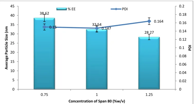
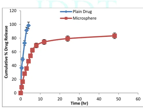
polymer concentration from 1.0 to 3.0 % w/v. In the present investigation a 2.5% w/v kondagogu gum concentration was found to be optimized which give the required size of microspheres. The average particle size of microspheres increased with increasing polymer concentration, since at higher concentrations the polymer solution dispersed into larger droplets due to increasing the viscosity of polymer solution and it was the reason behind the enhancement of average particle size of microsphere. Mean particle size and size distribution were studied to observe the effect of drug concentration. It was found from previous study that there was no major change observed on particle size and size distribution of microsphere with varying concentration of the cross linking agent. Percent encapsulation efficiency has increased up to 77.37±3.15% with increasing polymer drug concentration from 15% to 25 % w/w. But further increasing the concentration of drug, there was no significant enhancement was found in entrapment efficiency. The in vitro dissolution profile of acyclovir in PBS pH 7.4 was found 83.46% after 48 hr for optimized formulation (KMTSD-13) and follow the matrix diffusion Higuchi release kinetics Fig 1-6.
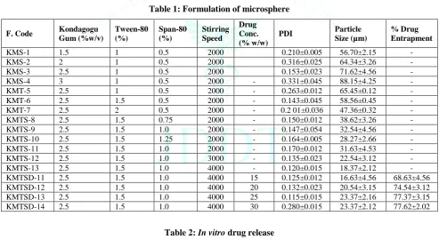
Table 1: Formulation of microsphere
F. Code Kondagogu Gum(%w/v)
Tween-80 (%)
Span-80 (%)
Stirring Speed
Drug Conc. (% w/w)
PDI Particle
Size (µm)
% Drug Entrapment
KMS-1 1.5 1 0.5 2000 0.210±0.005 56.70±2.15 - KMS-2 2 1 0.5 2000 0.316±0.025 64.34±3.26 - KMS-3 2.5 1 0.5 2000 0.153±0.023 71.62±4.56 - KMS-4 3 1 0.5 2000 - 0.331±0.045 88.15±4.25 - KMT-5 2.5 1 0.5 2000 - 0.263±0.012 65.45±0.12 - KMT-6 2.5 1.5 0.5 2000 - 0.143±0.045 58.56±0.45 - KMT-7 2.5 2 0.5 2000 - 0.2 01±0.036 47.36±0.32 - KMTS-8 2.5 1.5 0.75 2000 - 0.150±0.012 38.62±3.26 - KMTS-9 2.5 1.5 1.0 2000 - 0.147±0.054 32.54±4.56 - KMTS-10 2.5 1.5 1.25 2000 - 0.164±0.005 28.27±2.66 - KMTS-11 2.5 1.5 1.0 2000 - 0.170±0.012 31.63±4.53 - KMTS-12 2.5 1.5 1.0 3000 - 0.135±0.023 22.54±3.12 - KMTS-13 2.5 1.5 1.0 4000 - 0.120±0.015 18.37±2.12 - KMTSD-11 2.5 1.5 1.0 4000 15 0.125±0.012 16.63±4.56 68.63±4.56 KMTSD-12 2.5 1.5 1.0 4000 20 0.132±0.023 20.54±3.15 74.54±3.12 KMTSD-13 2.5 1.5 1.0 4000 25 0.115±0.015 23.37±2.16 77.37±3.15 KMTSD-14 2.5 1.5 1.0 4000 30 0.280±0.015 23.37±2.12 77.62±2.02
Table 2: In vitro drug release
S. No. Time interval
(h)
Plain drug Acyclovir Loaded
Microsphere
1 0.5 36.59 08.43
2 1 49.15 16.53
3 2 72.79 28.26
4 3 91.38 35.68
5 4 98.49 46.35
6 5 54.23
7 6 62.45
8 8 69.38
9 12 74.43
10 24 79.34
Figure 1: Effect of Kondagogu gum concentration on average particle size and PDI of microsphere
Figure 2: Effect of concentration of Tween 80 on average particle size and PDI of microsphere
Figure 3: Effect of Span 80 concentration on average particle size and PDI of microsphere
56.7
64.34
71.62
88.15 0.22
0.21
0.153
0.131
0 0.05 0.1 0.15 0.2 0.25
0 10 20 30 40 50 60 70 80 90 100
1.5 2 2.5 3
P
D
I
A
ve
rage
P
arti
cl
e
Si
ze
(nm
)
Concentration of Polymer (%)
Average Particle Size (µm) PDI
65.45
58.56
47.36 0.263
0.143
0.126
0 0.05 0.1 0.15 0.2 0.25 0.3
0 10 20 30 40 50 60 70 80
1 1.5 2
P
D
I
A
ve
rage
P
arti
cl
e
Si
ze
(nm
Concentration of Tween 80 (%w/v)
%EE PDI
38.62
32.54
28.27
0.15 0.147
0.164
0 0.02 0.04 0.06 0.08 0.1 0.12 0.14 0.16 0.18 0.2
0 5 10 15 20 25 30 35 40 45
0.75 1 1.25
P
D
I
A
ve
rage
P
arti
cl
e
Si
ze
(nm
Concentration of Span 80 (%w/v)
Figure 4: Effect of stirring speed concentration on average particle size and PDI of microsphere
Figure 5: Effect of drug concentration on average particle size and % entrapment efficiency of microsphere
Figure 6: Cumulative % acyclovir release
CONCLUSION
It was concluded that from this study that the microsphere can be prepared from kondagogu gum by emulsifying solvent evaporation method and can be loaded with drug acyclovir for it sustained delivery in GIT system. The prepared microspheres were optimized
for different formulation and process variables concentration and found that microsphere was uniform and acceptable size range. They were found smooth and spherical in shape. The optimized formulation was found significant loading efficiency of acyclovir that can release the acyclovir in sustained manner which was followed matrix diffusion Higuchi release kinetic. 31.63
22.54
18.37 0.17
0.135
0.12
0 0.02 0.04 0.06 0.08 0.1 0.12 0.14 0.16 0.18 0.2
0 5 10 15 20 25 30 35
2000 3000 4000
P
D
I
A
ve
rage
P
arti
cl
e
Si
ze
(nm
Stirring Speed (rpm)
% EE PDI
16.63
20.54
23.37 23.37
68.63
74.54 77.37 77.62
0 10 20 30 40 50 60 70 80 90
0 5 10 15 20 25 30
15 20 25 30
%
Ent
rapm
e
nt
E
ff
ici
e
ncy
A
ve
rage
P
A
rt
icl
e
S
iz
e
(nm
)
Drug Concentration (%)
Average Particle Size (nm) % EE
0 20 40 60 80 100 120
0 10 20 30 40 50 60
C
um
ul
at
iv
e
% Drug
R
e
le
ase
REFERENCES
1. Choi BY, Park HJ, Hwang SJ. Preparation of alginate beads
for floating drug delivery system effects of CO2 gas forming agents, International Journal of Pharmaceutics, 2002; 239:81-91.
2. Tekade BW, Jadhao UT, Thakare VM, Yogita A, Chaudhari,
Vaishali DP, Chaudhari C S. Design and In-vitro evaluation
of ethyl cellulose based floating microspheres containing
antidiabetic drug, Asian Journal of Biomedical and
Pharmaceutical Sciences, 2013; 3 (23):33-37.
3. Malaekeh-Nikouei B, Tabassi SAS, Jaafari MR, Davies NM.
Preparation and characterization of PLGA microspheres
loaded by cyclosporine-cyclodextrin complex, Iranian
Journal of Pharmaceutical Sciences, 2005; 1(4):195-201.
4. Karal-Yılmaz O, Serhatl M, Baysal K, Baysal BM.
Preparation and in vitro characterization of vascular endothelial growth factor (VEGF)-loaded poly(D,L-lactic-co-glycolic acid) microspheres using a double emulsion/solvent evaporation technique, Journal of Microencapsulation, 2011; 28(1):46-54.
5. El-Bagory IM, Hosny EA, Al-Suwayeh SA, Mahrous GM,
Al-Jenoobi FI. Effects of sphere size, polymer to drug ratio and plasticizer concentration on the release of theophylline from ethylcellulose microspheres, Saudi Pharmaceutical Journal, 2007; 15(3-4):213-217.
6. Liu H, Pan W, Ke P, Dong Y, Ji L. Preparation and
evaluation of a novel gastric mucoadhesive sustained-release acyclovir microsphere, Drug Development and Industrial Pharmacy, 2010): 36(9):1098-1105.
7. Basu SK, Adhiyaman R. Preparation and characterization of
nitrendipine loaded eudragit rl 100 microspheres prepared by an emulsion-solvent evaporation method, Tropical Journal of Pharmceutical Research, 2008; 7 (3):1033-1041.
8. Lakshmana PS, Shirwaikar AA, Shirwaikar A, Kumar A.
Formulation and evaluation of sustained release microspheres
of rosin containing aceclofenac, ARS Pharmaceutica,
2009;50 (2):51-62.
9. Kataria S, Middha A, Sandhu P, Bilandi A, Kapoor B.
Microspheres- a review, International Journal of Research in
Pharmacy and Chemistry, 2011; 1(4):1184-1198.
10. Verbeken D, Dierchx S, Dewettinck K. Exudate gums:
occurrence, production and applications, Applied
Microbiology and Biotechnology, 2003; 63:10-21.
11. Phillips GO, Williams PA. Tree exudates gums: natural and
versatile food additives and ingredients, Food Ingredients
Anal Int,2001; 23:26-28.
12. FDA (Food, Drug Administration). Sterculia (karaya) gum,
Federal Register, 1974; 39:34209–34211.
13. FAO (Food Agriculture Organization). Compendium of Food
Additive Specifications; FAO: Rome, Italy, 1991; 11:821- 823.
14. Janaki B, Sashidhar RB. Physico-chemical analysis of gum
kondagogu (Cochlospermum gossypium): A potential food
additive, Food Chemistry, 1998; 61:231-236.
15. Janaki B, Sashidhar RB. Sub-chronic (90-day) toxicity study
in rats fed gum kondagogu (Cochlospermum gossypium),
Food Chemistry Toxicology, 2000; 38:523-534.
16. Vinod VTP, Sashidhar RB, Suresh KI, Rama Rao B, Vijaya
Saradhi UVR, Prabhakar Rao T. Morphological, physico-chemical and structural characterization of gum kondagogu (Cochlospermum gossypium): a tree gum from India, Food Hydrocolloids, 2008; 22 (5):899-915.
17. Hayden FG. Antiviral agents. In: Hardman JG, Limbird LE,
editors. Goodman & Gilman's The Pharmacological Basis of
Therapeutics, 9th ed., McGraw-Hill, USA, 1996; 1195.
18. Celum C, et al. Effect of aciclovir on HIV-1 acquisition in
herpes simplex virus 2 seropositive women and men who have sex with men: a randomised, double-blind,