INTEGRATING GENOMICS AND TRANSCRIPTOMICS
TO UNDERSTAND THE VIRULENCE AND BIOFILM
FORMING MECHANISM OF SELECTED
VANCOMYCIN-RESISTANT Enterococcus faecium
LIM SHU YONG
FACULTY OF SCIENCE
UNIVERSITY OF MALAYA
KUALA LUMPUR
INTEGRATING GENOMICS AND
TRANSCRIPTOMICS TO UNDERSTAND THE
VIRULENCE AND BIOFILM FORMING MECHANISM
OF SELECTED VANCOMYCIN-RESISTANT
Enterococcus faecium
LIM SHU YONG
DESSERTATION SUBMITTED IN FULFILMENT OF
THE REQUIREMENTS FOR THE DEGREE OF MASTER
OF SCIENCE
INSTITUTE OF BIOLOGICAL SCIENCES
FACULTY OF SCIENCE
UNIVERSITY OF MALAYA
KUALA LUMPUR
UNIVERSITY OF MALAYA
ORIGINAL LITERARY WORK DECLARATION
Name of Candidate: LIM SHU YONG
Registration/Matric No: SGR140051
Name of Degree: MASTER OF SCIENCE
Title of Project Paper/Research Report/Dissertation/Thesis (“this Work”):
INTEGRATING GENOMICS AND TRANSCRIPTOMICS TO UNDERSTAND THE VIRULENCE AND BIOFILM FORMING
MECHANISM OF SELECTED VANCOMYCIN-RESISTANT Enterococcus faecium
Field of Study:
MICROBIOLOGY
I do solemnly and sincerely declare that:
(1) I am the sole author/writer of this Work;
(2) This Work is original;
(3) Any use of any work in which copyright exists was done by way of fair dealing
and for permitted purposes and any excerpt or extract from, or reference to or reproduction of any copyright work has been disclosed expressly and sufficiently and the title of the Work and its authorship have been acknowledged in this Work;
(4) I do not have any actual knowledge nor do I ought reasonably to know that the
making of this work constitutes an infringement of any copyright work;
(5) I hereby assign all and every rights in the copyright to this Work to the
University of Malaya (“UM”), who henceforth shall be owner of the copyright in this Work and that any reproduction or use in any form or by any means whatsoever is prohibited without the written consent of UM having been first had and obtained;
(6) I am fully aware that if in the course of making this Work I have infringed any
copyright whether intentionally or otherwise, I may be subject to legal action or any other action as may be determined by UM.
Candidate’s Signature: Date:
Subscribed and solemnly declared before,
Witness’s Signature: Date:
Name: PROFESSOR DR THONG KWAI LIN
INTEGRATING GENOMICS AND TRANSCRIPTOMICS TO UNDERSTAND THE VIRULENCE AND BIOFILM FORMING MECHANISM OF SELECTED VANCOMYCIN-RESISTANT Enterococcus faecium
ABSTRACT
Vancomycin-resistant enterococcus (VRE) is an emerging nosocomial pathogen
which causes outbreaks in hospitals worldwide. It is, therefore, important to understand
the virulence and how this organism persists as a nosocomial pathogen. Whole genome sequencing (WGS) provides a wealth of information to elucidate the genetic relationship, virulence potential, and resistance factors of VRE. However, such genomic studies are lacking in Malaysia. Moreover, since enterococci are often recovered from difficult-to-treat biofilm-mediated infections, a detailed study on the biofilm formed by VRE is useful to better understand its pathogenicity. The objectives of this study are to perform
comparative genomics analysis on the four local vancomycin-resistant Enterococcus
faecium (VREfm) and to elucidate the transcriptomic profile of biofilm cells in respect to the planktonic cells. Four VREfm strains were isolated from two fatal cases of nosocomial infections in a tertiary hospital in Kuala Lumpur. One of these strains (VRE2) was isolated from an index case (patient X), whereas the other three (VREr5, VREr6, VREr7) were isolated from different body sites of another patient (patient Y) at around one-week interval. WGS and comparative genomics analyses revealed that the four strains have different sequence types (STs), ST80 and ST203. Subsequent phylogenomic study showed that VREr5 was more closely related to VRE2, but was distantly related to VREr6 and VREr7 derived from the same host. Moreover, the genomic contents of VREr5 was also more similar to VRE2. The genomic data and clinical records suggested that patient Y was most probably infected by multiple strains of different clones. Alternatively, the strain that infected patient Y (VREr5) could have derived from the same clone from
patient X (VRE2), given their high genomic similarity. The four local strains were
multidrug resistant. All of them carried the vanA genotype and showed indistinguishable
Tn1546 structure. Virulence profiling revealed that these strains harbored a total of 13
virulence genes mainly associated with adherence and biofilm formation. The transcriptomic analysis focused on the initial stage of biofilm formation to examine genes that are involved during the transition from planktonic to biofilm cells. Differential gene expression analysis revealed that the up-regulated genes in biofilm cells involved mainly in adherence, plasmid replication, and carbohydrate metabolism. Genes that have been reported to negatively regulate biofilm formation, such as the quorum sensing systems,
fsr and luxS, and a transcriptional regulator gene spx, were highly down-regulated. The
unique bee homolog of VREr5 was found to be down-regulated, implying a negative
association of this locus to biofilm formation in VREr5. The results obtained from the gene expression study clearly reflected the attachment stage of biofilm development, including the preparation to enter the maturation stage. In conclusion, this study has contributed to the understanding of the genetic basis and diversity of local clinical strains which can be helpful to control the spread of VRE. This study also provides insight into the molecular mechanism of biofilm formation in VRE which might be useful in the development of new drugs.
INTEGRASI GENOMIK DAN TRANSKRIPTOMIK DALAM MEMAHAMI
KEVIRULENAN DAN PEMBENTUKAN BIOFILEM DALAM Enterococcus
faecium TERPILIH
ABSTRAK
Enterococcus yang resistan terhadap vancomycin (vancomycin-resistant enterococcus atau VRE) adalah patogen nosokomial yang menyebabkan wabak di hospital-hospital di seluruh dunia. Oleh itu, pengetahuan mengenai kevirulenan dan bagaimana organisma ini tegar sebagai patogen nosokomial adalah penting. Penjujukan Genome Kesuluruhan (whole genome sequencing, WGS) menyediakan pelbagai maklumat bagi mengkaji hubungan genetik, potensi virulens, dan faktor-faktor resistan VRE. Namun begitu, kajian genomik tersebut masih kurang di Malaysia. Selain itu, enterococci sering
diperolehi daripada jangkitan berkaitan dengan biofilem yang sukar untuk dirawat. Oleh itu, satu kajian terperinci mengenai biofilem yang dibentuk oleh VRE adalah berguna bagi lebih memahami pathogeniciti organisma ini. Objektif-objektif kajian ini adalah untuk melaksanakan analisis genomik perbandingan kepada empat Enterococcus faecium tempatan yang resistan terhadap vancomycin (VREfm) dan untuk mengkaji profil transkriptomik sel-sel biofilem yang berkait dengan sel-sel plankton. Keempat-empat strain yang dikaji diperolehi daripada dua kes maut jangkitan nosokomial di hospital pengajian tinggi di Kuala Lumpur. Salah satu strain yang dikaji (VRE2) diperolehi daripada kes indeks (pesakit X) manakala tiga strain lain (VREr5, VREr6, VREr7) diperolehi daripada pesakit tunggal (pesakit Y), daripada bahagian badan yang berbeza sekitar selang satu minggu. WGS dan analisis perbandingan genomik
menunjukkan bahawa keempat-empat strain mempunyai pelbagai jenis jujukan (ST) iaitu ST80 dan ST203. Kajian phylogenomic seterusnya menunjukkan bahawa VREr5
berasal daripada pesakit yang sama. Selain itu, kandungan genom VREr5 juga lebih serupa dengan VRE2. Data genomik dan rekod klinikal mencadangkan bahawa pesakit Y mungkin dijangkiti oleh pelbagai jenis klon yang berbeza. Sebagai alternatif, strain yang menjangkiti pesakit Y (VREr5) boleh diperolehi daripada klon yang sama seperti strain pesakit X (VRE2) memandangkan persamaan genomik yang tinggi. Semua strain yang dikaji mempunyai resistan terhadap pelbagai ubat. Mereka membawa genotip
vanA dan mempunyai struktur Tn1546 yang sama. Profil virulens mendedahkan bahawa strain ini menangandungi tiga belas gen virulens yang berkaitan dengan perekatan dan pembentukan biofilem. Analisis transkriptomik berfokus kepada peringkat awal pembentukan biofilem bagi mengkaji gen-gen yang terlibat semasa peralihan dari plankton kepada biofilem. Analisis perbezaan expresi gen mendedahkan penglibatan gen “up-regulated” dalam perekatan, replikasi plasmid, dan metabolisme karbohidrat dalam sel-sel biofilem. Gen-gen yang mempunyai pegawalan negatif terhadap
pembentukan biofilem, seperti sistem kuorum sensing, fsr dan luxS dan transkripsi gen pengatur spx, adalah “down-regulated”. Di samping itu, homolog bee telah didapati “down-regulated”, mencadangkan bahawa locus ini mungkin tidak berkaitan dengan pembentukan biofilem bagi VREr5. Keputusan yang diperolehi daripada kajian transkriptomik menunjukkan peringkat perekatan pembentukan biofilem termasuk persediaannya untuk memasuki peringkat matang.
Secara ringkasnya, kajian ini telah menyumbang kepada pemahaman asas genetik dan kepelbagaian genetik dalam strain klinikal tempatan. Pengetahuan ini boleh digunakan untuk mengawal penyebaran VRE. Kajian ini juga menyumbang kepada pengetahuan baru berkenaan mekanisme molekul pembentukan biofilem di dalam VRE yang mungkin berguna untuk pembuatan ubat baru.
Kata Kunci:Enterococcus yang resistan terhadap vancomycin, genomik perbandingan, transkriptomik
ACKNOWLEDGEMENTS
I would like to thank University of Malaya (UM), High Impact Research (HIR) grant, and Postgraduate Research Fund (PPP) for funding this research. Thanks to Institute of Biological Sciences, Faculty of Sciences, Dean of faculty and fellow members for approving this postgraduate study. I am extremely grateful to my supervisor and co-supervisor, Professor Dr. Thong Kwai Lin and Dr. Cindy Teh Shuan Ju for their supervision, guidance, and invaluable advice throughout this period. Also, I am grateful to Dr. Kartini Abdul Jabar who helped by providing clinical data and guidance in interpreting the data. To my parents who always support and encourage me, I offer my deepest thanks and appreciation. My sincere thanks to my seniors, Tony and Wing Sze, for their support and advice for completing this study. To my research buddies in lab A407 --- Hui Key, Shiang Chiet, Hannah, Soo Tein, Soo Ling, Ah Loong, Xiu Pei, Pei Yee, Sarah, Anis --- thanks for giving me a memorable postgraduate experience.
TABLE OF CONTENTS
Abstract ... iii
Abstrak ... v
Acknowledgements ... vii
Table of Contents ... viii
List of Figures ... xii
List of Tables... xiii
List of Symbols and Abbreviations ... xiv
List of Appendices ... xvi
CHAPTER 1: INTRODUCTION ... 1
1.1 General Introduction ... 1
1.2 Objectives...3
CHAPTER 2: LITERATURE REVIEW ... 4
2.1 The genus Enterococcus 4
2.2 Emergence of vancomycin-resistant enterococci (VRE) 4
2.3 Molecular subtyping of E. faecium 7
2.4 Whole genome sequencing of E. faecium 8
2.5 Comparative genomics 9
2.5.1 Core and accessory genome 9
2.5.2 Insertion sequence, phage, and CRISPR 10
2.5.3 Virulence factors 12
2.6 Antibiotic resistance in E. faecium 14
2.7 Biofilm formation by E. faecium 19
CHAPTER 3: METHODOLOGY ... 24
3.1 Clinical data collection and patients' background 24
3.2 Whole genome sequencing 25
3.2.1 Bacterial strains 25
3.2.2 Pulsed-field gel electrophoresis (PFGE) 25
3.2.3 DNA extraction 26
3.2.4 Genome sequencing, assembly, and annotation 27
3.2.5 Genome analyses and comparative studies 27
3.2.6 Phylogenomic analysis 28
3.2.7 GenBank accession number 29
3.3 Virulence factors 29
3.3.1 Virulence genes identification 29
3.3.2 Biofilm assay 29
3.3.3 Confocol laser screen microscopy (CLSM) 30
3.3.4 Determination of the plasmid origin of E. faecalis bee homolog 31
3.4 Antibiotic resistance 32
3.4.1 Antibiotic susceptibility test (AST) 32
3.4.2 MIC determination for vancomycin, teicoplanin, and gentamicin 33
3.4.3 Identification and confirmation of vancomycin-resistant subtype 33
3.4.4 Antibiotic resistance genes determination 34
3.4.5 Tn1546 structural analysis 34
3.5 Transcriptomic analyses of E. faecium biofilm 35
3.5.1 Confirmation of biofilm developmental stage using CLSM 35
3.5.2 Total RNA extraction 35
3.5.3 RNA-seq and data processing 37
3.5.5 Raw transcriptome data deposition 40
CHAPTER 4: RESULTS ... 41
4.1 Whole genome sequencing 41
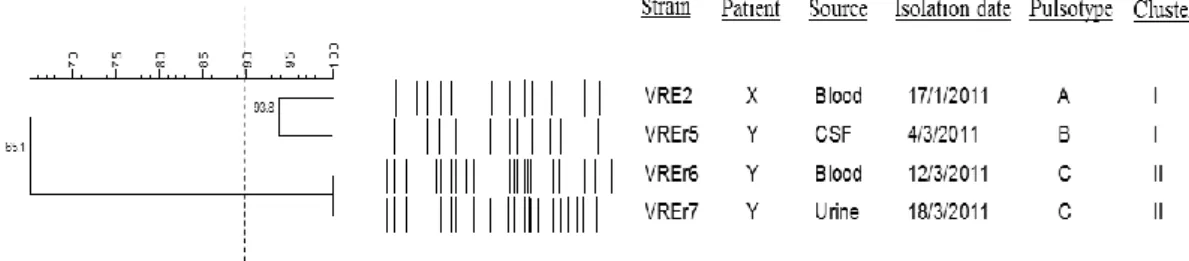
4.1.1 Pulsotypes of VREfm 41
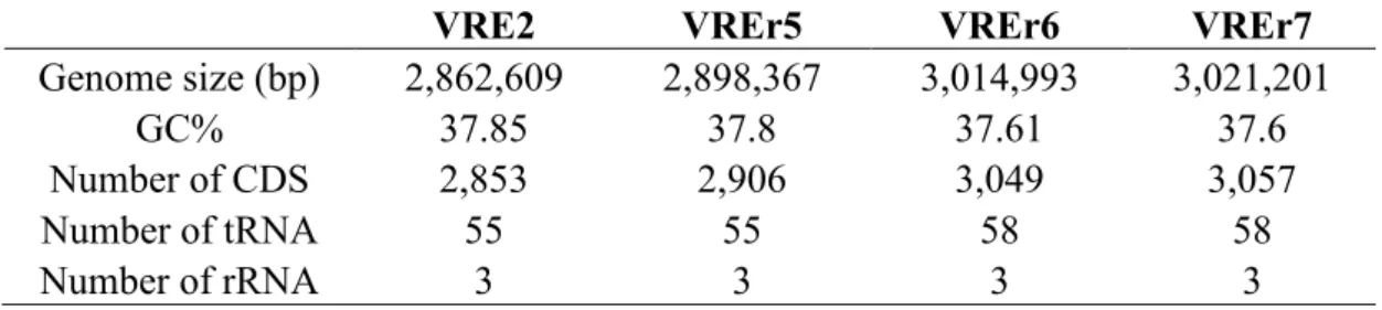
4.1.2 Genome features 42
4.1.3 Multilocus sequence typing (MLST) 42
4.1.4 Comparative genomics 43
4.1.5 Phylogenomic relationship of selected E. faecium strains 47
4.1.6 Genomic plasticity 49
4.2 Virulence factors 50
4.2.1 Virulence genes profiling 50
4.2.2 Biofilm formation 53
4.2.3 E. faecalis bee homolog 55
4.3 Antibiotic resistance 57
4.3.1 Antibiotic susceptibility profile 57
4.3.2 Antibiotic resistance determinants 60
4.4 Transcriptomic analysis 63
4.4.1 Biofilm developmental stage after 24 hours of growth 63
4.4.2 Transcriptome assembly and annotation 64
4.4.3 Transcriptome profiles of biofilm cells 67
CHAPTER 5: DISCUSSION ... 74
5.1 Whole genome sequencing 74
5.2 Virulence factors 78
5.3 Antibiotic resistance 80
CHAPTER 6: CONCLUSION ... 92
References 94
List of Publications and Papers Presented 112
Publications 112
Proceedings and Poster Presented 115
Appendices 116
LIST OF FIGURES
Figure 2.1: VanA-type vancomycin resistance mechanism ... 15 Figure 4.1: Dendrogram showing the cluster analysis of four VREfm strains based
on PFGE patterns of the SmaI-digested chromosomal DNA 41
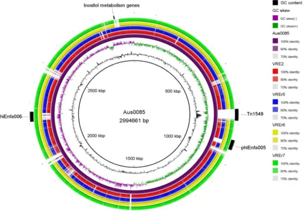
Figure 4.2: Circular genomic map and genome comparison of Aus0085, VRE2,
VREr5, VREr6, and VREr7 45 Figure 4.3: Venn diagram showing the distribution and number of core, dispensable
and strain-specific genes of the Malaysian VREfm strains 46 Figure 4.4: Phylogenomic tree inferred from approximately-maximum-
likelihood method from aligned core genomes 48 Figure 4.5: CLSM images of the four Malaysian VREfm strains grown in TSB 54
Figure 4.6: PCR results showing amplification of bee-1, bee-2 and bee-3 gene using
total and plasmid DNA as template 56
Figure 4.7: PCR results showing amplification of vanA gene 61
Figure 4.8: CLSM image of the biofilm formed by VREr5 after 24 hr of incubation 64
Figure 4.9: COG distribution of the assembled CDSs 66 Figure 4.10: Principal component analysis (PCA) of gene expression in biofilm
and planktonic cells 68 Figure 4.11: Heat map depicting the gene expression level of the six samples
LIST OF TABLES
Table 3.1: DNase incubation mix setup ... 36
Table 3.2: Master mix setup for reverse transcription 38
Table 3.3: PCR running condition for reverse transcription 38
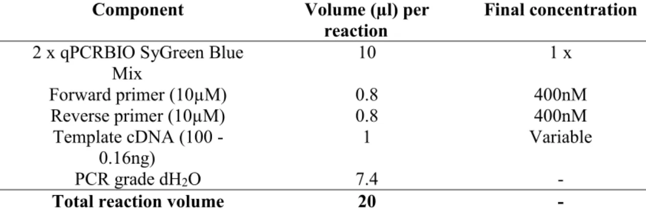
Table 3.4: Master mix setup for real-time PCR 39
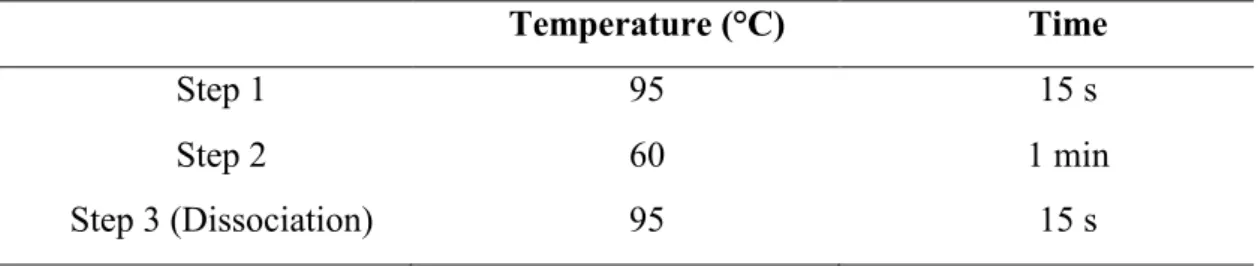
Table 3.5: Real-time PCR cycling condition 40
Table 3.6: Melt curve running condition 40
Table 4.1: General genome features of VREfmstrain VRE2, VREr5, VREr6, and VREr7 42
Table 4.2: Selected virulence-associated genes identified in the four Malaysian VREfm strains 52
Table 4.3: Average results of three replicates of the crystal violet assay to determine biofilm forming potential 53
Table 4.4: Antimicrobial resistance profile and the presence of corresponding resistance genes 58
Table 4.5: Summary of annotations of the assembled CDSs in VREr5 65
Table 4.6: Selected up-regulated genes in biofilm relative to planktonic cells 71
Table 4.7: Selected down-regulated genes in biofilm relative to planktonic cells 72
Table 4.8: Expression levels of selected up- and down-regulated genes in planktonic and biofilm cells as measured by qPCR 73
LIST OF SYMBOLS AND ABBREVIATIONS
AST : Antibiotic susceptibility test
BLAST : Basic Local Alignment System Tool
BRIG : BLAST Ring Image Generator
CA : Community-associated
CC17 : Clonal complex 17
COG : Cluster of Orthologous Group
CSF : Cerebrospinal fluid
CLSM : Confocal laser screen microscopy
CLSI : Clinical and Laboratory Standard Institute
CDS : Coding sequences
CRISPR : Clustered Regularly Interspaced Short Palindromic Repeat
°C : Degree celcius
EVD : Ventricular drain insertions
GC : Guanine-cytosine GO : Gene Ontology HA : Hospital-associated hr : Hour I : Intermediate IS : Insertion sequence
KEGG : Kyoto encyclopaedia of genes and genomes
M : Mega
MLST : Multilocus sequence typing
MIC : Minimum inhibitory concentration
mg/ml : Milligram per milliliter
mM : Milli Molar
min : Minute
μl : Micro liter
μg/ml : Micrograms per milliliter
MSCRAMM : Microbial surface components recognizing adhesive matrix molecules
NCBI : National Center of Biotechnology Information
NGS : Next generation sequencing
ORF : Open reading frame
PAI : Pathogenicity island
PBP5 : Penicillin-binding protein 5
PCR : Polymerase chain reaction
PCA : Principal component analysis
PFGE : Pulsed-field gel electrophoresis
RAST : Rapid Annotation using Subsystem Technology
RNA-seq : RNA sequencing
RealPhy : Reference Sequence Alignment-based Phylogenic Builder
R : Resistant
S : Susceptible
ST : Sequence type
UMMC : University Malaya Medical Centre
VRE : Vancomycin-resistant enterococci
VREfm : Vancomycin-resistant Enterococcus faecium
VFDB : Virulence Factors of Pathogenic Bacteria Database
LIST OF APPENDICES
Appendix 1: Media, Buffers and Solutions ……… 116
Appendix 2: Strains’ information ……….. 121
Appendix 3: Strains used in phylogenomic analysis ………. 122
Appendix 4: PCR primers and cycling conditions ……… 123
Appendix 5: Primers for gap closing and sequencing of Tn1546-like transposon ………. 124
Appendix 6: Primers for qPCR ……….. 125
Appendix 7: Supplementary data ……….. 126
DNA sequencing BLAST result of vanA……… 126
DNA sequencing BLAST result of bee-1 homolog ………… 127
DNA sequencing BLAST result of bee-2 homolog ………… 128
DNA sequencing BLAST result of bee-3 homolog ………… 128
Complete list of up-regulated genes ……… 129
CHAPTER 1: INTRODUCTION
1.1 General Introduction
Enterococcus faecium has been associated with several serious or life-threatening nosocomial diseases such as urinary tract infections, surgical-related wound infections,
bacteremia, and endocarditis (Higuita & Huycke, 2014). As an opportunistic pathogen, E.
faecium mainly targets elderly patients with underlying diseases, immunocompromised patients, and patients who have been hospitalized for prolonged periods or treated with
invasive devices. The clinical significance of E faecium becomes more prominent with
the increasing antimicrobial resistance among the clinical isolates, including high-level resistance to ampicillin, aminoglycosides, and glycopeptides (Cattoir & Giard, 2014; Leclercq et al., 1988; Padmasini et al., 2014; Uttley et al., 1988). The rapid spread of
vancomycin resistant E. faecium (VREfm) is of particular concern as VRE is often
multidrug resistant. According to a recent report from the National Healthcare Safety Network (NHSN), 82.2% and 85.1% of enterococci recovered from bloodstream and urinary tract infections are resistant to vancomycin in the year 2014 (Sievert et al., 2016). The emergence of VRE and outbreaks that occur around the world indicate the success of
E. faecium in adapting to and surviving in the hospital environment. Worryingly, resistance to antibiotics that are used to treat VRE infections, such as linezolid, daptomycin, and tigecycline, has been reported (Edelsberg et al., 2014; Tsai et al., 2012).
With the rapid advances in next generation sequencing (NGS) technology, different
NGS platforms such as Roche/454, SoLiD, and Illumina enable the entire genomes of
bacterial pathogen including E. faecium to be sequenced (Lam et al., 2013, 2012; Qin et
al., 2012). This technology generates massive information that is helpful in exploring the
differences between strains of E. faecium at a genome-wide level. The whole genome
contents, resistance determinants and mechanisms, as well as pathogenicity and evolution of E. faecium. This information is important in revealing factors that contribute to its adaptation and persistence in the clinical settings, and, hopefully, solutions to control and
treat infections. To date, a number of genome sequences of E. faecium have been reported
(García-Solache & Rice, 2016; Khan et al., 2015; Lam et al., 2013, 2012; Qin et al., 2012). Genome analyses revealed that clinical isolates are different from non-clinical isolates in that the number of mobile genetic elements, resistance and virulence genes are significantly higher in the genomes of clinical isolates (Kim & Marco, 2014; Qin et al.,
2012). Apart from that, specific elements such as the esp gene and IS16 are found almost
exclusively in the hospital-associated isolates (Heikens et al., 2012; Leendertse et al., 2009; Werner et al., 2011; Willems et al., 2001). The findings from these genomic analyses have
shed light on the virulence and persistence of clinical E. faecium which is important in
controlling the spread of this nosocomial pathogen.
The clinical relevance of infections caused by VREfm can also be attributed to the difficult-to-treat biofilm-associated diseases. It has been reported that majority of the device-associated infections, such as infections due to central lines, urinary catheters, and ventilators, are caused by VREfm (Sievert et al., 2016). Since biofilms are highly resistant to antibiotics and phagocytosis, a detailed insight into the process and molecular mechanism of biofilm formation is pivotal for the development of new drugs against biofilm-mediated infections. Transcriptomic analysis enables the quantification of gene expression of a full transcriptome, thereby help in interpreting the functional elements of a genome that are expressed at a specific physiological condition or developmental stage (Wang et al., 2009). The introduction of RNA-seq technology provides a more sensitive and dynamics approach to study the transcriptome of various organisms as compared to the traditional hybridization method (microarray) (Hinton et al., 2004; Wang et al., 2009). In the case of biofilm formation, this high-throughput technology has generated
informative data which is helpful in understanding the molecular mechanisms of biofilm biogenesis that are important for drugs development (Rumbo-Feal et al., 2013; Tan et al., 2015).
1.2 Objectives
To the best of our knowledge, a complete genome analysis of clinical VREfm has not been reported in Malaysia. Hence, such study is needed to better understand the overall biology, and the potential resistance and virulence of the locally isolated VREfm strains.
Moreover, transcriptomic studies on the biofilm formation of E. faecium is also lacking in
Malaysia or Southeast Asia. Understanding the transcriptome of biofilm cells is important in the development of new control or treatment methods against biofilm-mediated infections.
Therefore, the objectives of this study were:
1. To perform comparative genome analyses of four selected VREfm from Malaysia; 2. To elucidate the gene expression profile of biofilm cells in respect to the planktonic cells.
CHAPTER 2: LITERATURE REVIEW
2.1 The Genus Enterococcus
The genus Enterococcus was previously classified as part of the genus Streptococcus.
It is not until 1984 when the Enterococcus was recognized as a separate genus (Schleifer
& Kilpper-Balz, 1984). Streptococcus faecalis and Streptococcus faecium were the first
two species to be transferred to the new genus as Enterococcus faecalis and Enterococcus
faecium. To date, there are more than 50 species of enterococcus being described [http://www.bacterio.net/index.html].
Enterococci compose of Gram-positive cocci that are often arranged in pairs or chains. They are catalase-negative, non-spore-forming facultative anaerobes which can survive in harsh conditions, including high salinity (6.5% NaCl) and a wide range of temperature (10°C to 45°C) (Facklam & Collins, 1989). As such, enterococci are ubiquitous in nature. They can be found in water, soil, plants, as well as fermented food and dairy products. Enterococci are also commensals in the gastrointestinal tracts of human and animals. Although accounted for only 1% of the human gut microflora (Sghir et al., 2000), enterococci can occasionally cause diseases such as urinary tract infection and endocarditis, especially in immunocompromised patients.
2.2 Emergence of vancomycin-resistant enterococci (VRE)
Enterococci have long been considered as harmless inhabitants of the human gut flora. However, in the past few decades, this organism has emerged as one of the leading cause of hospital-associated infections. The extensive use of antibiotics in the clinical settings has contributed remarkably to the transition of this organism from commensal to the
nosocomial pathogen. Following the development of resistance to beta-lactam drugs and to high concentration aminoglycosides in the 1980s, vancomycin was among the last available antibiotic for enterococcal infections.
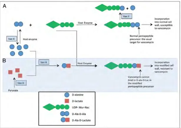
Vancomycin acts by disrupting cell wall synthesis. Once enterococci get in contact with the compound, vancomycin binds to the D-ala-D-ala terminus of the peptidoglycan precursor, thereby inhibiting cell wall development (Courvalin, 2006). Due to its disruptive effect on peptidoglycan, vancomycin is commonly used together with other antibiotics such as aminoglycosides to effectively get rid of the pathogen. Although the use of vancomycin has successfully controlled enterococcal infections, it was later discovered that the acquisition of vancomycin resistance has become increasingly prevalent (Bonten et al., 2001; Kuo et al., 2014).
Vancomycin-resistant enterococci (VRE) was first reported in Europe in the 1980s (Leclercq et al., 1988; Uttley et al., 1988). Since then, VRE has spread worldwide, including in the United States, Europe, and East Asia regions (Bonten et al., 2001; Kuo et al., 2014). In Malaysia, the first confirmed case of hospital acquired VRE was reported in 2006 in Hospital Kuala Lumpur (HKL) in a patient with chronic renal failure (Zubaidah et al., 2006). Other local studies reported a low prevalence of VRE (1-2%) in Malaysia (Ibrahim et al., 2010, 2011). A study collecting enterococci from different countries shows
that vancomycin resistance was more prevalent in E. faecium (Putnam et al., 2010). A
similar result was observed by Edelsberg et al. (2014) who examined resistance percentage of clinically significant bacterial pathogens in 19 US hospitals. The higher rate of
vancomycin resistance in E. faecium indicates the increasing clinical importance of this
The use of avoparcin had been suggested to contribute to the dissemination of
vancomycin resistance in E. faecium. This correlation can be shown through the different
epidemiology of VRE in Europe and the United States. In Europe, where avoparcin was massively used as a growth promoter in animal farms (Wegener, 1998), VRE are more prevalent in livestock and healthy people compared to patients (Devriese et al., 1996; Stobberingh et al., 1999; Van Braak et al., 1998). However, this type of community-reservoir in animals and healthy people is not observed in the United States, where avoparcin is banned in the animal farms. In contrast, VRE are the second most common nosocomial pathogen in the United States (Sievert et al., 2016). Since avoparcin can confer cross-resistant to vancomycin (Bager et al., 1997), the extensive use of this antibiotic in the animal farms may select for vancomycin resistant strains. Ultimately, these resistant strains could be transmitted to human through the food chain or to the farm workers due to poor hygiene practice in the animal farms. The possible association of avoparcin and the dissemination of vancomycin resistance was further strengthened by the reduction of VRE rate in Europe after the ban of avoparcin in 1997 (Aarestrup et al., 2001; Boggard & Stobberingh, 2000).
There is evidence indicating that glycopeptide-producing microorganisms, such as
Amycolatopsis orientalis and Streptomyces toyocaensis, are the sources of vancomycin resistance genes (Marshall et al., 1997). The production of these resistance genes is presumed as a self-defence mechanism of these organisms. These resistance genes then can be transferred horizontally via one or more bacterial intermediates, ultimately to enterococci. In the case of Europe, resistance gene may ultimately be transferred to genogroups that colonize animals and healthy people, resulted in a large community-based reservoir. On the contrary, in the United States, vancomycin resistance gene may be transferred to genogroups which had been resistant to multiple antibiotics, thereby
increasing the chance of acquired resistance due to the selective effects of antibiotics (Bonten et al., 2001).
2.3 Molecular subtyping of E. faecium
For many years, majority of the human enterococcal infections were caused by E.
faecalis (80-90%) (Jones et al., 2004; Moellering, 1992). However, in the past three
decades, E. faecium has increasingly become predominant as a leading cause of
hospital-acquired infections, particularly vancomycin-resistant E. faecium (VREfm). As such,
molecular subtyping is important for epidemiological studies to assist in infection control as well as to trace the dissemination of this pathogen. Various subtyping methods have
been developed for E. faecium. These include repetitive element sequence (REP)-PCR
typing, plasmid typing, pulsed-field gel electrophoresis (PFGE), amplified fragment length polymorphism (AFLP), multiple locus variable number of tandem repeat analysis (MLVA), and vancomycin resistance gene cluster typing. However, each of these typing methods has its own drawbacks, mainly in terms of the ease of use, discriminatory power, cost, data reproducibility, as well as data exchange (Werner, 2013).
PFGE is a highly discriminative typing method that can distinguish strains of the same species from different sources, time, and space. This macrorestriction-based typing method utilizes rare-cutting enzymes to fragment chromosomal DNA, and an alternating electric field (i.e. pulsed field) to separate the resulting DNA fragments, generating distinct patterns which can be used to differentiate closely related bacteria. Due to its high discriminatory power, PFGE remains a “gold standard” for epidemiological studies of a
large number of bacteria, including E. faecium. However, since the rate of recombination
is relatively high in enterococci (Willems et al., 2005), extreme genomic variations may be found in strains from the same outbreak over a period of time. As such, PFGE is less suitable for long-term epidemiological study of enterococci. Apart from that,
inter-laboratory transfer of PFGE data is challenging as a standardized protocol of enterococcal PFGE has not been established.
Multi-locus sequence typing (MLST) is a recently widely accepted typing method for population and evolutionary study (Urwin & Maiden, 2003). This method determines the alleles of the internal fragment sequences of multiple housekeeping genes and assigns each
allele a numerical value. In E. faecium, seven housekeeping genes, atpA, ddl, gdh, purK,
gyd, pstS, and adk, are used (Homan et al., 2002). The combination of the seven allelic
numbers yields a sequence type (ST) that classifies different strains. MLST allows for the exchange of data throughout the globe through the use of uniform typing method and the establishment of a large, publicly accessible database. However, this method can be relatively costly if sequencing of the seven housekeeping genes of a large number of samples is to be performed.
A distinct clonal lineage comprised of mostly clinical E. faecium isolates was identified
when the MLST data was analyzed using the enhanced based upon related sequence types (eBURST) algorithm (Willems et al., 2005). This lineage is termed clonal complex 17 (CC17). Members of the CC17 are characterized by ampicillin resistance, a pathogenicity island and an association with hospital outbreak (Willems et al., 2005). Shortly, this lineage turns into the major genetic complex associated with hospital infections and is found to be disseminated around the world (Matsushima et al., 2012; Ryan et al., 2015; Valdezate et al., 2009; Yu et al., 2015).
2.4 Whole genome sequencing of E. faecium
The increased medical importance of E. faecium over E. faecalis as a nosocomial
However, there are still gaps in our understanding of its virulence, pathogenicity, resistance mechanisms, and even adaptability. The availability of whole genome sequencing (WGS)
allows scientists to explore in more detail the biological features of E. faecium to provide
answers to the emergence of this pathogen.
The first complete genome of E. faecium was available in March 2012 from a
vancomycin-resistant strain Aus0004. Aus0004 was isolated from the bloodstream of a patient in Melbourne, Australia, in 1998 (Lam et al., 2012). Following this, the second complete genome was published by (Qin et al., 2012) and was determined by strain TX16 (also named TX0016 or DO) isolated from the blood of an endocarditis patient in 1992. More genomes were later being published, which provide chances for comparative genomics to unveil the genomic variations among strains from different niches.
There are currently more than 400 publicly available E. faecium genomes, with 17
genomes being completely sequenced
(https://www.ncbi.nlm.nih.gov/genome/genomes/871?). However, an overrepresentation
of strains from Europe and North America is observed. Hence, more genomes from strains of various regions, such as from the Southeast Asia, are needed to fully understand the
global diversity of E. faecium.
2.5 Comparative genomics
2.5.1 Core and accessory genome
Genome comparison between several E. faecium strains has identified a set of genes
known as “core” genome that are conserved in all strains. This core genome consists of genes that are mainly responsible for housekeeping functions such as DNA and carbon
metabolism, cell structural biosynthesis, and substrate transport. The non-core genes make up the “accessory” genome which are partially shared or strain-specific. These accessory genes may contribute to specific adaptive traits or even pathogenicity of different strains from different environmental niches. For instance, comparative analyses have revealed differences in genome content between the clinical and non-clinical strains. The genome of clinical strains composes of significantly more mobile genetic elements, virulence factors and antibiotic resistance genes compared to that of non-clinical strains (Kim &
Marco, 2014). The esp gene, encodes for the enterococcal surface protein (Esp) which is
associated with endocarditis and urinary tract infections, is specifically enriched in the clinical strains (Heikens et al., 2012; Leendertse et al., 2009; Willems et al., 2001).
Moreover, acquired antibiotic resistance genes, such as van gene that confers vancomycin
resistance, are also part of the accessory genome of clinical E. faecium strains.
Several studies suggested that the pan-genome of E. faecium is open, indicating that this
organism is able to acquire and incorporate novel genes efficiently into the collective gene pool of the species (Qin et al., 2012). This helps to explain the accumulation of a large number of accessory genes in the clinical strains that contribute to their pathogenicity. An
open pan-genome also implies that E. faecium can readily acquire new genes that
contribute to fitness, allowing it to adapt to various environmental niches, which might facilitate its transition from commensal to nosocomial pathogen.
2.5.2 Insertion sequence, phage, and CRISPR
Insertion sequence (IS) elements are short DNA fragments (0.7-2.5kb) that usually encode for one or two genes needed for transposition (Siguier et al., 2015). These elements
faecium (Lam et al., 2013, 2012; Mikalsen et al., 2015; Qin et al., 2012). IS elements can
play a vital role in evolution and diversification of genomes. IS16, for example, is almost
exclusively present in the hospital-associated (HA) strains and was suggested as a marker
for identification of HA E. faecium strains (Werner et al., 2011). Furthermore,
transposition of IS elements can also lead to gene rearrangement such as duplication, deletion, and inversion, which contributes to genome plasticity among the species (Lee et al., 2016; Ooka et al., 2009). Apart from that, there have been evidence showing that IS
elements can affect gene expression (Aubert et al., 2003; Sóki et al., 2013). In E. faecium,
for example, insertion of IS elements in the vancomycin resistance gene cluster has led to reduced resistance in certain VRE strains (Gagnon et al., 2011).
Bacteriophage (phage) is another mobile genetic element which can also drive genome diversification. Phages integrated into the bacterial genomes (prophages) can introduce new genes that modify the genome content of strains within the species. A number of
prophages have been identified in the genome of E. faecium (Lam et al., 2013, 2012; Qin
et al., 2012; van Schaik et al., 2010). These prophages are distinct from strain to strain,
demonstrating phage diversity of E. faecium (Lam et al., 2013). In a comparison between
seven E. faecium strains, van Schaik et al. (2010) reported that most of the phages
identified can be activated by the DNA cross-linking agent mitomycin. These induced
phages had the typical morphology of Siphoviridae, which is a common bacteriophage
family found in lactic acid bacteria. It remains to be determined to what extent prophages
contribute to the fitness of E. faecium but the variety of prophages identified suggest that
they play a major role in shaping the genome of E. faecium. Nevertheless, phages may aid
in the transmission of antibiotic resistance genes among enterococci species, as reported by Mazaheri Nezhad Fard and his co-workers (Mazaheri Nezhad Fard et al., 2011).
Clustered regularly interspaced short palindromic repeats (CRISPR) are short repetitive DNA sequences that are found in both archaea and bacteria. These repeats are characterized by direct repeats of varying size interspaced by non-repetitive sequences of a similarly size known as “spacer” and flanked on one side by a conserved leader sequence (Jansen et al., 2002). The CRISPR loci are usually associated with genes known as
CRISPR-associated (cas) genes which encode for nucleases or proteins involved in DNA
and RNA processing. This CRISPR-cas system has been shown to provide bacteria with
adaptive immunity against the integration of foreign genetic materials such as phages and
plasmids (Marraffini & Sontheimer, 2011). CRISPR-cas system appears to be found less
frequently in the HA strains, which are usually multidrug resistant (Palmer & Gilmore,
2010). This observation is consistent with the defensive role of CRISPR-cas system to
limit the acquisition of mobile genetic elements which might carry antibiotic resistance
genes. The absence of CRISPR-cas system in HA strains also explains the abundance of
phages and other mobile elements in their genomes, highlighting the lack of barriers to horizontal gene exchange in the species.
2.5.3 Virulence factors
Comparison between the genomes of hospital-associated (HA) and community-associated (CA) strains reveals several virulence factors that are enriched in the HA strains.
The most significant one being the presence of esp gene which is found more abundantly
in the clinical isolates than in food or environmental isolates (Abriouel et al., 2008;
Willems et al., 2001). The esp gene was initially suggested to involve in initial adherence
and biofilm formation in E. faecium (van Wamel et al., 2007). These roles were later
proven by a biofilm deficient esp insertion-deletion mutant (Heikens et al., 2007).
infections and endocarditis (Heikens et al., 2012; Leendertse et al., 2009). The esp gene of
E. faecium is located on a large transferable pathogenicity island (PAI) which ranges from
64 to 104 kb in size (van Schaik et al., 2010). Comparative genomics of three clinical E.
faecium strains reveals that although the general architecture of esp PAI is conserved in all the three studied strains, variations are observed in the gene content of the PAI which are likely caused by the independent acquisition of genes through horizontal gene transfer (van Schaik et al., 2010).
A putative virulence gene hylEfm was also found to be highly prevalent in clinical E.
faecium strains (Freitas et al., 2010; Rice et al., 2003; Soheili et al., 2014). The hylEfm gene was initially suggested to encode for hyaluronidases but was later annotated as a putative
glycoside hydrolase based on sequence comparison with spy1600 gene in Streptococcus
pyogenes (Rice et al., 2003; Sheldon et al., 2006). It has been previously shown that the
hylEfm gene is carried on transferable megaplasmids which can also carry genes conferring
resistance to glycopeptides (Arias et al., 2009; Freitas et al., 2010). Some of these hylEfm
-containing plasmids have been shown to enhance gastrointestinal colonization and increase virulence in an experimental peritonitis mouse model, implicating potential virulence of
the hylEfm gene in E. faecium (Arias et al., 2009; Rice et al., 2009). However, a more recent
study demonstrates that hylEfm does not mediate the increased virulence conferred by the
hylEfm-containing plasmid in murine peritonitis (Panesso et al., 2011). It remains to be
determined if hylEfm plays any role in other infections such as endocarditis or urinary tract
infections.
In addition to esp and hyl, several virulence factors associated with surface adhesion
and biofilm formation are also significantly enriched in the HA strains. These include the
and fms21-fms20 locus. These virulence factors will be discussed in detailed in the biofilm section.
2.6 Antibiotic resistance in E. faecium
The clinical importance of E. faecium can be linked to its resistance to a broad range of
antibiotics, which can be either intrinsic or acquired. Among all, acquisition of glycopeptides resistance, particularly to vancomycin, is the most significant event
contributing to the transition of E. faceium from commensal to nosocomial pathogen.
Glycopeptides inhibit bacterial growth by disrupting cell wall synthesis. Resistance to glycopeptides is caused by either elimination of the high-affinity peptidoglycan precursors originally produced by the host, which removes the target sites; or modification of the termini of peptidoglycan precursors from D-Ala to either D-Lac or D-Ala-D-Ser, which lowers the binding affinity of the antibiotics to the target sites (Courvalin, 2006; Guzman Prieto et al., 2016; Hollenbeck & Rice, 2012). To date, nine distinct gene
clusters (vanA, vanB, vanC, vanD, vanE, vanG, vanL, vanM, and vanN) conferring
glycopeptides resistance have been described in enterococci (Boyd et al., 2008; Courvalin,
2006; Lebreton et al., 2011; Xu et al., 2010), with vanA and vanB being the most
predominant gene clusters found in clinical VREfm.
The vanA gene cluster confers a high-level resistance to vancomycin and teicoplanin.
This cluster is typically encoded on Tn1546 or related transposons and consists of seven
genes. The vanR and vanS genes encode for a two-component regulatory system which
regulates the expression of vanHAXYZ. The vanH gene encodes for a dehydrogenase
which converts cellular pyruvate to D-Lac. The vanA-encoding ligase then links the
tripeptide precursor by host enzymes, yielding the low-affinity pentapeptide precursor. For a full resistance to vancomycin, elimination of normal precursors is required. This is
done by vanX and vanY genes. vanX encodes a D-D-dipeptidase which hydrolyses the
original D-Ala-D-Ala dipeptide, making D-Ala-D-Lac the sole substrate for
peptidoglycan synthesis. On the other hand, vanY encodes a D-D-carboxypeptidase
which eliminates the terminal D-Ala from the normal pentapeptides, rendering them useless for normal cell wall synthesis (Courvalin, 2006; Hollenbeck & Rice, 2012). The
vanZ gene encodes for a protein with yet unknown function. However, an association of
VanZ with low-level teicoplanin resistance had been documented before (Arthur et al.,
1995). Additionally, the vanA cluster also includes two genes (orf1 and orf2) which are
responsible for transposition.
Figure 2.1: VanA-type vancomycin resistance mechanism. A) Normal cell wall
synthesis pathway and disruption of the pathway by VanX and VanY. B) Construction of a modified cell wall that is resistant to vancomycin. Figure adapted from Hollenbeck and Rice (2012).
Studies that characterized the vanA gene clusters of E. faecium from different
geographical regions and sources revealed great structural variations of the Tn1546-like
elements that are mainly caused by point mutations, deletions and IS insertions (Hashimoto et al., 2000; Huh et al., 2004; Kuo et al., 2014; Willems et al., 1999). Most of these modifications are observed in the genes that are not involved directly in
vancomycin resistance (orf1, orf2, vanY, vanZ) and in the intergenic regions. Notably,
insertion of IS1216V, IS1542, and IS1251, as well as truncation of the orf1 are the most
frequently observed events (Gu et al., 2009; Huh et al., 2004; Kuo et al., 2014; Schouten et al., 2001). Some of these structural changes, especially those caused by IS integrations, have been shown to contribute to changes in the vancomycin and teicoplanin resistance level (Gagnon et al., 2011; Gu et al., 2009; Hashimoto et al., 2000; Sivertsen et al., 2016).
Despite its high diversity, similar vanA cluster variants has been found in strains from
different geographical regions and sources (human, animals, environments), indicating possible horizontal spread of the vancomycin resistance elements through conjugative plasmids or as part of a larger mobilized genetic unit (Huh et al., 2004; Sletvold et al.,
2010; Willems et al., 1999). In fact, polymorphism of the vanA gene cluster has been used
as a typing tool which, coupled with epidemiological data, provides a better
understanding on the dissemination of vancomycin resistance in E. faecium (Xu et al.,
2011).
Unlike the vanA gene cluster, the vanB gene cluster confers moderate to high-level
resistance to vancomycin, but not to teicoplanin (Hollenbeck & Rice, 2012). The VanB
locus is usually found on Tn1549-like transposons, which are encoded on plasmid or
chromosome. The genetic organization and resistance mechanism of VanB locus is similar to that of VanA locus. Homologs of VanH (VanHB), VanX (VanXB), VanY (VanYB), and VanA (VanB) are found in the VanB locus. Genes encoding the
related (34% and 24% amino acid identity, respectively) to the vanRS found in the VanA locus (Evers & Courvalin, 1996). This VanRSB system regulates differently from its
counterparts of the Tn1546 in that only vancomycin, but not teicoplanin, induces
resistance of the VanB locus (Evers & Courvalin, 1996). Apart from that, a gene related
to vanZ is lacking in the VanB locus but an additional vanW gene with unknown function
is found. Based on sequence analysis, three vanB subtypes (vanB1, vanB2, vanB3) are
identified. However, there is no correlation between the different genotypes and the level of resistance to vancomycin (Dahl et al., 1999).
High-level resistance to ampicillin is another important feature of clinical E. faecium.
Ampicillin, like other beta-lactams, binds covalently to the penicillin-binding proteins (PBPs) and disrupts cross-linking of peptidoglycan precursors, thereby impairing cell
wall synthesis. E. faecium exhibits intrinsic resistance to ampicillin due to the expression
of low-affinity penicillin-binding protein 5 (PBP5). Increased resistance to ampicillin is
mediated by either acquisition of beta-lactamase or mutations in the pbp5 gene
(Hollenbeck & Rice, 2012). In E. faecium, high-level ampicillin resistance is mainly due
to the accumulation of various point mutations in the penicillin binding regions of PBP5 (Galloway-Peña et al., 2011; Rice et al., 2004). A combination of two point mutations, a methionine-to-alanine change in position 485 and an insertion of serine at position 466 of
pbp5, has been shown to markedly enhance ampicillin resistance (Rice et al., 2004). A
later study showed that the chromosomally encoded pbp5 is able to transfer among E.
faecium strains through conjugation (Rice et al., 2005). This suggests that mutated pbp5
with enhanced resistance could be transmitted in the same way among the clinical isolates. On the other hand, high-level ampicillin resistance due to overproduction of
beta-lactamase is relatively rare in E. faecium compared to in E. faecalis (Hollenbeck & Rice,
Another group of antibiotics to which E. faecium exhibits intrinsic and high-level
acquired resistance to is aminoglycosides. Aminoglycosides act by inhibiting ribosomal
protein synthesis. Generally, all enterococci are intrinsically resistant to low-level of aminoglycosides due to poor uptake of the antibiotics (Bryan & Van Den Elzen, 1977). The combination of cell-wall active agents such as ampicillin or vancomycin with aminoglycosides increases the uptake of the antibiotics, thereby enhances the killing
effect on enterococci (Moellering & Weinberg, 1971). Other than the natural uptake
barrier, E. faecium also poses chromosomally encoded enzymes such as
6′-N-aminoglycoside acetyltransferase encoded by aac(6′)-Ii and rRNA methyltransferase
encoded by efmM that confer low to moderate intrinsic resistance to aminoglycosides
(Hollenbeck & Rice, 2012).
The acquisition of various genes encoding aminoglycoside-modifying enzymes
(AMEs) results in high-level aminoglycosides resistance in E. faecium, which abolish the
synergistic killing effect that is important for the treatment of severe enterococcal
infections. The bi-functional gene aac(6´)-Ie-aph(2´´)-Ia is the most clinically significant
as strains carrying this gene is virtually resistant to all clinically available
aminoglycosides except streptomycin (Chow, 2000). aac(6´)-Ie-aph(2´´)-Ia is the most
prevalent gene that confers high-level gentamicin resistance in enterococci, although
other genes such as aph(2´´)-Ib, aph(2´´)-Ic, and aph(2´´)-Id also confer resistance to
gentamicin (Chow, 2000). The enzymes encoded by aac(6´)-Ie-aph(2´´)-Ia inactivate
gentamicin by phosphorylating the 2´ hydroxyl position of gentamicin (Ferretti et al.,
1986). This modification renders the antibiotic unable to bind to its target on the 30S ribosome, thereby loses its antibacterial activity. Streptomycin can be used in the
synergistic therapy against enterococci when aac(6´)-Ie-aph(2´´)-Ia is present, provided
that there is no resistance to high levels of streptomycin (MIC ≥ 1000 μg/ml). Enzymatic modification of the antibiotic or single-step point mutations in the ribosome can
contribute to high-level streptomycin resistance in enterococci (Hollenbeck & Rice,
2012). The ant(6´)-Ia that encodes for an adenylyl transferase is one of the well-known
resistance determinants that can inactivate streptomycin. The ant(6´)-Ia gene is often
found as part of a multi-resistance gene cassette ant(6´)-sat4-aph(3´) which confers
resistance to streptomycin, streptothricin, and kanamycin. This gene cluster is encoded
on Tn5405 and other related transposons that are widely found in Staphylococci and
Enterococci (Derbise et al., 1997). Other AMEs that contribute to acquired
aminoglycosides to enterococci include those that are encoded by aph(3´)-IIIa and
ant(4´´)-Ia, which confer resistance to kanamycin, tobramycin, amikacin, and neomycin. Due to the growing problem and clonal spread of VREfm, new drugs such as linezolid, daptomycin, and tigecycline have been increasingly used to treat infections caused by VREfm. However, resistance has also been observed in these antibiotics (Edelsberg et al., 2014; Tsai et al., 2012). With very few effective therapeutic options left to treat VREfm,
development of newer drugs targeting different structures in the E. faecium cell, as well
as strict infection control measures are utmost important to combat this pathogen.
2.7 Biofilm formation by E. faecium
Biofilm formation is an important virulence feature responsible for the pathogenesis of enterococci. Several difficult-to-treat enterococcal diseases were often biofilm-mediated, including those associated with indwelling medical devices and urinary catheters, as well as endocarditis (Donlan et al., 2002). Several studies have also reported the association of biofilm formation to enterococci isolated from clinical settings (Baldassarri et al., 2001; Mohamed et al., 2004; Toledo-arana et al., 2001). Although
formation by clinical isolates is still relatively high compared to those isolated from other sources (Almohamad et al., 2014).
Multiple biofilm-associated genes have been identified, mostly originated from E.
faecalis. In E. faecium, these genes are mostly associated with adhesins or pili which are involved in the initial attachment step in biofilm formation. The enterococcal surface protein (Esp) is among the first cell-wall associated proteins that are shown to involve in
biofilm formation in E. faecium (Heikens et al., 2007). The levels of esp expression on
cell surface have been reported to affect initial attachment and biofilm formation (van
Wamel et al., 2007), thereby explaining the conflicting observations that some esp
positive isolates are unable to form biofilm (Dupre` et al., 2003). Further genetic analysis
identifies a gene encoding an AraC-type transcriptional regulator, known as ebrB
(enterococcal biofilm regulator B), which is found upstream of the esp gene. Deletion of
ebrB resulted in reduced expression of esp, as well as reduced biofilm formation (Top et
al., 2013). These results indicate that ebrB is involved in regulation of esp expression and
is implicated in biofilm formation in E. faecium.
Microbial surface components recognizing adhesive matrix molecules (MSCRAMMs) are a group of surface proteins that plays an important role in host-pathogen adherence, as well as binding of bacteria to abiotic surfaces coated with host-derived extracellular
matrix components. Two E. faecium MSCRAMMs that are involved in biofilm formation
are Acm and SgrA. Acm, a cell wall-anchored collagen adhesin which binds to collagen
type I and type IV is highly prevalent in E. faecium clinical isolates (Nallapareddy et al.,
2008a). Although this adhesin is also found in non-clinical isolates, the gene encoded for
this protein (acm) shows an insertion element disruption, rendering the encoded protein
non-functional (Nallapareddy et al., 2008a). An acm deletion mutant has been shown to
in a rat endocarditis model, indicating the contribution of acm to E. faecium pathogenesis (Nallapareddy et al., 2008). The surface adhesin SgrA, on the other hand, binds to extracellular matrix molecules nidogen 1 and nidogen 2. Although SgrA does not mediate
binding of E. faecium to biotic surfaces such as human bladder and intestinal epithelial
cells, its contribution to biofilm formation on polystyrene surface was demonstrated (Hendrickx et al., 2009).
Another group of surface expressed proteins that contributes to E. faecium biofilm
formation is pili. Two pilus-like structures, PilA and PilB, are frequently found in clinical
E. faecium isolates, including those isolated from endocarditis patients (Hendrickx et al.,
2010, 2008). The pilA gene is part of the pilA (fms21)-fms20 gene cluster which is
encoded on a large transferable plasmid (Kim et al., 2010). On the other hand, pilB, also
known as ebpC or fms9, is part of the ebpABCfm cluster and encodes for a major pilus
PilB. Deletion of ebpABCfm eliminates cell surface expression of PilB-containing pili and
reduces biofilm formation. Additionally, the deletion mutant displays reduced
colonization in a murine UTI model, signifying the role of ebpABCfm in the pathogenesis
of E. faecium (Sillanpää et al., 2011).
Other than factors involving in initial attachment, several proteins that are associated
with maturation of biofilm are also identified in E. faecium. These include a major
autolysin, AltEfm, and a secreted protein, SagA. AltEfm involves in the release of extracellular DNA (eDNA), which is one of the important components of extracellular polymeric substances. The importance of AltEfm in biofilm formation has been demonstrated through insertion disruption of the autolysin gene (Paganelli et al., 2013). Besides eDNA, secreted proteins are also an important component in the biofilm matrix.
(Paganelli et al., 2015). Proteolytic degradation of SagA has been shown to markedly
reduce biofilm formation of clinical E. faecium strains (Paganelli et al., 2015).
2.8 Transcriptome study
Transcriptome is the total mRNA in a cell that represents genes that are actively expressed at a specific growing stage or physiological condition (Wang et al., 2009). Transcriptomic aims to elucidate the functional elements of a genome; to determine the transcriptional structure of genes; and to quantify gene expression levels under different conditions (Wang et al., 2009). Understanding the transcriptome is, therefore, important to unravel the underlying molecular mechanisms of bacterial development and pathogenesis.
Microarray technology has been widely used in the transcriptomic analyses of different microorganisms. However, this hybridization-based method has several limitations, including a limited dynamic range of transcript detection due to problems with background noise and saturation of signals, dependency on the existing knowledge of genomes or transcripts, and complex normalization methods in comparing different experiments (Hinton et al., 2004; Wang et al., 2009). With the advent of next generation sequencing technologies, a more recent technology for transcriptome profiling termed RNA-seq (RNA sequencing) has been developed. In contrast to the microarray, transcriptomic studies using RNA-seq are not limited to previously known genes as this method directly determines the cDNA sequences (Wang et al., 2009). Moreover, RNA-seq provides better resolution and higher reproducibility compared to microarray (Wang et al., 2009). With all these advantages, RNA-seq technology has provided a large amount
of data which refined our understanding in the gene expression profile of an organism under different treatments or environmental stresses.
CHAPTER 3: METHODOLOGY
3.1 Clinical data collection and patients’ background
Clinical data including the patients’ ward, samples source, samples isolation date, antibiotic treatments and surgical procedures of the studied patients was collected. Ethical approval had been obtained from the University of Malaya Research Ethics Committee (UMREC) with ethical approval number 20159-1661.
Patient X was admitted into cardiology ward in 24th October 2010. Subsequently after
the isolation of first VRE (VRE2) from this patient, VRE infection cases were frequently reported in the hospital. Patient Y was admitted into neuro-ICU ward due to basal ganglia
bleed approximately three months (21st January 2011) after patient X was admitted.
Microbiological investigation revealed that patient Y was infected by multiple pathogens,
including multidrug-resistant Acinetobacter baumannii, methicillin-resistant
Staphylococcus aureus (MRSA), and Pseudomonas aeruginosa. VREr5 was isolated from patient Y after one month of treatment with vancomycin. Following this, another two strains, VREr6 and VREr7, were isolated from the same patient who was undergoing an antibiotic treatment with meropenem, colistin, ceftazidime, and linezolid. After the linezolid treatment, VRE had not been isolated from patient Y. To note, patient Y underwent several surgical procedures such as intracranial pressure monitoring and external ventricular drain insertions (EVD) during hospitalization. These procedures may serve as the route of bacterial transmission although this association is unclear. Eventually, patient Y died of sepsis due to infective endocarditis and pneumonia.
3.2 Whole genome sequencing
3.2.1 Bacterial strains
Four clinical strains of vancomycin-resistant E. faecium (VRE2, VREr5, VREr6,
VREr7) were collected in the year 2011 from University Malaya Medical Centre (UMMC), Kuala Lumpur. These strains were selected based on previous PFGE results, which were reconfirmed in this study. The strains were isolated and validated through PCR-amplification and sequencing of the 16S rRNA at the clinical microbiology laboratory. VRE2 was isolated from patient X and was the first VREfm isolated in the studied period. VREr5, VREr6, and VREr7 were isolated from the cerebrospinal fluid (CSF), blood, and urine of patient Y, respectively. These three strains were isolated at one-week interval, with VREr5 being the first isolate, followed by VREr6 and VREr7. A single colony of each strain was then cultured into Brain Heart Infusion broth (Oxoid Ltd., Basingstoke, UK) and incubated at 37°C for 24 hr. The culture was stored at -80°C in 50% glycerol until further experiments.
3.2.2 Pulsed-field gel electrophoresis (PFGE)
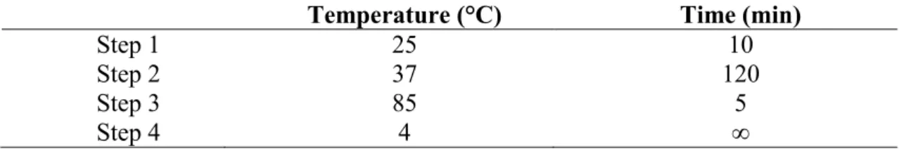
PFGE was performed as previously described (Turabelidze et al., 2000), with slight modifications. Briefly, the bacteria were first lysed in a combination of lysozyme (100mg/ml) and mutanolysin (10kU/ml) at 37°C for four hours. Chromosomal DNA was
then prepared in agarose gel block and digested with restriction enzyme SmaI (Promega,
Madison, WI, USA) at room temperature. The restriction fragments were separated by electrophoresis in 0.5 x TBE buffer for 20 hr at 14°C in a CHEF Mapper system
(Bio-Rad, CA, USA) using pulsed times of 3.5-25 s and 1-5 s. XbaI-digested Salmonella
analyzed using BioNumerics version 6.0 software (Applied Maths, Kortrijk, Belgium). The quantitative differences in the banding patterns were defined by the Dice coefficient. Cluster analysis was determined based on the unweighted pair group method with averages (UPGMA), using a position tolerance 1.5%.
3.2.3 DNA extraction
Genomic DNA of the four VREfm strains were extracted using the Wizard Genomic DNA Purification Kit (Promega, Madison, Wisconsin, United States). Briefly, 1 ml of the bacterial culture was harvested by centrifugation for 2 min at 14,000 rpm. To weaken the cell wall of gram-positive bacteria, the cell pellet formed was re-suspended in 480 µl of EDTA (50mM), after which 120 µl of lysozyme (10mg/ml) was added. The sample was incubated at 37°C for one hour, followed by centrifugation for 2 min at 14,000 rpm. The supernatant was removed and the cell pellet was re-suspended in 600 µl of lysis solution. The sample was further incubated at 80°C for 5 min. Following this, 3 µl of RNase solution was added to the re-suspended pellet, mixed, and incubated at 37°C for 15 to 60 min. After incubation, 200 µl of protein precipitation solution was added and vortexed to mix. The sample was incubated on ice for 5 min prior to centrifugation for 3 min at 14,000 rpm. The resultant supernatant was transferred to a clean microcentrifuge tube containing 600 µl of room temperature isopropanol and then followed by centrifugation at 14,000 rpm for 2 min to recover the precipitated DNA. The supernatant was discarded and 600 µl of room temperature 70% ethanol was added to clean the DNA pellet. The solution was centrifuged for another 2 min at 14,000 rpm. The ethanol was carefully aspirated and the pellet was air-dried for 10 to 15 min. Finally, the dried DNA pellet was re-suspended in 100 µl of sterile distilled water. Extracted DNA was quantified using the
spectrophotometer at OD260 and the purity was determined by OD260/OD280 ratio. The extracted DNA was stored at -20°C for long term storage.
3.2.4 Genome sequencing, assembly, and annotation
Whole genome sequencing of the VRE2, VREr5, VREr6, and VREr7 was carried out by a commercial vendor using the Illumina Miseq platform, version 2.0 with reads coverage ranged from 78x to 108x. The genome sequences were then assembled using CLC Genomic Workbench version 5.1 (CLC Bio, Aarhus, Denmark). Open reading frame (ORF) prediction was performed using Prodigal (Hyatt et al., 2010). Functional annotated of the genomes was performed using RAST (Rapid Annotation using Subsystem Technology) (Aziz et al., 2008) and Blast2GO (Conesa et al., 2005).
3.2.5 Genome analyses and comparative studies
The sequence types of the four sequenced strains were determined in silico using the
PubMLST database (http://pubmlst.org/). Genomes alignment and comparison were
performed using Mauve 2.3.1 using E. faecium Aus0085 as the reference (Darling et al.,
2004). The circular genomic map was constructed using BLAST ring image generator (BRIG) (Alikhan et al., 2011). Insertion sequence (IS) elements, prophages, and clustered regularly interspaced short palindromic repeats (CRISPR) were identified using IS Finder (Siguier et al., 2006), PHAST (Zhou et al., 2011), and CRISPRfinder (Grissa et al., 2008), respectively.