169
© Global Society of Scientific Research and Researchers http://asrjetsjournal.org/
LMS Based Adaptive Algorithm for Breast Cancer
Detection using Mammogram Images
Hafeez Ahmed
a*, Abdul Haseeb
ba,bInstitute of Space Technology, Main Expressway, Islamabad 44000, Pakistan a
Email: [email protected]
b
Email: [email protected]
Abstract
According to the annual report published by American Cancer Society (ACS) in 2018, about 0.3 million women are diagnosed with the deadly disease of breast cancer, among them, 40,000 were died due to breast cancer. For early detection of breast cancer, mammography screening is the most widely used method. It can improve survival rates. The sensitivity of mammogram depends upon the quality of the X-ray image. During acquisition and screening process the image is corrupted by noise, which can lead to the wrong diagnosis or minor tumors may not be marked by the radiologist. Moreover, due to low contrast nature of mammogram images, detection of specific diagnostic signs like masses and microcalcifications is a challenging problem. To cop up with this type of errors and discrepancies, computer-aided diagnosis (CAD) tools along with mammogram images is the only way forward. The advancement in the field of information and computing technology coupled with efficient image processing algorithms explores new ways for early detection of the breast tumor and image enhancement. In this paper, we have evaluated the performance of state of the art adaptive algorithm i.e. LSM combined with the connected components technique for the enhancement and segmentation of mammogram and detection of masses or lesions respectively. Additionally, we considered feature vector based on gray features i.e. gray value, window mean and standard deviation, which describes the extraction area i.e. region of interest (ROI). Results are evaluated in terms of sensitivity, specificity and accuracy. Proposed system exhibited encouraging results of 80% accuracy in the detection of clustered masses and tumors. The mammogram images used in our research are taken from Digital Database for Screening Mammograms (DDSM).
Keywords: Breast Cancer; Mammography; Computer Aided Diagnosis/Detection; Adaptive Algorithms; Image Enhancement.
--- * Corresponding author.
170
1.IntroductionOver the past few decades, breast cancer has become a major cause of deaths around the globe. According to breast cancer statistics of National Research Council USA, every year 40,000 women died due to this deadly disease only in USA. Unfortunately, there is no standardized way to prevent from breast cancer, except early detection of tumor [1]. Mammography is the widely accepted tool for the screening tests. Mammography is the technique used for the detection of abnormalities in the breast which may cause breast cancer or any breast related diseases. Cancer is referred to the unwanted extra cells growth in the specific area of the body and theses cells form a tumor which categorized in two classes i.e. non-cancerous (benign) and cancerous (malignant) [2,3,4,5]. A group of dividing cells in the form of lump, clustered micro calcifications (MCs) or architecturally distorted asymmetric regions commonly known as tumor. Tumor in its early development stages is known as benignant and when it is developed classified as malignant. Breast cancer is known as malignant which develops from breast cells and is a major cause of deaths among women across the globe. Fortunately, with the advancement of computing technology and after the adaptation of the computer aided diagnosis techniques, a 39% decrease in breast cancer mortality has been recorder during 1989-2015 [6]. In recent report of ACS, it is advised for women of age 40 and above to undergo screening mammography at least once a year [7].
Mammography screening is of two types a) film based mammography, in which beast x-ray taken on the screen film which is a traditional way and b) digital mammography which uses the latest full field digital mammography (FFDM) technology and gives better resolution and higher SNR which provides more benefits to the later [8]. According to [8,9], the screening results of both technologies i.e. screen film based and FFDM based mammography, particularly for asymptotic women are considerably same. Mammogram X-ray images are corrupted by noise having low contrast, which may lead to wrong clinical diagnosis. Screening process is highly dependent on humans, so there is a high probability of detection errors [10]. According to researchers, error rate of 10%-30% to detect cancer is due to the radiologists’ misjudgments [10,11,12].
One globally adopted way to detect the breast cancer is X-ray imaging of breast known as screening mammography. Screening mammography test is performed on asymptotic women for detection of suspected breast cancer. Diagnostic mammography is the next test conducted after suspicions are detected in screening mammography. Screening mammography is solely based on radiologist’s observations. Although the most important factor in diagnostic procedures is the interpretation of data by medical experts, yet expert classification system based on CAD technologies aid a great extent to radiologist [13]. Automated screening of mammograms also called CAD based screening is an open and extensive area of research and during the past two decades many researchers [10,14,15,16,17] presented a detailed study on computer aided diagnosis of mammograms. Diagnostic procedure can be improved by utilizing the CAD based detection techniqueswhich tries to locate the masses and any asymmetric region by processing the mammogram image on computers [14]. CAD system as the “second pair of eyes of the radiologists” has been proven very effective in the sense of time, man power and cost of the screening examination because only one radiologist can do the job instead of two as a second reader. According to Brem and his colleagues [18], radiologist’s sensitivity has been increased by 21.2% by practicing the CAD based cancer detection systems.
171
The abnormalities which attract radiologists’ attentions are asymmetric regions formed in breast which can be masses or micro calcifications (MCs) [3]. These detected asymmetric regions give strong view of the region of interest (ROI) i.e. lesion, which gives specialist more confident to decide whether it is a malignant or benign [13,19]. MCs are gatherings of calcium deposits which may be tiny enough to be missed while examining mammograms [10]. On the other hand, due to the compactness, variations in intensity, disperse shapes and dense surrounding tissues it become difficult for radiologists to point them on mammogram. According to [4], the appearance of MCs on the mammogram may be the only sign of cancer, consequently, the detection of MCs can be vital in treatment as well as precluding of the disease.
This paper is organized as follows. Section 2 consists of a detailed description of proposed system for breast cancer detection. Results, discussion and performance evaluation are presented in Section 3. Finally, Conclusion and future work is given in Section 4.
2.Proposed system
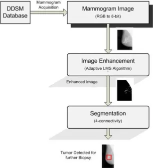
Figure 1 given below illustrates the two approaches use in our proposed system. In this research, we proposed a modified LMS algorithm with image feature vector for mammogram enhancement. Then by using 4-pixel connectivity technique, detection route is completed in the enhanced mammogram image.
Figure 1: Proposed system flow diagram
To develop the system for breast cancer detection, we imply the idea of signal detection problem in radar communication where our signal of interest is aircraft reflected signal and echoes and ambient noise are consider as noise. In this paper, healthy tissues in mammogram images are consider as noise and tumor or damaged tissues as signal of interest [20]. We proposed a hybrid algorithm which uses adaptive least mean square (LMS) algorithm combined with connected component technique called 4-connectivity. Additively we also design a feature vector of size V=3. All techniques and algorithms are explained in next section in details.
172
By comparing our results with ground truth the proposed computer aided scheme achieved adequate results with 80 % system accuracy.
We considered a very important application of adaptive filters i.e. signal enhancement, in which the LMS filter by iteratively updating the filter coefficients enhances the corrupted signal by removing the noise unless we obtained noise free signal that is supposed to be received at receiver end. We select a mask size of 5×1 which slide through all the image in row by row fashion and scan whole image to clean the image. We additionally compute a feature vector of size {V=3} with intensity value, mean and standard deviation value of cleaned window applied to whole image. Finally after we got enhanced image which is now only consists of only suspected part of the breast which may or may not have tumor. To separate the clusters of affected tissues and healthy part of the breast, we apply connected components technique called 4-connectivity.
Detailed step by step explanation of the proposed system is given below.
2.1.Image Enhancement using adaptive LMS algorithm
During data acquisition and transmission, the mammogram images are often corrupted with noise. Adaptive filters are widely used to model the noisy patterns in time varying environment. An adaptive filter tries to minimize the error 𝒆𝒆(𝒊𝒊) between a noisy signal 𝒚𝒚(𝒊𝒊) and a reference input 𝒅𝒅(𝒊𝒊).
𝒆𝒆(𝒊𝒊) = 𝒅𝒅(𝒊𝒊)− 𝒚𝒚(𝒊𝒊)
𝒚𝒚(𝒊𝒊) =𝑿𝑿𝑻𝑻(𝒊𝒊)𝒘𝒘(𝒊𝒊)
(1)
(2)
Where 𝒅𝒅(𝒊𝒊) the desired response, X(𝒊𝒊)is filter input and 𝒚𝒚(𝒊𝒊)is filter output.
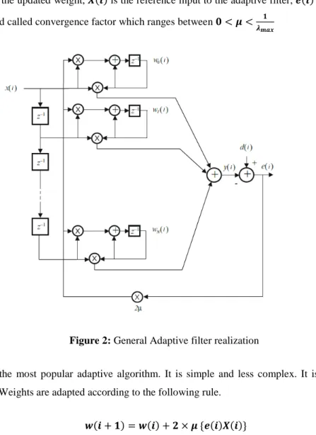
The structure for adaptive LMS filter is shown in Fig 2. Since in the mammogram processing, our objective is to detect the malignant tumors, we generate a noisy signal correlated with healthy tissues. As the algorithm runs iteratively, after some iterations, the algorithm converges and all the healthy tissues are cancelled out. The output of the filter reflects the malignant tumor. Filters weights 𝒘𝒘(𝒊𝒊) initialized, the weights are adapted according to some updation rule involving error signal 𝒆𝒆(𝒊𝒊).
LMS is perhaps the most popular adaptive algorithm. It is simple and less complex. It is stochastic gradient based algorithm. Weights are adapted according to the following rule.
𝒘𝒘(𝒊𝒊+𝟏𝟏) =𝒘𝒘(𝒊𝒊) +𝟐𝟐×𝝁𝝁 {𝒆𝒆(𝒊𝒊)𝑿𝑿(𝒊𝒊)}
173
Here 𝒘𝒘(𝒊𝒊+𝟏𝟏) is the updated weight, 𝑿𝑿(𝒊𝒊) is the reference input to the adaptive filter, 𝒆𝒆(𝒊𝒊) is error signal and 𝝁𝝁 is the step size and called convergence factor which ranges between 𝟎𝟎<𝝁𝝁< 𝟏𝟏
𝝀𝝀𝒎𝒎𝒎𝒎𝒎𝒎
Figure 2: General Adaptive filter realization
LMS is perhaps the most popular adaptive algorithm. It is simple and less complex. It is stochastic gradient based algorithm. Weights are adapted according to the following rule.
𝒘𝒘(𝒊𝒊+𝟏𝟏) =𝒘𝒘(𝒊𝒊) +𝟐𝟐×𝝁𝝁 {𝒆𝒆(𝒊𝒊)𝑿𝑿(𝒊𝒊)}
(3)
Here 𝒘𝒘(𝒊𝒊+𝟏𝟏) is the updated weight, 𝑿𝑿(𝒊𝒊) is the reference input to the adaptive filter, 𝒆𝒆(𝒊𝒊) is error signal and 𝝁𝝁 is the step size and called convergence factor which ranges between 𝟎𝟎<𝝁𝝁< 𝟏𝟏
𝝀𝝀𝒎𝒎𝒎𝒎𝒎𝒎
2.2.Feature Vector {𝑽𝑽=𝟑𝟑}
After mammogram enhancement, we computer feature vector which consists of { 𝒎𝒎𝒊𝒊𝑾𝑾,𝒋𝒋,𝒎𝒎𝑾𝑾𝒊𝒊,𝒋𝒋,𝒔𝒔𝒊𝒊𝑾𝑾,𝒋𝒋 }
, where 𝒎𝒎𝒊𝒊𝑾𝑾,𝒋𝒋 is the pixel intensity value, 𝒎𝒎𝒊𝒊𝑾𝑾,𝒋𝒋 is the mean value of cleaned window we got after enhancement and 𝒔𝒔𝒊𝒊𝑾𝑾,𝒋𝒋 is the standard deviation value of cleaned window.
174
𝑿𝑿𝒊𝒊,𝒋𝒋= { 𝒎𝒎𝒊𝒊𝑾𝑾,𝒋𝒋,𝒎𝒎𝑾𝑾𝒊𝒊,𝒋𝒋,𝒔𝒔𝒊𝒊𝑾𝑾,𝒋𝒋 } (4) 𝝁𝝁𝒎𝒎= 𝒎𝒎𝒊𝒊𝑾𝑾,𝒋𝒋= ∑ 𝒎𝒎𝒊𝒊 𝑵𝑵 𝒊𝒊=𝟏𝟏 𝑵𝑵 (5) 𝝈𝝈=𝒔𝒔𝒊𝒊𝑾𝑾,𝒋𝒋=��(𝒎𝒎𝒊𝒊− 𝝁𝝁𝒎𝒎) 𝟐𝟐 𝑵𝑵 𝑵𝑵 𝒊𝒊=𝟏𝟏 (6) 2.3.SegmentationTo separate out the tumor from the enhanced image, we performed segmentation by using 4-connectivity technique which works in a way that it computes the distances from its neighboring pixels iff located in its four standard directions. It is prohibited to check pixel availability in its diagonals, as follows:
𝑫𝑫𝟒𝟒(𝒑𝒑,𝒒𝒒) = |𝒎𝒎 − 𝒔𝒔| + |𝒚𝒚 − 𝒕𝒕|
(7)
Grouping the pixels meeting the criterion:
1. At pixel (𝑖𝑖,𝑗𝑗) Only see 𝑖𝑖 −1 and j−1 2. For [𝑖𝑖 −1 or j−1] > 0 same component 3. For [𝑖𝑖 −1 andj−1] > 0, assign 𝑖𝑖 −1
Component
3.Experimental results
All experiments are performed by using Matlab 2013a with machine specifications of Intel® Core™ i3-4010U CPU @ 1.70 GHz 64-bit Windows 10 OS. For experiments we acquired images from Digital Database for Screening Mammograms (DDSM) database, which is a collective effort of the University of South Florida and Massachusetts General Hospital and initially funded by the US army Breast Cancer Research Program. We took images of 26 patients randomly without considering age group, sex, region and tumor size. The evaluation of proposed system is based on sensitivity, specificity and accuracy of the system. Below is given their mathematical expressions in terms of true positives (TP), false positive (FP), true negative (TN) and false negative (FN) values.
175
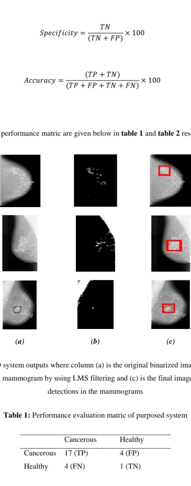
𝑆𝑆𝑆𝑆𝑆𝑆𝑆𝑆𝑖𝑖𝑆𝑆𝑖𝑖𝑆𝑆𝑖𝑖𝑆𝑆𝑆𝑆=(𝑇𝑇𝑇𝑇𝑇𝑇𝑇𝑇+𝐹𝐹𝐹𝐹) × 100 (8) 𝑆𝑆𝑆𝑆𝑆𝑆𝑆𝑆𝑖𝑖𝑆𝑆𝑖𝑖𝑆𝑆𝑖𝑖𝑆𝑆𝑆𝑆=(𝑇𝑇𝐹𝐹𝑇𝑇𝐹𝐹+𝐹𝐹𝑇𝑇) × 100 (9) 𝐴𝐴𝑆𝑆𝑆𝑆𝐴𝐴𝐴𝐴𝐴𝐴𝑆𝑆𝑆𝑆=(𝑇𝑇𝑇𝑇+(𝑇𝑇𝑇𝑇𝐹𝐹𝑇𝑇++𝑇𝑇𝐹𝐹𝑇𝑇𝐹𝐹)+𝐹𝐹𝐹𝐹) × 100 (10)Experimental results and performance matric are given below in table 1 and table 2 respectively.
(a) (b) (c)
Figure 3: proposed CAD system outputs where column (a) is the original binarized image to be processed, (b) is the adaptively enhanced mammogram by using LMS filtering and (c) is the final image output after distortions
detections in the mammograms
Table 1: Performance evaluation matric of purposed system
Cancerous Healthy Cancerous 17 (TP) 4 (FP) Healthy 4 (FN) 1 (TN)
176
Table 2: System performance-Percentage (%)
Sensitivity Specificity Accuracy
81% 80% 80%
4.Conclusion and future work
The proposed CAD based system is a good choice for double reading of mammogram images to assist the radiologists in order to enhance the sensitivity of the radiologist. We have successfully applied the adaptive LMS algorithm to detect the tumor in mammogram images. Our experiments with 80% overall system accuracy, show that adaptive filtering algorithms have huge potential to address the problem of cancer tumor detection in mammogram images by exhibiting 81% true positive and 80% true negative rates.
The future extensions of this work is in pipeline, first to compare the results of proposed system with existing statistical detection systems and then validate our system for its practical significance by testing our system by the expert radiologists or clinicians. We further interested to see whether advanced adaptive filtering algorithms like modified LMS and CRLB Based advanced detectors produce more better results. Further we can consider morphological features of tumor by using multiresolution techniques which are more robust for feature extraction.
References
[1] N. R. &. o. Council, Mammography and beyond: developing technologies for the early detection of breast cancer, National Academic Press, 2001.
[2] M. F. Akay, "Support vector machines combined with feature selection for breast cancer diagnosis," Expert Systems with Applications, vol. 36, p. 3240–3247, (2009.
[3] M. a. M. P.Sampat, Computer-Aided Detection and Diagnosis in Mammography, Handbook of Image and Video Processing, London, U.K.: Elsevier, 2003.
[4] W. Schulz, Molecular biology of human cancers: an advanced student's textbook, New York, USA: Springer, 2005.
[5] M. Biltawi and N. &. T. S. Al-Najdawi, "Mammogram enhancement and segmentation methods: classification, analysis, and evaluation," in The 13th international Arab conference on information technology, 2012.
[6] S. Komen, "SusanG. Komen," SusanG. Komen, 13 01 2018. [Online]. Available: https://ww5.komen.org/. [Accessed 13 01 2018].
[7] A. C. Society, Cancer Facts & Figures 2018, Atlanta: American Cancer Society, 2018
[8] B. S. L. H. H. C. N. P. M. H. R. J. G. a. C. Z. J. Wei, "Computer aided detection of breast masses on full field digital mammograms," Medical Physics, vol. 32, no. 9, pp. 2827-2837, 2005.
177
"American college of radiology imaging network digital mammographic imaging screening trial: Objectives and methodology," Radiology, vol. 236, no. 2, pp. 404-412, 2005.
[10] K. Ganesan, U. R. Acharya, C. K. Chua, L. C. Min and K. T. &. N. K.-H. Abraham, "Computer-aided breast cancer detection using mammograms: a review," IEEE Reviews in Biomedical Engineering, IEEE, vol. 6, pp. 77-98, 2013.
[11] R. E. Bird and T. W. &. Y. B. C. Wallace, "Analysis of cancers missed at screening mammography," Radiology, vol. 184, pp. 613-617, 1992.
[12] K. Kerlikowske, P. A. Carney, B. Geller, M. T. Mandelson, S. H. Taplin, K. Malvin, V. Ernster, N. Urban, G. Cutter and R. &. o. Rosenberg, "Performance of screening mammography among women with and without a first-degree relative with breast cancer," Annals of Internal Medicine, Am Coll Physicians, vol. 133, pp. 855-863, 2000.
[13] V. Ponomaryov, "Computer-aided detection system based on PCA/SVM for diagnosis of breast cancer lesions," in Conference on Electrical, Electronics Engineering, Information and Communication Technologies (CHILECON), CHILEAN, 2015.
[14] R. M. R. I. E. N. a. Y. Y. JInshan Tang, "Computer-Aided Detection and Diagnosis of Breast Cancer With Mammography: Recent Advances," IEEE TRANSACTIONS ON INFORMATION TECHNOLOGY IN BIOMEDICINE, vol. 13, no. 2, pp. 236-251, 2009.
[15] F. J. A. J. L. D. Rangaraj M. Rangayan, "A review of computer-aided diagnosis of breast cancer: Toeard the detection ofsubtle signs," Journal of The franklin Institute, vol. 244, no. 3-4, pp. 312-348, May–July 2007.
[16] M. L. G. C. J. V. a. C. E. M. Zhimin Huo, "Breast Cancer: Effectiveness of Computer-aided Diagnosis— Observer study with Independent Database of Mammograms," Radiology, vol. 224, pp. 560-568, 2002. [17] M. E. A. a. M. W. Nadia El Atlas, "Computer-Aided Breast Cancer Detection Using Mammograms: A
Review," in 2014 Second World Conference on Complex Systems (WCCS), Agadir, Morocco, 2014. [18] J. B. M. L. S. K. S. S. L. N. a. J. H. R. Brem, "Improvement in sensitivity of screening mammography with
computer-aided detection: A multiinstitutional trial," American JOurnal of Roentgenology, vol. 181, no. 3, p. 687–693, 2003.
[19] J. S. C. Bose and K. S. &. K. M. Kumar, "Detection of Microcalcification in Mammograms using soft computing techniques," European journal of scientific research, vol. 86, pp. 103-122, 2012.
[20] N. B. a. W. O. Hacer Varol, "Breast Cancer Detection Using Communications Technology," in International Conference on Industrial and Intelligent Information (ICIII 2012), Singapore, 2012.