ORIGINAL ARTICLE
Clinical application of massively parallel sequencing-based prenatal
noninvasive fetal trisomy test for trisomies 21 and 18 in 11 105
pregnancies with mixed risk factors
Shan Dan1,2†, Wei Wang1†, Jinghui Ren3, Yali Li4, Hua Hu5, Zhengfeng Xu6, Tze Kin Lau7, Jianhong Xie8, Weihua Zhao9, Hefeng Huang10,11, Jiansheng Xie12, Luming Sun13, Xiaohong Zhang14, Weipeng Wang15, Shixiu Liao16, Rong Qiang17, Jiangxia Cao18, Qiufang Zhang19, Yulin Zhou20, Haiyan Zhu21, Mei Zhong22, Yi Guo23, Linhua Lin3, Zhiying Gao4, Hong Yao5, Hongyun Zhang1, Lijian Zhao1, Fuman Jiang1, Fang Chen1, Hui Jiang1, Songgang Li1, Yingrui Li1, Jun Wang1, Jian Wang1, Tao Duan13*, Yue Su2* and Xiuqing Zhang1*
1BGI-Shenzhen, Shenzhen, China
2Department of Perinatology, Beijing Obstetrics and Gynecology Hospital, Capital University of Medical Sciences, Beijing, China 3The Center of Prenatal Diagnosis, Shenzhen People’s Hospital, 2nd Clinical Medical College of Jinan University, Shenzhen, China 4Department of Obstetrics and Gynecology, Chinese PLA General Hospital, Beijing, China
5Southwest Hospital, The Third Military Medical University, Chongqing, China
6Center of Prenatal Diagnosis, Nanjing Maternal and Child Health Hospital, Nanjing Medical University, Nanjing, China 7Fetal Medicine Centre, Paramount Clinic, Hong Kong
8Zhuhai Institute of Medical Genetics, Zhuhai Municipal Maternal and Child Healthcare Hospital, Zhuhai, China
9Department of Obstetrics, The Second People’s Hospital of Shenzhen, The First Affiliated Hospital of Shenzhen University, Shenzhen, China 10Key Laboratory of Reproductive Genetics, Zhejiang University, Ministry of Education, Hangzhou, China
11Department of Reproductive Endocrinology, Women’s Hospital, Zhejiang University School of Medicine, Hangzhou, China
12Central for Prenatal Diagnosis, Shenzhen Maternity and Child Healthcare Hospital, Affiliated Southern Medical University, Shenzhen, China 13Department of Obstetrics, Shanghai 1st Maternity and Infant Hospital, Tongji University, Shanghai, China
14Department of Obstetrics and Gynecology, Peking University People’s Hospital, Beijing, China 15Department of Clinical Laboratory, Hubei Maternal and Child Health Hospital, Wuhan, China
16Medical Genetic Center, Henan Provincial People’s Hospital, Zhengzhou University People’s Hospital, Zhengzhou, China 17Prenatal Diagnosis Center, Maternal and Child Health Hospital of Shanxi Province, Xi’an, China
18Department of Obstetrics and Gynecology, Wuhan Medical Care Center for Women and Children, Wuhan, China 19Department of Perinatal Medicine, Peking University Third Hospital, Beijing, China
20Xiamen Prenatal Diagnosis Center, Xiamen Maternal and Child Health Care Hospital, Xiamen, China
21Prenatal Diagnosis Center, Department of Obstetrics and Gynecology, Nanjing Drum Tower Hospital, Nanjing University Medical School, Nanjing, China 22Department of Gynecology and Obstetrics, Nanfang Hospital, First Military Medical University, Guangzhou, China
23Department of Perinatal Medicine, Dalian Obstetrics and Gynecology Hospital, Dalian, China
*Correspondence to: Xiuqing Zhang. E-mail: [email protected] or Yue Su. E-mail: [email protected] or Tao Duan. E-mail: [email protected]
†These authors contributed equally to this work.
ABSTRACT
Objective To report the performance of massively parallel sequencing (MPS) based prenatal noninvasive fetal trisomy test based on cell-free DNA sequencing from maternal plasma in a routine clinical setting in China.
MethodThe MPS-based test was offered as a prenatal screening test for trisomies 21 and 18 to pregnant women in 49 medical centers over 2 years. A total of 11 263 participants were recruited and the MPS-based test was performed in 11 105 pregnancies. Fetal outcome data were obtained after the expected date of confinement.
ResultsOne hundred ninety cases were classified as positive, including 143 cases of trisomy 21 and 47 cases of trisomy 18. With the karyotyping results and the feedback of fetal outcome data, we observed one false positive case of trisomy 21, one false positive case of trisomy 18 and no false negative cases, indicating 100% sensitivity and 99.96% specificity for the detection of trisomies 21 and 18.
Conclusion Our large-scale multicenter study proved that the MPS-based test is of high sensitivity and specificity in detecting fetal trisomies 21 and 18. The introduction of this screening test into a routine clinical setting could avoid about 98% of invasive prenatal diagnostic procedures. © 2012 John Wiley & Sons, Ltd.
Funding sources: The study was funded by Shenzhen Birth Defect Screening Project Lab (JZF No. [2011] 861) approved by Shenzhen Municipal Commission for Development and Reform and Key Laboratory Project in Shenzhen (CXB200903110066A and CXB201108250096A) and Key Laboratory of Cooperation Project in Guangdong Province (2011A060906007).
Conflicts of interest: Wei Wang, Hongyun Zhang, Lijian Zhao, Fuman Jiang, Fang Chen, Hui Jiang, Songgang Li, Yingrui Li, Jun Wang, Jian Wang, Xiuqing Zhang are employees of BGI-Shenzhen and none of the other authors have anyfinancial relationship with BGI-Shenzhen.
INTRODUCTION
Each year there are about 16 million newborns in China, of whom 4% to 6% are affected by some form of birth defects.1,2 Chromosomal abnormalities, with an incidence of 1 in 160 births, are one of the most important causes of birth defects, and there are no curative treatments at present.3 Although invasive prenatal tests allow accurate diagnosis, wide spread clinical use is limited by cost and a 0.5% to 1% risk of procedure-related miscarriage.4–6Over the last three decades, many screening tests have been developed to identify the high-risk groups for the most common chromosomal abnormalities, such as trisomy 21 (T21), 18 (T18) and 13 (T13), by various combinations of medical history, maternal age, ultrasound markers and maternal serum biochemistry. To date thefirst trimester combined screening has become widely accepted, being able to detect about 90% of trisomy 21 fetuses and 50% to 85% of other chromosomal defects.7,8 However, at a false positive rate of 4% to 5% for current screening approaches, only about 5% of the‘high-risk’pregnant women indeed carry a fetus with trisomy 21 while the remaining are normal, giving a positive predictive value of about 1 in 20.9,10 Therefore, there is a continuous drive to search for a diagnostic test without risk of miscarriage, or a screening test with better performance.
A variety of strategies have been explored using maternal blood, urine, and saliva samples to diagnose fetal gender and trisomy 21.11,12The discovery of the presence of fetal cell-free DNA and RNA in maternal plasma, combined with PCR technologies and mass spectrometric analysis, has enabled noninvasive detection of trisomies 21 and 18 with acceptable sensitivity and specificity.13–17However, these methods mainly detect specific alleles in a certain population, which has restricted its wide and robust application in clinical practice. In 2008 the rapid development of massively parallel sequencing (MPS) technology made it feasible to use maternal plasma cell-free DNA to detect trisomy 21 without using polymorphic markers.18,19Studies from multiple centers so far have shown that this is a highly reliable approach with a detection rate of 99% to 100% and a specificity of 98% to 100% for trisomy 21.20–23More recent studies have suggested that, with improved bioinformatic analysis, similar accuracies are achievable for the detection of trisomies 18 and 13.24–26Several research groups have published their clinical trial data with 397 to 4664 cases.22,23,27,28 Here we reported the results of a prospective large-scale multicenter study in a routine clinical setting in a Chinese population in which 11 105 participants were recruited and took the MPS-based test. Our experience may provide useful information to clinicians and medical practitioners who are interested in the application and integration of this new technology with established prenatal screening and diagnosis workflow.
METHODS
Participants and overall design
In a 2-year period from thefirst quarter of 2010 to thefirst quarter of 2012, the MPS-based test was offered to pregnant women as a clinical screening test in 49 medical centers in 15 provinces and 4 centrally administered municipalities, all of which are prenatal screening or diagnosis centers certified by the Ministry of Health (MOH) of China.
The inclusion criteria for participants were: (1) pregnant women, at least 18 years old or above, (2) with a singleton live fetus and (3) a gestational age of 9 to 28 weeks. Women with multiple pregnancies or intra-uterine fetal demise at the time of sampling or unknown gestational age were excluded from this study. Cases with an expected date of confinement before 31th March 2012 or those with karyotyping results were included for the current analysis.
Approvals were obtained from the institutional review board of BGI-Shenzhen. Informed written consent was obtained from all pregnant women who agreed to take this screening test. All MPS-based tests were performed prior to the recording of karyotyping information and the sequencing lab in Shenzhen was blinded.
Each collaborating site was responsible for obtaining fetal outcome data for their own study subjects. To minimize under-reporting of false negative cases among the negative cases, and to motivate participants to report undiagnosed trisomies 21 and 18, we have established an ‘insurance scheme’for this clinical study, as described below.
THE MPS-BASED TEST
Pretest counselingCareful pretest counseling was provided to each participant. All participants were informed of the limitations of the MPS-based test as follows:
(1) This test is mostly intended for the prenatal detection of fetal trisomies 21 and 18 at the gestational age of 12 to 24 weeks.
(2) Although the reported sensitivities and specificities for trisomies 21 and 18 were as high as 99% or above, the MPS-based test is still a screening test. All test positive pregnancies should receive confirmatory invasive testing. (3) About 1% of pregnancies would need resampling because
of failure in the quality control criteria.
(4) This test cannot be applied to the following situations: (a) when the pregnant woman is affected with trisomy 21 or 18, (b) at an earlier gestational age than 9 weeks, or (c) women with multiple pregnancies.
(5) This test is not designed to identify fetuses with mosaicism, triploidy or chromosomal microdeletions/microduplications. Besides the information described above, on the consent form all participants were informed that they automatically joined the insurance scheme purchased by BGI-Shenzhen on their behalf, if they accept our test. In the event that a pregnant woman had a test negative result but eventually delivered a baby with trisomy 21 or 18, the insurance company would pay her CNY 200 000 (equal to $31 658) (China Life Insurance Company, Ltd.).
Massively parallel DNA sequencing
Five milliliters of peripheral venous blood sample were collected into tubes containing Ethylene Diamine Tetraacetic Acid from each participant. All blood samples were obtained before any invasive prenatal diagnostic test, and were processed within 8 h by a double-centrifugation protocol. Blood samples werefirst centrifuged at 1600g for 10 min, and the supernatant was recentrifugated at 16 000g for 10 min to remove residual cells. The resultant cell-free plasma was stored at–80C, and then shipped to BGI-Shenzhen on dry ice. Most samples arrived at the lab within 24 h and very few arrived no more than 72 h. Each plasma sample was frozen and thawed only once.
For DNA extraction, 600mL of maternal plasma was used. The DNA libraries were prepared according to a modified protocol from Illumina. After end-repairing, A-base tailing and adaptor ligation, standard multiplex primers were introduced by 17-cycle PCR. The size distribution of the libraries was analyzed using an Agilent Bioanalyzer and quantified with real-time PCR. Four or 12 barcoded libraries were equally pooled and sequenced with 36-cycles single-end multiplex sequencing strategy on an Illumina GAIIx or HiSeq 2000 platform, respectively.
All molecular tests and procedures were performed in an ISO/IEC 17025 certified and MOH accredited clinical laboratory at BGI-Shenzhen.
Quality control in sampling and sequencing
We set up a quality control criteria system for each step from sampling to reporting. A barcode tracking system was employed during the whole process. Blood samples with evidence of hemolysis or those that were processed beyond 8 h after sample collection were excluded. The quality parameters of acceptable samples were as follows: the peak size of a qualified DNA library was between 290 and 303 bp, and the yield more than 30 nM. The sequencing quality value (Q20) was over 90% for each base, and the GC content was around 401.5%. The minimal amount of unique sequencing reads was no less than 2 million after alignment, which corresponded to about 5 million raw sequencing reads. Only qualified sequencing data were used for subsequent analyses.
Trisomies 21 and 18 analysis
Thirty-five base sequencing reads were trimmed and aligned back to a universal unique read set incised from the human reference genome (HG 18, NCBI build 36). A binary hypothesis
t-test and logarithmic likelihood ratioL-score between the two
t-tests were used to classify whether the fetus had trisomy 21 or
18 or not (a detailed description of the methodology is available in supplementary materials). If both thet-score>2.5 and the
L-score>1, the sample was located in the high risk zone. If either thet-score>2.5 or theL-score>1 the sample was located in the warning zones. If thet-score<2.5 and theL-score<1, samples were located in the low-risk zone. Samples located in the high risk zone or warning zones were classified as test positive, while samples located in low risk zone as test negative.
Data and statistical analysis
The sensitivity and specificity of the MPS-based test was evaluated, using the result of fetal karyotyping as the gold standard. 95% confidence intervals (CIs) were calculated on the bases of a standard normal distribution.
Report delivery and posttest counseling
The report of our test would be issued within 15 working days from blood drawing. Posttest counseling was provided by clinicians and careful advice was given according to the test results.
Follow up investigation
In the case of MPS-based test positive results, participants were advised to have prenatal fetal karyotyping. If refused, the outcome of the case was monitored intensively to ensure a complete follow-up of the pregnancy.
In the case of MPS-based test negative results, the decisions for prenatal fetal karyotyping were made according to other clinical parameters and the wish of individual participants.
Fetal outcome data were obtained by questionnaires from participants.
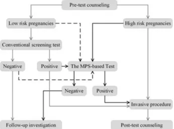
Figure 1The workflow of the MPS-based test. The MPS-based test was offered as a screening test with careful pre-test and posttest counseling. The test was performed in an ISO/IEC17025 certified and MOH accredited clinical laboratory, where a laboratory information management system was employed throughout the entire workflow of activities to guarantee end-to-end traceability. In this study, test positive cases were referred for conventional invasive procedures while negative cases for intensive monitoring to ensure a complete follow-up data
RESULTS
WorkflowFigure 1 summarizes the workflow of the MPS-based test. In general, 95% of cases received the report within 10 working days from blood sampling, 99% of our test reports were issued within 15 working days.
Resampling of pregnancies
During the study, 0.87% (97/11 105) pregnancies needed resampling because of failure in quality control criteria mostly because of early gestational age. The test for these cases usually took another 10 to 15 working days from resampling to report delivery.
Study participants
Forty-nine medical centers participated in this clinical study, and a total of 11 263 pregnant women were recruited in two years (Figure 2). Forty-two participating centers offered the test to
‘high risk’pregnant women identified by a conventional Down syndrome screening test, and the remaining 7 centers enrolled participants regardless of prior risk assessment. The MPS-based test was not performed in 79 cases because the recruitment criteria were not met. Another 79 cases failed in the quality control in the plasma separation, DNA extraction, sample preparation or sequencing steps mentioned in the methods section. Overall, our test was performed on the remaining 11 105 cases. Also, 1.5% (158/11 263) of the women did not
receive results because of recruitment criteria failure or quality control failure.
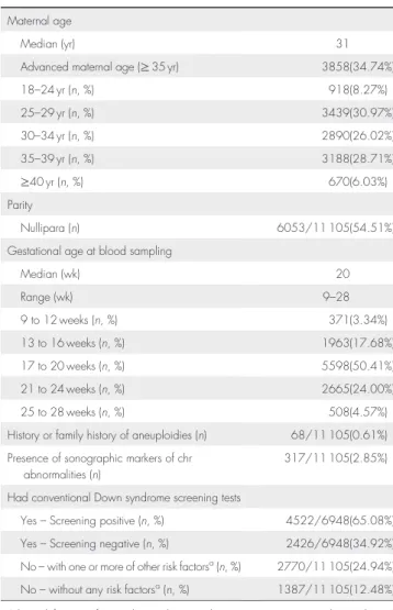
Basic characteristics of the study population are shown in Table 1. The maternal age ranged from 18 to 49 years old with a median of 31. The median gestational age at the time of blood sampling was 20 weeks. 34.74% (3858/11 105) pregnancies were 35 years old or above. 62.57% (6948/11 105) received eitherfirst or second trimester conventional Down syndrome screening tests, in which 65.08% (4522/6948) were screen positive. 2.85% (317/11 105) had ultrasound markers of chromosomal abnormalities, and 0.61% (68/11 105) had a family history of chromosomal abnormalities. No specific risk factor for chromosomal abnormalities was present in 12.49% of the tested cases (1387/11 105).
Identification of trisomies 21 and 18 by the MPS-based test
An average of 5 million sequencing reads per sample was obtained using the Illumina GAIIx or HiSeq 2000 sequencing platforms. After removal of low quality reads and alignment, at least 2 million unique reads for each sample were used for the detection of trisomies 21 and 18.
One hundred ninety cases were classified as test positive, giving a test positive rate of 1.71% (95% CI, 1.47%–1.95%). Of those, 143 cases were positive for trisomy 21 and 47 cases for trisomy 18, corresponding to an incidence of 1.29% (95% CI, 1.08%–1.50%) for trisomy 21, and 0.42% (95% CI, 0.30%–0.54%) for trisomy 18 in this cohort (Figure 3).
Figure 2Participants recruitment and overall design. A total of 11 263 pregnant women were recruited in this multicenter study. Seventy-nine cases failed to meet the recruitment criteria and another 79 cases failed in the quality control. The MPS-based test was performed on the remaining 11 105 cases, among which 3000 cases had performed full karyotyping and 4524 pregnancies had follow-up fetal outcome data
Follow-up investigation of test positive cases
One hundred forty of 143 T21-positive cases were confirmed by prenatal karyotyping, including 138 cases of typical trisomy 21, one case of mosaic trisomy 21 (23 of 50 cells studied were trisomic), and one case with euploid karyotype. Three test positive cases did not have karyotype analysis because of spontaneous abortion, intra-uterine fetal demise, or induced abortion, respectively.
Forty-two of 47 T18-positive cases were validated by prenatal karyotyping, and 41 cases were confirmed to be typical trisomy 18 and one case with euploid karyotype. Karyotyping was not performed infive test positive cases, including four cases of induced abortion, and one case of intra-uterine infection. Among thesefive cases, one had nuchal translucency>6 mm, one case had abnormal ultrasound markers and the other three cases were at high risk of trisomy 18 based on maternal biochemical screening.
Follow-up investigation of test negative cases
Overall, 10 915 cases were classified as test negative for both trisomies 21 and 18. Among them, 2818 also had fetal karyotyping performed. The indications for the invasive procedures were: (1) positive conventional Down syndrome screening tests in 1178 subjects, (2) abnormal sonographic findings in 56 cases, (3) advanced maternal age in 757 cases, and (4) other reasons such as maternal anxiety or family history in 827 cases. Trisomy 21 or 18 was not detected in any of these 2818 karyotyping studies.
In the remaining 8097 test negative cases without karyotyping, fetal outcome data and detailed information from the newborn examination was available for 4524 pregnancies, which showed that no woman gave birth to a trisomy 21 or 18 newborn. So far, no claim has been received for the insurance coverage for the birth of an affected newborn missed by the MPS-based test.
Table 1Basic characteristics of the 11 105 pregnant women who took the MPS-based test
Maternal age
Median (yr) 31
Advanced maternal age (≥35 yr) 3858(34.74%)
18–24 yr (n, %) 918(8.27%) 25–29 yr (n, %) 3439(30.97%) 30–34 yr (n, %) 2890(26.02%) 35–39 yr (n, %) 3188(28.71%) ≥40 yr (n, %) 670(6.03%) Parity Nullipara (n) 6053/11 105(54.51%)
Gestational age at blood sampling
Median (wk) 20 Range (wk) 9–28 9 to 12 weeks (n, %) 371(3.34%) 13 to 16 weeks (n, %) 1963(17.68%) 17 to 20 weeks (n, %) 5598(50.41%) 21 to 24 weeks (n, %) 2665(24.00%) 25 to 28 weeks (n, %) 508(4.57%)
History or family history of aneuploidies (n) 68/11 105(0.61%) Presence of sonographic markers of chr
abnormalities (n)
317/11 105(2.85%) Had conventional Down syndrome screening tests
Yes–Screening positive (n, %) 4522/6948(65.08%) Yes–Screening negative (n, %) 2426/6948(34.92%) No–with one or more of other risk factorsa(n, %) 2770/11 105(24.94%)
No–without any risk factorsa(n, %) 1387/11 105(12.48%) aOther risk factors refer to advanced maternal age, presence sonographic markers of
a positive history of affected pregnancies.
Figure 3Identification of fetal aneuploidy by the MPS-based test. The risk of fetal aneuploidy was described by theL-score (x-axis) andt-score (y-axis). The true aneuploidy and negative sample by fetal karyotpying is labeled by a solid triangle (▲) and hollow circles (○). The high-risk zone is defined by L-score>1 andt-score>2.5. (A) 143 cases (including one T21 false positive case) were located in the high-risk zone for trisomy 21 and classified as T21-positive. (B) 47 cases (including one T18 false positive case) were located in the high-risk zone for trisomy 18 and classified as T18-positive
The estimation of sensitivity and specificity of the MPS-based test
Overall, 3000 cases had full karyotyping performed. There were 139 cases of trisomy 21, 41 cases of trisomy 18, and 2820 euploid cases. We observed one false positive case of trisomy 21 and one false positive case of trisomy 18 in our test. Therefore, the detection rate of the MPS-based test in identifying fetal trisomy 21 and 18 were 100% (139/139) and 100% (41/41), whereas the false positive rate were 0.03% (1/2820) and 0.03% (1/2820), respectively. The estimated sensitivities and specificities were 100% and 99.96% for both trisomies 21 and 18, respectively.
DISCUSSION
In this study 11 263 pregnant women from 49 medical centers were recruited in China, to evaluate the performance of the MPS-based test as a screening test in a clinical setting. This multicenter study recruited a relatively large sample size involving participants with various clinical risk factors. It was performed in the routine and complicated clinical settings. With proper pretest and posttest counseling, our test results were shown to be highly accurate, helping clinicians to offer better medical advices to pregnant women who then can make well-informed pregnancy decisions.
The detection rate (or sensitivity) and the false positive rate (or specificity) are critical parameters in evaluating a new clinical testing technology. In this study, 182 of 190 test positive cases had fetal karyotyping performed. Our test successfully identified all true trisomies 21 and 18 cases; however, there were two false positive cases. For those eight cases identified as test positive but did not have karyotyping, we believe it was unlikely to be false positive, because these cases showed high-risk indications in conventional screening tests and ended with abnormal pregnancy outcomes, such as spontaneous abortion, and intra-uterine fetal demise. Taken together, the estimated specificity of 100% for trisomies 21 and 18 was likely to be reasonable. Even if all these eight cases were false positive, the false positive rate would still be very low, less than 1/1000.
Among the 10 915 test negative cases, 2818 (25.82%) women still requested prenatal fetal karyotyping results. This was probably because many pregnant women were still uncertain about the reliability of our test at that moment, and needed reassurance from conventional invasive tests. On the other hand, this gave us the opportunity to exclude false negativity of our test with certainty among these cases, because the karyotyping results were all normal for both chromosome 21 and 18. Six hundred twenty-five of 2818 cases were still pregnant at the time this manuscript was written.
However, follow-up investigation of the remaining 8097 test negative cases without karyotyping was a challenge. In China, follow-up data are difficult to obtain in such a large-scale multicenter clinical study because of an uncompleted medical tracking system. In this study all 49 participating sites were referral centers mainly located in big cities, whereas pregnant women prefer to give birth in local hospitals, making follow-up assessment difficult. For these reasons, we were only able to obtain fetal outcome data on 4524 participants, even though we used all possible means to contact these women through both the participants’centers and referring centers. Also, we came up with an incentive program to encourage these patients to report
to us in case of false negative results. By introducing a formal insurance policy from a listed company, any reported false negative case would receive CNY 200,000 ($31 658 ) of insurance coverage, which was equivalent to 8.3 years of the average household income in China (http://www.stats.gov.cn/). Therefore, it is unlikely that anyone with a false negative result would go unnoticed without making a claim to the insurance company. Because we have not received any claim for insurance so far, we believe that the chance of unreported false-negative case was very low.
The sensitivity and specificity of over 99% for the MPS-based test for both trisomies 21 and 18 in this study was consistent with the results of previous studies.21,22Only 1.17% (190/11 105) of the study population which were classified as test positive for trisomy 21 or 18, needed to perform conventional karyotyping analysis, suggesting that over 98% of invasive procedures could be avoided. This is not only cost-saving, but also is a much safer option, avoiding unnecessary fetal losses because of invasive tests. If all of these 11 105 pregnant women had received the invasive tests, 55 to 111 cases procedure-related miscarriage might have happened based on the 0.5% to 1% incidence found in prior studies.3–5 These hypothetical miscarriages could be avoided with the introduction of our test to the established prenatal screening and diagnosis workflow.
In principle, MPS-based methods could detect all kinds of fetal aneuploidy and microdeletions/microduplications. The testing for trisomy 21/18 is the most clinically relevant because other numerical autosomal chromosome aberrations are rare after 12 weeks.29–31 On the other hand, the detection of other chromosomal abnormalities such as mosaic or structural variations still remains challenging for this new MPS-based test.
Figure 4 Recommended pipeline to integrate the MPS-based test with established screening-diagnosis model in China. The pipeline with gray solid line refers to the established clinical procedures in China. The intensive route labeled with dark solid lines represents the preferred time of interference into conventional clinical practice. The dark dashed lines indicate our test can also be applied to pregnant women in the low-risk subgroup and those with negative results from conventional screening tests. Whether the MPS-based test is used as a primary or secondary screening test, it should be given careful pretest/posttest counseling based on informed consent and choice
For the two false positive cases, one of the possible explanations is that there was microduplication in chromosome 21/18 that could be detected by karyotyping. Another possibility is that it was a placenta mosaic that was hard to detect by conventional invasive procedure. Further studies are needed to explore the application of the MPS-based test in these situations.
With the revolutionary progress of noninvasive testing technology, new ethical questions are emerging.32One of the most difficult issues relates to the fact that such new approaches can count sex chromosomes and therefore contain information of fetal gender. At present, fetal sex is not reported in China because sex-selection is banned by the Law on Maternal and Infant Health of China. Would the availability of fetal sex information result in sex selection behavior? Because part of sex chromosomal abnormalities are not associated with major disabilities, should a woman be informed if there is a suspicion of sex chromosomal abnormalities? Thus, the key concern is what information could or should be provided to pregnant women. As for sex chromosomal abnormalities, it may be time to consider a more comprehensive informed consent process to allow pregnant women to make well-informed decisions on requesting this additional information or not.
There are several limitations of the current study. First, the majority of subjects were recruited after 17 weeks of gestation, and only 371 pregnant women had a gestational age between 9 and 12 weeks, which may be related with the high accuracy of detection of trisomies 21 and 18 and low ratio of repeat test in this study. This was due to the fact that in most Obstetrics and Gynaecology departments in China, medical records for pregnant women begin from 12 gestational weeks, and this is also an important reason we introduce our test at this time point and beyond in this study. Further large scale studies focusing on subjects in thefirst trimester of pregnancy may be required to evaluate the performance of the MPS-based testing at an earlier gestation (9–12 weeks or even earlier) before its complete integration into current obstetrical practice. Second, although the inadequacy of clinical follow-up could partially be overcome by the use of an insurance coverage scheme, further long-term investigations still need to be carried out for more comprehensive evaluation of this new technology.
CONCLUSION
Massively parallel sequencing-based test as a screening method is undoubtedly more efficient than any other existing noninvasive screening tests for fetal trisomies 21 and 18. Although wide-spread use of this technology is limited by the cost and reporting time at the moment, the situation probably will change quickly. The cost of MPS-based noninvasive test varies between countries, even states. In the United States, this MPS-based test is charged $795 to $2762, while it takes around $500 to $1000 in China. With the rapid development of high throughput sequencing technology, the cost of this test will drop to levels lower than that of conventional invasive diagnosis procedures in the near future. The reporting time of the MPS-based test could be reduced to three days or less.33,34Also, with increasing sequencing depth, our test has the potential to detect more fetal chromosomal abnormalities including structural variations or even single-gene disorders.35–38It is clear that this new technology will radically change the current clinical model of prenatal screening and diagnosis in the near future. However, the proper application of the MPS-based test in clinical settings needs careful consideration to integrate with established obstetric practice and workflow, and the right of informed consent and choice of pregnant women should be fully respected in this process. Here we also recommend a possible integration of the MPS-based test with the established screening-diagnosis model based on our experience in China (Figure 4).
WHAT’S ALREADY KNOWN ABOUT THIS TOPIC?
• Massively parallel sequencing has been proved to be feasible for the noninvasive prenatal diagnosis of trisomy 21. This is a large scale multicenter study of massively parallel sequencing-based noninvasive prenatal testing for trisomies 21 and 18 in China.
WHAT DOES THIS STUDY ADD?
• We proved that our noninvasive fetal trisomy test is successful as a screening test for detection of trisomy 21 and 18 in the Chinese population.
REFERENCES
1. Statistics NBo. The population change of China in 2011 [WWW document]. URL http://www.gov.cn/gzdt/2012-01/18/ content_2047892.htm [accessed on 18 January 2012].
2. Health CsMo. Report on Women and Children’s Health Development in
China [WWW document]. URL http://www.gov.cn/gzdt/2011-09/21/ content_1952953.htm [accessed on 21 September 2011].
3. Dai L, Zhu J, Liang J,et al. Birth defects surveillance in China. World J Pediatr 2011;7:302–10.
4. Corrado F, Cannata ML, La Galia T,et al. Pregnancy outcome following
mid-trimester amniocentesis. J Obstet Gynaecol 2012;32:117–9.
5. Enzensberger C, Pulvermacher C, Degenhardt J,et al. Fetal Loss Rate and
Associated Risk Factors After Amniocentesis, Chorionic Villus Sampling and Fetal Blood Sampling. Ultraschall Med 2012. [Epub ahead of print]
6. Kollmann M, Haeusler M, Haas J,et al. Procedure-Related
Complications after Genetic Amniocentesis and Chorionic Villus Sampling. Ultraschall Med 2012. [Epub ahead of print]
7. Dhaifalah I, Dusek L, Santavy J. Implementation of One-Stop-Clinic for
Risk Assessment of chromosomal abnormalities in thefirst trimester,
evaluating the effectiveness and making it part of our daily practice.
Ceska Gynekol 2011;76:292–306.
8. Ghaffari SR, Tahmasebpour AR, Jamal A,et al. First-trimester screening
for chromosomal abnormalities by integrated application of nuchal translucency, nasal bone, tricuspid regurgitation and ductus venosus
flow combined with maternal serum free beta-hCG and PAPP-A: a
5-year prospective study. Ultrasound Obstet Gynecol 2012;39:528–34.
9. Canick J. Prenatal screening for trisomy 21: recent advances and
guidelines. Clin Chem Lab Med 2011; 50:1003–8.
10. Morris JK, Waters JJ, de Souza E. The population impact of screening for Down syndrome: audit of 19 326 invasive diagnostic tests in England and Wales in 2008. Prenat Diagn 2012;32:596–601.
11. Majer S, Bauer M, Magnet E,et al. Maternal urine for prenatal
diagnosis--an analysis of cell-free fetal DNA in maternal urine and
plasma in the third trimester. Prenat Diagn 2007;27:1219–23.
12. Devaney SA, Palomaki GE, Scott JA,et al. Noninvasive fetal sex
determination using cell-free fetal DNA: a systematic review and
13. Lo YM, Lun FM, Chan KC,et al. Digital PCR for the molecular detection of fetal chromosomal aneuploidy. Proc Natl Acad Sci U S A
2007;104:13116–21.
14. Tsui NB, Chiu RW, Ding C,et al. Detection of trisomy 21 by quantitative
mass spectrometric analysis of single-nucleotide polymorphisms. Clin Chem 2005;51:2358–62.
15. Tong YK, Ding C, Chiu RW,et al. Noninvasive prenatal detection of fetal
trisomy 18 by epigenetic allelic ratio analysis in maternal plasma:
Theoretical and empirical considerations. Clin Chem 2006;52:2194–202.
16. Lo YM, Tsui NB, Chiu RW,et al. Plasma placental RNA allelic ratio
permits noninvasive prenatal chromosomal aneuploidy detection. Nat Med 2007;13:218–23.
17. Papageorgiou EA, Karagrigoriou A, Tsaliki E,et al. Fetal-specific DNA
methylation ratio permits noninvasive prenatal diagnosis of trisomy 21.
Nat Med 2011;17:510–3.
18. Chiu RW, Chan KC, Gao Y,et al. Noninvasive prenatal diagnosis of fetal
chromosomal aneuploidy by massively parallel genomic sequencing of
DNA in maternal plasma. Proc Natl Acad Sci U S A 2008;105:20458–63.
19. Fan HC, Blumenfeld YJ, Chitkara U,et al. Noninvasive diagnosis of fetal
aneuploidy by shotgun sequencing DNA from maternal blood. Proc
Natl Acad Sci U S A 2008;105:16266–71.
20. Ehrich M, Deciu C, Zwiefelhofer T,et al. Noninvasive detection of fetal
trisomy 21 by sequencing of DNA in maternal blood: a study in a clinical
setting. Am J Obstet Gynecol 2011;204:205 e1–11.
21. Chiu RW, Akolekar R, Zheng YW,et al. Non-invasive prenatal
assessment of trisomy 21 by multiplexed maternal plasma DNA sequencing: large scale validity study. BMJ 2011;342:c7401.
22. Palomaki GE, Kloza EM, Lambert-Messerlian GM,et al. DNA
sequencing of maternal plasma to detect Down syndrome: an international clinical validation study. Genet Med 2011;13:913–20.
23. Ashoor G, Syngelaki A, Wagner M,et al. Chromosome-selective
sequencing of maternal plasma cell-free DNA forfirst-trimester detection
of trisomy 21 and trisomy 18. Am J Obstet Gynecol 2012;206:322 e1–5.
24. Chen EZ, Chiu RW, Sun H,et al. Noninvasive prenatal diagnosis of fetal
trisomy 18 and trisomy 13 by maternal plasma DNA sequencing. PLoS One 2011;6:e21791.
25. Lau TK, Chen F, Pan X,et al. Noninvasive prenatal diagnosis of common
fetal chromosomal aneuploidies by maternal plasma DNA sequencing.
J Matern Fetal Neonatal Med 2012;25:1370–4.
26. Palomaki GE, Deciu C, Kloza EM,et al. DNA sequencing of maternal
plasma reliably identifies trisomy 18 and trisomy 13 as well as Down
syndrome: an international collaborative study. Genet Med 2012;14:296–305.
27. Lau TK, Chan MK, Salome Lo PS,et al. Clinical utility of noninvasive
fetal trisomy (NIFTY) test - early experience. J Matern Fetal Neonatal Med 2012;25(10):1856–9.
28. Bianchi DW, Platt LD, Goldberg JD,et al. Genome-wide fetal aneuploidy
detection by maternal plasma DNA sequencing. Obstet Gynecol 2012;119:890–901.
29. Aboussair N, Jaouad IC, Dequaqui SC,et al. Cytogenetic analysis of 5572
patients referred for suspected chromosomal abnormalities in morocco.
Genet Test Mol Biomarkers 2012;16:569–73.
30. Mademont-Soler I, Morales C, Clusellas N,et al. Prenatal cytogenetic
diagnosis in Spain: analysis and evaluation of the results obtained from
amnioticfluid samples during the last decade. Eur J Obstet Gynecol
Reprod Biol 2011;157:156–60.
31. Leung TY, Chan LW, Leung TN,et al. First-trimester combined
screening for trisomy 21 in a predominantly Chinese population.
Ultrasound Obstet Gynecol 2007;29:14–7.
32. Deans Z, Newson AJ. Ethical considerations for choosing between possible models for using NIPD for aneuploidy detection. J Med Ethics 2012;38(10):614–8.
33. Phimister EG, Feero WG, Guttmacher AE. Realizing genomic medicine. N Engl J Med 2012;366:757–9.
34. Rothberg JM, Hinz W, Rearick TM,et al. An integrated
semiconductor device enabling non-optical genome sequencing.
Nature 2011;475:348–52.
35. Jensen TJ, Dzakula Z, Deciu C,et al. Detection of Microdeletion 22q11.2
in a Fetus by Next-Generation Sequencing of Maternal Plasma. Clin
Chem 2012;58:1148–51.
36. Lo YM, Chan KC, Sun H,et al. Maternal plasma DNA sequencing reveals
the genome-wide genetic and mutational profile of the fetus. Sci Transl
Med 2010;2:61ra91.
37. Kitzman JO, Snyder MW, Ventura M,et al. Noninvasive whole-genome
sequencing of a human fetus. Sci Transl Med 2012;4:137ra76.
38. Yan TZ, Mo QH, Cai R,et al. Reliable detection of paternal SNPs within
deletion breakpoints for non-invasive prenatal exclusion of homozygous alpha-thalassemia in maternal plasma. PLoS One 2011;6:e24779.