The role of the ETS factor ELF3 in the
normal and malignant prostate
Leanne K Archer
PhD
University of York
Biology
Abstract
The ETS transcription factor family members are involved in multiple cancers, including prostate cancer. Whilst extensive literature exists on the highly prevalent TMPRSS2-ERG gene fusion, the role of other ETS factors in prostate cancer is less well understood. ELF3 has been ascribed both oncogenic and tumour suppressive roles in prostate cancer and has been highlighted as a regulator of epithelial cell differentiation in other tissues. The overarching aim of this project was to elucidate the role of ELF3 in the context of both normal prostate development and prostate cancer.
This study details an extensive expression profile of ELF3 in prostate epithelial cell lines, primary prostate cell subpopulations and prostate tissue. ELF3 expression was restricted to the basal compartment of epithelial glands, specifically to the committed basal cell subpopulation of the basal epithelial hierarchy. Appropriate cell models were used to investigate the role of ELF3 in the prostate. By silencing ELF3 in BPH-1 and PC3 cells, ELF3 was established as a regulator of the cell cycle. This manifested as a decrease in colony forming ability, migration and cell viability caused by G2 cell cycle arrest. Furthermore, manipulating ELF3 expression altered the differentiation status of cell lines and primary cells, highlighting that a balance of ELF3 expression is required to maintain proper differentiation of the epithelial hierarchy. Finally, a possible link of ELF3 to more advanced prostate tumours using tissue microarray analysis, a potential association with neuroendocrine differentiation and putative survival advantage of ELF3 expression in cancer vs normal cells, was identified.
These results suggest that ELF3 acts as an oncogene in the prostate cancer setting. However, its importance in normal prostate epithelial cell growth and differentiation has also been demonstrated. Work should now focus on identifying appropriate downstream effectors that could be targeted to exploit these properties for prostate cancer treatment.
List of Contents
Abstract
... 2List of Contents
... 3List of Figures
...10List of Tables
...13Acknowledgements
...14Author’s Declaration
...151. Introduction
...161.1 Function and anatomy of the prostate
...161.2 Cellular organisation of the normal prostate
...181.3 Disorders of the prostate
...201.3.1 Benign prostatic hyperplasia...20
1.3.2 Prostatic intraepithelial neoplasia ...20
1.3.3 Prostatitis ...20
1.4 Prostate cancer
...201.4.1 Epidemiology and risk factors ...20
1.4.2 Diagnosis and grading ...23
1.4.3 Treatment of localised prostate cancer ...25
1.4.4 Metastasis and epithelial-to-mesenchymal transition ...26
1.4.5 Treatment of metastatic prostate cancer ...28
1.4.6 The androgen receptor and the emergence of castration-resistant prostate cancer ...29
1.4.7 Treatment of CRPC ...29
1.5.1 A basal cell is the cell of origin of prostate cancer ...32
1.6 Cancer stem cells and treatment resistance in prostate cancer
...341.6.1 Drug efflux transporters and anti-apoptotic molecules...36
1.6.2 DNA damage response ...36
1.6.3 The tumour microenvironment and CSC niche ...36
1.6.4 Other CSC targets ...37
1.7 Transcriptional regulation in the prostate
...381.8 ETS transcription factors control of cell differentiation and the stem cell phenotype in prostate
cancer
...381.8.1 Aberrant expression of ETS factors ...41
1.8.2 ETS fusion genes ...42
1.8.3 Epithelial-specific ETS factors ...45
1.8.3.1 PDEF...45
1.8.3.2 ELF3 ...46
1.8.3.3 ESE3 ...47
1.9 Models of the prostate
...481.9.1 Prostate epithelial cell lines ...48
1.9.2 Primary prostate epithelial cultures ...49
1.9.3 3D models of the prostate ...49
1.9.4 In vivo models of the prostate ...49
1.10 Aims of research
...502. Materials and Methods
...512.1 Mammalian cell culture
...512.1.1 Maintenance of cell lines...51
2.1.2 Primary prostate tissue processing and cell culture ...52
2.1.4 Cryopreservation of cell cultures ...53
2.1.5 Live cell count using haemocytometer ...53
2.1.6 Enrichment of basal cell subpopulations from primary cell cultures ...53
2.1.7 Vorinostat treatment of primary cells...54
2.2 Cell transfection
...542.2.1 siRNA transfection of prostate cell lines ...54
2.2.2 siRNA transfection of primary prostate cells ...55
2.3 Mammalian cell RNA analysis
...562.3.1 RNA extraction ...56
2.3.2 cDNA synthesis and PCR purification ...56
2.3.3 Quantitative reverse-transcriptase PCR (qRT-PCR)...57
2.4 Gene expression microarray analysis
...582.4.1 Sample preparation ...58
2.4.2 Data analysis ...58
2.5 Protein analysis
...602.5.1 Protein extraction ...60
2.5.2 Protein quantification ...60
2.5.3 Cytoplasmic and nuclear fractionation ...60
2.5.4 SDS-PAGE gel electrophoresis ...61
2.5.5 Western blot ...61
2.5.6 Western blot stripping ...62
2.6 Paraffin-embedding and sectioning of prostate tissue
...632.6.1 Preparation of prostate tissue for paraffin-embedding ...63
2.6.2 Preparation of cell pellets for paraffin-embedding ...63
2.6.3 Paraffin-embedding of cell pellets and prostate tissue ...63
2.6.4 Sectioning of paraffin-embedded samples ...63
2.7.1 Immunohistochemistry (IHC) – prostate tissue and cell pellets...64
2.7.2 IHC using the ImmPRESS Excel Amplified HRP Polymer Staining Kit ...65
2.7.3 Immunocytochemistry (ICC) – fixed cells ...66
2.8 Lentiviral cloning and virus production
...672.8.1 attB sequence flanking of ELF3...67
2.8.2 Gel extraction...68
2.8.3 BP reaction ...69
2.8.4 Transformation and bacterial cultures ...69
2.8.5 Miniprep ...69
2.8.6 LR reaction...70
2.8.7 PCR ...70
2.8.8 Restriction digest ...71
2.8.9 Plasmid transfection ...71
2.8.10 Lentivirus production and concentration ...71
2.8.11 Lentivirus titre ...72
2.8.12 Lentiviral transduction of primary prostate epithelial cells ...72
2.9 Cell function assays
...722.9.1 Cell viability assay ...72
2.9.2 Cell adhesion assay ...73
2.9.3 Wound healing assay following ELF3 knockdown in prostate epithelial cell lines ...73
2.9.4 Wound healing assay following ELF3 overexpression in primary prostate epithelial cells ...73
2.9.5 Colony forming assay ...73
2.9.6 Cell cycle analysis ...73
3. Results
...753.1 ELF3 expression in prostate epithelial cell lines
...753.1.1 ELF3 is ubiquitously expressed in a range of prostate cell lines ...75
3.2 ELF3 expression in primary prostate epithelial cells
...783.2.1 ELF3 is more highly expressed in the committed basal cell population of primary prostate cultures as detected by microarray analysis ...78
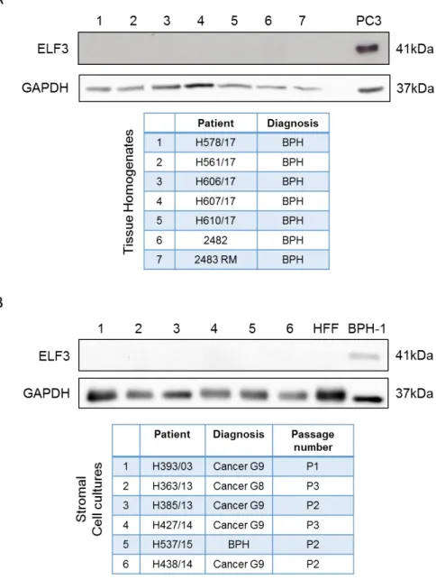
3.2.2 ELF3 is expressed in the CB population of primary prostate cultures at the protein level ...80
3.3 Cellular and tissue localisation of ELF3
...833.3.1 ELF3 is expressed in both the cytoplasmic and nuclear compartments of prostate epithelial cells ...83
3.3.2 ELF3 expression is restricted to the basal layer of the prostate epithelium in tissue...88
3.4 The effects of ELF3 knockdown on prostate epithelial cell lines
...963.4.1 siRNA transfection maintains long-term ELF3 knockdown in prostate epithelial cell lines ...96
3.4.2 ELF3 knockdown significantly reduces the cell viability of prostate epithelial cell lines by a process other than apoptosis ...98
3.4.3 ELF3 knockdown significantly decreases cell motility in prostate epithelial cell lines ... 101
3.4.4 ELF3 knockdown significantly decreases colony forming ability in prostate epithelial cell lines ... 104
3.4.5 ELF3 knockdown alters the morphology and colony formation of prostate epithelial cells... 106
3.4.6 ELF3 knockdown alters the expression of differentiation markers ... 111
3.5 Generating lentiviral vectors for ELF3 overexpression studies
... 1143.5.1 Invitrogen Gateway Cloning Technology strategy ... 114
3.5.2 Validation of ELF3 expression vectors ... 117
3.5.3 Determining lentiviral titre of ELF3 overexpression vectors ... 121
3.6 The effects of ELF3 overexpression on primary prostate epithelial cells
... 1233.6.1 Localisation of ELF3 in lentiviral transduced primary prostate epithelial cells ... 123
3.6.2 ELF3 overexpression differentially alters the viability of primary normal and cancer prostate epithelial cells ... 125
3.6.3 ELF3 overexpression does not significantly alter primary prostate cell migration... 127
3.6.4 ELF3 overexpression alters the differentiation state of primary prostate epithelial cells ... 129
3.7 ELF3 regulatory networks
... 1353.7.1 ELF3 expression is upregulated by histone deacetylase inhibitor Vorinostat in primary prostate epithelial cells but does not induce a neuroendocrine phenotype ... 135
3.7.2 Global gene expression changes following ELF3 knockdown in prostate epithelial cell lines. 138 3.7.3 ELF3 knockdown alters the expression of key cell cycle regulatory genes in prostate epithelial cell lines at the protein level and results in a block at the G2 phase ... 147
3.7.4 ELF3 knockdown does not significantly alter the cell cycle in primary prostate epithelial cells152 3.7.5 ELF3 overexpression has no effect on the cell cycle of primary prostate epithelial cells ... 156
4. Discussion
... 1584.1 Expression pattern of ELF3 in prostate cells and tissue
... 1584.2 Subcellular localisation of ELF3
... 1604.3 ELF3 and the neuroendocrine phenotype
... 1614.4 The role of ELF3 in the prostate and prostate cancer
... 1624.4.1 ELF3 and the stem cell phenotype ... 162
4.4.2 ELF3 as a regulator of the cell cycle ... 164
4.4.3 The differential role of ELF3 in benign and cancerous prostate ... 165
4.4.4 The role of ELF3 in differentiation and EMT ... 166
4.5 Targeting ETS factors in cancer
... 1694.5.1. Direct targeting of ETS factors ... 169
4.5.2. Indirect targeting of ETS factors ... 170
4.5.3. Expression of suppressive ETS factors ... 171
4.5.4 Differentiation therapy ... 171
Appendices
... 174 Appendix 3.1: Videos of migration assay of primary prostate cells with ELF3 overexpression (see attached CD). ... 174 Appendix 3.2: ELF3 knockdown does not induce apoptosis in primary prostate committed basal cells. ... 174 Appendix 3.3: Gene ontology (GO) terms associated with siSCR vs siELF3 in BPH-1 and PC3 cells combined.. ... 175 Appendix 3.4: Expression changes of cell cycle-related genes following ELF3 knockdown from gene expression microarray.. ... 179 Appendix 3.5: Expression graphs of genes that show different behaviour upon ELF3 knockdown between BPH-1 and PC3 cells from gene expression microarray.. ... 184Abbreviations
... 190List of Figures
Figure 1.1. The zonal anatomy of the prostate. ...17
Figure 1.2. Differentiation hierarchy of the normal prostate. ...19
Figure 1.3. Prostate cancer is highly associated with age. ...22
Figure 1.4. Gleason grading system of prostate adenocarcinoma. ...24
Figure 1.5. The invasion-metastasis cascade. ...27
Figure 1.6. The cancer stem cell hypothesis and progression of prostate cancer. ...31
Figure 1.7. Prostate cancer cell response and resistance to treatment. ...35
Figure 1.8. Domain structure of ETS factors which have proposed roles in prostate cancer. ...39
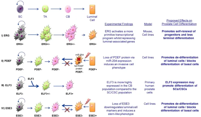
Figure 1.9. Proposed mechanisms of ETS factor-induced de-differentiation of prostate cancer epithelial cells. ...43
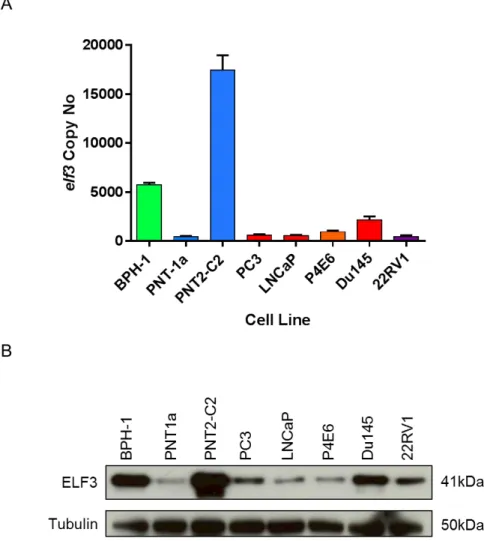
Figure 3.1. mRNA and protein expression of ELF3 in prostate epithelial cell lines. ...77
Figure 3.2. Affymetrix gene expression microarray data analysis from benign and malignant prostate suggests ELF3 is expressed at higher levels in the committed basal cell subpopulation compared to stem cells. ...79
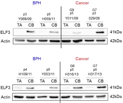
Figure 3.3. Protein expression of ELF3 in the fractionated cell subpopulations from primary prostate benign and tumour samples. ...81
Figure 3.4. Protein expression of ELF3 in prostate tissue homogenates and stromal cells...82
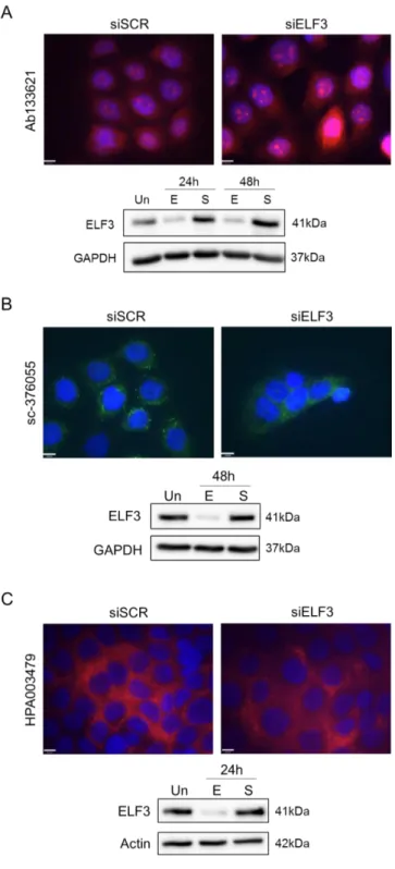
Figure 3.5. Testing ELF3 antibodies for immunocytochemistry using BPH-1 cells with ELF3 knockdown. 84 Figure 3.6. Testing ELF3 antibodies for immunocytochemistry using ELF3-negative cell line U-87 MG. ...85
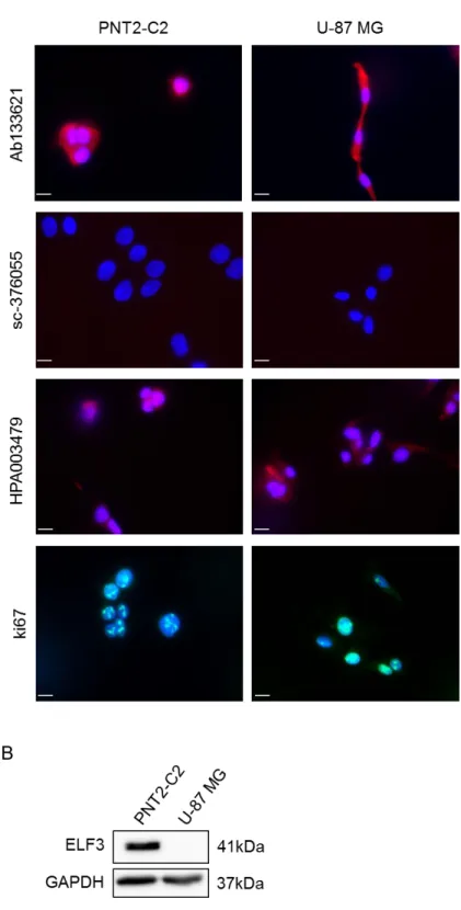
Figure 3.7. Testing ELF3 antibodies for immunocytochemistry using alternative fixation method. ...86
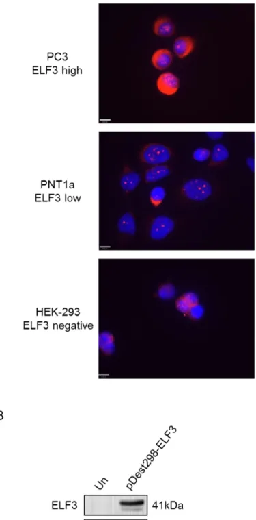
Figure 3.8. ELF3 cellular localisation in prostate cell lines. ...87
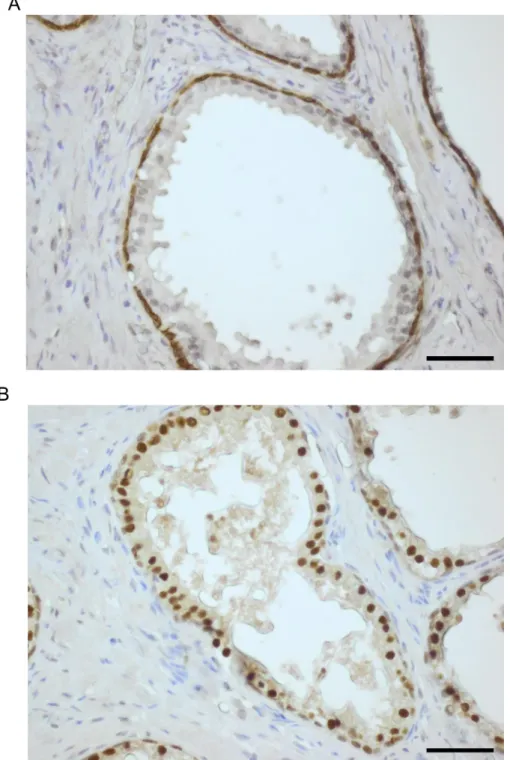
Figure 3.9. ELF3 expression in BPH tissue by immunohistochemistry (immunofluorescence). ...89
Figure 3.10. ELF3 expression in BPH tissue using the ImmPRESS Excel Amplified HRP Polymer Staining Kit. ...90
Figure 3.11. ELF3 expression in BPH tissue microarrays (TMAs)...91
Figure 3.12. ELF3 expression in Cancer tissue microarrays (TMAs) – Low Gleason grade prostate cancer. ...93
Figure 3.13. ELF3 expression in Cancer tissue microarrays (TMAs) – High Gleason grade prostate cancer.
...94
Figure 3.14. Time course of ELF3 knockdown in benign (BPH-1) and cancer (PC3) prostate epithelial cell lines...97
Figure 3.15. ELF3 knockdown decreases the viability of prostate epithelial cell lines after replating. ...99
... 100
Figure 3.16. ELF3 knockdown reduces cell adhesion but does not cause cell death via apoptosis. ... 100
Figure 3.17. ELF3 knockdown decreases the migration of BPH-1 cells. ... 102
Figure 3.18. ELF3 knockdown reduces the migration of PC3 cells. ... 103
Figure 3.19. ELF3 knockdown decreases the colony forming ability of benign and cancer prostate epithelial cell lines. ... 105
Figure 3.20. ELF3 knockdown alters the morphology of BPH-1 cells... 107
Figure 3.21. ELF3 knockdown alters the morphology of PC3 cells. ... 108
Figure 3.22. ELF3 knockdown alters colony formation in BPH-1 cells. ... 109
Figure 3.23. ELF3 knockdown does not alter colony formation in PC3 cells. ... 110
Figure 3.24. ELF3 knockdown alters the expression of differentiation markers in BPH-1 cells. ... 112
Figure 3.25. ELF3 knockdown alters the expression of differentiation markers in PC3 cells. ... 113
Figure 3.26. Lentivirus cloning strategy for ELF3 overexpression. ... 116
Figure 3.27. Vector maps of lentiviral ELF3 overexpression constructs... 118
Figure 3.28. Validation of ELF3-WT lentiviral vectors... 119
Figure 3.29. Validation of ELF3-ΔAT lentiviral vectors. ... 120
Figure 3.30. Gating strategy for lentiviral titration. ... 122
Figure 3.31. ELF3 localisation in primary prostate epithelial cells. ... 124
Figure 3.32. ELF3 overexpression differentially alters the viability of normal and cancer cells over time. ... 126
Figure 3.33. The effects of ELF3 overexpression on primary prostate cell migration. ... 128
Figure 3.34. Heterogeneity of primary prostate epithelial cells with ELF3 overexpression. ... 130
Figure 3.35. ELF3 overexpression significantly alters the median cell area of primary prostate epithelial cells. ... 131
Figure 3.36. ELF3 overexpression significantly alters the median cell perimeter of primary prostate epithelial
cells. ... 132
Figure 3.37. Statistical analysis of median cell area and median cell perimeter of primary prostate cells with ELF3 overexpression. ... 133
Figure 3.38. ELF3 overexpression alters the differentiation state of primary prostate cells. ... 134
Figure 3.39. ELF3 expression is upregulated in primary prostate epithelial cells following vorinostat treatment. ... 137
Figure 3.40. Validation of ELF3 knockdown at the protein level in array samples. ... 145
Figure 3.41. Gene expression changes and gene ontology in prostate epithelial cells following ELF3 knockdown... 146
Figure 3.42. ELF3 knockdown alters the expression of key cell cycle regulator genes. ... 149
Figure 3.43. ELF3 knockdown causes a progressive accumulation of cells in the G2 phase of the cell cycle. ... 150
Figure 3.44. ELF3 knockdown reduces the number of BPH-1 cells in mitosis... 151
Figure 3.45. Efficiency of ELF3 knockdown in primary prostate committed basal cells. ... 153
Figure 3.46. ELF3 knockdown does not alter the cell cycle in primary prostate committed basal cells. .. 155
Figure 3.47. ELF3 overexpression does not have a distinct effect on cell cycle... 157
Figure 4.1. ELF3 expression in the prostate epithelial hierarchy. ... 168
Figure 4.2. The benefits and disadvantages of inhibiting ETS factors in cancer. ... 170
Figure 4.3. Multidimensional scaling of prostate epithelial cells... 173
List of Tables
Table 1.1. ETS transcription factor subfamilies………...…40
Table 2.1. Cell lines and their culture conditions...…...51
Table 2.2. Culture media constituents………..……52
Table 2.3. Components of siRNA transfection mixes for cell lines………55
Table 2.4. Components of siRNA transfection mixes for primary cells………55
Table 2.5. Mastermix components for cDNA synthesis……….……57
Table 2.6. Antibodies used for protein detection by western blot………..……62
Table 2.7. Antibodies used for protein detection by immunohistochemistry………...……65
Table 2.8. Antibodies used for protein detection by immunocytochemistry………....67
Table 2.9. Components used for attB flanking of ELF3 by PCR………68
Table 2.10. Primer sequences for sequential attB flanking of ELF3……….68
Table 2.11. ELF3 primer sequences for PCR………..70
Table 3.1. Origin and phenotypic characteristics of prostate epithelial cell lines………76
Table 3.2. Characteristics of retroviruses and lentiviruses as vectors for gene expression………115
Table 3.3. Number of differentially expressed genes in each gene expression microarray analysis……….138
Table 3.4. Expression changes of ETS transcription factors following ELF3 knockdown………...139
Table 3.5. Expression changes of genes involved in differentiation following ELF3 knockdown…………...141
Table 3.6. Expression changes of transcription factors involved in epithelial-to-mesenchymal transition (EMT) following ELF3 knockdown………..142
Table 3.7. Expression changes of stem cell markers following ELF3 knockdown………...143 Table 3.8. Genes that show different behaviour upon ELF3 knockdown between BPH-1 and PC3 cells….144
Acknowledgements
This work is dedicated to my mum, the strongest woman I know and provider of exceptional potato scones when needed. Everything I am is because of you.
Thank you to Norman and Prostate Cancer UK for giving me the opportunity to be part of the CRU and providing essential funding.
Thanks to Hannah, Michelle, Dominika and the rest of the CRU, past and present, for their support and always being a friendly face.
Thank you to Mandy, for truly being my lab mum and supplying biscuits on tap during desperate times. Thank you to Georgie, my partner in crime, for sharing that bottle of prosecco with me and always making me feel clever, may you always be bright and shiny.
Thank you to John P., Ban, Conor and John H., for providing endless banter and always laughing at my jokes.
Last and foremost, thank you to Fiona, for being the best mentor I could have ever hoped for. This would not have been achievable without your guidance, pep talks and friendship.
Author’s Declaration
I declare that this thesis is a presentation of my own unaided work, except where acknowledged otherwise in the text. This work has not previously been presented for an award at this, or any other, University. All sources are acknowledged as References.
This work has been presented at the following meetings:
Oct 2016 23rd Meeting of the EAU Section of Urological Research, Parma, Italy.
Poster presentation
Sep 2017 European Society for Medical Oncology Congress, Madrid, Spain
Poster presentation
The following review article was published during this PhD and has been used in part in this thesis: ARCHER, L. K., FRAME, F. M. & MAITLAND, N. J. 2017. Stem cells and the role of ETS transcription factors in the differentiation hierarchy of normal and malignant prostate epithelium. J Steroid Biochem Mol Biol, 166, 68-83.
1. Introduction
1.1 Function and anatomy of the prostate
The prostate is a male secretory gland comprised of a series of glandular epithelial acini and muscular stroma. It is responsible for producing proteins, such as prostatic acid phosphatase (PAP) and prostate specific antigen (PSA) and secreting the fluid which constitutes semen via ducts which lead to the urethra. The prostate is located below the bladder, surrounding the urethra, and is in close proximity to a collection of nerves called the prostatic plexus which is responsible for controlling urinary output as well as erection and ejaculation. As a result of this position, enlargement of the prostate can lead to both urinary and sexual symptoms (Figure 1.1). The prostate can be divided into separate zones, each with its own propensity for disease. The peripheral zone is found at the base of the prostate and forms ~70% of the tissue. This area is where the majority of prostate cancers (PCa) arise (>70%) and can be assessed during a digital rectal examination. The central zone lies just below the bladder. Prostate tumours rarely arise from the central zone however they are associated with an aggressive phenotype and poor prognosis (Lee et al., 2011). Finally, the transitional zone is located in front of the central zone surrounding the urethra and is the site where the majority of benign enlargement of the prostate occurs, known as benign prostatic hyperplasia (BPH) (McNeal, 1981).
Figure 1.1. The zonal anatomy of the prostate.
The prostate gland is situated below the bladder, surrounding and linking the urethra to the ejaculatory duct. The prostate consists of four zones; the peripheral zone, anterior zone, central zone and transitional zone, each of which has its own propensity for prostate pathology. Reprinted from (Sathianathen et al., 2018), with Copyright permission from Springer Nature.
1.2 Cellular organisation of the normal prostate
The normal prostate epithelium is composed of an hierarchy of cells that can be split into a basal compartment, which is attached to the basement membrane (BM), and a luminal compartment, which is the inner layer lining the lumen (Figure 1.2). Androgen-independent basal cells comprise the proliferative compartment of the adult prostate and can be distinguished by their expression of specific cytokeratins (CK) including CKs 5 and 14 (Schalken and van Leenders, 2003), and other cell markers such as p63 (Signoretti et al., 2000), CD44 (Terpe et al., 1994) and bcl-2 (McDonnell et al., 1992). The fully differentiated luminal cells are the secretory cells of the prostate. These are androgen-responsive, expressing high levels of the androgen receptor (AR) and PSA. Luminal cells also express CKs 8 and 18 (Schalken and van Leenders, 2003) and CD57 (Liu et al., 1997b). Distinct differentiation states have been found within the basal compartment, adding another level of complexity and heterogeneity to the prostate epithelium. The base of the hierarchy consists of normal basal stem cells (SC) which are largely quiescent (Oldridge et al., 2012). Some SCs asymmetrically divide into a proliferative, intermediate progenitor cell population which are termed transit amplifying (TA) cells. A third population committed to differentiation called committed basal (CB) cells completes the basal hierarchy (Oldridge et al., 2012, Isaacs and Coffey, 1989). Significantly, several studies have shown that both basal and luminal cells derive from the same rare SC precursors found in the basal compartment, providing further evidence for the presence of a differentiation hierarchy in the prostate (Isaacs and Coffey, 1989, Hudson et al., 2001, Lang et al., 2001b, van Leenders et al., 2001). A rare neuroendocrine (NE) cell population is also scattered throughout the basal compartment of the prostate epithelium. These granular cells secrete peptide hormones such as serotonin and chromogranin A (ChrA) that regulate epithelial cell proliferation and differentiation (di Sant'Agnese, 1998, Rumpold et al., 2002). On the opposite side of the basal basement membrane lies the network of fibromuscular stroma which includes smooth muscle cells, fibroblasts, collagen fibres, endothelial cells and nerve fibres (Barron and Rowley, 2012). The stroma is important for maintaining prostate tissue growth and homeostasis through interactions with epithelial cells and secretion of growth factors.
Figure 1.2. Differentiation hierarchy of the normal prostate.
The normal human prostate epithelium is composed of an hierarchical differentiation pathway. SCs differentiate into rapidly dividing transit amplifying cells by asymmetric division and thus also maintain themselves. Multipotent transit amplifying cells differentiate into committed basal cells, which then commit to terminal differentiation into secretory luminal cells. This tightly regulated pathway generates a highly organised glandular structure consisting of the luminal layer, the basal layer attached to the basement membrane and the surrounding stroma. Adapted from (Archer et al., 2017).
1.3 Disorders of the prostate
1.3.1 Benign prostatic hyperplasia
There are a number of disorders of the prostate which have all been linked by some means to the development of PCa. BPH involves an androgen-dependent abnormal increase in proliferation of the epithelial and stromal cells within the prostate (Roehrborn, 2008). This disorder primarily occurs in the transitional zone and can result in the obstruction of urine in the urethra which can be somewhat alleviated by transurethral resection of prostate (TURP) (Simpson, 1997). Like PCa, the presence of BPH increases with age, with 88% of autopsies of men over 80 presenting with BPH (Boyle, 1994). There is accumulating evidence that inflammation plays a key role in the development of BPH. The presence of immune cell infiltrates and cytokines in BPH lesions has been observed and this is thought to promote tissue remodelling and further proliferation and enlargement (Kramer et al., 2007, Bostanci et al., 2013, Fibbi et al., 2010).
1.3.2 Prostatic intraepithelial neoplasia
Prostatic intraepithelial neoplasia (PIN) is defined by accumulating abnormalities in the prostatic epithelium with progressive similarities to adenocarcinoma. These include an increase in proliferation in luminal epithelial cells and a decrease in basal cells (Shen and Abate-Shen, 2010). Whilst it does not increase the levels of prostate PSA, the presence of PIN is considered an early stage of carcinogenesis and the majority of patients presenting with PIN develop cancer within 10 years. The incidence of PIN also increases with age, but predominantly occurs in the peripheral zone of the prostate. Tissue with high-grade PIN lesions share both morphological and genetic similarities to PCa (Bostwick et al., 2004).
1.3.3 Prostatitis
Inflammation of the prostate, known as prostatitis, is divided into four categories; acute and chronic bacterial prostatitis, most commonly caused by E.coli and Enterococcus spp, asymptomatic inflammatory prostatitis and chronic prostatitis, also known as chronic pelvic pain syndrome (CPPS) (Murphy et al., 2009, Cai et al., 2011). It is estimated that 15% of US males will be affected by symptomatic prostatitis in their lifetime (Krieger, 2004). However, the presence of inflammation in prostatic biopsies and TURPs suggest that the prevalence of asymptomatic prostatitis may be much higher and that inflammation may be an important factor in prostate carcinogenesis (De Marzo et al., 2007, Nickel et al., 1999, Nickel et al., 2008).
1.4 Prostate cancer
1.4.1 Epidemiology and risk factors
PCa is the most common cancer of men in the UK, accounting for 26% of all new cancer cases. It is currently the second most common cause of cancer-related death (13%), following lung cancer and it is estimated that 1 in 8 males in the UK will develop the disease in their lifetime (Cancer Research UK, 2016).
Epidemiological and genetic studies have defined clear risk factors. PCa is most highly associated with age, with over a third of new cases found in men ≥75 (Figure 1.3). PCa is also more prevalent in black males than white males, with Asian males possessing significantly lower risk (Quinn and Babb, 2002).
Other PCa risks include familial factors which was first explored by Morganti et al. in 1956, and describes several members of the same family developing the disease due to environmental factors, genes or a combination of the two, accounting for 10-20% of all PCa cases (Morganti et al., 1956, Stanford and Ostrander, 2001). Hereditary PCa is a subset of familial PCa caused by the Mendelian inheritance of rare susceptibility genes (Potter and Partin, 2000). Hereditary PCa is considerably more prevalent in relatively younger diagnosed patients, accounting for 43% of cases diagnosed below the age of 55. There are now over 70 susceptibility loci that confer an increased risk for PCa, identified through genome wide association studies (Eeles et al., 2013). An example of susceptibility genes that are of current interest are BRCA1 and BRCA2, gene mutations of which are most widely associated with a predisposition for breast cancer. BRCA2 mutations have now been confirmed as a risk factor for early and aggressive PCa and patients show poorer outcomes following localised PCa treatment than those who do not harbour mutations (Mottet et al., 2017, Castro et al., 2015). Phase II clinical trials have found patients with BRCA2 mutations and other DNA repair defects respond favourably to PARP inhibitors such as olaparib, highlighting the need for stratified therapies (De Felice et al., 2017).
Figure 1.3. Prostate cancer is highly associated with age.
The average number of new cases per year and incidence rate with age from 2013-2015, compiled by Cancer Research UK. https://www.cancerresearchuk.org/health-professional/cancer-statistics/statistics-by-cancer-type/prostate-cancer/incidence#heading-One.
1.4.2 Diagnosis and grading
PSA testing of men over 50 remains the gold standard for the screening of PCa. Normal levels of PSA in the blood is <4ng/ml, however this figure rises naturally with age. Although the introduction of PSA testing led to a dramatic increase in PCa incidence in the late 1980s, this method of detection remains imperfect. PSA levels not only rise in the incidence of PCa but also due to infection, prostatitis and BPH (Obort et al., 2013). Furthermore, although PSA is termed a prostate-specific protein, detectable expression has also been found in other tissues including the breast, lung, ovary and salivary glands (Smith et al., 1995). PSA detection and subsequent PCa diagnosis can often lead to the overtreatment of many patients. Several alternative biomarkers specific to PCa have been investigated, such as PCA3 and TMPRSS2:ERG (Romero Otero et al., 2014, McGrath et al., 2016). However, PSA still remains the most advantageous screening method to date.
Following high level PSA detection, multiple transrectal ultrasound (TRUS) biopsies are usually taken. The Gleason grading system is the most widely used technique for grading prostate carcinomas (Gleason, 1966). H&E stained histological sections of prostate are graded according to the most common patterns of cells observed under the microscope. The system has been updated in recent years to reduce ambiguity between grades for pathologists (Figure 1.4) (Epstein, 2010). Patterns are graded 1-5 by their degree of differentiation and visual abnormalities compared to normal prostate tissue, with 1 representing normal glandular structures and 5 representing abnormal sheets of undifferentiated tissue. The sum of the most common (primary) and second most common (secondary) patterns produces the total Gleason score. A new classification system divides patients into the grade groups defined in Figure 1.4 (Epstein et al., 2016). The Gleason system has been a powerful tool in predicting prognosis and aiding the decision of treatment options. Furthermore, the upgraded classification has resulted in a reduction in overtreatment of Gleason 6 cancers (as more aggressive patterns are now associated with a Gleason score of 7) and distinguishes the prognostic differences in Gleason scores 3 + 4 and 4 + 3 (Epstein et al., 2016). However, its limitations include the absence of consideration of the heterogeneity of PCa, as several different Gleason patterns have been found in single prostate tumours (Humphrey, 2004). Discovery of a PCa-specific biomarker capable of determining progression and aggressiveness would aid to avoid overtreatment and allow for a more appropriate treatment regime for individual patients.
Figure 1.4. Gleason grading system of prostate adenocarcinoma.
Updated Gleason grading system of prostate adenocarcinomas. Histological patterns are scored 1 to 5 according to degree of disorder. Reprinted from (Epstein, 2010), with Copyright permission from Elsevier.
1.4.3 Treatment of localised prostate cancer
A patient with low Gleason grade, localised PCa has the option to either receive treatment or undergo active surveillance (Kollmeier and Zelefsky, 2012). Both tumour grade and patient preference will impact this decision.
As some low Gleason grade adenocarcinomas will never progress, overtreatment of these men is an ongoing problem. A case for active surveillance as an alternative option is growing, whereby the patient receives routine PSA level checks and biopsies. The inability to determine which patients will undergo disease progression is a cause for reservation. However, the quality of life of the patient following treatment must also be taken into consideration (Bul et al., 2013, Klotz, 2006, Xia et al., 2012, Bellardita et al., 2014). If immediate treatment is favoured, options include surgical removal of the prostate, known as radical prostatectomy (RP), radiotherapy or focal treatment. The most common treatment is RP, which risks undesirable side effects, most notably incontinence and impotence.
Radiotherapy is an option for local and locally advanced PCa in conjunction with hormone therapy, and can be delivered by external beam radiation therapy (EBRT) or directed brachytherapy. EBRT includes three-dimensional conformal radiation therapy (3D-CRT) and image-guided intensity-modulated radiation therapy (IMRT) (Heidenreich et al., 2014a). IMRT is presently favoured due to the capacity for higher doses of radiation without increasing off-target toxicity (Bauman et al., 2012). Brachytherapy involves direct radiation of the prostate by either a temporary high dose implant or permanent low dose radioactive seeds. An advantage to brachytherapy is the limited radiation to the surrounding tissues, whereas EBRT risks rectal toxicity (Crook, 2011). However, many authors have demonstrated that a high dose of radiation is required for a better outcome in patients (Zelefsky et al., 2001, Kupelian et al., 2005). A ten year follow-up study monitoring patients undergoing active surveillance, RP or EBRT found there was no significant difference in PCa-related deaths (Hamdy et al., 2016). However, RP and ERBT were associated with lower risk of disease progression and metastases compared to active surveillance.
At present there are various focal therapies for PCa available which aim to target a tumour directly and limit damage to the surrounding tissue in order to avoid the undesirable side effects of the alternative therapies already discussed. Given PCa is often a multifocal disease, focal therapy is generally selected for patients considered low-risk of progression, where the tumour is restricted to one lobe and unifocal (Eggener et al., 2010). High-intensity focused ultrasound (HIFU) utilises targeted thermal damage with magnetic resonance imaging (MRI) as guidance (Napoli et al., 2013). It is delivered transrectally and so avoids invasive surgery. As HIFU is a relatively new therapy, there is a lack of studies with long term follow up. However, the current cancer-free survival rates and low rates of serious side effects are promising (Cordeiro et al., 2012, Ramsay et al., 2015). Extreme low temperature and thawing cycles characterise cryotherapy which can be used to treat localised PCa or as salvage therapy following relapse (de la Taille et al., 2000). A transperineal argon gas or liquid nitrogen probe is used, guided by TRUS (Nomura and Mimata, 2012). Cryoablation is a relatively invasive treatment and also requires warming of the surrounding tissues to avoid damage (Cohen
and Miller, 1994). Photodynamic therapy (PDT) involves intravenous injection of photo-sensitive drugs which specifically act in the prostate by administration of light. This therapy induces necrosis of tumour material and largely avoids damaging other tissues (Koudinova et al., 2003). However, since the drug is administered intravenously, patients must avoid direct sunlight for a period of time depending on the half-life of the drug used (Moore et al., 2009). Results from a recent clinical trial show that after 3.5 years follow up, 75% of patients maintained complete tumour ablation in the treated lobe (Noweski et al., 2018).
1.4.4 Metastasis and epithelial-to-mesenchymal transition
Metastasis is the cause of over 90% of deaths from solid tumours (Valastyan and Weinberg, 2011, Gupta and Massague, 2006). Despite local therapy, up to 40% of men will develop metastatic PCa, most commonly to the bone (Beltran et al., 2011). Activation of invasion and metastasis is one of the hallmarks of cancer described by Hanahan and Weinberg, where transformed cells progress through a coordinated series of steps, coined the invasion-metastasis cascade, summarised in Figure 1.5 (Hanahan and Weinberg, 2000, Hanahan and Weinberg, 2011).
Following formation of a primary tumour, cells undergo a number of changes to facilitate local invasion of the BM and surrounding stroma. Epithelial-to-mesenchymal transition (EMT) is a process which occurs during normal organ development and wound healing (Thiery et al., 2009). EMT has also become recognised as a major mechanism by which transformed epithelial cells acquire the ability to invade surrounding tissue and disseminate to form distant metastases. It is a molecular switch from an epithelial cell phenotype to a mesenchymal cell phenotype, culminating in the loss of cell polarity and tight junctions between cells. The most well characterised alteration is the loss of epithelial cell marker E-cadherin and induced expression of mesenchymal cell marker N-cadherin, which results in the loss of cell-to-cell contacts and promotes a migratory mesenchymal cell phenotype (Valastyan and Weinberg, 2011). This switch in gene expression is controlled by several master transcription factors of EMT including Snail, Slug, ZEB1/2 and Twist (Thiery et al., 2009). EMT transcription factors are capable of promoting a cell phenotype which facilitates almost the entirety of the invasion-metastasis cascade. Mesenchymal-to-epithelial transition (MET), the reversal of EMT, may occur at distant tumour sites to permit establishment of invading tumour cells (Polyak and Weinberg, 2009).
Figure 1.5. The invasion-metastasis cascade.
Schematic detailing the stages of the invasion-metastasis cascade. A metastatic cell must invade the local tissue and enter the circulation via intravasation. Should the cell survive transit, it must then invade the distant tissue site and adapt in order to survive and form metastatic lesions in the new foreign microenvironment. Reprinted from (Valastyan and Weinberg, 2011), with Copyright permission from Elsevier.
Whilst bone is the most prominent site of PCa metastases, occurring in 90% of patients with advanced PCa, they are also commonly known to occur in the lymph nodes, lungs and liver (Bubendorf et al., 2000, Kelly and Yin, 2008). This tropism for bone is thought to be regulated by chemokines. Chemokines are a class of chemotactic cytokine which are important for immune cell development and recruitment of immune cells to sites of inflammation. The chemokine CXCL12 is highly expressed at sites of PCa metastases, including bone (Mognetti et al., 2013). Its receptor, CXCR4, has been shown to be expressed in PCa cell lines as well as increased expression in tissue sections derived from primary prostate tumours and mestastases compared to normal prostate (Sun et al., 2003, Akashi et al., 2008, Chetram et al., 2011).
The surrounding stroma, both locally in the prostate and at sites of metastases, provide factors such as cytokines and growth factors, which interact with tumour cells and cooperatively facilitate invasion into the circulation from the primary tumour and also survival at the new tumour site (Josson et al., 2010, Morrissey and Vessella, 2007). The tumour-associated stroma (known as “reactive stroma”) demonstrates changes in composition which can be distinguished from normal stroma, such as a switch from largely smooth muscle cell content in normal prostate stroma to a more myofibroblast and fibroblast cell content in reactive stroma (Tuxhorn et al., 2002). This process is thought to be regulated in part by TGF-β and Wnt signalling, which are also known regulators of EMT (Barron and Rowley, 2012).
1.4.5 Treatment of metastatic prostate cancer
High-grade and metastatic PCa is generally treated with pharmacological drugs which aim to deprive the tumour of androgens which are essential for growth, known collectively as androgen-deprivation therapy (ADT). The standard categories of ADT are as follows:
1) Anti-androgen therapy: prevents activation of target genes by interacting with the AR, e.g.
bicalutamide (Iversen et al., 2000).
2) Oestrogen treatment: prevents testosterone production by inhibiting 5-α reductase, e.g.
diethylstilbestrol (McLeod, 2003).
3) Gonadotrophin-releasing hormone (GnRH) agonist: inhibits androgen production by
desensitising hormone feedback systems in the pituitary, initially used in conjunction with anti-androgens to prevent tumour flare, e.g. goserelin (Peeling, 1989).
4) GnRH antagonist: binds to GnRH receptor and inhibits testosterone production, e.g. degarelix
(Shore, 2013).
Many different agents have been trialled with different mechanisms of action. Oestrogen therapy via diethylstilbestrol (DES) was the first hormonal therapy used for PCa in the 1960s. However, the overall mortality rate among patients treated with DES was higher due to increased cardiovascular disease, highlighting the need for alternative treatments (McLeod, 2003). GnRH agonists, such as goserelin, are used at present, which avoid the need for surgical castration (Peeling, 1989, Heidenreich et al., 2014b, Seidenfeld et al., 2000). The use of nonsteroidal anti-androgen therapy such as bicalutamide for locally advanced PCa does not have significant survival benefits over castration but does provide the patient with a better quality
of life (Tyrrell et al., 1998, Iversen et al., 2000, Iversen, 2003). Other agents include GnRH antagonists, which have been shown to decrease testosterone and PSA levels at a much faster rate than GnRH agonists (Klotz et al., 2008, Trachtenberg et al., 2002).
1.4.6 The androgen receptor and the emergence of castration-resistant prostate cancer
Androgens are necessary factors for pre- and post-natal development and growth of the prostate (Kellokumpu-Lehtinen et al., 1979, Aumuller, 1991). The AR is a member of the nuclear receptor superfamily and is a vital transcriptional regulator for both the normal prostate and PCa. The AR is expressed by luminal epithelial cells, which comprise the bulk of a prostate tumour and are therefore the main target of ADT. In its unactivated form, the AR exists in the cytoplasm in complex with several proteins from the heat shock protein family. Upon ligand binding, most commonly to dihydrotestosterone (DHT), the AR undergoes a conformational change which triggers nuclear translocation. Here, the AR binds to the promoter/enhancer regions of genes which contain androgen response elements (ARE) such as PSA and PAP. AR-mediated transcription can be positively and negatively regulated in several ways. This includes interaction with co-regulators which modulate histones and either promote/prevent AR binding, and also by interaction with other transcription factors (Heinlein and Chang, 2004).
Patients with high grade PCa undergoing androgen-ablation treatment may show promising tumour regression and lowering of PSA initially. However, they eventually become hormone resistant and the cancer recurs, known as castration-resistant prostate cancer (CRPC). Several androgen-related mechanisms of resistance have been investigated including:
1) AR mutations: mutations can result in a receptor that is more sensitive to small amounts of
hormone and increase sensitivity to other steroid hormones and molecules (Taplin et al., 1999). 2) AR variants: several AR splice variants have been identified in CRPC patients. Variants lacking a
ligand-binding domain result in constitutive activation of the AR (Watson et al., 2010).
3) AR amplification: multiple copies of the AR gene resulting in an increase of AR protein (Linja et
al., 2001).
An alternative hypothesis for the emergence of CRPC suggests that androgen-independent, and therefore ADT-resistant, cell populations of the prostate epithelium can survive treatment and subsequently reconstitute the tumour with other androgen-independent cells (Shen and Abate-Shen, 2010).
1.4.7 Treatment of CRPC
Therapy for CRPC includes treatment with cytotoxic chemotherapy agents such as docetaxel or cabazitaxel, which target rapidly dividing cells. Unfortunately, not all PCa patients respond to chemotherapeutic agents and others often become resistant, as the survival benefit with current agents is between a few months to 2 years (Seruga and Tannock, 2011, Bahl et al., 2013, Pezaro et al., 2014). Several novel therapies have now
been developed which are not curative but aim to prolong progression-free survival and improve quality of life, these include:
1) Abiraterone acetate: second-line anti-androgen that inhibits CYP17A, an enzyme involved in
androgen synthesis (de Bono et al., 2011).
2) Enzalutamide: second-line anti-androgen that inhibits AR signalling by powerful blockade of the
AR (Scher et al., 2012).
3) Sipuleucel-T: a personalised immunotherapy whereby the patients antigen-presenting cells are
externally activated by granulocyte-macrophage colony stimulating factor (GM-CSF) which is fused to PAP as the antigen. The vaccine is then infused into the patient to stimulate an immune response against PAP-expressing cells (Kantoff et al., 2010, Anassi and Ndefo, 2011).
4) Radium-223 dichloride: an α-emitting radionuclide with a tropism for bone metastases (Deshayes
et al., 2017).
The median life expectancy of a patient with CRPC is 2 years despite the development of these next generation therapies, which only increase life expectancy by a matter of months relative to a placebo (de Bono et al., 2011, Kantoff et al., 2010, Scher et al., 2012). Ongoing clinical trials aim to determine the best time at which to introduce these drugs during disease progression and the combinations which yield the greatest effects possible (James et al., 2016, James et al., 2017, Attard et al., 2018). Although the principal focus of drug design has been on androgen depletion, the use of these drugs has in fact now been linked to an increased risk of developing advanced PCa by the U.S. Food and Drug Administration (FDA) (Thompson et al., 2003, Andriole et al., 2010, FDA, 2011). Resistance suggests that AR-positive cells are not the only important cell type within the tumour. These current PCa drugs have been designed to target androgen-responsive luminal cells which comprise the bulk of a tumour, whilst overlooking the AR-negative basal cell populations which includes a stem-like population.
1.5 Cancer stem cells
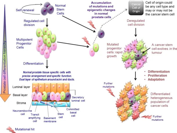
The cancer stem cell (CSC) hypothesis proposes that only a subpopulation of cells within a tumour is able to initiate, propagate and maintain tumour growth (Greaves, 2010). In addition to self-renewal and multipotency, CSCs also have dysregulated proliferation and differentiation. Thus, a CSC can maintain itself, whilst also differentiating into the distinct heterogeneous cell types that constitute the bulk of a tumour (Figure 1.6). These bulk tumour populations are considered non-tumourigenic, although it should be noted that CSC properties may not only be originally acquired by normal tissue SCs but also by a more differentiated cell type within the hierarchy. The original cell type which is targeted for genetic mutation and transformation, whether or not it is a SC, is known as the cell of origin.Since CSCs are the only cells capable of driving tumour growth, these cells therefore require targeting to achieve long term cancer therapy. However, consistent with normal tissue SCs, CSCs possess inherent resistance mechanisms which allow them to evade standard cancer treatments including chemotherapy and radiation (Alison et al., 2012, Morrison et al., 2011, Ishii et al., 2008).
Figure 1.6. The cancer stem cell hypothesis and progression of prostate cancer.
This model shows the progression from a normal prostate differentiation hierarchy to the deregulated growth of a prostate tumour. Although it is more likely for the cancer stem cell to arise from a normal stem cell, due to its longevity and self-renewal properties, this model does not exclude the possibility that a progenitor cell could become the cancer stem cell following a series of mutations. This cancer stem cell is then able to maintain tumour growth and differentiate into the different cell types that compose the tumour. In addition, it is possible for other mutations to occur within bulk tumour cells altering their behaviour and contributing to a more aggressive cancer phenotype. Taken from (Archer et al., 2017).
Multiple studies, going back over a hundred years implied the existence of CSCs (Maitland and Collins, 2014). The first “modern age” evidence presenting CSCs as the cause and maintenance of cancer was elegantly demonstrated in acute myeloid leukaemia (AML) by John Dick’s laboratory in 1994 (Lapidot et al., 1994). Since then our understanding of CSCs has been refined and their identification in multiple other leukaemias and solid tumours has supported the CSC hypothesis of cancer (Visvader and Lindeman, 2008). However, accumulating evidence for the CSC hypothesis has posed significant challenges, including the identification of markers that can accurately distinguish CSCs and expedite isolation of these relatively rare cells from a complex tumour environment. The two functional caveats for identifying CSCs are that they must 1) be tumourigenic, forming heterogeneous tumours reminiscent of those from which they were derived and 2) be serially transplantable when xenografted in mice. An array of markers, including CD133, CD44, CD24 and ALDH1, have been used to isolate CSCs from different solid tumours. Moreover, as there is no single CSC marker for each tumour type, multiple markers are generally used to isolate as homogeneous a population as possible. These markers usually include normal SC markers of the same tissue (Park et al., 2007). Whilst it has previously been hypothesised that CSCs constitute a rare population of tumour cells, studies in melanoma have shown that the current models could be significantly underestimating the size of the CSC population. In NOD/SCID mice the proportion of cells with tumourigenic capacity was 0.1-0.0001%, which rose to 25% in more immunocompromised NOD/SCID/IL2Rγnull mice (Quintana et al., 2008). However, the original hypothesis seems true in tumours of other tissues such as human pancreatic adenocarcinoma, lung squamous cell carcinoma, lung adenocarcinoma, and head and neck squamous cell carcinoma (HNSCC). In these tumours, the CSC population only accounted for 0.0028-0.04% of total tumour cells in NOD/SCID/IL2Rγnull mice (Ishizawa et al., 2010). The proportion of tumour cells with a CSC phenotype is therefore likely to be a quality that is dependent on tissue of origin and cancer subtype within that tissue, and in some cases tumour grade may also be important (Marcato et al., 2011, Thirant et al., 2011). Although serial transplantation in mice is considered to be the gold standard as the means of identifying CSCs, this data also highlights the host’s immune response and differences in vital growth factors as limiting factors in studying CSCs in model systems.
1.5.1 A basal cell is the cell of origin of prostate cancer
Substantial evidence exists for a basal SC origin of PCa. By introducing ERG expression and constitutive PI3K signalling into the basal and luminal epithelial cells of mice, Lawson et al. demonstrated that whilst luminal cells were unresponsive, the basal population was able to form fully differentiated tumours reminiscent to those seen in humans (Lawson et al., 2010). Significantly, evidence from human prostate cells also indicates a basal SC origin of PCa. The CD44+/CD133+/α2β1hi markers in basal cells identify normal prostate SCs (Collins et al., 2001, Richardson et al., 2004). These cells showed self-renewal and differentiation properties, were highly proliferative and reconstituted prostate glands in vivo (Richardson et al., 2004). To isolate primary prostate CSCs which constitute approximately 0.1% of tumour cells from PCa biopsies the same markers were used; cells were first selected for high α2β1 integrin expression by rapid adhesion to collagen I-coated plates and CD133+ cells were then enriched from α2β1hi cells (Collins et al.,
2005). These CD133+/α2β1hi cells exhibited enhanced proliferative potential and secondary colony-forming efficiency in vitro compared to other populations, i.e. they were able to expand and self-renew through several generations as the proportion of CD133+ cells remained constant. As well as possessing the fundamental SC properties, the cells also demonstrated classic cancer cell characteristics of invasion and anchorage-independent growth, confirming that the cells were of tumour origin. Furthermore, CD133+/α
2β1hi cells were able to differentiate into AR+ luminal cells, recapitulating the characteristics of a primary prostate tumour (Collins et al., 2005). It should also be noted that these cells are normally studied at as low a passage as possible, minimising any changes that may occur due to long-term culture. Patrawala et al also demonstrated that primary human PCa CD44+ basal cells had increased tumourigenic and proliferative capabilities in vitro and in vivo than CD44- cells (Patrawala et al., 2006). Then, similarly to Lawson, Goldstein et al. confirmed that human benign basal cells, but not luminal cells, could produce tumours with a luminal phenotype in NOD/SCID/IL2Rγnull mice following AKT, ERG and AR expression (Goldstein et al., 2010). Furthermore, it was recently shown by high-throughput RNA sequencing that a basal SC phenotype was associated with more aggressive types of PCa (Smith et al., 2015). Various combinations of markers including CD133, CD49f, CD44 and Trop2 are commonly used to detect prostate CSCs (Trerotola et al., 2010, Patrawala et al., 2006, Goldstein et al., 2010).
In contrast, Wang et al identified castration-resistant Nkx3.1-expressing cells (CARNs) which were defined as a subset of luminal cells in the mouse prostate (Wang et al., 2009). These cells comprised 1% of cells in the total prostate and were able to survive following androgen deprivation. CARN cells were proposed to be the origin of PCa, due to the presence of high grade PIN and carcinoma lesions following PTEN deletion and their ability to form organoids in culture (Wang et al., 2009, Chua et al., 2014). Similarly, luminal progenitors have also been found in other mouse models of PCa (Korsten et al., 2009, Liu et al., 2011, Agarwal et al., 2015). However, the normal mouse and human prostates display significant differences. For instance, the murine prostate consists of a single layer of epithelia where luminal cells contact the basement membrane directly, and has a lower basal cell content compared to human. It is therefore possible that the development of PCa differs between the two species and this CARN progenitor could be a unique cell found in mice. However, a putative luminal CSC was found in the BM18 xenograft model derived from a human PCa bone metastasis (Germann et al., 2012). A quiescent population of cells co-expressing stem-like (NANOG or ALDH1A1) and luminal (NKx3.1 and CK18) markers was able to reconstitute a tumour in the presence of androgen following previous castration. However, these specific cells were not selected and serially transplanted into different mice, so it remains unclear whether the cells truly have tumour-initiating properties. It should also be noted that this xenograft model only represents one patient and it is well known that PCa is a very heterogeneous disease. Furthermore, the xenograft had been passaged many times since it was established in 2005 and is likely to have evolved significant changes since then (McCulloch et al., 2005). Critically, a luminal CSC has yet to be isolated from a primary human prostate tumour (Collins et al., 2005).
1.6 Cancer stem cells and treatment resistance in prostate cancer
The CSC hypothesis also logically explains the development of metastases. Complex sequencing studies have shown that the cancer cell clones present in a primary prostate tumour are also present in metastases (Hong et al., 2015, Haffner et al., 2013). Theoretically, in order to maintain the founder mutations present in the original tumour through to metastasis, this must be carried out by a primitive, long-lived cell that is capable of both clonal expansion and the ability to accumulate further abnormalities that allow the cell to develop independence from the extracellular matrix and migrate to extraprostatic sites (Gundem et al., 2015). Prostate CSCs possess enhanced invasive qualities in comparison to other tumour cell populations (Smith et al., 2015, Klarmann et al., 2009). Successfully targeting the CSC population alongside cytotoxic hormone therapies to kill the rapidly dividing tumour bulk may be curative, and should prevent the initiation of lethal secondary metastases (Figure 1.7). However, heterogeneity within the CSC population itself makes drug design for the rare CSC population even more challenging (Guenechea et al., 2001, Greaves, 2010). Furthermore, CSCs are thought to be the cause of cancer recurrence following radiation and chemotherapy, and the emergence of CRPC following ADT (Frame and Maitland, 2011, Rane et al., 2012). This seems logical, given that a characteristic of normal SCs is their robustness and inherent resistance to toxic substances. The quiescent nature of SCs poses a further challenge, since most cancer therapies target actively dividing cells. So, whilst conventional cancer therapies reduce the bulk population of tumour cells, these treatments consequently have the potential to enrich for CSCs (Dylla et al., 2008, Freitas et al., 2014, Li et al., 2008).
Figure 1.7. Prostate cancer cell response and resistance to treatment.
Whilst current PCa therapies can effectively eradicate the differentiated luminal cells which compose the bulk of the tumour, cancer stem cells can survive via extensive therapy resistance mechanisms. The CSCs are then able to initiate the growth of new tumours and metastases and may themselves have evolved to thrive in the post-treatment microenvironment. The discovery of novel CSC-directed therapies may be curative and eliminate all tumour cells in conjunction with conventional therapies. Taken from (Archer et al., 2017).
1.6.1 Drug efflux transporters and anti-apoptotic molecules
Normal SCs and CSCs express drug efflux transporters of the ABC transporter superfamily that can efficiently pump chemotherapeutic drugs out of the cells (Moitra et al., 2011). ABCG2 has been shown to be expressed in prostate CSCs and is more highly expressed in prostate tumours which have recurred following treatment compared to non-recurrent tumours, suggesting that the transporter does indeed play a part in drug resistance in PCa (Pascal et al., 2007, Guzel et al., 2014). There have been numerous clinical trials for ABC inhibitors, but outcomes have been poor due to high toxicity and low efficacy (Fletcher et al., 2010). Many CSCs also express anti-apoptotic molecules, essentially allowing them to bypass signals which would ordinarily lead to cell death. This includes members of the Bcl2 family (Madjd et al., 2009, Konopleva et al., 2002) and inhibitor of apoptosis (IAP) family (Liu et al., 2006). Affymetrix gene-expression arrays comparing the gene expression of human primary prostate CSCs and committed basal cells fractionated from benign and cancerous prostate tissue showed that SCs and CSCs also have significantly higher expression of IAP family members survivin and BIRC6 (Birnie et al., 2008).
1.6.2 DNA damage response
Enhanced DNA damage response is another characteristic of CSCs, which allows them to survive exposure to radiation and chemotherapy. Under normal circumstances, when DNA damage is irreversible, cell death pathways are activated, resulting in the upregulation of pro-apoptotic molecules. However, given the DNA damage repair mechanisms and upregulation of cell cycle checkpoint molecules in CSCs, they are able to survive such genotoxic stresses (Maugeri-Sacca et al., 2012). In glioma, CD133+ CSCs were more resistant to radiation than the CD133- tumour cell populations. Inhibitors of checkpoint kinases Chk1 and Chk2 induced radiosensitivity, demonstrating that glioma CSCs have a permissive DNA damage checkpoint in response to radiation (Bao et al., 2006). In cell line models, prostate CSCs also have increased expression of DNA damage repair molecules (Yan and Tang, 2014, Kim et al., 2013). In human prostate tissue the CD133+/α
2β1hi CSCs exhibited higher levels of heterochromatin, which rendered them more resistant to the lethal double strand breaks induced by radiation. (Frame et al., 2013). Radio-sensitivity could be induced by combination treatment with the histone deacetylase (HDAC) inhibitor Trichostatin A, i.e. CSC therapy resistance in this case is defined at the chromatin level. Radiotherapy produces reactive oxygen species (ROS), causing oxidative stress and subsequently DNA damage in cells. Breast CSCs possess low levels of ROS compared to normal SCs due to increased expression of free radical scavengers and thus survive following radiation (Diehn et al., 2009).
1.6.3 The tumour microenvironment and CSC niche
As well as possessing several mechanisms that confer drug resistance, CSCs may also persist due to signals from the local microenvironment. The normal SC niche is a distinct area within a tissue that supports and provides SCs with the factors they require to maintain stemness (Li and Xie, 2005). A special niche may also exist for CSCs (Sneddon and Werb, 2007). For instance, CSCs are often found in niches in close proximity to vasculature, where VEGF-secreting endothelial cells can promote CSC-induced angiogenesis
and metastasis (Beck et al., 2011, Alvero et al., 2009, Veeravagu et al., 2008). The stroma surrounding solid tumours is altered from its normal state, sometimes referred to as “reactive stroma” in the prostate (Dakhova et al., 2009, Thalmann et al., 2010). The reactive stroma (or cancer-associated fibroblasts, CAFs) isolated from primary prostate tumours was sufficient to induce tumorigenesis in selected basal cells from the benign BPH-1 cell line, resulting in tumour formation in immunocompromised mice (Taylor et al., 2012). Additionally, with help from the reactive stroma, PCa cells are able to continuously adapt to their environment promoting EMT which allows cells to become motile and disseminate. This inevitably results in invasion and metastasis with a particular tropism for bone (Thalmann et al., 2010, Josson et al., 2010, Scheel and Weinberg, 2011, van der Pluijm, 2011). The innate plasticity of CSCs, which allows them to thrive in distant environments from the original tumour also makes them a difficult therapeutic target. Treatments to target the surrounding microenvironment may also be required to treat PCa more effectively (Bracarda et al., 2011).
1.6.4 Other CSC targets
Given the extensive therapy resistance mechanisms of CSCs discussed above, it is therefore likely that both the CSC and clonal evolution models occur in unison in PCa (Greaves and Maley, 2012). Whilst the tumour is probably initiated by a long-lived CSC, following treatment, the selection pressures will dictate which CSCs will mutate and thrive and cause metastases by clonal evolution. This makes the continually adapting CSCs an essential but challenging target. Inhibitors of key developmental pathways such as Wnt, Notch and Hedgehog, all crucial for CSC maintenance, are currently being tested (Morrison et al., 2011, Alison et al., 2012, Takebe et al., 2011). For instance galiellalactone, a STAT3 inhibitor which blocks the binding of activated STAT3 to target gene DNA, decreased the proportion of ALDH+ cells in PCa cell lines, another commonly used CSC marker (Hellsten et al., 2011). Other inhibitors of the STAT3 pathway have also proved successful in human primary PCa cultures and xenograft models (Kroon et al., 2013). Siltuximab (anti-IL-6) and LLL12, a specific inhibitor of activated STAT3, supressed the colony forming ability of prostate CSCs. Following ex vivo treatment, LLL12 also prevented tumour initiation of a xenograft derived from a castrate-resistant patient (Kroon et al., 2013).
It should be noted that since these pathways are also crucial in many normal cell types, including SCs, the off-target effects and toxicity must be carefully monitored. Two phase 2 clinical trial targeting CSCs in late stage pancreatic cancer (ALPINE trial) and small cell lung cancer (PINNACLE trial) have so far provided disappointing results (OncoMed Pharmaceuticals, 2016, OncoMed Pharmaceuticals, 2017). The lack of success with tarextumab, a Notch2 and Notch3 receptor inhibitor, to produce a beneficial response in patients is most likely due to the off-target side effects of blocking this critical signalling pathway. However, encouragingly, NF-κB pathway blockade by parthenolide treatment has been shown to decrease the viability of human primary prostate CSCs without affecting the normal SC population, highlighting that selective cell death of prostate CSCs may be possible (Birnie et al., 2008).
1.7 Transcriptional regulation in the prostate
To maintain normal prostate homeostasis, a delicate balance of cell renewal, differentiation and cell death within the prostate epithelium must occur. This equilibrium is dictated by specific gene expression patterns which are in part controlled by transcription factors.
As mentioned previously, androgens and the AR are essential for both prostate development and maintenance of the adult prostate. Specifically, androgens are required for the survival of adult luminal epithelial cells, as demonstrated by involution–regeneration experiments in rats (English et al., 1987). Other important factors include Nkx3.1 and FoxA1, which are also required for normal prostate luminal cell differentiation (Dutta et al., 2016, Gao et al., 2005). In addition, p63 is essential for maintaining the basal progenitor cell populations of the prostate (Signoretti et al., 2005).
There are several key signalling pathways involved in the development and maintenance of the complex differentiation hierarchy of the prostate including Notch, Wnt and Hedgehog signalling (Leong and Gao, 2008, Deng et al., 2016, Simons et al., 2012, Shaw and Bushman, 2007). These vital signalling pathways are also involved in regulating SC survival (Shahi et al., 2011, Bisson and Prowse, 2009, Chang et al., 2011). It should be noted that signals from the surrounding stroma also play an important role in prostate cell differentiation and homeostasis (Hall et al., 2002, Berry et al., 2008).
1.8 ETS transcription factors control of cell differentiation and the stem cell
phenotype in prostate cancer
The E26 transformation-specific (ETS) family of transcription factors are critical for the control and regulation of a variety of cellular processes such as haematopoiesis, differentiation, survival, and the immune response (Kastner and Chan, 2008, Russell and Garrett-Sinha, 2010, Kar and Gutierrez-Hartmann, 2013, Scott et al., 1994). Notably, ETS factors are also important in the maintenance of SCs of different tissues (Hock et al., 2004, Chen et al., 2005). Many ETS factors are ubiquitously expressed in several different cell types, whilst some factors are restricted to specific cell lineages and tissues (Oikawa and Yamada, 2003). ETS proteins are exclusively found in metazoans and there are at least 30 human genes, which have been divided into specific subfamilies according to the gene homology of their characteristic ETS domains (Table 1.1). All ETS family members contain the unique 85 amino acid ETS DNA-binding domain (Figure 1.8) which consists of a winged-helix-turn-helix (WHTH) structure and binds to core purine-rich sequences of GGAA/T in DNA target genes. This domain is also involved in protein-protein interactions which regulate DNA binding (Sharrocks, 2001). ETS factors vary widely outside the ETS domain and variations within the ETS domain itself between the transcription factors can affect the proteins that can bind and also how they are regulated, demonstrating the vast potential for regulation within this family.
Figure 1.8. Domain structure of ETS factors which have proposed roles in prostate cancer.
The amino acid number and positions of known domains of ETS factors which have been shown to have a role in PCa are displayed above. ETS factors are characterised by a unique 85 amino acid long sequence known as the ETS DNA binding domain (ETS). A subset of ETS factors also possess a pointed domain (Pointed) involved in protein-protein interactions. The transactivation domain (TAD) is essential for activation of transcription factor activity. Other regions important for DNA binding have also been identified such as the serine-rich region (SRR) of ETS1 and the AT hook domain (AT) of ELF3. ELF3 also contains a serine- and aspartic acid-rich domain (SAR) which may play a role in cellular transformation. It s