CD Spectral Studies of Pb
2+
Interference with
5-Fluorouracil Induced DNA Structural Changes
Siva Sankar.K
1,*, Veda V Ramineni
2, Kaiser Jamil
3, Deepak Bhatnagar
41Dept of Pediatrics, Health Science Center, State University of New York (SUNY), Stony Brook, New York, USA – 11794 2Abramson Research Center, Department of Anesthesiology, The Children's Hospital of Philadelphia (CHOP), University of Pennsylvania,
Philadelphia, Pennsylvania, USA – 19018
3Department of Genetics, Bhagwan Mahavir Medical Research Center, Hyderabad, India 4School of Biochemistry, Devi Ahilya University, Indore, MP, India
*Corresponding Author: [email protected]
Copyright © 2013 Horizon Research Publishing All rights reserved.
Abstract
Lead (Pb2+) a common pollutant and knowntoxicant, and other various anti cancer drugs such as 5-fluorouracil bind to DNA competitively. Their efficacy as anti cancer drugs was brought by their mechanism of involvement with DNA and their interactive geometries. In this perspective we have designed CD spectral studies to investigate the interference of Pb2+ with DNA-Drug
interaction geometries. The change in elliptical intensities at around 260 nm to 280 nm and 245 nm shows the conformational changes in B-form of DNA. Amplitude changes observed with in B-form indicates the hairpin formation which is an intra-molecular variant of the B-form. Calf thymus DNA with 42% of G-C content has tendency towards hairpin transitions. In this study we have found that hairpin transitions might be involved in mechanism of Pb2+
interference with 5-fluorouracil induced structural changes.
Keywords
Lead (Pb2+), 5- Fluorouracil (5FU),CalfThymus DNA, Circular Dichroism (CD) Spectral Studies
1. Introduction
Lead (Pb2+) is an ubiquitous metal that has been used by
humans for more than 3 millionnia. Human exposure to metals is a frequent occurrence due to their environmental persistence and wide usage in industry [1]. Lead (Pb2+) is the
most common metal in the environment that has known adverse effects on biological systems. Lead, a heavy metal with atomic mass of 207.2 amu is considered to be one of the most toxic metals in our society [2]. At the cellular level, Lead (Pb2+) accumulates in cell nuclei and associates with
nuclear proteins and chromatin [3].
Lead (Pb2+) had been recognized as an environmentally
toxic chemical, which causes a wide range of biological and biochemical consequences such as enzyme inhibition, fidelity of DNA synthesis, mutation, chromosome aberration,
cancer and birth defects [4-8].
Interaction of Lead (Pb2+) with DNA can be of direct and
indirect [9,10]. Direct interaction may involve covalent binding between Lead (Pb2+) and DNA. Indirect interaction
may be associated with oxidative damage to DNA, thereby increasing cellular oxidants in the cells and producing free radicals. In addition, indirect interaction also involves the impairment of DNA repair processes via formation of DNA–protein and DNA–amino acid crosslinks [11].
The conformational changes mediated by divalent cations within a discrete polynucleotide helical geometries of nucleic acid enzyme system and interference of common metallic pollutants with DNA interacting therapeutics is of biological importance [12]. The binding affinities and conformational effects of metal ions on RNA and DNA have been well studied and reported [13-16]. Studies had revealed that interaction of divalent cations with phosphate backbone stabilizes polymer conformation where as transition metal ions induces random coil formation due to their affinity towards bases [13,14,17-21].
Lead (Pb2+) ions which act as Lewis acids are of
intermediate strength. Consequently, it binds preferentially to the charged phosphate groups of DNA, stabilizing its double-helical structure without affecting its conformation. It interacts also weakly with the nucleic bases, most likely at the N7 positions of guanine and adenine in the major groove and to the O2 atom of thymine in the small groove of the
macromolecular duplex. However, confirmation of its binding to the O2 atom of cytidine, which has been suggested
as a major site of interaction of the nucleotide in Me2SO has
not been concluded [22].
synthetic phases of DNA [23,24].
In this study we have investigated the structural changes of calf thymus DNA brought by Lead (Pb2+) and 5-Fluoro
Uracil (5-FU) individually and in combination under solvent affect. Secondary structural changes of the DNA have been investigated by Circular Dichroism Spectroscopy.
2. Materials & Methods
2.1. Preparation of Calf Thymus DNA Sample
A stock solution of 100µg/ml of calf thymus DNA was prepared by dissolving in deionized double distilled water. 1ml from the above stock was used as working solution.
2.2. Sample Preparation for Combinational Treatments 2.2.1. Preparation of Lead Acetate Solution
A stock of 1% Lead Acetate solution was prepared by dissolving 1g of Lead Acetate in 100 ml of distilled water. Different concentrations of 0.032, 0.064 and 0.32 mM of Lead Acetate solutions from the stock (1% Lead Acetate solution in distilled water) were used for treating 1ml of 100µg/ml of calf thymus sample for a period of 1 Hour to study the interaction of Lead with calf thymus DNA. 2.2.2. Preparation of 5- Fluorouracil
A stock of 10mg/ml of 5- Fluorouracil has been dissolved in DMSO and then diluted to 1mg/ml using injection grade water.
1ml working solution of Calf thymus DNA sample was treated with 0.08 and 0.16 nM of the drug sample from the above stock and was used as working solution for CD Spectroscopy.
2.3. Sample Preparation for Combinational Treatments 1ml working solution of Calf Thymus DNA sample was treated with 0.032 mM concentration of Lead Acetate and was titrated with 0.08 and 0.16 nM concentrations of drugs 5-Fluorouracil for a period of 1 Hour (the threshold time which we observed that the secondary structural changes were stable) to study the interaction of Lead with Calf thymus DNA.
2.4. CD Spectroscopy
The Circular Dichroism (CD) spectroscopy of the sample was determined by M/S JASCO – 810 Circular Dichroism Spectrophotometer. Since, CD spectroscopy distinguishes two-state conformational isomerizations between distinct conformers from gradual changes within arrangements characterized by a single energetic minimum [25,26]. CD spectral studies had been designed to investigate the interference of Pb2+ with Drug 5-Fluorouracil induced
structural changes in DNA geometries.
The samples were subjected to Circular Dichroism Spectroscopy .We have performed the analysis on JASCO-810 circular dichroism spectrophotometer at the scan speed of 20nm/min using 0.2cm cuvette at room temperature. The scan has been performed from 220 nm-340 nm and all the readings are an average of 4 scans. The spectrum obtained has been smoothened using Origin package.
3. Results
Since our study was to check the effect of Lead Pb2+ at
most lower threshold levels of action, we have selected 0.032 mM for combinational treatments, which has brought the intensity variations compared to controls. In combinational treatments DNA was prior incubated with Lead Acetateand then titrated with different concentrations of and 5-fluorouracil.
[image:2.595.324.539.318.495.2]The results obtained are as below.
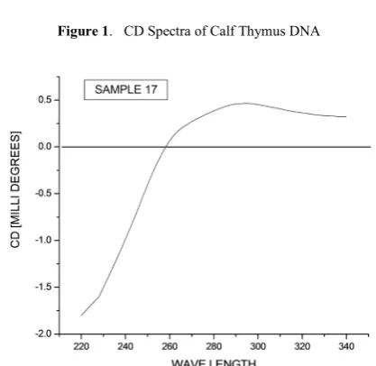
[image:2.595.327.536.497.700.2]Figure 1. CD Spectra of Calf Thymus DNA
Figure 3. Calf Thymus DNA treated with 0.064mM of Lead Acetate
Figure 4. Calf Thymus DNA treated with 0.32mM of Lead Acetate
[image:3.595.323.542.297.468.2]Figure 5. Calf Thymus DNA treated with 0.08nM of 5-Fluorouracil
[image:3.595.68.284.321.488.2]Figure 6. Calf Thymus DNA treated with 0.16nM of 5- Fluorouracil.
Figure 7. Calf Thymus DNA treated with 0.032 mM of Lead Acetate &
0.08nM of 5-Fluorouracil
Figure 8. Calf Thymus DNA treated with 0.032mM of Lead Acetate &
[image:3.595.325.560.528.693.2] [image:3.595.70.287.541.704.2]4. Discussion
In many biological and biochemical systems divalent cations are important for the activity of some integrated structural requirements of nucleic acids [27]. CD spectra of nucleic acids provide valuable information about conformational changes induced by variation of the suspending mediumor by effects of interactions with the polymers [28,29].
The change in elliptical intensities at around 260 nm to 280 nm and 245 nm shows the conformational changes in B-form of DNA30. At the same time a negative ellipticity was
observed at 260 to 280 nm with 0.32 mM of Lead Acetate treatment in Fig 4. A negative ellipticity was also observed for all viz 0.08 nM and 0.16 nM concentrations of 5-Fluorouracil Fig 5 and Fig 6. The negative ellipticity that was observed at 290 nm at 0.32 mM of Pb2+ Fig 4 indicates
the possibility of B - Z transitions [28]. Also the negative ellipticity that was observed at 260 – 280 nm for 0.08 nM & 0.16 nM treatments of 5-fluorouracil Fig 5 and Fig 6, indicates the topological variations.
In combinational treatments DNA was prior incubated with 0.032 mM Pb2+ and then titrated with different
concentrations of 5-Fluorouracil. A positive ellipticity was observed at 260-280 nm in combinational treatment of 5-fluorouracil with Pb2+ Fig 7 to Fig 8. And a negative
ellipticity at 245 nm with all the treatments support that, the predominant structure was B-form and the variations observed in elliptical intensities explains the varied geometry. The absence of negative ellipticity with other individual treatments Fig 2 and for combination treatments with Lead Acetate and the drug 5-Flurouracil Fig 7 and Fig 8 shows the variations in intensities suggesting varied adduct geometries in presence of Pb2+.
Calf thymus DNA with 42% of G-C content [24] has tendency towards hairpin transitions. The amplitude changes observed in this study indicates the hairpin formation which is an intra-molecular variant of the B-form [28]. With these results, it is anticipated that, hairpin transitions might be involved in mechanism of Pb2+
interference with 5-Fluorouracil induced structural changes. B-form is the most frequently observed conformation of DNA with weak chirality showing characteristic positive ellipticity around 260-280 nm and negative ellipticity around 245 nm [28]. CD spectrum also depends on number of base pairs per turn corresponding to varied elliptical properties and geometries [28].
With the results obtained, we can presume the interacting competence of Pb2+ and Pb2+ - 5-fluorouracil which brings
the changes in geometries of their interactions with DNA. Also, we have investigated the interference of this important pollutant (Pb2+) which exists in various physiological
systems and its role in altering the geometries of drug induced DNA secondary structural changes.
Acknowledgements
SSK strongly acknowledge Bhagawan Mahavir Medical Research Center and Devi Ahilya University for their support.
REFERENCES
[1] Shaik A.P, Siva Sankar, Satish C. Reddy, Prabhavathy G. Das & Kaiser Jamil (2006) Drug and Chemical Toxicology 29,111–124.
[2] Hitzfeld B & Taylor DM (1989) Mol Toxicol 2,151–162. [3] Yakovlev V.V and Sona E.L (2005) in Studies on the
interaction between Human Serum Albumin and Lead Ions (Pb+2), Department of physics, University of Wisconsin Milwankee, Milwankee, USA.
[4] Gebhart, E., (1984) Toxicol. Environ. Chem. 8, 253–265. [5] Zelikoff JT, Li JH, Hartwig A, wang XW, Costa M and
Rossman TG (1988) Carcinogenesis 9: 1727 – 1732. [6] Chen, Q., (1992) Chung Hua Yu Fang I Hsueh Tsa Chih 26,
334–335 .
[7] Hartwig, A., (1994) Environ. Health Perspect. 102 (3), 45–50.
[8] Johnson FM (1998).Mutation Res 410:123–140.
[9] Coogan, P.T., Bare, M.R., Waalkes, P.M., (1992) Toxicol. Appl. Pharmacol. 113, 227– 233.
[10] Hossain, Z., Huq, F., (2002) J. Inor. Biochem. 90, 85–96. [11] Waalkes M.P. & Misra R.R. (1996) in Toxicology of Metals
(ed.by L.W. Chang), pp. 231- 244. CRC Press, Boca Raton, FL, USA.
[12] Zimmer Ch, Luck G & Holy A (1976) Nucleic Acid Research 3, 2757 – 2770.
[13] Eichhorn, G.L.(1973) in Inorganic Biochemistry, pp. 1210-1243, Elsevier Scientific Publishing Comp., Amsterdam.
[14] Zimmer, Ch, (1971) Z. Chem. 11, 441-458.
[15] Eichhorn, G.L, Berger, N.A, Butzow, J.J, Clark, P.C, Rifkiad, J.M, Shin, Y.A & Tarien, E (1971) Adv. Chem. Ser. 100, 135.
[16] Schreiber, J.P & Daune, M (1969) Biopolymers 8, 139-152. [17] Shin, Y.A, Heim, J & Eichhorn, G.L (1972) Bioinorganic
Chem. 1, 149- 163.
[18] Shin, Y.A (1973) Biopolymers 12, 2459-2467.
[19] Venner, H. & Z.immer, Ch (1966) Biopolymers 4, 321-335. [20] Eichhorn, G.L & Shin, Y.A (1968) J. Amer. Chem. Soc. 90,
7323-7328.
[22] Tajmir-Riahi H.A, Langlais M & Savoie R(1988) Nucleic Acid Research 16, 751-762.
[23] Noordhuis P, Holwerda U, Van der wilt C.L, Van Groeningen C.L, Smid K, Meijer S, Pinedo H.M & Peters G.J (2004) Annals of Oncology 15, 1025 – 1032.
[24] Seymour S Cohen, Joel G Flaks, Hazel D Barner, Marlyin L Loeb & Janet Lichtenstein ( 1958) Proc Natl Acad Sci U.S.A 44, 1004 – 1012.
[25] Kypr,J. and Vorlickova,M. (1986) Gen. Physiol. Biophys.5, 415–422.
[26] Mostafa R T, Seyed H M, Bijan R, Mojtaba A & Sayed – Amir M (2006) J Biochem and Mol Biol 39, 530 – 536.
[27] Luck, G. & Zimmer, Ch. (1972) Eur. J. Biochem. 29, 528-536.
[28] Jaroslav Kypr, Iva Kejnovska,Daniel Renciuk & Michaela
Vorlıc kova (2009) Nucleic Acids Research,1 –13.
[29] Brahms, J. & Brahms, S. (1970) in Biological Macromolecules (Fasman, G. D. & Timasheff, s. N., eds) Vol. 4, pp. 191, Marcel Dekker, New York.