0095-1137/11/$12.00 doi:10.1128/JCM.00713-11
Copyright © 2011, American Society for Microbiology. All Rights Reserved.
Colorimetric Nucleic Acid Testing Assay for RNA Virus Detection
Based on Circle-to-Circle Amplification of Padlock Probes
䌤
Rongqin Ke,
1,7Anna Zorzet,
2† Jenny Go
¨ransson,
1,7‡ Gunnel Lindegren,
3Batool Sharifi-Mood,
4Sadegh Chinikar,
5Masoud Mardani,
6Ali Mirazimi,
3* and Mats Nilsson
1,7*
Department of Immunology, Genetics and Pathology, The Rudbeck Laboratory, Uppsala University, 75185 Uppsala, Sweden1;
Department of Medical Biochemistry and Microbiology, Uppsala University, Box 582, 75123 Uppsala, Sweden2;
Swedish Institute for Communicable Disease Control, 171 82 Solna, Sweden3; Research Center for
Infectious Diseases and Tropical Medicine, Boo-Ali Hospital, Zahedan University of Medical Sciences,
Zahedan, Iran4; Laboratory of Arboviruses and Viral Haemorrhagic Fevers
(National Reference Laboratory), Pasteur Institute of Iran, Tehran, Iran5;
Infectious Diseases and Tropical Medicine Research Center, Shahid Beheshti University of Medical Sciences,
Tehran, Iran6; and Science for Life
Laboratory, Uppsala University,
751 85 Uppsala, Sweden7
Received 10 April 2011/Returned for modification 9 July 2011/Accepted 13 September 2011
We developed a molecular diagnostic method for detection of RNA virus based on padlock probes and colori-metric readout. The feasibility of our approach was demonstrated by using detection of Crimean-Congo hemor-rhagic fever (CCHF) virus as a model. Compared with conventional PCR-based methods, our approach does not require advanced equipment, involves easier assay design, and has a sensitivity of 103
viral copies/ml. By using a cocktail of padlock probes, synthetic templates representing different viral strain variants could be detected. We analyzed 34 CCHF patient samples, and all patients were correctly diagnosed when the results were compared to those of the current real-time PCR method. This is the first time that highly specific padlock probes have been applied to detection of a highly variable target sequence typical of RNA viruses.
Outbreaks of viral diseases highlight the need for sensitive, rapid, reliable, and convenient diagnostic methods. Diagnostic methods based on nucleic acid testing, such as PCR, can fulfill the requirements of being sensitive and reliable. However, PCR-based methods are very unlikely to be convenient when the test needs to be carried out far away from a well-equipped laboratory. This problem is even more pronounced in low-income and developing countries. Therefore, new methods are required for this purpose. In this paper, we present an ap-proach for RNA virus detection based on padlock probes (16) and colorimetric readout, which can potentially overcome the drawbacks of PCR-based nucleic testing methods. We used Crimean-Congo hemorrhagic fever virus (CCHFV) as a model for establishing the assay.
CCHFV is a member of the Bunyaviridae family (genus
Nairovirus) and is known to cause severe disease and mortality
(3 to 50%) in humans. The disease, for which the underlying molecular mechanisms are poorly understood, is manifested by
symptoms like fever, prostration, and severe hemorrhage (6). CCHFV is widely distributed throughout large areas of sub-Saharan Africa, Bulgaria, the former Yugoslavia, northern Greece, European Russia, Pakistan, the Xinjiang Province of northwest China, the Arabian Peninsula, Iraq, and Iran. In addition, in areas of endemicity, viremia and the presence of antibodies have been documented in a long list of domestic and wild vertebrates, including horses, sheep, goats, pigs, cam-els, donkeys, mice, and dogs. Thus, CCHFV circulates unno-ticed in nature in an enzootic tick-vertebrate-tick cycle. Enzo-otic foci of the virus mainly occur where one or several
Hyalommaspecies are the predominant ticks feeding on
do-mestic and wild animals. The virus is also mainly transmitted to humans through ixodid ticks of the genusHyalomma, in par-ticular,Hyalomma marginatum marginatum. Human infection also occurs through contact with blood or tissue material from infected animals or humans. In addition, person-to-person transmission can occur via bloody vomit, body fluids, or aero-sols from patients in advanced stages of disease (20–22).
Current CCHFV diagnostic methods involve the detection of viral genomic RNA and/or viral antigen and detection of specific IgM and IgG antibodies (7, 10, 17). Conventional re-verse transcription (RT)-PCR protocols take several hours for reverse transcription, amplification, and analysis, and in some cases, a second round of nested amplification is necessary to achieve the desired sensitivity. Designing primers and probes for RT-PCR for CCHFV is hampered by a remarkable genetic variability among virus strains. In the last couple of years, several RT-PCR protocols with a higher sensitivity and a shorter running time than conventional PCR analysis have
* Corresponding author. Mailing address for Mats Nilsson: Depart-ment of Immunology, Genetics and Pathology, The Rudbeck Laboratory, Uppsala University, 75185 Uppsala, Sweden. Phone: 46184714816. Fax: 46184714808. E-mail: [email protected]. Mailing address for Ali Mirazimi: Swedish Institute for Communicable Disease Control, 171 82 Solna, Sweden. Phone: 4684572573. Fax: 468307957. E-mail: ali.mirazimi @smi.se.
† Present address: ReAct/Uppsala University, Box 256, 751 05 Upp-sala, Sweden.
‡ Present address: Q-linea, AB, Dag Hammarskjo¨lds Va¨g 58A, SE-75237, Uppsala, Sweden.
䌤Published ahead of print on 28 September 2011.
4279
on May 16, 2020 by guest
http://jcm.asm.org/
been developed. However, the requirement for expensive RT-PCR equipment is still a problem. The disease is endemic in low-income and developing countries, and outbreaks mostly occur in villages far away from equipped laboratories. Many locations experience sudden power outages that can damage delicate equipment, which is then costly to repair or replace. This has created an urgent demand for a sensitive detection method that does not require expensive equipment.
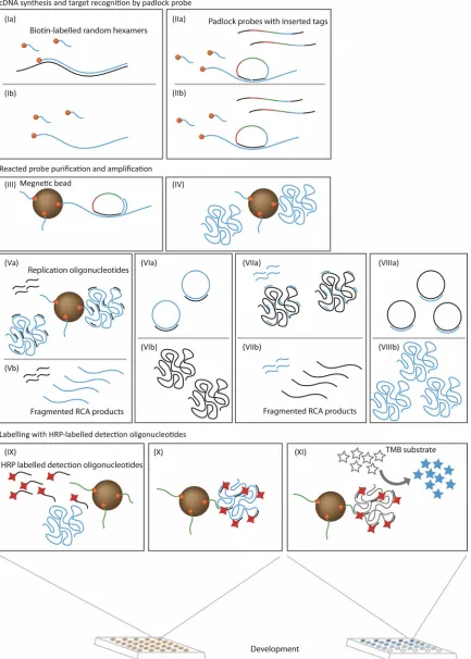
In this study, a sensitive detection assay for CCHFV using padlock probes (16) and circle-to-circle amplification (C2CA) technology (4) combined with enzymatic readout has been developed (Fig. 1). A padlock probe is a linear oligonucleotide that contains half of a unique target recognition sequence at each end and a target-independent sequence in between. The target recognition sequences are designed to bind head to tail on a genomic target DNA. Upon the hybridization to target DNA, the probes become circularized by enzymatic ligation, while if there is no target DNA present, the padlock probes remain linear. Padlock probes have previously been used for genotyping (9, 13), gene copy number (23), expression analysis (14), target sequencing (5), and mRNA splicing (3), as well as for detection of bacteria and other infectious pathogens (1, 8, 15). Only the circularized padlock probes are then amplified by the exponential rolling-circle amplification (RCA)-based C2CA reaction (2, 4). The ligation reaction is highly specific and will not occur if there is a single mismatch between the target and the probe at the ligation site. This ensures high specificity but may be a problem when addressing highly vari-able target sites. The main aim of this study was to investigate whether it is feasible to detect highly variable target sequences by using cocktails of padlock probes covering the range of variation. In the current study, the amplification product is finally detected using a horseradish peroxidase (HRP)-cata-lyzed colorimetric readout to eliminate the use of sophisticated amplification and detection instruments.
We show that our method can detect CCHFV with high sensitivity. Using a combination of padlock probes, CCHFV strains with known sequences can be detected. The colorimet-ric readout makes it possible to read the results with the naked eye or semiquantitatively in a simple absorbance reader. Our approach is not based on PCR amplification and does not require sophisticated or expensive instrumentation. It is possi-ble to use this method for field-based diagnosis and monitoring applications in developing countries.
MATERIALS AND METHODS
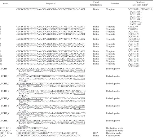
Probe sequences.All oligonucleotide sequences are shown in Table 1. The polarity of the padlock probe is referred to as positive (⫹), and that of its complement is referred to as negative (⫺). Padlock probes were designed using ProbeMaker software (18) to target the most conserved sequences in the L segment of the CCHFV genome. Due to the high genetic variability of the viral genome, 7 additional probes were designed to ensure detection of all strains (total of 8 probes).
Serum samples.Serum samples from a total of 20 patients were included in the study. Samples were taken from patients displaying symptoms of CCHFV infection, and where possible, both acute-phase samples (A) and convalescent-phase samples (B) were taken from the patients. All samples were also analyzed by conventional real-time RT-PCR as described by Wolfel et al. (24).
Virus propagation.Viral RNA for the concentration standard curve was pre-pared from the CCHFV Iranian strain. Confluent Vero cells (ATCC CCL-81) were inoculated with virus and grown for 2 to 3 days in minimal essential medium (MEM; Invitrogen, Life Technologies, Paisley, United Kingdom) supplemented
with penicillin-streptomycin solution (Gibco), HEPES (Gibco), and 2% heat-inactivated fetal calf serum (FCS; Gibco) at 37°C. The supernatant was collected after clarification by centrifugation at 1,000⫻gfor 10 min and subsequently aliquoted and stored at⫺80°C prior to RNA extraction. All handling of live virus was carried out in biosafety level 4 (BSL4) facilities.
RNA extractions.The CCHFV supernatants and the serum samples were treated with TRIzol LS reagent (ratio, 1⫹3; Invitrogen) in the BSL4 laboratory for a minimum of 5 min, before decontamination of the tubes and transport to a BSL2 laboratory following the safety instructions. After phase separation by chloroform (Merck) treatment and centrifugation, viral RNA was purified from the aqueous phase using a QIAamp viral RNA minikit (Qiagen), according to the manufacturer’s instructions.
RT-PCR.The extracted RNA was analyzed by CCHFV real-time RT-PCR, positive reactions were plotted against a standard curve, and the genome con-tent/ml was calculated (24). The cycling reactions were performed in a Roche LightCycler 2.0 or 480 apparatus.
Synthesis of biotin-labeled cDNA.In a 21-l reaction volume, a mixture of 1.5
l (100 ng/l) 5⬘biotin-labeled random hexamers (Gene Link, Hawthorne, NY), 1l 10 mM deoxynucleoside triphosphate (dNTP) mix (Invitrogen), 2.5 l distilled water, and 5l RNA was incubated at 65°C for 5 min and then immediately chilled on ice. A mixture of 2l 10⫻RT buffer, 4l MgCl2(25
mM), 2l dithiothreitol (DTT; 0.1 M), 1l (40 U/l) RNaseOUT, and 1l SuperScriptIII reverse transcriptase (200 U/l; Invitrogen) was added and the mixture was incubated at 25°C for 10 min, 50°C for 50 min, and 65°C for 5 min and then kept at 4°C. Finally, 1l RNase H (2 U/l; Invitrogen) was added and the mixture was incubated at 37°C for 20 min. The biotinylated cDNA was stored at⫺20°C until use. The cycling reactions were performed in a thermocycler (Gene Amp 2700; ABI).
PCR analysis of cDNA.RNA and cDNA were analyzed simultaneously by the previously described RT-PCR, and the genome content/ml was calculated (24). The cycling reactions were performed in a Roche LightCycler 480 apparatus.
Padlock probe ligation and circle-to-circle amplification.All padlock probes were phosphorylated prior to use. Briefly, 100l of a phosphorylation mixture containing 10M padlock probes, 1⫻PNK buffer A (Fermentas), 1 mM ATP, and 0.1 U/l T4 polynucleotide kinase was incubated at 37°C for 30 min and 60°C for 20 min. The phosporylated probes were then stored at⫺20°C until use. A schematic illustration of the following protocol can be found in Fig. 1. First, 10
l biotinylated cDNA from either clinical samples or the cultivated Iranian reference strain was added to 10l ligation mix in a 96-well PCR plate (Thermo Electron). Final concentrations were 1⫻Ampligase buffer (Epicentre), 100 nM each padlock probe, and 5 U Ampligase (Epicentre). The ligation reaction was carried out by incubation at 55°C or 60°C for 5 min, forming DNA circles. Then, 10l 10-mg/ml streptavidin-coupled MyOne T1 magnetic beads (Invitrogen) suspended in 3⫻wash buffer (15 mM Tris-HCl [pH 7.5], 1.5 mM EDTA, 3 M NaCl, 0.1% Tween 20) was added to each sample, followed by gentle vortexing and incubation at room temperature for 5 min. Unbound material was then separated from the beads using a magnetic rack (Invitrogen). The beads were washed once with wash buffer containing 5 mM Tris-HCl (pH 7.5), 0.5 mM EDTA, 1 M NaCl, and 0.1% Tween 20, and the wash buffer was discarded. The DNA circles captured by the beads were primed by adding 20l RCA mix containing 1⫻phi29 DNA polymerase buffer (33 mM Tris-acetate [pH 7.9 at 37°C], 10 mM magnesium acetate, 66 mM potassium acetate, 0.1% [vol/vol] Tween 20, 1 mM DTT; Fermentas), 100M dNTPs, 0.2 mg/ml bovine serum albumin (BSA), 50 nM primer, and 4 U phi29 DNA polymerase. The reaction mixture was incubated at 37°C for 20 min, followed by 1 min at 65°C to inactivate the phi29 DNA polymerase. The amplified single molecules were then mono-merized by adding 5l restriction digestion mixture containing 1 U/l AluI restriction enzyme (New England BioLabs), 1⫻phi29 DNA polymerase buffer, 400 nM replication oligonucleotides CCHF_RO⫹, and 0.2 mg/ml BSA. The reaction mixture was incubated at 37°C for 5 min, and AluI was inactivated at 65°C for 3 min. The monomers were then recircularized and amplified by addi-tion of 10l ligation and RCA mix containing 1⫻phi29 DNA polymerase buffer, 0.2 mg/ml BSA, 3 mM ATP, 250M dNTP, 0.05 U/l T4 DNA ligase (Fermen-tas), and 0.3 U/l phi29 DNA polymerase. To initiate the second restriction digestion, 5l restriction digestion mix containing 1 U/l AluI, 1⫻phi29 DNA polymerase buffer, 1.6 M replication oligonucleotides CCHF_RO⫺, and 0.2 mg/ml BSA was added. Finally, the third RCAs were then carried out by adding the same ligation and RCA mix mentioned above.
HRP detection.Magnetic beads were preincubated in capture probe solution (50 nM capture probe in hybridization buffer containing 20 mM EDTA, 20 mM Tris-HCl, 1.4 M NaCl, and 0.1% Tween 20) at a 1:3 volume ratio. The mixture was then incubated for 5 min at room temperature and placed in a magnetic rack (Invitrogen) for 1 min, and the supernatant containing unbound capture probes
4280 KE ET AL. J. CLIN. MICROBIOL.
on May 16, 2020 by guest
http://jcm.asm.org/
FIG. 1. Schematic illustration of CCHFV detection using padlock probes and enzymatic readout. (I) Synthesis of biotin-labeled cDNA from CCHF mRNA fragments. (II) Padlock probes are added to samples and specifically circularized by DNA ligase when hybridized to the correct template. (III) Ligated padlock probes are captured by streptavidin-coated magnetic beads, whereas unbound probes are removed by washing. (IV) Ligated padlock probes are amplified by RCA. (V) RCA products are digested by restriction enzyme to generate monomers. (VI) The monomers are recircularized and amplified by RCA to generate second-generation RCA products. (VII) Second digestion of RCA products. (VIII) Monomers are recircularized and again amplified by RCA to generate third-generation RCA products. (IX to X) Third-generation RCA products are hybridized to HRP-labeled detection probes and bound by capture probes immobilized on magnetic beads. (XI) Signal is developed by adding TMB substrates to the beads.
4281
on May 16, 2020 by guest
was aspirated. The plate was removed from the rack, and beads were resus-pended in 5l hybridization buffer per sample to be detected. Five microliters of this bead-capture probe complex was added to each sample well along with 50l detection probe (30 nM HRP-coupled probes in hybridization buffer), and the plate was incubated at 55°C for 15 min in a heating block. The beads were then washed two times with 150 and 250l washing buffer (phosphate-buffered saline plus 0.05% Tween 20) with the aid of the magnetic rack (Invitrogen). After the last washing, buffer was aspirated, 100l tetramethylbenzidine (TMB; Thermo Scientific) was added, and the plate was incubated at 37°C for 20 min. The result was quantified in an enzyme-linked immunosorbent assay reader (Multiscan FC; Thermo Scientific) at 650 nm but could also be examined visually.
RESULTS
Method design. Padlock probes targeting the most con-served sequence in the L segment of the CCHFV genome were
designed. The padlock probes in the current study are equipped with two end sequences that recognize the target sequence and three sequence elements that are used for dif-ferent purposes: a detection sequence for hybridization with an HRP-labeled detection oligonucleotide, a capture sequence for hybridization to capture oligonucleotide immobilized on magnetic beads, and finally, a replication sequence for C2CA (Fig. 1).
Viral CCHF cDNA was synthesized from viral mRNA by reverse transcription using 5⬘biotin-labeled random hexamers. The cDNA was probed using a high concentration of padlock probes to promote hybridization and ligation kinetics. Excess unreacted padlock probes were eliminated by washing enabled by capture of the target sequence to streptavidin-coated
mag-TABLE 1. Oligonucleotides for CCHFV detection
Name Sequencea 5⬘end
modification Function
GenBank accession no(s).b
T CTCTCTCTCTCTCTAAACCAAGCCTGACCAT兩GTTGATACAGACT Biotin Template GQ337055.1, EU044832.1, DQ211623.1,
DQ211618.1, DQ211617.1, DQ211614.1, AY389361.2, AY995166.2 T1 CTCTCTCTCTCTCTAAACCAAGCCTGACTAT兩GTTGATACAGACT Biotin Template AY675240 T2 CTCTCTCTCTCTCTAAACCTAGCCTGACCAT兩GTTGATACAGACT Biotin Template AY720893 T3 CTCTCTCTCTCTCTAAACCAAGCCTGACCAT兩GTTAATACAGACT Biotin Template DQ211621 T4 CTCTCTCTCTCTCTAAACCAAGCCTGACCAT兩GCTGATACAGACT Biotin Template DQ076417.1
DQ076414.1 T5 CTCTCTCTCTCTCTAAACCAAGTCTGACCAT兩GTTGATACAGACA Biotin Template HM452307.1 AY947890.1 T6 CTCTCTCTCTCTCTAAACCAAGCCTGACCAT兩GTTGATACAAACT Biotin Template DQ211613.1 T7 CTCTCTCTCTCTCTAAACCAAGCCTGACCAT兩GTTAATACAGACC Biotin Template DQ211622.1 DQ211620.1 T8 CTCTCTCTCTCTCTAAACCAAGTCTGACCAT兩GCTGATACAGACG Biotin Template DQ211619.1 T9 CTCTCTCTCTCTCTAAACCAAGTTTGACCAT兩GTTGATACAAACC Biotin Template DQ076412.1 T10 CTCTCTCTCTCTCTAAACCAAGCCTAACCAT兩GTTAATACAGACC Biotin Template DQ211615.1 T11 CTCTCTCTCTCTCTAAACCAAGTTTAACCAT兩GTTGATACAGACA Biotin Template DQ211616.1 T12 CTCTCTCTCTCTCTAAACCGAGTCTAACAAT兩GTTGATACAGACC Biotin Template DQ211612.1 T13 CTCTCTCTCTCTCTAAACCGAGTTTGACCAT兩GCTGATACAAACC Biotin Template DQ211624.1 DQ099335.2 P_CCHF ATGGTCAGGCTTGGTTTGTGGATAGTGTCTTACACGAAGAGTG
TACCGACCTCAGTAAGTCTCCTAGCTCGGTGAACTAGTCTGT ATCAAC
Padlock probe
P_CCHF_1 ATGGTCAGACTTGGTTTGTGGATAGTGTCTTACACGAAGAGTG TACCGACCTCAGTAAGTCTCCTAGCTCGGTGAACTAGTCTGT ATCAGC
Padlock probe
P_CCHF_2 ATTGTTAGACTTGGTTTGTGGATAGTGTCTTACACGAAGAGTG TACCGACCTCAGTAAGTCTCCTAGCTCGGTGAACTAGTCTGT ATCAAC
Padlock probe
P_CCHF_3 ATGGTCAGGCTTGGTTTGTGGATAGTGTCTTACACGAAGAGTG TACCGACCTCAGTAAGTCTCCTAGCTCGGTGAACTAGTCTGT ATTAAC
Padlock probe
P_CCHF_4 ATGGTCAAACTTGGTTTGTGGATAGTGTCTTACACGAAGAGTG TACCGACCTCAGTAAGTCTCCTAGCTCGGTGAACTAGTCTGT ATCAAC
Padlock probe
P_CCHF_5 ATGGTCAAACTCGGTTTGTGGATAGTGTCTTACACGAAGAGTG TACCGACCTCAGTAAGTCTCCTAGCTCGGTGAACTGGTTTGT ATCAGC
Padlock probe
P_CCHF_6 ATGGTCAGGCTTGGTTTGTGGATAGTGTCTTACACGAAGAGTG TACCGACCTCAGTAAGTCTCCTAGCTCGGTGAACTAGTCTGT ATCAGC
Padlock probe
P_CCHF_7 ATAGTCAGGCTTGGTTTGTGGATAGTGTCTTACACGAAGAGTG TACCGACCTCAGTAAGTCTCCTAGCTCGGTGAACTAGTCTGT ATCAAC
Padlock probe
CCHF_RO⫹ AGTCTCCTAGCTCGGTGAAC Replication probe
CCHF_RO⫺ GTTCACCGAGCTAGGAGACT Replication probe
HRP_1° RCA HRP-CTTGCGACGTCAGTGGATAGTGTCTTACACGATTT HRP Detection probe B2_CO TTTTCTGGATCGTCAGGGAAAGAGTGTACCGACCTCAGTA Biotin Capture probe
aItalics, inserted spacer sequences;兩, ligation site; underline, target complementary sequences; bold letters, SNPs. bGenBank accession number(s) for the representative virus strain(s) containing the sequence of synthetic templates.
4282 KE ET AL. J. CLIN. MICROBIOL.
on May 16, 2020 by guest
http://jcm.asm.org/
[image:4.585.47.544.81.527.2]netic beads. Reacted padlock probes were then amplified by three cycles of RCA. The C2CA products were thereafter hybridized with HRP-labeled detection oligonucleotides, fol-lowed by separation of free detection oligonucleotides by cap-turing the labeled product in a sandwich hybridization reaction using capture oligonucleotides immobilized on magnetic beads. Finally, the signal was developed by adding HRP col-orimetric substrate.
Two different probing approaches were taken. The first aimed to target as many viral strains as possible with a single padlock probe (P_CCHF). The second approach involved the addition of seven different padlock probes (P_CCHF_1 to P_CCHF_7) targeting the same site for detection of variants of other CCHFV strains (in total, 8 probes).
Sensitivity.The sensitivity of the approach was investigated by detection of serial dilutions of cDNA from cultivated CCHFV of an Iranian strain, whose concentration was vali-dated by real-time RT-PCR. The limit of detection for the current method is 103copies/ml using the single-padlock-probe
approach (Fig. 2).
Patient samples.We applied our method for analysis of 34 samples from 20 patients, which were also analyzed by RT-PCR. By applying an absorbance value higher than 0.5 as a threshold, we scored eight of the samples positive and the rest negative, results which are in agreement with the RT-PCR results (Fig. 3).
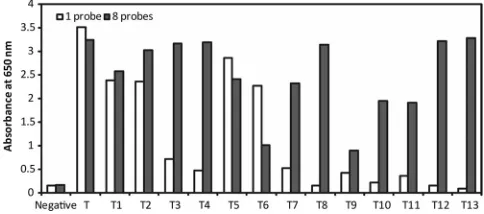
Capability of the approach.We investigated the capability of our approach by detection of synthetic templates that repre-sented different CCHFV strains found in different regions. We found that when we used a single padlock probe for detection, some strains gave false-negative results. To overcome this problem, we designed seven additional padlock probes with slightly different sequences (Table 1). The eight padlock probes were then pooled and applied for detection. Using this approach, all 14 synthetic templates representing the target sequences of the different strains were detected at a concen-tration of 6⫻104copies per ml, which is equal to 600 copies
per reaction (Fig. 4). The negative controls were either double-distilled H2O or Rift Valley virus, which also belongs to the
Bunyaviridaefamily (12), and neither gave a positive signal.
FIG. 2. Typical standard curve of CCHFV detection using diluted cDNA samples. Error bars,⫾1 standard deviation;n⫽3.
[image:5.585.299.541.71.178.2]FIG. 3. Detection of CCHF patient samples. The results for all samples were confirmed by RT-PCR; circles, positive signals from RT-PCR. The cutoff value for oversaturated signals is set to 4.⫹, positive results;⫺, negative results.
FIG. 4. Detection of different CCHFV variants by using either a single padlock probe or a set of eight padlock probes. T to T13 correspond to variants of the target sequence (listed in Table 1).
on May 16, 2020 by guest
http://jcm.asm.org/
DISCUSSION
We have established a non-PCR-based nucleic acid detec-tion assay applying padlock probe and C2CA for the detecdetec-tion of CCHFV. The isothermal amplification strategy and simple colorimetric readout together make the assay independent of sophisticated instrumentation. Therefore, it is potentially suit-able for field-based diagnosis and monitoring applications in developing countries. Even though our approach is PCR inde-pendent, the sensitivity is comparable to that of PCR-based methods. In a typical PCR-based detection method, a pair of primers, sometimes along with a TaqMan probe, is needed to amplify the target sequence. Hence, every target sequence needs two to three hybridization sequences to set up a detec-tion assay. However, the CCHFV genome is highly variable due to high mutation rates. Thus, it may be difficult to find conserved sequences for all the required probe binding sites. For our padlock probe-based detection, only one hybridization sequence is required, which increases the chance of finding a suitable target sequence.
In our experience, even though the reagents need to be added into the reaction tubes several times during the proto-col, cross contamination was not observed, which was in accor-dance with the findings of previous studies (4). This property is important, because for simple readout assays, open-tube ma-nipulation of amplified products to generate a signal is often required. In contrast, PCR-based assays are more sensitive to open-tube manipulations. The reason why our method is less prone to cross contamination may be because the RCA prod-ucts are macromolecules, which are not as easily spread into the air. In addition, the C2CA generates amplified products with opposite polarity between two successive generations. Even though the risk of cross contamination is low, it is still possible. Therefore, careful handling of samples and reagents is important when performing the experiment.
In previous studies that involve C2CA technology, RCA products were detected by radiation-labeled detection oligo-nucleotides, fluorescence-labeled detection oligooligo-nucleotides, or paramagnetic beads (4, 11, 19). However, these methods often require advanced and expensive instrumentation, which made them unsuitable for field-based diagnosis. The only equipment absolutely required in the field using our method would be heating blocks. In this study, we used HRP-labeled oligonucleotides for detection of amplified RCA products (Fig. 1). The result can be determined either by a regular spectro-photometer or by the naked eye, which makes our method more suitable for on-site detection in developing countries.
To address the problem of high variability in the target sequence of CCHFV (Table 1), we designed seven additional padlock probes for detection of the variants in different viral strains (eight probes in total). By using a combination of all eight padlock probes for the detection of different CCHF strains, all 14 strains investigated could be detected with a minimal loss of sensitivity. The result indicates that when an-alyzing a new variant of CCHFV, the sensitivity is altered. However, once the new variant strain is sequenced, if the existing pool of padlocks cannot already detect it, a new pad-lock probe can be designed and added to the pool for high-sensitivity detection.
The limitation of the current approach is the multiple steps
for adding reagents in the protocol. However, even though the protocol has many steps and may seem complicated, it is made simpler by using premade mixes and can be learned quickly. Also, scaling up to analyze many samples at the same time does not add either much time or much effort to the protocol due to the 96-well format. In well-equipped laboratories, automated pipetting devices could be used, eliminating the problem com-pletely.
To summarize, we have developed a PCR-free but highly sen-sitive and high-capability CCHFV detection assay. This is the first time that highly specific padlock probes have been applied to detection of a highly variable target sequence, typical of RNA viruses. By designing new probes, our method could easily be adapted for detection of other RNA or DNA viruses.
ACKNOWLEDGMENTS
This work was supported by grants from the Wallenberg Foun-dation, VINNOVA, and the Swedish Research Council. This work is also part of the CCH Fever Network (Collaborative Project), supported by the European Commission under the Health Coop-eration Work Programme of the 7th Framework Programme (grant agreement 260427).
M.N. holds shares in the company Olink Bioscience, which holds commercial rights to the technology.
REFERENCES
1.Bane´r, J., et al.2007. Microarray-based molecular detection of foot-and-mouth disease, vesicular stomatitis and swine vesicular disease viruses, using padlock probes. J. Virol. Methods143:200–206.
2.Bane´r, J., M. Nilsson, M. Mendel-Hartvig, and U. Landegren.1998. Signal amplification of padlock probes by rolling circle replication. Nucleic Acids Res.26:5073–5078.
3.Conze, T., et al.2010. Single molecule analysis of combinatorial splicing. Nucleic Acids Res.38:e163.
4.Dahl, F., et al.2004. Circle-to-circle amplification for precise and sensitive DNA analysis. Proc. Natl. Acad. Sci. U. S. A.101:4548–4553.
5.Deng, J., et al.2009. Targeted bisulfite sequencing reveals changes in DNA methylation associated with nuclear reprogramming. Nat. Biotechnol.27: 353–360.
6.Ergonul, O.2006. Crimean-Congo haemorrhagic fever. Lancet Infect. Dis. 6:203–214.
7.Garcia, S., et al.2006. Evaluation of a Crimean-Congo hemorrhagic fever virus recombinant antigen expressed by Semliki Forest suicide virus for IgM and IgG antibody detection in human and animal sera collected in Iran. J. Clin. Virol.35:154–159.
8.Go¨ransson, J., et al.2010. Sensitive detection of bacterial DNA by magnetic nanoparticles. Anal. Chem.82:9138–9140.
9.Gyarmati, P., et al.2008. Simultaneous genotyping of all hemagglutinin and neuraminidase subtypes of avian influenza viruses by use of padlock probes. J. Clin. Microbiol.46:1747–1751.
10.Ibrahim, S. M., et al.2011. Detection of Crimean-Congo hemorrhagic fever, Hanta, and sandfly fever viruses by real-time RT-PCR. Methods Mol. Biol. 665:357–368.
11.Jarvius, J., et al.2006. Digital quantification using amplified single-molecule detection. Nat. Methods3:725–727.
12.Kortekaas, J., O. Ergonul, and R. J. Moormann.2010. Interventions against West Nile virus, Rift Valley fever virus, and Crimean-Congo hemorrhagic fever virus: where are we? Vector Borne Zoonotic Dis.10:709–718. 13.Kurt, K., et al.2009. Multiplexed genotyping of methicillin-resistant
Staph-ylococcus aureus isolates by use of padlock probes and tag microarrays. J. Clin. Microbiol.47:577–585.
14.Larsson, C., I. Grundberg, O. So¨derberg, and M. Nilsson.2010. In situ detection and genotyping of individual mRNA molecules. Nat. Methods 7:395–397.
15.Long, Y., X. Zhou, and D. Xing.2010. Sensitive and isothermal electrochemi-luminescence gene-sensing of Listeria monocytogenes with hyperbranching rolling circle amplification technology. Biosens. Bioelectron.26:2897–2904. 16.Nilsson, M., et al.1994. Padlock probes: circularizing oligonucleotides for
localized DNA detection. Science265:2085–2088.
17.Saijo, M., et al. 2002. Immunofluorescence technique using HeLa cells expressing recombinant nucleoprotein for detection of immunoglobulin G antibodies to Crimean-Congo hemorrhagic fever virus. J. Clin. Microbiol. 40:372–375.
18.Stenberg, J., M. Nilsson, and U. Landegren.2005. ProbeMaker: an
extensi-4284 KE ET AL. J. CLIN. MICROBIOL.
on May 16, 2020 by guest
http://jcm.asm.org/
ble framework for design of sets of oligonucleotide probes. BMC Bioinfor-matics6:229.
19.Stro¨mberg, M., et al.2008. Sensitive molecular diagnostics using volume-amplified magnetic nanobeads. Nano Lett.8:816–821.
20.Swanepoel, R., A. J. Shepherd, P. A. Leman, and S. P. Shepherd.1985. Investigations following initial recognition of Crimean-Congo haemorrhagic fever in South Africa and the diagnosis of 2 further cases. S. Afr. Med. J. 68:638–641.
21.van de Wal, B. W., J. R. Joubert, P. J. van Eeden, and J. B. King.1985. A nosocomial outbreak of Crimean-Congo haemorrhagic fever at Tygerberg
Hospital. Part IV. Preventive and prophylactic measures. S. Afr. Med. J. 68:729–732.
22.van Eeden, P. J., et al.1985. A nosocomial outbreak of Crimean-Congo haemorrhagic fever at Tygerberg Hospital. Part II. Management of patients. S. Afr. Med. J.68:718–721.
23.Wang, Y. K., et al.2005. Allele quantification using molecular inversion probes (MIP). Nucleic Acids Res.33:e813.
24.Wolfel, R., et al.2007. Virus detection and monitoring of viral load in Crimean-Congo hemorrhagic fever virus patients. Emerg. Infect. Dis.13: 1097–1100.