(1)www.impactjournals.com/oncotarget/
Oncotarget, Vol. 5, No. 24
Classification of current anticancer immunotherapies
Lorenzo Galluzzi
1,2,3,4,*, Erika Vacchelli
1,2,3, José-Manuel Bravo-San Pedro
1,2,3,
Aitziber Buqué
1,2,3, Laura Senovilla
1,2,3, Elisa Elena Baracco
1,2,3,5, Norma Bloy
1,2,3,5,
Francesca Castoldi
1,2,3,5,6, Jean-Pierre Abastado
7, Patrizia Agostinis
8, Ron N.
Apte
9, Fernando Aranda
1,2,3,10, Maha Ayyoub
11,12, Philipp Beckhove
13, Jean-Yves
Blay
14,15, Laura Bracci
16, Anne Caignard
17,18, Chiara Castelli
19, Federica Cavallo
20,
Estaban Celis
21, Vincenzo Cerundolo
22, Aled Clayton
23,24, Mario P. Colombo
19,
Lisa Coussens
25, Madhav V. Dhodapkar
26, Alexander M. Eggermont
3, Douglas T.
Fearon
27, Wolf H. Fridman
2,4,28,29, Jitka Fučíková
6,30, Dmitry I. Gabrilovich
31, Jérôme
Galon
2,4,28,32, Abhishek Garg
8, François Ghiringhelli
33,34,35, Giuseppe Giaccone
36,37, Eli
Gilboa
38, Sacha Gnjatic
39, Axel Hoos
40, Anne Hosmalin
4,41,42,43, Dirk Jäger
44, Pawel
Kalinski
45,46,47, Klas Kärre
48, Oliver Kepp
1,2,49, Rolf Kiessling
50, John M. Kirkwood
51,
Eva Klein
48, Alexander Knuth
52, Claire E. Lewis
53, Roland Liblau
54,55,56, Michael T.
Lotze
45,46, Enrico Lugli
57, Jean-Pierre Mach
58, Fabrizio Mattei
16, Domenico Mavilio
57,59,
Ignacio Melero
60,61, Cornelis J. Melief
62,63, Elizabeth A. Mittendorf
64, Lorenzo
Moretta
65, Adekunke Odunsi
66, Hideho Okada
67, Anna Karolina Palucka
68, Marcus
E. Peter
69, Kenneth J. Pienta
70, Angel Porgador
9, George C. Prendergast
71,72,73,
Gabriel A. Rabinovich
74, Nicholas P. Restifo
75, Naiyer Rizvi
76, Catherine
Sautès-Fridman
2,4,28,29, Hans Schreiber
77, Barbara Seliger
78, Hiroshi Shiku
79, Bruno
Silva-Santos
80, Mark J. Smyth
81,82, Daniel E. Speiser
83,84, Radek Spisek
6,30, Pramod K.
Srivastava
85,86, James E. Talmadge
87, Eric Tartour
4,88,89,90, Sjoerd H. Van Der Burg
91,
Benoît J. Van Den Eynde
92,93,94, Richard Vile
95, Hermann Wagner
96, Jeffrey S.
Weber
97, Theresa L. Whiteside
46,98, Jedd D. Wolchok
99,100, Laurence Zitvogel
3,101,102,
Weiping Zou
103 and Guido Kroemer
1,2,4,49,104,*
1 Equipe 11 labellisée pas la Ligue Nationale contre le Cancer, Centre de Recherche des Cordeliers, Paris, France
2 INSERM, U1138, Paris, France
3 Gustave Roussy Cancer Campus, Villejuif, France
4 Université Paris Descartes/Paris V, Sorbonne Paris Cité, Paris, France
5 Faculté de Medicine, Université Paris Sud/Paris XI, Le Kremlin-Bicêtre, France
6 Sotio a.c., Prague, Czech Republic
7 Pole d’innovation thérapeutique en oncologie, Institut de Recherches Internationales Servier, Suresnes, France
8 Cell Death Research and Therapy (CDRT) Laboratory, Dept. of Cellular and Molecular Medicine, University of Leuven,
Leuven, Belgium
9 The Shraga Segal Dept. of Microbiology, Immunology and Genetics, Faculty of Health Sciences, Ben-Gurion University of
the Negev, Beer-Sheva, Israel
10 Group of Immune receptors of the Innate and Adaptive System, Institut d’Investigacions Biomédiques August Pi i Sunyer
(IDIBAPS), Barcelona, Spain
11 INSERM, U1102, Saint Herblain, France
12 Institut de Cancérologie de l’Ouest, Saint Herblain, France
13 Translational Immunology Division, German Cancer Research Center, Heidelberg, Germany
14 Equipe 11, Centre Léon Bérard (CLR), Lyon, France
15 Centre de Recherche en Cancérologie de Lyon (CRCL), Lyon, France
16 Dept. of Hematology, Oncology and Molecular Medicine, Istituto Superiore di Sanità, Rome, Italy
17 INSERM, U1160, Paris, France
18 Groupe Hospitalier Saint Louis-Lariboisière - F. Vidal, Paris, France
20 Molecular Biotechnology Center, Dept. of Molecular Biotechnology and Health Sciences, University of Torino, Torino, Italy
21 Cancer Immunology, Inflammation and Tolerance Program, Georgia Regents University Cancer Center, Augusta, GA, USA
22 MRC Human Immunology Unit, Weatherall Institute of Molecular Medicine, University of Oxford, Oxford, UK
23 Institute of Cancer & Genetics, School of Medicine, Cardiff University, Cardiff, UK
24 Velindre Cancer Centre, Cardiff, UK
25 Knight Cancer Institute, Oregon Health & Science University, Portland, OR, USA
26 Sect. of Hematology and Immunobiology, Yale Cancer Center, Yale University, New Haven, CT, USA
27 Cold Spring Harbor Laboratory, Cold Spring Harbor, NY, USA
28 Université Pierre et Marie Curie/Paris VI, Paris, France
29 Equipe 13, Centre de Recherche des Cordeliers, Paris, France
30 Dept. of Immunology, 2nd Faculty of Medicine and University Hospital Motol, Charles University, Prague, Czech Republic
31 Dept. of Pathology and Laboratory Medicine, Perelman School of Medicine, University of Pennsylvania, Philadelphia, PA,
USA
32 Laboratory of Integrative Cancer Immunology, Centre de Recherche des Cordeliers, Paris, France
33 INSERM, UMR866, Dijon, France
34 Centre Georges François Leclerc, Dijon, France
35 Université de Bourgogne, Dijon, France
36 Center for Cancer Research, National Cancer Institute (NCI), National Institutes of Health (NIH), Bethesda, MD, USA
37 Lombardi Comprehensive Cancer Center, Georgetown University, Washington, DC, USA
38 Dept. of Microbiology and Immunology, Sylvester Comprehensive Cancer Center, University of Miami, Miller School of
Medicine, Miami, FL, USA
39 Sect. of Hematology/Oncology, Immunology, Tisch Cancer Institute, Icahn School of Medicine at Mount Sinai, New York,
NY, USA
40 Glaxo Smith Kline, Cancer Immunotherapy Consortium, Collegeville, PA, USA
41 INSERM, U1016, Paris, France
42 CNRS, UMR8104, Paris, France
43 Hôpital Cochin, AP-HP, Paris, France
44 National Center for Tumor Diseases, University Medical Center Heidelberg, Heidelberg, Germany
45 Dept. of Surgery, University of Pittsburgh, Pittsburgh, PA, USA
46 University of Pittsburgh Cancer Institute, Hillman Cancer Center, Pittsburgh, PA, USA
47 Dept. of Immunology and Infectious Diseases and Microbiology, University of Pittsburgh, Pittsburgh, PA, USA
48 Dept. of Microbiology, Tumor and Cell Biology, Karolinska Institute, Stockholm, Sweden
49 Metabolomics and Cell Biology Platforms, Gustave Roussy Cancer Campus, Villejuif, France
50 Dept. of Oncology, Karolinska Institute Hospital, Stockholm, Sweden
51 University of Pittsburgh Cancer Institute Laboratory, Pittsburgh, PA, USA
52 National Center for Cancer Care and Research, Hamad Medical Corporation, Doha, Qatar
53 Academic Unit of Inflammation and Tumour Targeting, Dept. of Oncology, University of Sheffield Medical School, Sheffield,
UK
54 INSERM, UMR1043, Toulouse, France
55 CNRS, UMR5282, Toulouse, France
56 Laboratoire d’Immunologie, CHU Toulouse, Université Toulouse II, Toulouse, France
57 Unit of Clinical and Experimental Immunology, Humanitas Clinical and Research Institute, Rozzano, Italy
58 Dept. of Biochemistry, University of Lausanne, Epalinges, Switzerland
59 Dept. of Medical Biotechnologies and Translational Medicine, University of Milan, Rozzano, Italy
60 Dept. of Immunology, Centro de Investigación Médica Aplicada (CIMA), Universidad de Navarra, Pamplona, Spain
61 Dept. of Oncology, Clínica Universidad de Navarra, Pamplona, Spain
62 ISA Therapeutics, Leiden, The Netherlands
64 Research Dept. of Surgical Oncology, The University of Texas, MD Anderson Cancer Center, Houston, TX, USA
65 Istituto Giannina Gaslini, Genova, Italy
66 Center for Immunotherapy, Roswell Park Cancer Institute, Buffalo, NY, USA
67 Dept. of Neurological Surgery, University of California San Francisco, San Francisco, CA, USA
68 The Jackson Laboratory for Genomics Medicine, Farmington, CT, USA
69 Div. of Hematology/Oncology, Northwestern University, Feinberg School of Medicine, Chicago, IL, USA
70 The James Buchanan Brady Urological Institute, The Johns Hopkins Medical Institutions, Baltimore, MD, USA
71 Lankenau Institute for Medical Research, Wynnewood, PA, USA
72 Dept. of Pathology, Anatomy and Cell Biology, Sidney Kimmel Medical College, Philadelphia, PA, USA
73 Cell Biology and Signaling Program, Kimmel Cancer Center, Thomas Jefferson University, Philadelphia, PA, USA
74 Laboratorio de Inmunopatología, Instituto de Biología y Medicina Experimental (IBYME), Buenos Aires, Argentina
75 National Cancer Institute (NCI), National Institutes of Health (NIH), Bethesda, MD, USA
76 Memorial Sloan Kettering Cancer Center (MSKCC), New York, NY, USA
77 Dept. of Pathology, The Cancer Research Center, The University of Chicago, Chicago, IL, USA
78 Institute of Medical Immunology, Martin Luther University Halle-Wittenberg, Halle, Germany
79 Dept. of Immuno-GeneTherapy, Mie University Graduate School of Medicine, Tsu, Japan
80 Instituto de Medicina Molecular, Universidade de Lisboa, Lisboa, Portugal
81 Immunology in Cancer and Infection Laboratory, QIMR Berghofer Medical Research Institute, Herston, Queensland,
Australia
82 School of Medicine, University of Queensland, Herston, Queensland, Australia
83 Dept. of Oncology, University of Lausanne, Lausanne, Switzerland
84 Ludwig Cancer Research Center, Lausanne, Switzerland
85 Dept. of Immunology, University of Connecticut School of Medicine, Farmington, CT, USA
86 Carole and Ray Neag Comprehensive Cancer Center, Farmington, CT, USA
87 Laboratory of Transplantation Immunology, Dept. of Pathology and Microbiology, University of Nebraska Medical Center,
Omaha, NE, USA
88 INSERM, U970, Paris, France
89 Paris-Cardiovascular Research Center (PARCC), Paris, France
90 Service d’Immunologie Biologique, Hôpital Européen Georges Pompidou (HEGP), AP-HP, Paris, France
91 Dept. of Clinical Oncology, Leiden University Medical Center, Leiden, The Netherlands
92 Ludwig Institute for Cancer Research, Brussels, Belgium
93 de Duve Institute, Brussels, Belgium
94 Université Catholique de Louvain, Brussels, Belgium
95 Dept. of Molecular Medicine and Immunology, Mayo Clinic College of Medicine, Rochester, MN, USA
96 Institute of Medical Microbiology, Immunology and Hygiene, Technical University Munich, Munich, Germany
97 Donald A. Adam Comprehensive Melanoma Research Center, Moffitt Cancer Center, Tampa, FL, USA
98 University of Pittsburgh School of Medicine, Pittsburgh, PA, USA
99 Dept. of Medicine and Ludwig Center, Memorial Sloan Kettering Cancer Center (MSKCC), New York, NY, USA
100 Weill Cornell Medical College, New York, NY, USA
101 INSERM, U1015, Villejuif, France
102 Centre d’Investigation Clinique Biothérapie 507 (CICBT507), Gustave Roussy Cancer Campus, Villejuif, France
103 University of Michigan, School of Medicine, Ann Arbor, MI, USA
104 Pôle de Biologie, Hôpital Européen Georges Pompidou (HEGP), AP-HP, Paris, France
* share senior co-authorship
Correspondence to: Lorenzo Galluzzi, email: [email protected]
Keywords: adoptive cell transfer, checkpoint blockers, dendritic cell-based interventions, DNA-based vaccines,
immunostimula-tory cytokines, peptide-based vaccines, oncolytic viruses, Toll-like receptor agonists.
INTRODUCTION
Our perception of cancer has changed dramatically
during the past 3 decades. For instance, it has been
appreciated that tumors are not a purely clonal disorder,
although in some cases they do evolve from a single
(pre-)malignant cell [1-3]. It is now clear that established
neoplasms do not consist only of transformed cells, but
contain an abundant and heterogeneous non-transformed
component, including stromal, endothelial and immune
cells [4-6]. We no longer consider the metabolism of
cancer cells as completely distinct from that of their
normal counterparts [7-9]. We have shown that the survival
of transformed cells can critically depend on adaptive
responses that
per se
are non-tumorigenic, establishing
the concept of non-oncogene addiction [10, 11]. We
discovered mechanisms other than intrinsic apoptosis
that may be harnessed for therapeutic applications, such
as several forms of regulated necrosis [12-14]. Finally,
we obtained evidence indicating that the host immune
system can recognize (and sometimes react against) (pre-)
malignant cells as they transform, proliferate, evolve and
respond to therapy, founding the theoretical grounds of
anticancer immunosurveillance [15-17]. These conceptual
shifts have profound therapeutic implications, some of
which have already been translated into clinical realities.
For instance, several anticancer agents that are now
approved by the US Food and Drug Administration (FDA)
and European Medicines Agency (EMA) for use in cancer
patients inhibit tumor-associated angiogenesis, perhaps the
best characterized interaction between malignant and
non-malignant components of the tumor microenvironment
[18, 19].
Over the last decade, great efforts have been
dedicated to the development of interventions that mediate
antineoplastic effects by initiating a novel or boosting an
existing immune response against neoplastic cells (Table
1) [20-32]. This intense wave of preclinical and clinical
investigation culminated with the approval of various
immunotherapeutic interventions for use in humans
(Table 2). In 2013, the extraordinary clinical success of
immunotherapy was acknowledged by the Editors of
Science Magazine with the designation of “Breakthrough
of the Year” [33]. Nonetheless, we have just begun to
unravel the therapeutic possibilities offered by anticancer
immunotherapy. Clinical studies are being initiated at an
ever accelerating pace to test the safety and efficacy of
various immunotherapeutic regimens in cancer patients,
either as standalone interventions or combined with
other antineoplastic agents [34]. The hopes generated by
this approach are immense, and several other forms of
immunotherapy are expected to obtain regulatory approval
within the next few years (Figure 1).
Anticancer immunotherapies are generally classified
as “passive” or “active” based on their ability to (re-)
activate the host immune system against malignant cells
[35]. From this standpoint, tumor-targeting monoclonal
antibodies (mAbs) and adoptively transferred T cells
(among other approaches) are considered passive forms
of immunotherapy, as they are endowed with intrinsic
antineoplastic activity [23, 24, 36, 37]. Conversely,
anticancer vaccines and checkpoint inhibitors exert
anticancer effects only upon the engagement of the host
immune system, constituting clear examples of active
immunotherapy [22, 27, 28, 32, 38]. An alternative
classification of immunotherapeutic anticancer regimens
is based on antigen-specificity. Thus, while
tumor-targeting mAbs are widely considered antigen-specific
This is an open-access article distributed under the terms of the Creative Commons Attribution License, which permits unrestricted use,
distribu-tion, and reproduction in any medium, provided the original author and source are credited.
ABSTRACT
interventions, immunostimulatory cytokines or checkpoint
blockers activate anticancer immune responses of
unknown (and generally broad) specificity [27, 39-42].
Herein, we critically revise these classifications while
discussing the clinical relevance of various forms of
anticancer immunotherapy.
Passive immunotherapy
Tumor-targeting mAbs
Tumor-targeting mAbs are the best-characterized
form of anticancer immunotherapy, and perhaps the most
widely employed in the clinic [43-46]. The expression
“tumor-targeting” refers to mAbs that (1) specifically
alter the signaling functions of receptors expressed on
the surface of malignant cells [47-49]; (2) bind to, and
hence neutralize, trophic signals produced by malignant
cells or by stromal components of neoplastic lesions [50,
51]; (3) selectively recognize cancer cells based on the
expression of a “tumor-associated antigen” (TAA), i.e., an
antigen specifically (or at least predominantly) expressed
by transformed cells but not (or at least less so) by their
non-malignant counterparts [30, 52]. Tumor-targeting
mAbs exist in at least 5 functionally distinct variants.
First, naked mAbs that inhibit signaling pathways required
for the survival or progression of neoplastic cells, but not
of their non-malignant counterparts, such as the epidermal
growth factor receptor (EGFR)-specific mAb cetuximab,
which is approved by the US FDA for the treatment of
head and neck cancer (HNC) and colorectal carcinoma
(CRC) [47, 48, 53]. Second, naked mAbs that activate
potentially lethal receptors expressed on the surface
of malignant cells, but not of their non-transformed
counterparts, such as tigatuzumab (CS-1008), a mAb
specific for tumor necrosis factor receptor superfamily,
member 10B, (TNFRSF10B, best known as TRAILR2
or DR5) that is currently under clinical development [49,
54]. Third, immune conjugates, i.e., TAA-specific mAbs
coupled to toxins or radionuclides, such as gemtuzumab
ozogamicin, an anti-CD33 calicheamicin conjugate
currently approved for use in acute myeloid leukemia
patients [55, 56]. Fourth, naked TAA-specific mAbs
that opsonize cancer cells and hence activate
antibody-dependent cell-mediated cytotoxicity (ADCC) [44,
57-59], antibody-dependent cellular phagocytosis [60],
and complement-dependent cytotoxicity [61], such as
the CD20-specific mAb rituximab, which is currently
approved for the treatment of chronic lymphocytic
leukemia (CLL) and non-Hodgkin lymphoma [62, 63].
Fifth, so-called “bispecific T-cell engagers” (BiTEs),
i.e.
, chimeric proteins consisting of two single-chain
variable fragments from distinct mAbs, one targeting a
TAA and one specific for a T-cell surface antigen (
e.g.
,
blinatumomab, a CD19- and CD3 BiTE recently approved
for the therapy of Philadelphia chromosome-negative
precursor B-cell acute lymphoblastic leukemia) [64-69].
The therapeutic activity of opsonizing mAbs
and BiTEs clearly relies on the host immune system,
implying that these molecules should be considered
active immunotherapeutics. Conversely, tumor-targeting
mAbs of the first two classes are endowed with intrinsic
antineoplastic activity, and have been considered for a
long time as passive forms of immunotherapy. However,
growing evidence indicates that the actual antineoplastic
potential of these molecules does not simply reflect
their direct tumor-inhibitory activity, but also involves
(at least to some degree) the activation of an anticancer
immune response. For instance, cetuximab does not only
inhibit EGFR signaling [53], but also promotes ADCC
[70], and mediates immunostimulatory effects [71, 72].
Similarly, bevacizumab, a vascular endothelial growth
factor A (VEGFA)-neutralizing mAb approved for the
treatment of glioblastoma multiforme, CRC, as well as
cervical carcinoma, renal cell carcinoma (RCC) and lung
carcinoma, not only exerts anti-angiogenic effects [50, 73],
but also boosts tumor infiltration by B and T lymphocytes,
[74, 75], while inhibiting CD4
+
CD25
+
FOXP3
+
regulatory
T cells (Tregs) [76]. Moreover, polymorphisms in the
genes coding for the receptors mainly responsible for
ADCC,
i.e.
, Fc fragment of IgG, low affinity IIa, receptor
(FCGR2A, also known as CD32) and FCGR3A (also
known as CD16a), have been shown to influence the
response of cancer patients to most tumor-targeting
mAbs [77]. Thus, it is possible (although not formally
demonstrated) that tumor-targeting mAbs operate as active
immunotherapeutics. Irrespective of this possibility, 18
distinct tumor-targeting mAbs are currently approved by
the US FDA for use in cancer patients (source http://www.
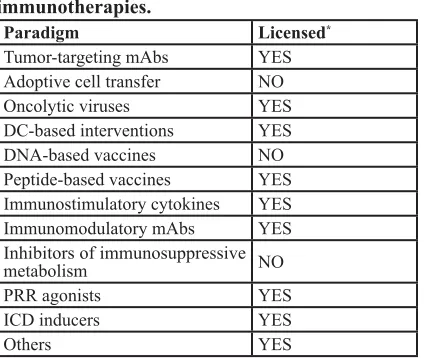
Table 1: Currently available anticancer
immunotherapies.
Paradigm Licensed*
Tumor-targeting mAbs YES
Adoptive cell transfer NO
Oncolytic viruses YES
DC-based interventions YES
DNA-based vaccines NO
Peptide-based vaccines YES
Immunostimulatory cytokines YES
Immunomodulatory mAbs YES
Inhibitors of immunosuppressive
metabolism NO
PRR agonists YES
ICD inducers YES
Others YES
[image:5.612.60.275.65.244.2]
fda.gov) [45, 46], demonstrating the extraordinary success
of this immunotherapeutic paradigm.
Adoptive cell transfer
[image:6.612.114.489.278.642.2]
The term “adoptive cell transfer” (ACT) refers to a
particular variant of cell-based anticancer immunotherapy
that generally involves: (1) the collection of circulating
or tumor-infiltrating lymphocytes; (2) their selection/
modification/expansion/activation
ex vivo
; and (3)
their (re-)administration to patients, most often after
lymphodepleting pre-conditioning and in combination
with immunostimulatory agents [23, 24, 78-80].
Other anticancer (immune)therapies involving the (re)
infusion of living cells, such as hematopoietic stem cell
transplantation (HSCT), conceptually differ from ACT.
ACT involves the (re-)introduction of a cell population
enriched in potentially tumor-reactive immune effectors
[23, 24, 81]. HSCT is employed as a means to reconstitute
a healthy, allogeneic (and hence potentially
tumor-reactive) immune system in patients with hematological
malignancies previously subjected to myelo- and
lymphoablating treatments (which aim at eradicating
the majority of neoplastic cells) [82]. Dendritic cell
(DC)-based interventions should also be conceptually
differentiated from ACT for two reasons. First, (re-)
infused DCs are not endowed with intrinsic anticancer
activity, but act as anticancer vaccines to elicit a
tumor-targeting immune response [83, 84]. Second, DCs are
not administered in the context of lympho/myeloablating
chemo(radio)therapy [85-87].
Several strategies have been devised to improve the
therapeutic potential of ACT [79, 80, 88]. For instance,
genetic engineering has been employed to endow
peripheral blood lymphocytes (PBLs) with features
such as a unique antigen specificity [89], an increased
proliferative potential and persistence
in vivo
[90-93],
[image:7.612.57.556.59.570.2]
Table 2: Anticancer immunotherapeutics currently approved by regulatory agencies worldwide.
Paradigm Agent Indication(s) Year* Proposed mechanism of action
Dendritic cell-based
immunotherapies Sipuleucel-T Prostate carcinoma 2010 Priming of a PAP-specific immune response
Immunogenic cell
death inducers
Bleomycin Multiple hematological and solid tumors <1995 DNA-damaging agent
Bortezomib Mantle cell lymphoma Multiple myeloma 2003 Proteasomal inhibitor
Cyclophosphamide Multiple hematological and solid tumors <1995 Alkylating agent
Doxorubicin Multiple hematological and solid tumors <1995 DNA-intercalating agent
Epirubicin Breast carcinoma 1999 DNA-intercalating agent
Mitoxantrone Acute myeloid leukemiaProstate carcinoma <1995 DNA-intercalating agent
Oxaliplatin Colorectal carcinoma 2002 DNA-damaging agent
Photodynamic
therapy Multiple hematological and solid tumors 1996 Induction of oxidative stress with damage to (intra)cellular membranes
Radiation therapy Multiple hematological and solid tumors <1995 DNA-damaging agent and oxidative stress inducer
Immunostimulatory
cytokines
IL-2 Melanoma Renal cell carcinoma <1995 Non-specific immunostimulation
IFN-α2a Chronic myeloid leukemia Hairy cell leukemia
Melanoma 1999 Non-specific immunostimulation
IFN-α2b Multiple hematological and solid tumors <1995 Non-specific immunostimulation
Immunomodulatory
mAbs
Ipilimumab Melanoma 2011 Blockage of CTLA4-dependent immunological checkpoints
Nivolumab Melanoma 2014 Blockage of PDCD1-dependent immunological checkpoints
Pembrolizumab Melanoma 2014 Blockage of PDCD1-dependent immunological checkpoints
Oncolytic viruses Oncorine H101 Head and neck cancer 2005 Selective lysis of malignant cells
Peptide-based
vaccines Vitespen Renal cell carcinoma 2008 Activation of a tumor-specific immune response
PRR agonists
Bacillus
Calmette-Guérin Non-invasive bladder transitional cell carcinoma <1995 TLR2/TLR4 agonist
Imiquimod Actinic keratosis Condylomata acuminata
Superficial basal cell carcinoma 1997 TLR7 agonist
Mifamurtide Osteosarcoma 2009 NOD2 agonist
Monophosphoryl
lipid A Prevention of HPV-associated cervical carcinoma 2009 TLR2/TLR4 agonist
Picibanil
Gastric carcinoma
Head and neck cancer
Lung carcinoma
Thyroid carcinoma
Tumor-targeting
mAbs
Alemtuzumab Chronic lymphocytic leukemia 2001 Selective recognition/opsonization of CD52+ neoplastic cells
Bevacizumab
Colorectal carcinoma
Glioblastoma multiforme
Cervical carcinoma
Lung carcinoma
Renal cell carcinoma
2004 VEGFA neutralization
Brentuximab
vedotin Anaplastic large cell lymphomaHodgkin's lymphoma 2011 Selective delivery of MMAE to CD30+ neoplastic cells
Blinatumumab Acute lymphoblastic leukemia 2014 CD3- and CD19-specific BiTE
Catumaxomab Malignant ascites in patientswith EPCAM+ cancer 2009 CD3- and EPCAM-specific BiTE
Cetuximab Head and neck cancerColorectal carcinoma 2004 Inhibition of EGFR signaling
Denosumab Breast carcinomaProstate carcinoma
Bone giant cell tumors 2011 Inhibition of RANKL signaling
Gemtuzumab
ozogamicin Acute myeloid leukemia 2000 Selective delivery of calicheamicin to CD33+ neoplastic cells
Ibritumomab
tiuxetan Non-Hodgkin lymphoma 2002 Selective delivery of
90Y or 111In
to CD20+ neoplastic cells
Panitumumab Colorectal carcinoma 2006 Inhibition of EGFR signaling
Pertuzumab Breast carcinoma 2012 Inhibition of HER2 signaling
Obinutuzumab Chronic lymphocytic leukemia 2013 Selective recognition/opsonization of CD20+ neoplastic cells
Ofatumumab Chronic lymphocytic leukemia 2009 Selective recognition/opsonization of CD20+ neoplastic cells
Ramucirumab Gastric or gastroesophageal junction adenocarcinoma 2014 Inhibition of KDR signaling
Rituximab Chronic lymphocytic leukemiaNon-Hodgkin lymphoma 1997 Selective recognition/opsonization of CD20+ neoplastic cells
Siltuximab Multicentric Castleman’s disease 2014 IL-6 neutralization
Tositumomab Non-Hodgkin lymphoma 2003 Selective recognition/opsonization of, or selective delivery of 90Y or
111In to, CD20+ neoplastic cells
Trastuzumab Breast carcinomaGastric or gastroesophageal
junction adenocarcinoma 1998
Selective recognition/opsonization
of, or selective delivery of mertansine
to, HER2+ cancer cells
Others
Lenalidomide Mantle cell lymphomaMyelodysplastic syndrome
Multiple myeloma 2005
IKZF degradation and
immunomodulation
Pomalidomide Multiple myeloma 2013 IKZF degradation and immunomodulation
Thalidomide Multiple myeloma 2006 IKZF degradation and immunomodulation
Trabectedin Soft tissue sarcomaOvarian carcinoma 2007 Reprogramming of tumor-associated macrophages
an improved secretory profile [91], an elevated
tumor-infiltrating capacity [94, 95], and superior cytotoxicity
[96]. The specificity of PBLs can be altered prior to
(re-)infusion by genetically modifying them to express:
(1) a TAA-specific T-cell receptor (TCR) [89, 97-99],
or (2) a so-called “chimeric antigen receptor” (CAR),
i.e., a transmembrane protein comprising the
TAA-binding domain of an immunoglobulin linked to one or
more immunostimulatory domains [100-106]. The latter
approach is advantageous in that it renders T cells capable
of recognizing (and hence potentially killing)
TAA-expressing cells in an MHC-independent fashion. Several
clinical trials have already demonstrated the therapeutic
potential of CAR-expressing T cells, in particular (but not
only) for patients affected by hematological malignancies
[102, 107-111]. T cells expressing TAA-specific TCRs
have also been shown to provide objective benefit to
cancer patients [89, 97-99]. Conversely, in spite of
promising preclinical findings [112-117], the adoptive
transfer of purified natural killer (NK) cells to cancer
patients has been associated with limited therapeutic
activity [118-120]. To the best of our knowledge, the
adoptive transfer of purified B lymphocytes has not yet
been investigated in the clinic [121], possibly because B
cells (or at least some subsets thereof) can exert potent
immunosuppressive effects [122-125]. Of note, no ACT
protocol is currently approved by the US FDA for use in
cancer patients (source http://www.fda.gov).
Since (re-)infused T cells are endowed with intrinsic
antineoplastic activity, ACT is generally considered as a
passive form of immunotherapy. However, the survival,
expansion, migration and cytotoxic activity of adoptively
transferred T cells rely on several cytokines, some of
which are supplied by the host immune system. Current
ACT protocols involve indeed the administration of
exogenous interleukins (ILs), including 2, 15 or
IL-21 [126-130], but these stimulate a cytokine cascade in the
host that sustains the survival and activity of adoptively
transferred cells. Thus, ACT may not represent a
bona fide
paradigm of passive immunotherapy.
Oncolytic viruses
The term “oncolytic viruses” refers to
non-pathogenic viral strains that specifically infect cancer cells,
triggering their demise [131-133]. Oncolytic viruses must
be conceptually differentiated from so-called “oncotropic
viruses”, i.e., viruses that exhibit a preferential tropism for
malignant cells but no (or very limited) cytotoxic activity
[134, 135]. The antineoplastic potential of oncolytic
viruses can be innate and simply originate from the
so-called cytopathic effect, i.e., the lethal overload of cellular
metabolism resulting from a productive viral infection
[136, 137]. As an alternative, these viruses can mediate an
oncolytic activity because of (endogenous or exogenous)
gene products that are potentially lethal for the host cell,
irrespective of their capacity to massively replicate and
cause a cytopathic effect [131, 132]. Of note, genetic
engineering has been successfully employed to endow
oncolytic virus with various advantageous traits, including
sequences coding for (1) enzymes that convert an
innocuous pro-drug into a cytotoxic agent [138-143]; (2)
proteins that (at least theoretically) trigger lethal signaling
cascades in cancer cells only [144-146]; or (3)
short-hairpin RNAs that target factors that are strictly required
for the survival of transformed, but not normal cells [147,
148]. Of note, no oncolytic virus has been approved by
the US FDA for use in cancer patients (source http://
www.fda.gov). Conversely, a recombinant adenovirus
(H101, commercialized under the name of Oncorine
®
)
has been approved by the regulatory authorities of the
People’s Republic of China for the treatment of HNC (in
combination with chemotherapy) as early as in November
2005 [149, 150].
As oncolytic viruses are endowed with intrinsic
anticancer activity, they are generally viewed as passive
immunotherapeutics. Moreover, several effectors of
innate and adaptive immunity limit the efficacy of
oncolytic therapy because they can neutralize viral
particles before they reach neoplastic lesions [131,
132, 151]. This is particularly true for the mononuclear
phagocytic system of the liver and spleen, which is able
to sequester large amounts of oncolytic viruses upon
injection [152, 153]; the complement system, to which
oncolytic viruses are particularly sensitive [154, 155];
and neutralizing antibodies, which can exist in patients
prior to oncolytic virotherapy owing to their exposure to
naturally occurring variants of the viral strains commonly
employed for this purpose [156, 157]. This being said,
accumulating preclinical and clinical evidence indicates
that the therapeutic activity of oncolytic viruses stems, for
the most part, from their ability to elicit tumor-targeting
immune responses as they promote the release of TAAs in
an immunostimulatory context. In support of this notion,
oncolytic viruses engineered to drive the expression of
co-stimulatory receptors [158-160] or immunostimulatory
cytokines/chemokines [161-165] reportedly mediate
superior antineoplastic effects as compared to their
unmodified counterparts [131, 132]. Thus, conventional
oncolytic viruses also appear to be active, rather than
passive, immunotherapeutics.
Active immunotherapy
DC-based immunotherapies
therapeutically relevant anticancer immune responses
[168]. Several forms of DC-based immunotherapy have
been developed, most of which involve the isolation of
patient- or donor-derived circulating monocytes and their
amplification/differentiation
ex vivo
, invariably in the
presence of agents that promote DC maturation, such
as granulocyte macrophage colony-stimulating factor
(GM-CSF) [28]. This is particularly important because
immature DCs exert immunosuppressive, rather than
immunostimulatory, functions [169-171]. Most often,
autologous DCs are re-infused into cancer patients
upon exposure to a source of TAAs, including (1)
TAA-derived peptides [172-175]; (2) mRNAs coding for one
or more specific TAAs [176]; (3) expression vectors
coding for one or more specific TAAs [177-180]; (4) bulk
cancer cell lysates (of either autologous or heterologous
derivation) [181-186]; (5) or bulk cancer cell-derived
mRNA [187-191]. As an alternative, DCs are allowed to
fuse
ex vivo
with inactivated cancer cells, generating
so-called dendritomes [192-197]. The rationale behind all
these approaches is that DCs become loaded
ex vivo
with
TAAs or TAA-coding molecules, hence becoming able to
prime TAA-targeting immune responses upon reinfusion.
Additional DC-based anticancer immunotherapies
include the targeting of specific TAAs to DCs
in vivo
[169, 198-205], the use of DC-derived exosomes
[206-208], and the (re-)administration of autologous
or allogeneic DCs amplified, matured and optionally
genetically modified
ex vivo
, but not loaded with TAAs
[209-214]. In the former setting, TAAs are fused to
mAbs, polypeptides or carbohydrates that selectively
bind to DCs [169, 198-202, 215, 216], encapsulated
in DC-targeting immunoliposomes [217, 218], or (3)
encoded by DC-specific vectors [219-221]. In the latter
scenarios, DCs or their exosomes are administered as a
relatively non-specific immunostimulatory intervention
[209-213]. Interestingly, one cellular product containing
a significant proportion of (partially immature) DCs is
currently licensed for use in cancer patients, namely
sipuleucel-T (also known as Provenge
®
) (source http://
www.fda.gov). Sipuleucel-T has been approved by the
US FDA and the EMA for the therapy of asymptomatic
or minimally symptomatic metastatic castration-refractory
prostate cancer as early as in 2010 [222-224]. However,
the manufacturer of sipuleucel-T, Dendreon Co. (Seattle,
WA, US), filed for bankruptcy in November 2014 (source
http://dealbook.nytimes.com/2014/11/10/dendreon-maker-of-prostate-cancer-drug-provenge-files-for-bankruptcy/?_
r=0). This reflects the disadvantageous cost-benefit ratio
of such a cellular therapy, whose preparation requires a
relatively elevated quantity of each patient’s peripheral
blood mononuclear cells [25, 222, 223]. The safety and
efficacy of many DC-based cellular preparations other
than are sipuleucel-T are currently being investigated in
clinical settings, with promising results [225].
Although DCs isolated from cancer patients have
been shown to exert cytotoxic activity against malignant
cells [226], DC-based immunotherapies mediate
antineoplastic effects mainly because they engage the
host immune system against malignant lesions [227, 228].
Thus, all forms of DC-based anticancer interventions
constitute paradigms of active immunotherapy.
Peptide- and DNA-based anticancer vaccines
DCs and other antigen-presenting cells (APCs)
are also targeted by peptide- and DNA-based anticancer
vaccines [83, 84, 229-231]. In the former scenario,
full-length recombinant TAAs or peptides thereof are
administered to cancer patients, most often via the
intramuscular, subcutaneous or intradermal route, together
with one or more immunostimulatory agents commonly
known as adjuvants (which potently promote DC
maturation) [232-237]. The rationale behind this approach
is that resident DCs (or other APCs) acquire the ability to
present the TAA-derived epitopes while maturing, hence
priming a robust TAA-specific immune response [32,
238, 239]. The mechanisms underlying the priming of
anticancer immune responses by peptide-based vaccines,
and hence their efficacy, depend (at least in part) on their
size [38]. Thus, while short peptides (8-12 amino acids) are
conceived to directly bind to MHC molecules expressed
on the surface of APCs, synthetic long peptides (25-30
residues) must be taken up, processed and presented by
APCs for eliciting an immune response [38]. Normally, the
therapeutic activity of synthetic long peptides is superior
to that of their short counterparts, especially when they
include epitopes recognized by both cytotoxic and helper
T cells or when conjugated to efficient adjuvants [38, 240,
241]. This said, some commonly used immunostimulants
such as the so-called incomplete Freund’s adjuvant (IFA)
have recently been shown to limit the efficacy of
peptide-based anticancer vaccination [242], calling for the use of
alternative immunostimulants. A peculiar type of
peptide-based vaccines is constituted by autologous tumor lysates
complexed with immunostimulatory chaperones, most
often members of the heat-shock protein (HSP) family
[243]. This approach is advantageous in that it does not
rely on a single TAA but (at least hypothetically) on all
TAAs that bind to HSPs (including patient-specific
neo-TAAs) [243]. However, generating anticancer vaccines on
a personalized basis is associated with considerable costs
[243].
249]. A particularly interesting approach in this context
is represented by so-called “oncolytic vaccines”, i.e.,
oncolytic viruses genetically altered to code for a TAA
[250-252]. Promising results have also been obtained
with DNA-based vaccines administered
per os
[253-256].
In this setting, live-attenuated bacteria expressing a
full-length TAA are taken up by APCs in the intestinal mucosa,
resulting in the priming of a robust, TAA-specific immune
response in the so-called “mucosa-associated lymphoid
tissue” [253-256].
Both peptide- and DNA-based vaccines have been
associated with clinical activity in patients affected by
various neoplasms [83, 84, 229-231, 257]. For instance, a
peptide-based vaccine targeting the human papillomavirus
type 16 (HPV-16) proteins E6 and E7 have been shown to
promote complete, long-lasting responses in a significant
fraction of patients with vulvar intraepithelial neoplasia
[258]. Along similar lines, the administration of a
multipeptide vaccine after single-dose cyclophosphamide
(an immunogenic alkylating agent, see below) has been
shown to prolong overall survival in a cohort of RCC
patients [259]. No peptide- or DNA-based anticancer
vaccine is currently approved by the US FDA and EMA
for use in humans (sources http://www.fda.gov and
http://www.ema.europa.eu/ema/). However, vitespen
(Oncophage
®
), a heat shock protein 90kDa beta (Grp94),
member 1 (HSP90B1)-based anticancer vaccine, has been
approved in Russia for the treatment of RCC patients with
intermediate risk of recurrence as early as in 2008 [257].
Moreover, three DNA-based anticancer vaccines have
been licensed for veterinary use [260-263], one of which
relies on a human TAA (
i.e.
, tyrosinase) [263].
Similar to DC-based interventions, both peptide- and
DNA-based anticancer vaccines mediate antineoplastic
effects as they (re-)activate the host immune system
against malignant cells, hence constituting active forms of
anticancer immunotherapy.
Immunostimulatory cytokines
Taken as a family, cytokines regulate (via autocrine,
paracrine or endocrine circuits) virtually all biological
functions [264-267]. It is therefore not surprising that
various attempts have been made to harness the biological
potency of specific cytokines to elicit novel or reinvigorate
pre-existent tumor-targeting immune responses [268-271].
The administration of most immunostimulatory cytokines
to cancer patients as standalone therapeutic interventions,
however, is generally associated with little, if any, clinical
activity [272-275]. Thus, immunostimulatory cytokines
are generally employed as adjuvants for other anticancer
(immuno)therapeutics, either as recombinant molecules
or encoded within expression vectors [276-284]. Notable
exceptions include interferon (IFN)-α2b (also known
as Intron A
®
), and IL-2 (also known as aldesleukin and
Proleukin
®
), which mediate single agent therapeutic
activity in patients affected by melanoma, a tumor type
particularly sensitive to immunotherapy [274, 284].
IFN-α2b is currently approved by the US FDA and EMA
for the therapy of hairy cell leukemia (HCL),
AIDS-related Kaposi’s sarcoma, follicular lymphoma, multiple
myeloma, melanoma, external genital/perianal warts
(
condylomata acuminata
) and cervical intraepithelial
neoplasms (both as a recombinant, unmodified protein,
and as a pegylated variant), while IL-2 is licensed for
the treatment of metastatic forms of melanoma and
RCC. Moreover, IFN-α2a (also known as Roferon-A
®
)
is approved for use in subjects with HCL and chronic
phase, Philadelphia chromosome-positive chronic myeloid
leukemia, upon minimal pretreatment (within 1 year of
diagnosis). In Europe, IFN-α2a is also licensed for the
treatment of melanoma. Of note, GM-CSF (also known
as molgramostim, sargramostim, Leukomax
®
, Mielogen
®
or Leukine
®
) and granulocyte colony-stimulating
factor (G-CSF, also known as filgrastim, lenograstim or
Neupogen
®
) are approved by the US FDA and EMA for
use in humans, but not as part of anticancer regimens
[285-288]. Nonetheless, GM-CSF has been shown to potentiate
the clinical activity of several immunotherapeutics,
including (but not limited to) peptide-based vaccines
and immunomodulatory mAbs [259, 289]. Recombinant
tumor necrosis factor α (TNFα) is also licensed by several
regulatory agencies worldwide (but not by the US FDA),
for the treatment of limb-threatening soft tissue sarcoma
and melanoma [290-292]. However, in this setting
TNFα is not employed as an immunostimulatory agent
but administered in combination with melphalan (an
alkylating agent) to increment the local concentration of
the drug (and hence boost its cytotoxicity), and to promote
the selective destruction of the tumor vasculature [293].
The antineoplastic activity of immunostimulatory
cytokines is expected to depend on the host immune
system, implying that they underlie a
bona fide
paradigm
of active immunotherapy. However, the actual mode of
action of immunostimulatory cytokines has not yet been
fully explored. Moreover, some of these agents may
promote a cytokine cascade with unwarranted, potentially
lethal effects, and hence should be employed with caution.
Immunomodulatory mAbs
receptor (KIR) family [302-304]; (2) the inhibition of
the principal ligands of these receptors, such as the PD-1
ligand CD274 (best known as PD-L1 or B7-H1) [300,
305-307]; (3) the activation of co-stimulatory receptors
expressed on the surface of immune effector cells [308]
such as tumor necrosis factor receptor superfamily,
member 4 (TNFRSF4, best known as OX40)
[309-313], TNFRSF9 (best known as CD137 or 4-1BB) [58,
314, 315], and TNFRSF18 (best known as GITR)
[316-318]; and (4) the neutralization of immunosuppressive
factors released in the tumor microenvironment, such as
transforming growth factor β1 (TGFβ1) [319, 320].
The first of these approaches, which is commonly
referred to as “checkpoint blockade”, has been shown to
induce robust and durable responses in cohorts of patients
with a variety of solid tumors [39, 300, 321-327]. As it
stands, no less than three checkpoint-blocking mAbs are
currently approved by international regulatory agencies for
use in humans (source http://www.fda.gov): (1) the
anti-CTLA4 mAb ipilimumab (Yervoy™), which was licensed
by the US FDA for use in individuals with unresectable
or metastatic melanoma on 2011, March 25
th
[328-332];
the anti-PD-1 mAb pembrolizumab (Keytruda™), which
received accelerated approval by the US FDA for the
treatment of advanced or unresectable melanoma patients
who fail to respond to other therapies on 2014, September
4
th
[333-338]; and nivolumab (Opvido™), another
PD-1-targeting mAb licensed by the Japanese Ministry of Health
and Welfare for use in humans on 2014, July 07
th
[339].
Based on the results of a recently completed Phase III
clinical trial demonstrating that nivolumab significantly
improves the progression-free and overall survival of
patients with BRAF
WT
melanoma [340], the approval of
this mAb by the US FDA is expected within the next
few months. The safety and efficacy of ipilimumab,
pembrolizumab, nivolumab and other checkpoint-blocking
mAbs are being demonstrated in a steadily expanding
panel of oncological indications [45, 46, 341, 342]. Of
note, some co-stimulatory mAbs including urelumab
and PF-0582566 (both of which target CD137) are also
under clinical development, with promising results [46,
341]. Preclinical data suggest that combining checkpoint
blockers with co-stimulatory mAb mediates superior
antineoplastic effects [294, 343, 344]. At least in part, this
reflects the ability of co-stimulatory mAbs to promote NK
cell functions [58, 345, 346]. In line with this notion, a few
clinical trials testing checkpoint blockers in combination
with urelumab or lirilumab (a KIR-inhibiting mAb) have
just been initiated (source http://www.clinicaltrials.gov).
Designed to (re-)activate the host immune system
against malignant cells, immunomodulatory mAbs
constitute an established and clinically promising
paradigm of active immunotherapy. Interestingly, despite
their non-specific mechanism of action, the clinical
efficacy of immunomodulatory mAbs (and in particular
checkpoint blockers) may be profoundly influenced by the
panel of (neo-)TAAs specific to each neoplasm [347].
Inhibitors of immunosuppressive metabolism
Indoleamine 2,3-dioxigenase 1 (IDO1) catalyzes
the first, rate-limiting step in the so-called “kynurenine
pathway”, the catabolic cascade that converts
L
-tryptophan (Trp) into
L
-kynurenine (Kyn) [348].
Although this enzyme was initially believed to mediate
immunostimulatory effects (partly because inflammatory
cues including IFNγ promote its expression in cells of the
innate immune system) [349, 350], IDO1 mediates robust
immunosuppressive effects, in both physiological (
e.g.
,
tolerance during pregnancy) and pathological (mostly
oncological) settings [351-356]. IDO1 has been proposed
to inhibit both innate and adaptive immune responses (1)
by depleting immune effector cells of Trp, resulting in
irresponsiveness to immunological challenges [352, 353,
357-359]; (2) by favoring the accumulation of Kyn and
some of its derivatives, which exert cytotoxic effects on
immune effector cells while promoting the differentiation
of Tregs [360-364]; or (3) through various indirect
mechanisms mediated by IDO1-expressing DCs [124,
365-371]. Evidence accumulated during the last decade
indicates that both 1-methyltryptophan (an inhibitor
of IDO1 and IDO2) and genetic interventions targeting
IDO1 mediate antineoplastic effects while eliciting novel
or reinvigorating existent anticancer immune responses
[372-375]. No IDO1 inhibitor is currently approved by
the US FDA for use in humans (source http://www.fda.
gov). However, the results of recent Phase I-II studies
suggest that 1-methyl-
D
-tryptophan (an inhibitor of
the IDO pathway also known as indoximod), other
pharmacological blockers of IDO1 (such as INCB024360),
and IDO1-targeting vaccines are well tolerated by cancer
patients and mediate antineoplastic effects, at least in a
subset of individuals [376-382].
or inhibit adenosine receptors [392, 397]. Preclinical
evidence indicates that CD39- or CD79-targeting
agents (mostly mAbs) mediate antineoplastic effects as
standalone interventions and improve the efficacy of other
anticancer agents [397]. The clinical development of these
agents, however, has not yet been initiated. Conversely,
ADORA2A antagonists are currently being tested in
late-stage clinical trials, but as a therapeutic option against
Parkinsonism [397]. It will be interesting to determine the
safety and efficacy of inhibitors of adenosine generation or
signaling in cancer patients.
Although it remains unclear whether these agents
truly operate by altering the microenvironmental
availability of Trp and Kyn [398], the antineoplastic
effects of IDO inhibitors critically rely on the host immune
system, implying that this constitutes an instance of active
anticancer immunotherapy [399]. This also applies to
strategies aimed at limiting the extracellular availability
of adenosine.
PRR agonists
Pattern recognition receptors (PRRs) are
evolutionarily conserved proteins involved in the
recognition of danger signals [400, 401]. PRRs include
(but are not limited to) Toll-like receptors (TLRs) [402,
403] and nucleotide-binding oligomerization domain
containing (NOD)-like receptors (NLRs) [404, 405].
TLRs are transmembrane enzymatically-inactive
proteins expressed by most APCs, including monocytes,
macrophages and DCs, as well as by some types of
epithelial cells [402, 403]. NLRs are expressed by a
variety of cell types, including various components of the
innate and adaptive immune system [404, 405]. Taken
together, PRRs sense a wide panel of danger signals,
including exogenous “microbe-associated molecular
patterns” (MAMPs) like bacterial lipopolysaccharide
(LPS) or muramyl dipeptide (MDP), and endogenous
“damage-associated molecular patterns” (DAMPs), like
the non-histone nuclear protein high-mobility group box
1 (HMGB1) and mitochondrial DNA [406-410]. The
activation of various PRRs ignites a signal transduction
cascade with potent pro-inflammatory outcomes, including
the activation of NF-κB [411-413], and the secretion of
immunostimulatory cytokines, like type I IFNs and TNFα
[413-415]. Moreover, PRR signaling favors the maturation
of DCs as well as the activation of macrophages and
NK cells [416]. Besides being critical for the response
of the host to viral and bacterial challenges [402, 403],
some PRRs play a key role in the (re)activation of
anticancer immune responses by chemo-, radio- and
immunotherapeutic interventions [15, 413, 417-422].
Thus, PRR agonists have spurred interest not
only as adjuvants for conventional vaccines [423,
424], but also as immunotherapeutic interventions that
may mediate antineoplastic effects
per se
or boost the
therapeutic activity of other anticancer agents [34, 48,
425]. Three TLR agonists are approved by the US FDA
for use in cancer patients: (1) the bacillus
Calmette-Guérin (BCG), an attenuated variant of
Mycobacterium
bovis
that presumably operates as a mixed TLR2/
TLR4 agonist, which is currently used as a standalone
immunotherapeutic agent in subjects with non-invasive
transitional cell carcinoma of the bladder [426]; (2)
monophosphoryl lipid A (MPL), a TLR2/TLR4-activating
derivative of
Salmonella minnesota
LPS currently
utilized as adjuvant in Cervarix
®
, a vaccine for the
prevention of HPV-16 and -18 infection [427]; and (3)
imiquimod, an imidazoquinoline derivative that triggers
TLR7 signaling, currently employed for the treatment
of actinic keratosis, superficial basal cell carcinoma and
condylomata acuminata
[422, 426]. Of note, picibanil
(a lyophilized preparation of
Streptococcus pyogenes
that operates as a TLR2/TLR4 agonist has been licensed
for use in cancer patients by the Japanese Ministry of
Health and Welfare (but not by the US FDA) as early
as in 1975 [428, 429]; while mifamurtide (a synthetic
lipophilic glycopeptide that activates NOD2) has been
approved by the EMA for the treatment of osteosarcoma
in 2009 [430-432]. Moreover, the safety and efficacy of
several other PRR agonists are currently being evaluated
in clinical trials [433-435]. These molecules include
agatolimod (CpG-7909, PF-3512676, Promune
®
), an
unmethylated CpG oligodeoxynucleotide that activates
TLR9 [436]; polyriboinosinic polyribocytidylic acid
(polyI:C, Ampligen™, Rintatolimod), a synthetic
double-strand RNA that signals via TLR3 [437]; and Hiltonol™,
a particular formulation of polyI:C that involves
carboxymethylcellulose and poly-
L
-lysine [48, 438].
Some malignant cells express PRRs [439-445],
implying that PRR agonists may not be completely devoid
of intrinsic tumor-modulating functions. Nonetheless, a
large body of preclinical and clinical literature indicates
that the antineoplastic effects of PRR agonists stem from
their ability to engage the host immune system. Thus, PRR
agonists constitute active immunotherapeutics.
Immunogenic cell death inducers
optimal antineoplastic effects in immunocompetent, but
not in immunodeficient, mice [15, 451-454]. However, the
ability of a specific stimulus to trigger ICD can be properly
assessed only by means of vaccination experiments
involving immunocompetent mice and syngeneic tumor
models [15, 455]. As it stands, a few FDA-approved
therapies have been shown to constitute
bona fide
ICD
inducers, including: doxorubicin, mitoxantrone and
epirubicin (three anthracyclines currently employed
against various carcinomas) [186, 449], bleomycin (a
glycopeptide antibiotic endowed with antineoplastic
properties) [456], oxaliplatin (a platinum derivative
generally used for the therapy of colorectal carcinoma)
[453, 457], cyclophosphamide (an alkylating agent
employed against neoplastic and autoimmune conditions)
[458-460], specific forms of radiation therapy [419,
461-466], photodynamic therapy (an intervention that relies
on the administration of a photosensitizing agent coupled
to light irradiation) [448, 467, 468], and bortezomib (a
proteasomal inhibitor used for the treatment of multiple
myeloma) [469, 470].
These and other (hitherto experimental) ICD
inducers have been viewed as conventional forms
of anticancer therapy, exerting antineoplastic effects
via cytostatic or cytotoxic mechanisms. However,
accumulating evidence indicates that the full-blown
therapeutic potential of these molecules relies on the
host immune system [15, 471]. Thus, we propose to
classify ICD inducers as a form of active anticancer
immunotherapy.
Others
Other anticancer immunotherapies are approved by
regulatory agencies worldwide for use in cancer patients
or are currently being investigated for safety and efficacy
in preclinical or clinical settings.
Lenalidomide (Revlimid
®
, also known as
CC-5013) and pomalidomide (Pomalyst
®
, also known as
CC-4047) are two derivatives of thalidomide (Thalomid
®
)
originally developed in the 1990s to achieve improved
potency in the absence of significant side effects [472].
Thalidomide was indeed marketed as an
over-the-counter sedative, tranquilizer, and antiemetic for morning
sickness in various countries in the late 1950s, but was
rapidly withdrawn following a peak of infants born with
malformation of the limbs [473]. In spite of its pronounced
teratogenic activity, thalidomide raised renewed interest
as an inhibitor of TNFα secretion in the 1990s [474], and
was approved by the US FDA (under a strictly controlled
distribution program) for the therapy of erythema nodosum
leprosum (a complication of leprosy etiologically linked
to TNFα) in 1998 [475]. The combination of thalidomide
with dexamethasone (a glucocorticoid) rapidly turned
out to mediate therapeutic effects in patients with
hematological malignancies, eventually resulting in the
approval by the US FDA of this regimen for the treatment
of newly diagnosed multiple myeloma [476]. Alongside,
lenalidomide (which retains some degree of teratogenicity)
was licensed for use in patients with multiple myeloma
(also in combination with dexamethasone) and low or
intermediate-1 risk myelodysplastic syndromes that harbor
5q cytogenetic abnormalities (as a standalone intervention)
[477-480]. Conversely, pomalidomide (which is devoid of
teratogenic activity) has been approved for use in multiple
myeloma patients only in 2013, when the approval of
lenalidomide has been extended to mantle cell lymphoma
(MCL) [481-483]. Although the effects of thalidomide,
lenalidomide and pomalidomide, which are collectively
referred to as “immunomodulatory drugs” (IMiDs),
on the immune system have been characterized with
increasing precision throughout the past two decades
[484], the underlying molecular mechanisms remained
obscure [485]. Recent findings indicate that the therapeutic
activity of IMiDs depend, at least in part, on their ability
to bind the E3 ubiquitin ligase cereblon (CRBN) and
hence boost the proteasomal degradation of the B
cell-specific transcription factors IKAROS family zinc finger
1 (IKZF1) and IKZF3 [486, 487]. Of note, CRBN, which
is also involved in the teratogenic effects of thalidomide
and lenalidomide [488], regulates the abundance of
interferon regulatory factor 4, perhaps accounting for the
immunomodulatory functions of IMiDs [489]. Although
endowed with intrinsic antineoplastic activity, IMiDs
should be considered active immunotherapeutics.
As they progress and respond to treatment,
neoplastic lesions are infiltrated by a significant amount
of lymphoid and myeloid cells, including CD8
+
T
lymphocytes, Tregs, tumor-associated macrophages
(TAMs) and immunosuppressive B-cell populations
[122-124, 490, 491]. Robust tumor infiltration by CD8
+
T lymphocytes is generally associated with a good
prognosis, especially when the intratumoral levels of
Tregs are limited [124, 492]. Along similar lines, high
intratumoral levels of TAMs with a “classically-activated”
M1 phenotype (which exert tumoricidal functions,
stimulate NK cells and secrete T
H1-polarizing cytokines)
generally correlate with improved disease outcome [491,
493]. The contrary holds true when the myeloid tumor
infiltrate contains high levels of “alternatively-activated”
M2 TAMs or specific B-cell subsets, which can secrete
not only immunosuppressive cytokines like IL-10 and
TGFβ1, but also angiogenic mediators such as VEGFA
and enzymes that remodel the extracellular matrix [491,
493]. These observations prompted the development of
immunotherapeutic regimens based on the depletion/
inhibition of Tregs or B lymphocytes, as well as on the
conversion of M2 TAMs to their M1 counterparts.