International Journal of Emerging Technology and Advanced Engineering Website: www.ijetae.com (ISSN 2250-2459, Volume 2, Issue 5, May 2012)
334
Elemental Analysis of selected epidermal creams by X-ray
Fluorescence (XRF) Spectrometry
Margaret A. Briggs-Kamara
Department of Pure and Applied Physics, Veritas University Abuja, Obehie Campus, P.O.ox. 7084, Aba, Abia State, Nigeria
Abstract -X-ray Fluorescence Spectrometry (XRF) was used to determine the elemental composition of epidermal cosmetic creams. These creams are usually used by dark-skinned persons to lighten their skins. This research was undertaken with a view of assessing the health implications associated with the use of these creams. Eight of such creams were selected using the Questionnaire Method and then analyzed. The analysis of eighteen elements (Ca, Ti, Cr, Mn, Fe, Co, Ni, Cu, Zn, As, Br, Rb, Sr, Y, Zr, Nb, Mo and Pb) showed that only one element, titanium (Ti) was detected in two of the creams (Movate and Neutrotone) with concentrations of 8.986 ± 2.601 µg.cm-3 and 10.002 ± 2.749 µg.cm-3 respectively.
Keywords: elemental composition, epidermal cosmetic creams, health hazard, skin colour, X-ray Fluorescence Spectrometry (XRF)
I. INTRODUCTION
Bleaching, fade, skin-toning and skin-lightening are the common names given to epidermal creams. These creams are used principally to change the natural skin colour of the user from dark to a much lighter shade. The active ingredients in these creams include Allantoin; betamethasone 17, 21 dipropionate; clobetasol propionate; DEA (N,N-diethylaniline; hydroquinone, imidazolidnyl urea; liposomes; octyl dimethyl PABA; octyl salicylate; and sodium lauryl sulfate. The use of these creams for purposes other than short-term therapy is hazardous to health [1].
The colour of the human skin is determined by melanin, a pigment manufactured by specific dendritic cells called melanocytes. These cells reside below or between the basal cells of the epidermis.
The quality rather than the quantity of these cells determine the intensity of pigmentation of skin. Pigment production by melanocyte cells is a complex biological process. The copper-containing metallo-enzyme-tyrosinase system is influences not only by metabolic and hormonal changes in the body but also by local disease in the skin, especially inflammation. The normal stimulus to the enzyme is ultraviolet light of wavelength 290 to 315nm, producing suntan. The response is greatly increased by ingestion or local application of a group of chemicals called psolarens, which are found in many plant products and are now made synthetically. Inhibiting the tyrosine system with substances such as hydroquinones, 4-isopropylcaatechol and p-tertiary butyl-phenol can produce depigmentation [2]. Hence Caucasians can be made as black as Negroes by injections of a melanocyte-stimulating hormone [3], and Negroes can be made as white as albinos with hydroquinone; but there are hazards associated with such procedures.
Health implications of using epidermal creams on a long-term basis and for purposes other than prescribed and/or directed are:
Skin becomes susceptible to infection due to thinning of its protective barrier. Reduction in sensitivity of the blood
International Journal of Emerging Technology and Advanced Engineering Website: www.ijetae.com (ISSN 2250-2459, Volume 2, Issue 5, May 2012)
335
Abnormal increase in the rate of division of living cells leading to cancer.
Hampering of the healing capability of the skin.
Reduction in the skin’s capability to absorb and radiate heat.
The process of emissions of characteristic x-rays is called "X-ray Fluorescence," or XRF. When a primary x-ray excitation source from an x-ray tube or a radioactive source strikes a sample, the x-ray can either be absorbed by the atom or scattered through the material. The process in which an x-ray is absorbed by the atom by transferring all of its energy to an innermost electron is called the "photoelectric effect." During this process, if the primary x-ray had sufficient energy, electrons are ejected from the inner shells, creating vacancies. These vacancies present an unstable condition for the atom. As the atom returns to its stable condition, electrons from the outer shells are transferred to the inner shells and in the process give off a characteristic x-ray whose energy is the difference between the two binding energies of the corresponding shells. Because each element has a unique set of energy levels, each element produces x-rays at a unique set of energies, allowing one to non-destructively measure the elemental composition of a sample. Sometimes, as the atom returns to its stable condition, instead of emitting a characteristic x-ray it transfers the excitation energy directly to one of the outer electrons, causing it to be ejected from the atom. The ejected electron is called an "Auger" electron. This process is a competing process to XRF. Auger electrons are more probable in the low Z elements than in the high Z elements [4]. Analysis using x-ray fluorescence is called "X-ray Fluorescence Spectroscopy." In most cases the innermost K and L shells are involved in XRF detection.
A typical x-ray spectrum from an irradiated sample will display multiple peaks of different intensities (see Fig. 1).
The X-ray spectrum is taken using a semi-conductor detector usually a lithium-drifted silicon [Si (Li)] detector. The Si (Li) detector is a type of junction detector fabricated by applying a reverse bias to a pn junction consisting of a lithium-diffused n-type region on p-type silicon, causing the lithium ions to migrate toward the negative side and to compensate negative ions, fixed in the silicon crystal lattice, to create a wide depletion layer.
Fig.1: Spectrum taken using Amptek XR-100CR 25mm2X500µm
X-Ray Detector (20µs shaping time) and Amptek MCA8000A [5]
To maintain the width of the depletion layer the Si (Li) detector is usually kept at liquid nitrogen temperature of 77 K (-196 oC).
International Journal of Emerging Technology and Advanced Engineering Website: www.ijetae.com (ISSN 2250-2459, Volume 2, Issue 5, May 2012)
336
This work seeks to investigate the presence of any toxic and/or carcinogenic elements in some selected epidermal creams using X-ray Fluorescence (XRF) Spectrometry. This is one of the reliable and powerful analytical methods for trace element analysis, allowing the determination of many elements with good accuracy and low detection limits. Other advantages are the minimal sample handling and treatment before irradiation (minimizing risk of contamination), and very small sample quantities needed for the analysis. In most sample matrices, XRF can detect elements at concentration levels of less than one microgramme per gramme (1 ppm) of sample, or total amounts of a few tenths of one microgramme in a thin film sample [7]. In an earlier work using Instrumental Neutron Activation Analysis [8] all the creams were found to have traces of Al and Na. Eight of the elements (Zn, Hg, Al, V, Ba, Mn, Sb and Co) were found to be listed under priority chemicals recognized as hazardous to humans by the United States Environmental Protection Authority [9].
II. MATERIALS AND METHOD
For the choice of creams the Questionnaire method of research was adopted because of the statistical nature of the problem. The respondents were mostly fair-skinned ladies (University students and workers). Eight creams were identified and purchased from licensed cosmetic shops.
The XRF method used an Energy Dispersive XRF (EDXRF) system at the Centre for Energy Research and Training of the Ahmadu Bello University, Zaria (Nigeria) which consisted of a 925 MBq 109Cd annular isotopic source with Canberra Si (Li) detector having a resolution of 170 eV at 5.9 keV.
The Si (Li) detector was mounted at a sufficiently large angle to the direction of the exciting photons so that it can be effectively shielded against direct source radiation and the secondary radiation induced in assembly components other then the sample. Sample preparation was quite simple. Since the samples were creams, a thin film of each specimen was placed on a glass slide and then analyzed. Each cream was given a code number viz.: IS2068, IS2069, IS2070, IS2071, IS2072, IS2073, IS2074, and IS2075. A thin film of each of the samples was placed on separate glass slides and irradiated with 125I and 55Fe sources. Both sources decay by electron capture (EC). The 55Fe source was for determining low atomic-number elements (Na to Cr) using K-lines, and the 125I source was for determining elements from Cr to Mo (using K-lines) and of Pb and U (using L-lines). A vacuum system was incorporated in the experimental set-up when the
55
Fe source was used. This was to prevent interference lines from X-ray scattering by light elements and to reduce the background.
The spectra were collected by a computer basedacquisition system (ACCUSPEC). The AXIL® program was used for both spectral analysis and concentration determination.
The X-ray spectra were acquired with a computer based Multi-Channel Analyzer (MCA) card (Trump 8k). The setup provided for dead-time correction and pile-up rejection. Sensitivity calibration of the system was performed using thick foils of pure metals (Ti, Fe, Co, Ni, Cu, Zn, Zr, Nb, Mo, Sn, Ta, Pb) and stable chemical compounds (KCO3, CaCO3, Ce2O3, WO3, ThO2, U3O8).
International Journal of Emerging Technology and Advanced Engineering Website: www.ijetae.com (ISSN 2250-2459, Volume 2, Issue 5, May 2012)
337
depending on the elements to be analyzed and the composition of the samples.
III. RESULTS AND DISCUSSION
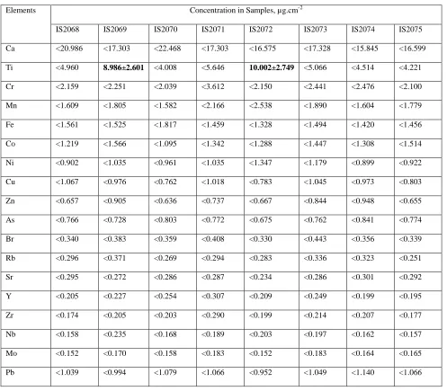
Eighteen elements (Ca, Ti, Cr, Mn, Fe, Co, Ni, Cu, Zn, As, Br, Rb, Sr, Y, Zr, Nb, Mo and Pb) were analyzed but only one element, titanium (Ti) was detected in two of the eight samples. All the other elements were below the detection limit. The two samples were Movate (IS2069) and Neutrotone (IS2072) having Ti concentrations of 8.986 ± 2.601 µg.cm-3 and 10.002 ± 2.749 µg.cm-3 respectively. The other epidermal creams selected for the analysis were: Tempovate cream (IS2068), Clear Essence (Maxitone) cream (IS2070), Tura (skin toning) cream (IS2071), Neu Clear cream (IS2073), Skin Success cream (IS2074), and A3 (triple action) cream (IS2075).
Table I shows the elemental concentrations of the samples analyzed. Fig. 2 is a graphical representation showing an exponential
relationship between the concentration and the atomic number of the elements.
The significance of the Ti levels found in this study lies not on the absolute values so determined but on the frequency and quantities applied to the skin each day (or regularly). The fact that most of the elements were below the detection limits implied that exposure time may have to be greatly increased, or alternatively, very large quantities of the creams may have to be used.
IV. CONCLUSION
International Journal of Emerging Technology and Advanced Engineering Website: www.ijetae.com (ISSN 2250-2459, Volume 2, Issue 5, May 2012)
[image:5.612.44.541.180.615.2]338
Table 1: Analyzed elements/concentrations using X-ray Fluorescence Spectrometry
Elements Concentration in Samples, µg.cm-2
IS2068 IS2069 IS2070 IS2071 IS2072 IS2073 IS2074 IS2075
Ca <20.986 <17.303 <22.468 <17.303 <16.575 <17.328 <15.845 <16.599
Ti <4.960 8.986±2.601 <4.008 <5.646 10.002±2.749 <5.066 <4.514 <4.221
Cr <2.159 <2.251 <2.039 <3.612 <2.150 <2.441 <2.476 <2.100
Mn <1.609 <1.805 <1.582 <2.166 <2.538 <1.890 <1.604 <1.779
Fe <1.561 <1.525 <1.817 <1.459 <1.328 <1.494 <1.420 <1.456
Co <1.219 <1.566 <1.095 <1.342 <1.288 <1.447 <1.308 <1.514
Ni <0.902 <1.035 <0.961 <1.035 <1.347 <1.179 <0.899 <0.922
Cu <1.067 <0.976 <0.762 <1.018 <0.783 <1.045 <0.973 <0.803
Zn <0.657 <0.905 <0.636 <0.737 <0.667 <0.844 <0.948 <0.655
As <0.766 <0.728 <0.803 <0.772 <0.675 <0.762 <0.841 <0.774
Br <0.340 <0.383 <0.359 <0.408 <0.330 <0.443 <0.356 <0.339
Rb <0.296 <0.371 <0.269 <0.294 <0.283 <0.336 <0.323 <0.251
Sr <0.295 <0.272 <0.286 <0.287 <0.234 <0.286 <0.301 <0.292
Y <0.205 <0.227 <0.254 <0.307 <0.209 <0.249 <0.199 <0.195
Zr <0.174 <0.205 <0.203 <0.290 <0.199 <0.214 <0.207 <0.177
Nb <0.158 <0.235 <0.168 <0.189 <0.203 <0.197 <0.162 <0.157
Mo <0.152 <0.170 <0.158 <0.183 <0.152 <0.183 <0.164 <0.165
International Journal of Emerging Technology and Advanced Engineering Website: www.ijetae.com (ISSN 2250-2459, Volume 2, Issue 5, May 2012)
339
Acknowledgement
The author wishes to thank Management and staff of the Centre for Energy Research and Training of the Ahmadu Bello University, Zaria (Nigeria) for their hospitality during the experimental aspect of this work. The contributions of Dr. I.I. Funtua are appreciated. The kind gesture of Dr. Abo Karibo of Biedomo Clinic and Skin Centre, Port Harcourt in
providing literature on dermatology is highly appreciated and for the fruitful discussions.
REFERENCES
[1]. Brooks, S. M., Gochfeld, M., Hertstein, J., Jackson, R. J. and Schenker, M. B. (1995). Environmental Medicine, Mosby - Year Book Inc., St. Louis.
[2]. Journal of Dermatological Science (2001). August Supplement, pp68-78.
0 5 10 15 20 25
Ca Ti Cr Mn Fe Co Ni Cu Zn As Br Rb Sr Y Zr Nb Mo Pb
C
o
n
ce
n
tr
a
ti
o
n
i
n
S
a
m
p
le
s,
µ
g
.c
m
-2
Elements
Fig. 1: Chart of Concentration versus Element for each Sample
IS2068 IS2069 IS2070
International Journal of Emerging Technology and Advanced Engineering Website: www.ijetae.com (ISSN 2250-2459, Volume 2, Issue 5, May 2012)
340 [3].
http://en.wikipedia.org/wiki/Melanocyte-stimulating_hormone.
[4]. Jenkens, R. (1974). An Introduction to X-ray Spectrometry, Heyden and Son Ltd., London.
[5]. http://www.amptek.com/xrf.html
[6]. X-ray Spectometry (1993). An International Journal, 22, No. 4, pp192-197.
[7]. Funtua, I. I., Ogunleye, P. O., Umar, I. M. and Elegba, S. B. (1998). Preliminary Studies on the Recovery of Uranium from Mika, Northeastern Nigeria. Journal of Mining and Geology, vol.34, No.2, pp219-224.
[8]. M. A. Briggs-Kamara, G. Tay and E.K. Osae (2002). Multi-elemental Analysis of Epidermal Creams by Instrumental Neutron Activation Analysis, International Journal of Science and Technology (IJST), vol. 1, Number 1, August 2002, pp. 60 – 63.
[9]. United States Environmental Protection Authority, USEPA, 1987): Report if the EPA workshop on the development of risk assessment methodologies for minor tumor promoters, Washington DC, Environmental Protection Agency EPA/600/9-87/013.