What does Stat3 do?
David E. Levy, Chien-kuo Lee
J Clin Invest.
2002;
109(9)
:1143-1148.
https://doi.org/10.1172/JCI15650
.
The Stat protein family was discovered in the course of studies of signaling specificity from
IFN receptors (1). The initial finding of a family of related proteins, each activated by a
different cytokine receptor, suggested that these proteins would fulfill the requirements
predicted for carriers of intracellular signaling information capable of retaining the specificity
inherent in cytokine-receptor interactions (2). While studies in cell culture systems belied
some of this early promise, the initial findings with genetically manipulated mice, especially
using gene-targeting approaches, generally suggested a high degree of specificity for the
various Stat proteins in individual signaling pathways. As reviewed elsewhere in this
Perspective series, the analysis of mice lacking one or more Stat genes has shown
relatively discrete phenotypes, assigning each Stat protein to a relatively specific pathway.
Not so with Stat3. Unlike all other members of the Stat gene family, ablation of Stat3 leads
to embryonic lethality (3). This finding, along with evidence of its activation by a wide variety
of cytokines, growth factors, and other stimuli (4, 5), implied that Stat3 might be more
generally deployed than its relatives and has led to the suggestion that it might represent a
primordial Stat protein. Recent data, especially from the analysis of conditional loss of Stat3
protein in adult tissues, confirm that Stat3 participates in a wide variety of […]
Perspective
Find the latest version:
PERSPECTIVE SERIES
Christian Schindler, Series Editor
JAK-STAT signaling in human disease
The Stat protein family was discovered in the course of studies of signaling specificity from IFN receptors (1). The initial finding of a family of related proteins, each activated by a different cytokine receptor, suggested that these proteins would fulfill the requirements pre-dicted for carriers of intracellular signaling informa-tion capable of retaining the specificity inherent in cytokine-receptor interactions (2). While studies in cell culture systems belied some of this early promise, the initial findings with genetically manipulated mice, especially using gene-targeting approaches, generally suggested a high degree of specificity for the various Stat proteins in individual signaling pathways. As reviewed elsewhere in this Perspective series, the analy-sis of mice lacking one or more Stat genes has shown relatively discrete phenotypes, assigning each Stat pro-tein to a relatively specific pathway. Not so with Stat3. Unlike all other members of the Stat gene family, abla-tion of Stat3leads to embryonic lethality (3). This find-ing, along with evidence of its activation by a wide vari-ety of cytokines, growth factors, and other stimuli (4, 5), implied that Stat3 might be more generally deployed than its relatives and has led to the suggestion that it might represent a primordial Stat protein. Recent data, especially from the analysis of conditional loss of Stat3 protein in adult tissues, confirm that Stat3 participates in a wide variety of physiological processes and even directs seemingly contradictory responses.
Discovery of Stat3
Stat3 was first described as a DNA-binding activity from IL-6–stimulated hepatocytes, capable of selec-tively interacting with an enhancer element in the pro-moter of acute-phase genes, known as the acute-phase response element (6–9). Molecular definition of this factor demonstrated that the same protein, a close rel-ative of Stat1, is activated by the entire family of IL-6–type cytokines, which signal through gp130 and related receptors (5, 10, 11). Moreover, at least in cell culture systems, Stat3 is also activated by such diverse agents as growth factors, oncogenes, and IFNs.
Structurally, Stat3 is similar to other Stat proteins, having a conserved amino-terminus involved in tetramerization, a DNA-binding domain with a sequence specificity for a palindromic IFN-γ–activated
sequence (GAS) element very similar to that of Stat1, an SH2 domain involved in receptor recruitment as well as Stat dimerization, and a carboxy-terminal trans-activation domain. As with other Stat proteins, Stat3 is activated by tyrosine phosphorylation at a single site close to the carboxy-terminus (Y705), as well as by ser-ine phosphorylation at a site within the transactivation domain (S727). Tyrosine phosphorylation in response to cytokine stimulation is mediated by a Janus kinase, most often JAK1 (12), and is required for Stat3 dimer-ization, nuclear translocation, and DNA binding. Tar-geted phosphorylation of Stat3 appears to follow the general paradigm of receptor recruitment first worked out for Stat1 activation in response to IFN-γ (13), involving a specific interaction between the Stat3 SH2 domain and a phosphotyrosine on the gp130 cytoplas-mic domain within the consensus sequence YxxQ. Ser-ine phosphorylation occurs within a mitogen-activat-ed protein kinase consensus site.
The identity of the serine kinase for Stat3 is somewhat controversial, most likely because different activation signals lead to serine phosphorylation by any of several kinases, including ERK1, ERK2, p38, JNK, and an H-7–sensitive kinase (14). Most evidence suggests a pos-itive role for S727 phosphorylation in Stat3 transcrip-tional activation, presumably through enhanced recruit-ment of necessary transcriptional cofactors, as is the case for serine phosphorylation of Stat1 (15). However, there is also evidence for a negative role for serine phosphory-lation, although its underlying mechanism is unclear. The function of Stat3 has been extensively studied in cell culture systems. IL-6–type cytokines evoke a num-ber of distinct responses in different cells, including induction of an acute-phase response in hepatoma cells, stimulation of proliferation in B lymphocytes, activa-tion of terminal differentiaactiva-tion and growth arrest in monocytes (11), and maintenance of the pluripotency of embryonic stem cells (16–19). The finding that Stat3 is involved in all these distinct functions has suggested that Stat3 is the major signal transducer downstream of gp130-like receptors. Moreover, this conclusion raises the interesting issue of how a single transcription factor can be involved in seemingly contradictory cell respons-es. The answer to this riddle may be found at least in part in the induction of distinct sets of target genes by
What does Stat3 do?
David E. Levy and Chien-kuo Lee
Department of Pathology and Kaplan Comprehensive Cancer Center, New York University School of Medicine, New York, New York, USA Address correspondence to: David E. Levy, Department of Pathology, New York University School of Medicine,
550 First Avenue, New York, New York 10016, USA.
Stat3 in different cells (5). For instance, part of the mechanism by which Stat3 stimulates B cell prolifera-tion is through inhibiprolifera-tion of apoptosis, a funcprolifera-tion mediated by induction of the antiapoptotic gene Bcl-2. In contrast, activation of Stat3 in monocytic cells leads to downregulation of c-myc and c-myb and induction of junB and IRF-1, a pattern of gene regulation consis-tent with differentiation and growth arrest. Likewise, a different set of Stat3-dependent genes are upregulated in IL-6–stimulated hepatocytes, genes for the secreted proteins of the acute-phase response (20).
Additional signaling systems also appear to rely heav-ily on Stat3, at least in cell culture systems. For instance, G-CSF receptor signaling during granu-lopoiesis leads to a striking activation of Stat3. More-over, the Stat3 requirement site on the receptor is required for G-CSF–driven proliferation, and expres-sion of dominant negative Stat3 impairs proliferation (21). HGF activates Stat3 during the process of tubule outgrowth in epithelial cells (22). IL-10 requires Stat3 activation for its anti-inflammatory properties on macrophages (23). How a common transcription fac-tor activates a distinct gene program dependent on cell type is an area of active investigation.
Stat3, like other Stat proteins (24), has also been implicated in cancer (see Bromberg, this Perspective series, ref. 25; and references therein). Following the seminal discovery that Stat3 is constitutively phos-phorylated in v-Src–transformed cells (26), consider-able evidence has accumulated suggesting a critical role for activated Stat3 during malignant transformation. Activated Stat3 has been observed in a variety of exper-imental malignancies, and its abrogation by use of dominant negative inhibitors or antisense oligonu-cleotides has led to reversal of the malignant pheno-type. Expression of a constitutively active version of Stat3 on its own can lead to fibroblast transformation, suggesting that Stat3 is an oncogene. Moreover, numerous mouse and human malignancies have shown activated Stat3, including many head and neck cancers, mammary carcinomas, multiple myelomas, and other hematological malignancies. In these situa-tions, Stat3 has been described as mediating largely a survival function, and induction of such antiapoptot-ic genes as Bcl-2 and Bcl-X has been suggested as a tar-get of Stat3 action.
Stat3 is required for embryogenesis
The seemingly contradictory responses mediated by Stat3 in cell culture have been somewhat difficult to resolve, partly because such studies are by their nature indirect. Often, these studies are limited to observing a correlation between Stat3 phosphorylation and cytokine action. More direct evidence has been obtained through the use of receptor mutants inca-pable of recruiting Stat3, through ectopic expression of potentially trans-dominant mutants of Stat3, and through mimicking cytokine responses with constitu-tively active versions of Stat3. Biological effects of Stat3 have also been evaluated by targeted gene ablation in transgenic mice. Unlike the results with ablation of other Stat family genes, all of which have produced
viable mice with relatively limited phenotypes, ablation of Stat3led to early embryonic lethality (3). In fact, loss of Stat3 is lethal even in embryonic stem cells (18, 19). Homozygous Stat3-null embryos degenerate rapidly between 6.5 and 7.5 days of embryogenesis, just after blastocyst implantation. Stat3 mRNA is present in both maternal and extraembryonic tissues during early postimplantation stages of murine development. Fur-thermore, activated Stat3 protein is present from embryonic days 4.5 to 9.5 in decidual swellings of the visceral endoderm (27). Because the visceral endoderm plays an important supportive role during early embryogenesis, fostering metabolic exchange between embryo and placenta, it has been hypothesized that Stat3 may be involved in a nutritional process that sup-ports the implanted blastocyst. It should be possible to test this hypothesis by tetraploid rescue experiments. Unfortunately, the inviability of Stat3-null embryonic stem cells would greatly complicate this experiment and would require the use of homozygous blastocysts obtained in vivo by intercrossing Stat3+/–mice.
Like Stat3, other components of the gp130 signaling system are also required for embryogenesis. For instance, loss of genes for various receptors, including gp130 and leukemia inhibitory factor receptor β (LIFRβ), leads to embryonic lethality (28), absence of LIF in female mice results in failure of embryo implan-tation (29), and cardiotrophin-1 (another gp130 ligand) is required to stimulate embryonic motor neuron sur-vival (30). Jak1, which appears to be the essential recep-tor-associated kinase for gp130 action (12, 31), is also essential (32). However, none of these components explains the early embryonic lethality observed in the absence of Stat3: LIF is required only in maternal tissue, not in the embryo proper, while absence of Stat3 in the embryo is lethal. Loss of gp130 and loss of LIFRβ, while producing embryonic lethality, do so at a much later stage than that observed with loss of Stat3, showing that there is an essential Stat3-dependent function that is not mediated by any of the known Stat3-activating cytokines or their receptors. Similarly, growth factor receptors capable of activating Stat3 appear to act at later stages of development than does Stat3, judging by the phenotypes of loss-of-function mutants (33, 34). Even Jak1, which appears to be necessary for all gp130-related functions, is not required at the early embryon-ic stage when Stat3 first acts, since embryos lacking Jak1 survive to term (32). Similarly, c-src, another tyrosine kinase implicated in Stat3 activation (26, 35), is not required for embryogenesis (36), and even embryos lack-ing all three common src family members survive longer than do those lacking Stat3 (37, 38). Therefore, the early embryonic process mediated by Stat3 remains mysteri-ous, as does the signaling pathway that leads to its acti-vation at this early developmental stage.
Pleiotropic requirements for Stat3 revealed by conditional knockout mice
processes at late stages in embryogenesis and beyond. This has been achieved by using the Cre-loxP recombina-tion system. Paired loxPsites have been targeted into introns surrounding critical regions of Stat3, including the site for JAK-dependent tyrosine phosphorylation and the SH2 domain (18, 20, 39). Following introduction of Cre recombinase, deletion of the DNA segment flanked by the loxPsites results in functional inactivation of the
Stat3gene. This approach allows the targeting of the Stat3
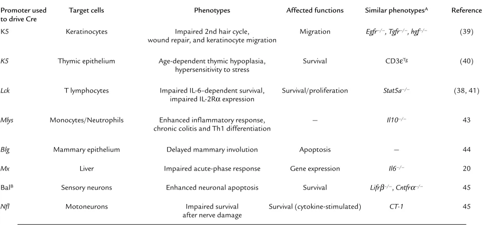
mutation to any desired adult or embryonic tissue where Cre expression can be induced. To date, this approach has been used to evaluate Stat3 function in skin, thymic epithelium, T cells, granulocytes, mammary gland, liver, nervous system, and bone marrow (Table 1).
Stat3 in skin
The keratin 5 (K5) promoter has been used to direct Cre expression to epidermal and follicular keratinocytes (40). The resulting mice are viable despite the absence of functional Stat3 protein in their epidermis. Hair cycle and wound healing processes are severely compromised in the mutant animals, which display sparse hair and spontaneously develop ulcers with age. However, the first hair cycle is essentially normal in the absence of Stat3. Defects become apparent only with the second cycle and grow more pronounced with age. This phe-notype is quite distinct from that seen in the absence of EGFR (33, 34), suggesting that a different ligand, per-haps HGF, is responsible for activating Stat3 in the skin. In addition, migration of keratinocytes in response to growth factor stimulation in vitro was impaired, although the cells displayed normal mitogenic respons-es. Interestingly, induction of genes necessary for cell migration also appeared normal. Therefore, Stat3 is necessary for a variety of functions in the skin, some of
which are dependent on growth factors such as EGF family members and TGF-α, ligands known to activate Stat3. Nonetheless, many responses to these ligands remain intact. These results do not fit a simple model of Stat3 function being restricted to cell growth, differen-tiation, or survival but rather suggest that Stat3 is involved in maintenance of postnatal interactions between epithelia and mesenchymal compartments.
Stat3 in the thymic epithelium
Keratin 5 is expressed in thymic epithelial cells, so the same mouse strain used to evaluate Stat3 function in skin has been examined for effects on thymic function (41). Alterations are only observed in adult mice, which develop severe thymic hypoplasia, including loss of thy-mocytes and of the normal thymic architecture. These changes appear at the time of normal thymic involu-tion, suggesting that Stat3 may play a role in the tim-ing or extent of this normal agtim-ing process. In young mice prior to developing any overt symptoms, the thy-mus shows hypersensitivity to apoptosis-inducing agents, such as steroids or γ-irradiation, although iso-lated thymocytes are no more sensitive than controls. Interestingly, it is the thymocytes that degenerate in the mutant animals, even though they are wild-type for
Stat3, since the conditional mutation affects only the neighboring epithelial cells. Therefore, while Stat3 has been proposed to mediate survival functions, in this case, these functions operate in an indirect manner involving the influence of the thymic microenviron-ment on T cell survival.
[image:4.576.57.545.479.704.2]Again, these results defy a simple explanation, such as absence of a set of Stat3 target gene products. Microarray analysis has been applied to identify mis-regulated genes in the Stat3-deficient thymus. Rather
Table 1
Summary of Stat3-deficiency phenotypes revealed by conditional gene targeting in mice
Promoter used Target cells Phenotypes Affected functions Similar phenotypesA Reference to drive Cre
K5 Keratinocytes Impaired 2nd hair cycle, Migration Egfr–/–, Tgfr–/–, hgf–/– (39) wound repair, and keratinocyte migration
K5 Thymic epithelium Age-dependent thymic hypoplasia, Survival CD3εTg (40)
hypersensitivity to stress
Lck T lymphocytes Impaired IL-6–dependent survival, Survival/proliferation Stat5a–/– (38, 41) impaired IL-2Rαexpression
Mlys Monocytes/Neutrophils Enhanced inflammatory response, — Il10–/– 43 chronic colitis and Th1 differentiation
Blg Mammary epithelium Delayed mammary involution Apoptosis — 44
Mx Liver Impaired acute-phase response Gene expression Il6–/– 20
BalB Sensory neurons Enhanced neuronal apoptosis Survival Lifrβ–/–, Cntfrα–/– 45
Nfl Motoneurons Impaired survival Survival (cytokine-stimulated) CT-1 45 after nerve damage
than a loss of a set of potential Stat3 target genes, mutant thymi showed aberrant cortical expression of mRNAs for keratin 5, the growth factor receptor ErbB2/neu, and the nonclassical MHC class II protein H2-O, all of which are otherwise expressed predomi-nantly in medullary regions of the thymic epithelium. Since these molecules are presumably downregulated in the cortex during differentiation, some degree of de-differentiation may have occurred in the absence of epithelial Stat3.
Stat3 in T cells
Possible functions of Stat3 in T cells have been exam-ined by specific gene ablation using Cre driven by the
Lckpromoter (39, 42). Prior experiments, provided lit-tle evidence for an important Stat3 function in T cells, since most T cell cytokines selectively activate other Stat family members, such as Stat4, Stat5, and Stat6. However, the effects of IL-6 are pleiotropic, including immunomodulatory functions onT cells, that include stimulation of T cell survival (11). Given the important role for Stat3 downstream of the IL-6 receptor, there-fore, it was not surprising that IL-6–mediated T cell survival is impaired in Stat3-deficient T cells (39). Con-versely, while it might have been expected that Stat3 would exert its antiapoptotic action through induction of survival genes, it was unexpected to discover that Bcl-2 is induced in the absence of Stat3, similar to its induction in wild-type cells. Therefore, an unknown, Bcl-2–independent mechanism must mediate the sur-vival effects of IL-6 in T cells.
Stat3-deficient T cells also display a reduced prolifer-ative response to IL-2 stimulation but this phenotype cannot be explained by reduced survival (42). Rather, this effect correlates with reduced expression of CD25, the inducible chain of the IL-2 receptor. Cd25is known to be a Stat target gene, but it has been previously shown to be induced by Stat5a following IL-2 stimula-tion. While Stat3 is a minor target for IL-2 signaling, these results demonstrate that it is nonetheless a rele-vant component of the signaling pathway.
Stat3 in the myeloid lineage
Stat3 function has been examined in macrophages and neutrophils by Cre-mediated gene ablation directed by the macrophage lysozyme (Mlys) promoter (43). Mutant mice display enhanced susceptibility to endo-toxic shock and develop chronic enterocolitis with age. The phenotype of these animals is reminiscent of that in mice lacking IL-10, including aberrant expression of MHC class II molecules and increased expression of inflammatory cytokines. IL-10 is an anti-inflammato-ry cytokine that suppresses induction of TNF-α through a Stat3-dependent pathway (23). Therefore, it is tempting to speculate that the phenotype of Stat3 -ablated macrophages derives from the inability to sup-press production of TNF-αonce it has been induced during bacterial infection.
The IFN-inducible Mxpromoter driving Cre recom-binase has been used to ablate Stat3 in hematopoietic progenitor cells to evaluate its role in granulopoiesis (C. Lee et al., unpublished observations). Despite the
con-siderable evidence from cell culture and transgenic mouse experiments that Stat3 would be critical for var-ious aspects of myeloid development, including the action of G-CSF (21), all major myeloid cell types devel-op in the absence of Stat3, and granulocytes proliferate and differentiate in response to G-CSF.
Stat3 in mammary development
Stat5 is critical for mammary development, due to its role as a signaling target for growth hormone and pro-lactin. In mice lacking Stat5, mammary lobuloalveolar outgrowth during pregnancy is curtailed and terminal differentiation does not occur, leaving females unable to lactate. Stat3 phosphorylation occurs coincident with mammary gland involution, and the absence of Stat3 due to Cre recombinase driven by the β-lactoglobulin (Blg) promoter delays the onset of involution (44). Mam-mary gland involution is thought to be triggered by absence of the survival factor IGF-1, which becomes sequestered by binding proteins, such as IGFBP5. Inter-estingly, expression of IGFBP5, which occurs during involution of control glands, is nearly abolished in mutant glands. However, other markers of apoptosis, such as Bcl-X and Bax, were unaffected, and aberrant expression of other survival molecules may occur as a consequence of the lack of apoptosis, rather than a cause. Therefore, it would appear that Stat3 is proapop-totic in the involuting mammary gland, possibly through a role in regulating the expression of IGFBP5. This role for Stat3 would appear to be opposite to the involution-suppressing effects of Stat3 in thymus (41).
Stat3 in the nervous system
Stat3 is known to be activated in the brain, particularly in response to ciliary neutrophic factor (CNTF) and leptin. A balancer strain of Cre transgenic mice has been used to examine the role of Stat3 in vivo by generating mosaic animals with differential gene inactivation in different tissues. Because deletion is particularly effective in the nervous system, Stat3 is lost in the brain, resulting in peri-natal lethality (45). Stat3 is required for the neurotroph-ic effects of CNTF and LIF on developing sensory neu-rons in vitro, consistent with the finding that few of these cells survive in vivo. Stat3-deficient neurons are unable to induce Akt phosphorylation in response to CNTF, sug-gesting a direct link between activation of Stat3 and of Akt. It is unclear what underlying mechanism would link Akt activation by CNTF through Stat3, although it has been suggested that Stat3 can function as an adaptor molecule that promotes phosphatidylinositol 3-kinase activation in response to IFN. A similar survival function has been found for Stat3 in injured motoneurons, by using the neurofilament light chain (Nfl) promoter to drive Cre (46). Motoneurons in these animals, unlike con-trols that maintain one wild-type Stat3allele, undergo cell death within weeks of axotomy. These findings are con-sistent with the requirement of Stat3 for signaling from gp130-type cytokines, such as CNTF, LIF, or CT-1.
Stat3 and the acute-phase response
inactivation of Stat3 in the liver led to significant impairment of the acute-phase response (20). Genes induced during the acute phase have been categorized according to their time of induction, their promoter structures, and their dependence on IL-6. In the absence of Stat3, genes exclusively regulated through Stat-binding sites are silent, while those dependent on other transcription factors, including CAAT/enhancer binding protein (C/EBP) family members and NF-κB, are affected to a lesser extent. These results clearly demonstrate the fundamental importance of Stat3 to the cytokine-mediated induction of acute-phase response genes in vivo. Stat3 appears to function as a classical transcription factor during this response, and its requirement is directly related to the presence of Stat-binding sites in the promoters of target sites.
It is interesting that Stat3 was discovered because of its role in the acute-phase response, and that this is the only capacity in which Stat3 function in vivo can be clearly ascribed to its activity as a transcription factor. Nevertheless, there are also surprises in the liver. The truncated Stat3βprotein produced by alternative splic-ing has not received much attention, because its lack of a transactivation domain implied no role in gene expression. However, recent results from specific abla-tion of this isoform in transgenic mice indicate that it is an important negative regulator, at least during the acute-phase response to endotoxic shock (47). Stat3β -deficient mice recover poorly from endotoxic shock and display significant hyperresponsiveness of a subset of inducible genes in the liver. Therefore, Stat3βmay be an important negative regulator of Stat3α-inducible genes. This negative regulation appears to be critical for normal recovery following systemic inflammation.
Physiological importance of Stat3
When first discovered, Stat3 appeared to be a straight-forward additional member of the growing Stat family that would function in the induction of a limited set of target genes in response to IL-6 released during inflam-mation. The list of its possible functions grew through subsequent studies in cell culture, showing that it is activated in a wide variety of signaling systems and mediates a bewildering complexity of responses. While similar confusion relating to other Stat family members has been somewhat laid to rest due to the restricted phe-notypes observed in knockout mice, Stat3 remains something of an enigma. Early embryonic lethality in the absence of Stat3 suggests an essential role in an early developmental process, but the nature of that process, its physiological activator, and even its site of action (in embryonic or extraembryonic tissue) remain to be defined. In particular, none of the known Stat3 activa-tors can account for this embryonic function.
Despite the clear importance of Stat3 during very early development, the ablation of Stat3in adult tissues leads to surprisingly mild phenotypes. All tissues, including all cells of the hematopoietic system, appear to develop normally, although their responses to par-ticular cytokines are impaired. In some cases, the action of Stat3 can be ascribed to the induction of a set of important target genes, but in others it may be acting
as a repressor (e.g., in thymic epithelium) or as a sig-naling adaptor without a transcriptional function (e.g., activation of Akt in neurons). Moreover, even in situa-tions where Stat3 appears to function as a transcrip-tional activator, the biological readout can be prolifer-ation, survival, or apoptosis, depending on the target tissue. Thus, although we possess a wealth of data on Stat3 functions in various contexts, we are still unable to describe fully what Stat3 does. The finding, dis-cussed in detail by Bromberg in this Perspective series (25), that Stat3 may be a key player in the pathogenesis of diverse human cancers makes this molecule a prime target for novel therapies and lends greater urgency to answering this complex question.
Acknowledgments
We thank our colleagues for helpful discussions and apol-ogize to all whose work was not properly cited due to the space constraints of a short review. Work in the authors’ laboratory was funded by the NIH, the American Heart Association, and the Mathers Charitable Foundation.
1. Darnell, J.E., Kerr, I.M., and Stark, G.R. 1994. Jak-STAT pathways and transcriptional activation in response to IFNs and other extracellular proteins. Science.264:1415–1421.
2. Levy, D.E., and Darnell, J.E. 1990. Interferon-dependent transcriptional activation: signal transduction without second messenger involvement? New Biol.2:923–928.
3. Takeda, K., et al. 1997. Targeted disruption of the mouse Stat3 gene leads to early embryonic lethality. Proc. Natl. Acad. Sci. USA.94:3801–3804. 4. Takeda, K., and Akira, S. 2000. STAT family of transcription factors in
cytokine-mediated biological responses. Cytokine Growth Factor Rev. 11:199–207.
5. Hirano, T., Ishihara, K., and Hibi, M. 2000. Roles of STAT3 in mediating the cell growth, differentiation and survival signals relayed through the IL-6 family of cytokine receptors. Oncogene.19:2548–2556.
6. Akira, S., et al. 1994. Molecular cloning of APRF, a novel IFN-stimulat-ed gene factor 3 p91-relatIFN-stimulat-ed transcription factor involvIFN-stimulat-ed in the gp130-mediated signaling pathway. Cell.77:63–71.
7. Lütticken, C., et al. 1994. Association of transcription factor APRF and protein kinase Jak1 with the interleukin-6 signal transducer gp130. Sci-ence.263:89–92.
8. Zhong, Z., Wen, Z., and Darnell, J.E. 1994. Stat3: a STAT family member activated by tyrosine phosphorylation in response to epidermal growth factor and interleukin-6. Science.264:95–98.
9. Raz, R., Durbin, J.E., and Levy, D.E. 1994. Acute phase response factor and additional members of the interferon-stimulated gene factor 3 fam-ily integrate diverse signals from cytokines, interferons, and growth fac-tors. J. Biol. Chem.269:24391–24395.
10. Taga, T., and Kishimoto, T. 1997. gp130 and the interleukin-6 family of cytokines. Annu. Rev. Immunol.15:797–819.
11. Heinrich, P.C., Behrmann, I., Müller-Newen, G., Schaper, F., and Graeve, L. 1998. Interleukin-6-type cytokine signalling through the gp130/Jak/STAT pathway1. Biochem. J.334:297–314.
12. Guschin, D., et al. 1995. A major role for the protein tyrosine kinase JAK1 in the JAK/STAT signal transduction pathway in response to inter-leukin-6. EMBO J.14:1421–1429.
13. Greenlund, A.C., Farrar, M.A., Viviano, B.L., and Schreiber, R.D. 1994. Ligand-induced IFN gamma receptor tyrosine phosphorylation couples the receptor to its signal transduction system (p91). EMBO J. 13:1591–1600.
14. Decker, T., and Kovarik, P. 2000. Serine phosphorylation of STATs. Onco-gene.19:2628–2637.
15. Zhang, J.J., et al. 1998. Ser727-dependent recruitment of MCM5 by stat1alpha in IFN-gamma-induced transcriptional activation. EMBO J. 17:6963–6971.
16. Boeuf, H., Hauss, C., Graeve, F.D., Baran, N., and Kedinger, C. 1997. Leukemia inhibitory factor-dependent transcriptional activation in embryonic stem cells. J. Cell Biol.138:1207–1217.
17. Niwa, H., Burdon, T., Chambers, I., and Smith, A. 1998. Self-renewal of pluripotent embryonic stem cells is mediated via activation of STAT3. Genes Dev.12:2048–2060.
19. Matsuda, T., et al. 1999. STAT3 activation is sufficient to maintain an undifferentiated state of mouse embryonic stem cells. EMBO J. 18:4261–4269.
20. Alonzi, T., et al. 2001. Essential Role of STAT3 in the control of the acute-phase response as revealed by inducible gene inactivation in the liver. Mol. Cell. Biol.21:1621–1632.
21. McLemore, M.L., et al. 2001. STAT-3 activation is required for normal G-CSF-dependent proliferation and granulocytic differentiation. Immuni-ty.14:193–204.
22. Boccaccio, C., et al. 1998. Induction of epithelial tubules by growth fac-tor HGF depends on the STAT pathway. Nature.391:285–288. 23. Riley, J.K., Takeda, K., Akira, S., and Schreiber, R.D. 1999. Interleukin-10
receptor signaling through the JAK-STAT pathway. Requirement for two distinct receptor-derived signals for anti-inflammatory action. J. Biol. Chem.274:16513–16521.
24. Levy, D.E., and Gilliland, D.G. 2000. Divergent roles of STAT1 and STAT5 in malignancy as revealed by gene disruptions in mice. Oncogene. 19:2505–2510.
25. Bromberg, J. 2002. Stat proteins and oncogenesis. J. Clin. Invest. 109:1139–1142. DOI:10.1172/JCI200215650.
26. Yu, C.-L., et al. 1995. Enhanced DNA-binding activity of a Stat3-related protein in cells transformed by the Src oncoprotein. Science.269:81–83. 27. Duncan, S.A., Zhong, Z., Wen, Z., and Darnell, J.E. 1997. STAT signaling is active during early mammalian development. Dev. Dyn.208:190–198. 28. Yoshida, K., et al. 1996. Targeted disruption of gp130, a common signal transducer for the interleukin 6 family of cytokines, leads to myocardial and hematological disorders. Proc. Natl. Acad. Sci. USA.93:407–411. 29. Stewart, C.L., et al. 1992. Blastocyst implantation depends on maternal
expression of leukaemia inhibitory factor. Nature.359:76–79. 30. Oppenheim, R.W., et al. 2001. Cardiotrophin-1, a muscle-derived
cytokine, is required for the survival of subpopulations of developing motoneurons. J. Neurosci.21:1283–1291.
31. Ernst, M., Oates, A., and Dunn, A.R. 1996. Gp130-mediated signal trans-duction in embryonic stem cells involves activation of Jak and Ras/mito-gen-activated protein kinase pathways. J. Biol. Chem.271:30136–30143. 32. Rodig, S.J., et al. 1998. Disruption of the Jak1 gene demonstrates obliga-tory and nonredundant roles of the Jaks in cytokine-induced biologic responses. Cell.93:373–383.
33. Sibilia, M., and Wagner, E.F. 1995. Strain-dependent epithelial defects in mice lacking the EGF receptor. Science.269:234–238.
34. Threadgill, D.W., et al. 1995. Targeted disruption of mouse EGF recep-tor: effect of genetic background on mutant phenotype. Science. 269:230–234.
35. Cirri, P., et al. 1997. c-Src activates both STAT1 and STAT3 in PDGF-stim-ulated NIH3T3 cells. Biochem. Biophys. Res. Commun.239:493–497. 36. Soriano, P., Montgomery, C., Geske, R., and Bradley, A. 1991. Targeted
disruption of the c-src proto-oncogene leads to osteopetrosis in mice. Cell. 64:693–702.
37. Klinghoffer, R.A., Sachsenmaier, C., Cooper, J.A., and Soriano, P. 1999. Src family kinases are required for integrin but not PDGFR signal trans-duction. EMBO J.18:2459–2471.
38. Stein, P.L., Vogel, H., and Soriano, P. 1994. Combined deficiencies of Src, Fyn, and Yes tyrosine kinases in mutant mice. Genes Dev.8:1999–2007. 39. Takeda, K., et al. 1998. Stat3 activation is responsible for IL-6-dependent T cell proliferation through preventing apoptosis: generation and char-acterization of T cell- specific Stat3-deficient mice. J. Immunol. 161:4652–4660.
40. Sano, S., et al. 1999. Keratinocyte-specific ablation of Stat3 exhibits impaired skin remodeling, but does not affect skin morphogenesis. EMBO J.18:4657–4668.
41. Sano, S., et al. 2001. Stat3 in thymic epithelial cells is essential for post-natal maintenance of thymic architecture and thymocyte survival. Immu-nity.15:261–273.
42. Akaishi, H., et al. 1998. Defective IL-2-mediated IL-2 receptor alpha chain expression in Stat3-defiient T lymphocytes. Int. Immunol.10:1747–1751. 43. Takeda, K., et al. 1999. Enhanced Th1 activity and development of chron-ic enterocolitis in mchron-ice devoid of Stat3 in macrophages and neutrophils. Immunity.10:39–49.
44. Chapman, R.S., et al. 1999. Suppression of epithelial apoptosis and delayed mammary gland involution in mice with a conditional knockout of Stat3. Genes Dev.13:2604–2616.
45. Alonzi, T., et al. 2001. Role of STAT3 and PI 3-kinase/Akt in mediating the survival actions of cytokines on sensory neurons. Mol. Cell. Neurosci. 18:270–282.