IJPSR (2014), Vol. 5, Issue 5 (Research Article)
Received on 09 December, 2013; received in revised form, 30 January, 2014; accepted, 09 March, 2014; published 01 May, 2014
CATHEPSIN D LEVEL IN THE HUMAN GINGIVAL TISSUES AND SERUM OF PERIODONTITIS SUBJECTS
D.S. Pushparani* and S. Nirmala
Department of Biochemistry, SRM Dental College, Ramapuram, Chennai-600089, Tamil Nadu, India
ABSTRACT: This study investigates the level of cathepsin D activity in the serum and gingival tissues of periodontitis subjects and healthy individuals. The cathepsin D activity (spectrophotometric assay) was measured in the gingival tissues and serum of control healthy individuals (group I) and periodontitis subjects (group II). Inflamed gingival tissue was obtained from 15 periodontitis patients (9 males and 6 females) and15 control individuals (8 males and 7 females) aged 32-52 yrs with healthy gingival who were undergoing other forms of oral surgery. Serum was obtained by centrifuging the blood at 1500 r.p.m for 10 minutes. The enzyme unit was expressed by micromoles of tyrosine liberated per minute / 1000 ml in serum and micromoles of tyrosine liberated per minute / mg in gingival tissues. Cathepsin D activity in the serum and gingival tissue was higher in periodontitis group than that of the control healthy group (p < 0.05). We found that serum cathepsin D in the serum of group II subjects was elevated 8 times and in the gingival tissues, its level is nearly 7 times higher when compared to group I subjects. There were correlations between periodontal parameters, and cathepsin D activity. The results suggest that cathepsin D activity increases in patients with periodontitis. The higher cathepsin D activity in the gingival and serum of periodontitis may be attributed to an adaptive mechanism in the tissue which increases oxidative stress and would be helpful for monitoring the periodontal condition.
INTRODUCTION: Periodontitis is a chronic inflammatory disorder mediated by host and has been implied as a risk factor for systemic diseases, including diabetes mellitus, hyperlipidemia and
atherosclerosis 1.
They have been proposed that it is the host response to the long-term bacterial challenge and manifested by damage to the periodontal tissues
which may progress to tooth loss 2.
QUICK RESPONSE CODE
DOI:
10.13040/IJPSR.0975-8232.5(5).1924-30
Article can be accessed online on: www.ijpsr.com
DOI link: http://dx.doi.org/10.13040/IJPSR.0975-8232.5(5).1924-30
Chronic periodontitis is an inflammatory disease initiated and maintained by bacterial plaque that
triggers a local infiltration of inflammatory cells 3.
The mechanisms by which periodontitis increases the likelihood of these systemic diseases are not fully understood, but the prerequisite is believed to be the host response to long-term exposure to
certain bacterial pathogens (e.g.,
lipopolysaccharide (LPS) and proteases) 4.
Cathepsin D (EC 3.4.23.5) is a lysosomal aspartic proteinase enzyme, belonging to the pepsin family and is widely distributed in almost all mammalian cells. It is the major lysosomal endopeptidase which plays an important role in physiological and pathological breakdown of intracellular and extracellular proteins at an acidic pH optimum between 3.5 and 4.5.
Keywords:
Cathepsin D, Gingival tissue, Oxidative stress,
Periodontitis
Correspondence to Author:
Mrs. D. S. Pushparani Department of Biochemistry, SRM Dental College, Ramapuram, Chennai – 600089, Tamil Nadu, India.
It inactivates many inhibitors of proteolytic enzymes. The proteolytic activity of the enzyme is regulated by various intra lysosomal factors such as pH, products of metabolism, hormones, growth factors and specific inhibitors. The enzyme has been found to be up regulated in many acute and chronic pathological conditions such as trauma,
sepsis and diabetes mellitus 5. The best known
aspartic proteinases are probably produced
digestive enzymes like pepsins, gastricsin and chymosin, and intracellular proteinases such as the lysosomal cathepsin D.
The mammalian lysosomal cathepsin D is synthesized in the rough endoplasmic reticulum as preprocathepsin D. Cathepsin D enzymes from mammalian sources are well known and the enzyme is probably one of the most important factors causative to the lysosomal digestive activity
6, 7
.Polymorphonuclear leucocytes located at sites
of microbial invasion are activated by
inflammatory mediators, thereby generating
increased levels of reactive oxygen species (ROS), which not only attack the pathogens but also host
surrounding tissues 8. Topical application of
lipopolysaccharides (LPS) and proteases into the gingival sulcus induces periodontitis and DNA
damage in periodontal tissue 9. This suggests that
the gingival response to bacterial pathogens generates excessive ROS.
A variety of ROS (e.g., superoxide and hydroxyl radicals, hydrogen peroxide, hypochlorous acid) are well characterized and are able to cause direct damage to proteins, DNA, carbohydrates and lipids
10
. It is known that ROS cause tissue necrosis, organ failure, atherosclerosis, infertility, birth
defects, premature ageing, mutations and
malignancy 11. The inflammatory gingival tissue
contains large numbers of different subpopulations of inflammatory cells. The gingival connective tissues play a major role in the gingival immune response 12.
As oxidative stress from periodontitis induces liver injury, improvement of periodontitis and reduction of gingival oxidative stress may be important for reducing the occurrence of circulating oxidative stress and systemic diseases.
As increased release of ROS from peripheral
neutrophils was observed in adult periodontitis 13,
generation of ROS in periodontally involved gingiva may increase circulating oxidative stress and result in systemic oxidative damage.
Polymorpho nuclear leucocytes (PMNs) produce ROS in periodontitis in response to LPS
stimulation 14, 15. Expression of ROS (H2O2) was
observed in junctional epithelial cells and infiltrated PMNs in the periodontitis group. These findings suggest that, in the periodontitis group,
epithelial cells and PMNs generate ROS (H2O2) by
LPS stimulation, the ROS cause DNA damage in the gingival fibroblasts and the cathepsin D level in the fibroblasts subsequently increases. Monitoring of ROS formation on several targets in vivo has focused on the biomarkers of oxidative stress so far. Free radical-catalysed products of lipid peroxidation, modified proteins, and indices of free radical-catalysed modification of DNA are widely
used to detect oxidative stress 16.
Our aim, therefore, was to determine the activities of lysosomal enzyme cathepsin D, in gingival tissues and serum of subjects with periodontal disease.
MATERIALS AND METHODS:
Study Subjects and Ethical Approval: The study consisted of a total of 30 subjects between the age group 32 to 52 years and are categorized into two groups as control healthy individuals (Group I, n=15), and periodontitis subjects (group II, n=15). Group I healthy individuals were selected from the general population and group II were selected from the outpatients attending the Department of Periodontology & Oral Implantology, SRM Dental College, India. Each subject gave their consent to participate in the study, the protocol of which had been approved by the Institutional Ethical Committee of Medical and Health Sciences, SRM University, Kattankulathur, India.
Clinical Assessment of study subjects:
The clinical assessment for periodontitis subjects included examination of gingiva, intra oral examination- number of teeth present and missing, pathological migration, and probing depth. Mean pocket probing depth (PPD), and clinical attachment loss (CAL) were measured using mouth mirror and William’s periodontal probe to assess the periodontal status. The periodontal status was examined by a trained Periodontist of SRM Dental College, Department of Periodontology, Chennai -600 089.
Inclusion and Exclusion Criteria: To be assigned to the periodontitis group, patients should have more than 30% of the sites with Clinical attachment level (CAL) ≥ 3mm, pocket probing depth (PPD) ≥ 4 mm, and at least 2 teeth in each quadrant with the condition of 20 teeth in all the subjects. The healthy controls were not on any kind of prescribed medication or dietary restrictions.
Patients allocated to the control group had no loss of attachment greater than 3 mm at all designated sites, with bleeding on probing at no more than 2 of the designated sites. Smokers, alcoholics, drug abused, patients who had periodontal therapy six months prior to the study, patients under antibiotics and having systemic disease, taking hormone drugs, lipid lowering drugs, oral contraceptives, and pregnant women were excluded from the study.
Preparation of Gingival Tissue: Inflamed gingival tissue was obtained from 15 periodontitis patients (9 males and 6 female) aged 32-52 yrs who had given informed consent for periodontal flap surgery. Fifteen subjects (8 males and 7 females) with healthy gingival, who were undergoing other forms of oral surgery, served as controls. Tissues were rinsed in normal saline and blotted to remove blood and saliva, and stored at -20°C.
Before surgery, the severity of disease at local sites were assessed by measurement of probing depth
and clinical attachment level. Tissue samples were
homogenized at a concentration of 2% w/v in ice-cold 0.1 M phosphate buffer, pH 6.0, containing 2 mM EDTA and 0.2% v/v Triton X-100 with a homogenizer. The homogenates were centrifuged in a cooling centrifuge at 4°C for 20 min at 13,000g and the supernatants stored at - 20°C.
Measurement of Cathepsin D Enzyme Activity:
Blood samples were collected after an overnight fast for each subject. Serum was obtained by centrifuging the blood at 1500 r.p.m for 10 minutes. Cathepsin D was determined in serum and
gingival tissues by the method of Anson17 as
modified by Barrett 18 the assay for cathepsin D
involved measurement of TCA-soluble, Folin-reactive products of hemoglobin digestion at pH 3.5 and 37°C. 1 ml of 1% hemoglobin dissolved in 0.2 M acetate buffer (pH 3.8), 0.8 ml of 0.05 M acetate buffer (pH 3.8) and 0.2 ml of the enzyme sample were mixed.
After incubation at 37°C for 60 min, the reaction was stopped by addition of 2 ml of 10% trichloroacetic acid solution and the mixture was centrifuged at 1000 g for 15 minutes. The liberated peptides in the presence of trichloroacetic acid
were measured in UV absorption
spectrophotometer at 280 nm and the cathepsin D activity was calculated. The enzyme unit was expressed by micromoles of tyrosine liberated per minute / 1000 ml in serum and micromoles of tyrosine liberated per minute / mg in gingival tissues.
Statistical Analysis: The data are presented as mean ± SD (standard deviation). An unpaired Student’s t test was used to evaluate the significance of differences, accepting P <0.05 as the level of significance. Comparisons between control group, and periodontitis group were made using ANOVA. Statistical analysis included Pearson’s correlations between cathepsin D with PPD and CAL in the 2 groups; P <0.05 were considered significant. All statistical analysis was performed using the statistical software package, Winks SDA 7.0.5 (Windows Kwik Stat).
RESULTS: The demographic data of the study population in the control (group I), and periodontitis (group II) was shown in Tables 1.
Pearson correlation data analysis is given in Table
2 and 3. The mean cathepsin D level in the serum and gingival tissues of healthy individuals and
periodontitis subjects are presented in Figures 1
and 2, respectively. The group II subjects exhibited higher levels of mean BMI, systolic and diastolic
TABLE 1: DEMOGRAPHIC DATA OF THE STUDY POPULATION
Parameters Control
Group I
Periodontitis Group II
No of samples 15 15
Gender (M/F) 8/7 9/6
Age, years 38.8 ± 1.30 42.3 ± 13.04***
BMI, kg/m2 22.72 ± 1.1 24.5 ± 1.25**
Systolic blood pressure, mm Hg 118.7 ± 3.22 124.0 ± 8.1**
Diastolic blood pressure, mm Hg 74.7 ± 0.89 72.91 ± 2.64**
Values are expressed as Mean ± SD; except for gender (Male, M / Female, F). Differences were considered significant level at *** p <0.0001; ** p <0.001 for parameters of group I vs group II. NS, non-significant
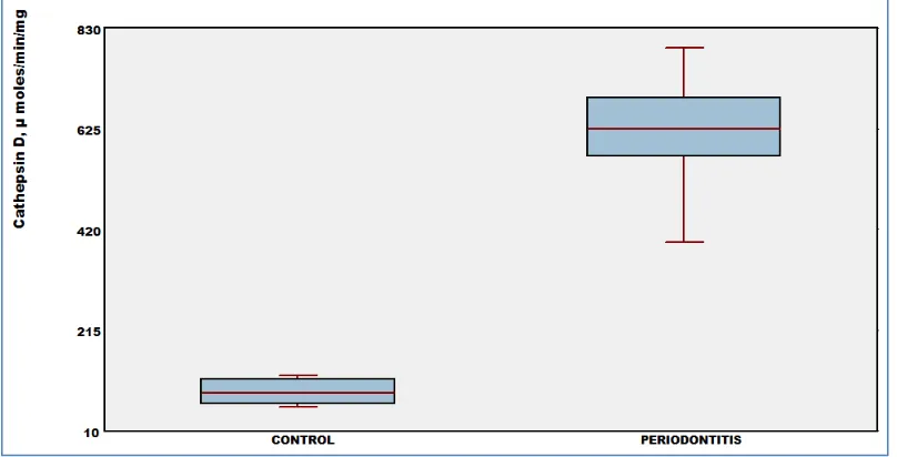
The activity of cathepsin D in the serum and gingival tissue of control group is lesser when
compared to periodontitis subjects, Fig 1 and 2
respectively. The serum cathepsin D of group II
(periodontitis) subjects was found to be 7 times higher than control group. The cathepsin D level in the gingival tissue was nearly 8 times elevated in group II compared to group I subjects.
FIGURE 1: CATHEPSIN D ACTIVITY IN THE GINGIVAL TISSUE OF CONTROL HEALTHY SUBJECTS AND PERIODONTITIS
[image:4.612.106.510.262.468.2] [image:4.612.102.511.513.716.2]
We observed a positive correlation between serum cathepsin D with PPD, CAL, BMI and it showed negative correlation with age whereas in the gingival tissue, cathepsin D showed negative correlation with PPD, CAL, BMI and positively correlated with age of healthy control individuals (group I). In the serum we observed a positive
correlation between cathepsin D with CAL, BMI and negative correlation with PPD and age whereas in the gingival tissue, cathepsin D showed negative correlation with CAL, BMI and positively correlated with PPD and age of periodontitis
subjects (group II), Table 2 and 3.
TABLE 2: CORRELATION BETWEEN CATHEPSIN D AND STUDY PARAMETERS IN THE SERUM AND HUMAN GINGIVAL TISSUE OF GROUP I SUBJECTS
GROUP I
PARAMETERS SERUM GINGIVAL TISSUE
PPD, mm r= 0.563
p= 0.323
r= -0.452 p=0.445
CAL, mm r= 0.587
p= 0.298
r= -0.987 p= 0.002**
BMI, kg/m2 r= 0.776
p= 0.123
r= -0.767 p= 0.130
AGE, Years r= -0.242
p= 0.694
r= 0.069 p= 0.913
Pocket probing depth, PPD; Clinical attachment level, CAL; Body mass index, BMI; * significant p value; Pearson’s coefficient, r
TABLE 3: CORRELATION BETWEEN CATHEPSIN D AND STUDY PARAMETERS IN THE SERUM AND HUMAN GINGIVAL TISSUE OF GROUP II SUBJECTS
GROUP II
PARAMETERS SERUM GINGIVAL TISSUE
PPD, mm r= -0.149
p= 0.596
r= 0.560 p= 0.03*
CAL, mm r= 0.416
p= 0.123
r= -0.264 p= 0.342
BMI, kg/m2 r= 0.435
p= 0.105
r= -0.215 p= 0.442
AGE, Years r= -0.163
p= 0.563
r= 0.341 p= 0.213
Pocket probing depth, PPD;Clinical attachment level, CAL; Body mass index,BMI; *significant p value; Pearson’s coefficient, r
DISCUSSION: Periodontal diseases are initiated by Gram-negative tooth associated bacterial biofilms that generate a host response, with resultant soft tissue destruction. In reaction to endotoxins resulting from periodontal pathogens,
several osteoclast-related mediators (matrix
metalloproteinases, cathepsins and other osteoclast-derived enzymes) target the destruction of alveolar
bone and sustaining connective tissues 19. The
factors which are able to alter mitochondrial efficiency can enhance ROS production, with a direct and critical effect on oxidative stress. The mitochondrion is the organelle that is most influenced by oxidative damage, and lysosomal enzymes have been found to act on mitochondria to promote ROS generation, thereby creating a feedback loop that leads to additional lysosomal damage.
Therefore, the highest cathepsin D enzyme activity was observed in periodontitis subjects (Group II) than the control group (Group I).
In the swollen periodontal tissues, ROS are produced as a result of complex interactions between pathogenic bacteria and the host’s immune response. A variety of proof indicates that AGEs might be concerned in a closed cycle of
inflammation, generation of ROS, elevated
production of AGEs, more inflammation, and so on
20
AGEs are among the factors which influence the periodontal disease development and increases oxidative stress in gingiva of diabetes and periodontitis patients 21, 22.
AGE formation on collagen proteins would lead to the thickening of basement membrane in gingival tissues, impairing the delivery of leukocytes and nutrients into the gingival and periodontal tissues. In a cell with high energy charge, acidification of lysosomes would be maximized due to maximal
activity of the proton pump, 23 and lysosomal pH
would be lowest. The pH optima for cathepsin D suggests that ATP activation of cathepsin D may be sturdily favored under certain conditions and the optimum for cathepsin D is about pH 4 and this is
not altered by ATP 24.
Thus, when lysosomal pH drops below 5 the catalytic activity of cathepsin D would increase. Under these conditions increased activation of cathepsin D by ATP would be enhanced. The periodontal tissues that encompass the tooth, consist of alveolar bone, periodontal ligament, cementum of the tooth, and gingiva, are the tooth supporting tissues. Periodontal ligament is an intense connective tissue, surrounding the dental root and connects it with alveolar bone. The most essential components of it are the principal fibers, which are collagenous, are prearranged in bundles, and the terminal areas of the fibers placed into
cementum and alveolar bone. Therefore,
periodontal ligament has the chief function of tooth support. It also has a vital function in the tissue rejuvenation of periodontal ligament itself, alveolar bone and cementum, keeping the homeostasis of sustaining tissues and offering the remedial process
25-28
.
The proteases that are released from the lysosomal compartment to the cytosol seem to be able to activate some steps of the death cascade leading to apoptosis. If apoptosis is delayed, however, this will lead to increased retention of neutrophils in the periodontal tissues. This in turn could lead to increased tissue damage and formation of more reactive oxygen species with the release of
destructive enzymes, cathepsin D by the
neutrophils.
Increased cathepsin D level among group II subjects indicates the end point of periodontal
tissue destruction 29. Accumulation of
glucose-mediated AGEs in diabetic patients impairs
chemotactic and phagocytic function of
polymorphonuclear leukocytes 30. It also impairs
the movement of metabolic waste of periodontal pathogens out of the tissue; causing decreased wound healing capacity and increased disease severity. Measurement of cathepsin D activities might thus have a role in monitoring the efficacy of periodontal treatment or in predicting future periodontal disease.
CONCLUSION: Raised levels of cathepsin D enzyme has been found in the gingival tissue and serum of periodontitis patients and thus its increased activity might prove of value in
monitoring periodontitis disease activity.
Proteolytic enzymes are believed to play a role in the pathogenesis of periodontal disease and therefore gingival cathepsin D may prove of value as indicators of disease activity. Gingival tissue and serum of cathepsin D levels was found to be higher in periodontitis patients than controls. This suggests that measurements of cathepsin D activity in gingival tissues and serum might ultimately prove of more value for monitoring the periodontal condition.
We conclude that cathepsin D could be potential as a marker of periodontal disease activity.
ACKNOWLEDGEMENT: We would like to
thank the Management of SRM Dental College for supporting this work and for providing laboratory facilities and Dr. P. Theagarayan MDS., Professor,
Department of Periodontology & Oral
Implantology for valuable discussion of the manuscript.
REFERENCES:
1. Keum Jin Baek, Youngnim Choi, and Suk Ji: Gingival fibroblasts from periodontitis patients exhibit inflammatory characteristics in vitro. Archives of Oral Biology 2013; 58(10): 1282-1292.
3. George Hajishengallis: Immunomicrobial pathogenesis of periodontitis: keystones, pathobionts, and host response. Trends in Immunology 2014; 35(1): 3-11
4. Purnima S. Kumar: Oral microbiota and systemic disease. Anaerobe 2013; 24: 90-93
5. Tomofuji T, Ekuni D, Yamanaka R, Kusano H, Azuma T, Sanbe T, et al. Chronic administration of lipopolysaccharide and proteases induces periodontal inflammation and hepatic steatosis in rats. J Periodontol 2007; 78:1999–2006.
6. Maryssa Canuel, Ann Korkidakis, Kristin Konnyu, Carlos R. Morales. Sortilin mediates the lysosomal targeting of cathepsins D and H. Biochemical and Biophysical Research Communications 2008; 373:292–297.
7. Ashapogu Venugopal, and Nadimpalli Siva Kumar: Biochemical characterization of cathepsin D from the mussel Lamellidens corrianus.Comparative Biochemistry and Physiology Part B: Biochemistry and Molecular Biology 2014; 169: 25-30
8. Waddington RJ, Moseley R, Embery G. Reactive oxygen species: a potential role in the pathogenesis of periodontal diseases. Oral Dis 2000; 6:138–151.
9. Maximiliano Schünke Gomes, Trevor Charles Blattner, Manoel Sant'Ana Filho, Fabiana Soares Grecca, Fernando Neves Hugo, Ashraf F. Fouad, and Mark A. Reynolds: Can Apical Periodontitis Modify Systemic Levels of Inflammatory Markers? A Systematic Review and Meta-analysis. Journal of Endodontics 2013; 39(10):1205-1217
10. Reejamol MK,and Mythili Swaminathan: Estimation of lipid peroxides and antioxidants in smokers and non-smokers with periodontitis. King Saud University Journal of Dental Sciences 2013; 4(2): 53-56
11. Chapple IL, and Matthews JB: The role of reactive oxygen and antioxidant species in periodontal tissue destruction. Periodontol 2000 2007; 43:160–232.
12. Valko M, Leibfritz D, Moncol J, Cronin MT, Mazur M, and Telser J: Free radicals and antioxidants in normal physiological functions and human disease. Int J Biochem Cell Biol 2007; 39:44–84.
13. Babior BM: Phagocytes and oxidative stress. Am J Med 2000; 109:33–44.
14. Zdařilová A, Rajnochová Svobodová A, Chytilová K, Šimánek V, and Ulrichová J: Polyphenolic fraction of Lonicera caerulea L. fruits reduces oxidative stress and inflammatory markers induced by lipopolysaccharide in gingival fibroblasts. Food and Chemical Toxicology 2010; 48(6): 1555-1561
15. Kasai H: Chemistry-based studies on oxidative DNA damage: formation, repair, and mutagenesis. Free Radic Biol Med 2002; 33:450–456.
16. Nemec A, Verstraete FJM, Jerin A, Šentjurc M, Kass PH, Petelin M, and Pavlica Z: Periodontal disease, periodontal treatment and systemic nitric oxide in dogs.Research in Veterinary Science 2013; 94(3): 542-544
17. Anson ML: The estimation of pepsin, trypsin, papain and cathepsin with haemoglobin. J Gen Physiol 1938; 22:79-89. 18. Barrett AJ: Proteinases in Mammalian Cells and Tissues.
North-Holland, Amsterdam 1977; 209–248.
19. Giannobile WV. Host-response therapeutics for periodontal diseases. J Periodontol 2008; 79:1592–1600.
20. Mediha Sefi, Hamadi Fetoui, Mohamed Makni, and Najiba Zeghal: Mitigating effects of antioxidant properties of Artemisia campestris leaf extract on hyperlipidemia, advanced glycation end products and oxidative stress in alloxan-induced diabetic rats .Food and Chemical Toxicology 2010; 48(7): 1986-1993
21. Southerland JH, Taylor GW, Moss K, Beck JD, and Offenbacher S: Commonality in chronic inflammatory diseases: periodontitis, diabetes, and coronary artery disease. Periodontol 2000 2006; 40:130–143.
22. Malene W. Poulsen, Rikke V. Hedegaard, Jeanette M. Andersen, Barbora de Courten, Susanne Bügel, John Nielsen, Leif H. Skibsted,and Lars O. Dragsted: Advanced glycation endproducts in food and their effects on health Review Article. Food and Chemical Toxicology 2013; 60: 10-37 23. Vincent Soubannier, Gian-Luca McLelland, Rodolfo Zunino,
Emelie Braschi, Peter Rippstein, Edward A. Fon, and Heidi M. McBride: A Vesicular Transport Pathway Shuttles Cargo from Mitochondria to Lysosomes. Current Biology 2012; 22(2): 135-141
24. Roman Buckow, Binh Quong Truong, and Cornelis Versteeg: Bovine cathepsin D activity under high pressure. Food Chemistry 2010; 120(2): 474-481
25. Hirohito Kato, Yoichiro Taguchi, Kazuya Tominaga, Makoto Umeda, and Akio Tanaka: Porphyromonas gingivalis LPS inhibits osteoblastic differentiation and promotes pro-inflammatory cytokine production in human periodontal ligament stem cells. Archives of Oral Biology 2014; 59(2): 167-175
26. Li Wang, Tingle Wang, Meng Song, and Jinsong Pan: Rho plays a key role in TGF-β1-induced proliferation and cytoskeleton rearrangement of human periodontal ligament cells Original. Archives of Oral Biology 2014; 59(2): 149-157 27. Rui Mauricio Santos de Araujo, Yasuo Oba, Shingo Kuroda, Eiji Tanaka, and Keiji Moriyama: RhoE regulates actin cytoskeleton organization in human periodontal ligament cells under mechanical stress. Archives of Oral Biology 2014; 59(2): 187-192
28. Sunita P. Ho, Michael P. Kurylo, Kathryn Grandfield, Jonathan Hurng, Ralf-Peter Herber, Mark I. Ryder, Virginia Altoe, Shaul Aloni, Jian Q. Feng, Samuel Webb, Grayson W. Marshall, Donald Curtis, Joy C. Andrews, and Piero Pianetta: The plastic nature of the human bone–periodontal ligament– tooth fibrous joint. Bone 2013; 57(2): 455-467
29. Olivier Masson, Anne-Sophie Bach, Danielle Derocq Christine Prébois, Valérie Laurent-Matha, Sophie Pattingre, and Emmanuelle Liaudet-Coopman: Pathophysiological functions of cathepsin D: Targeting its catalytic activity versus its protein binding activity? Biochimie 2010; 92(11):1635-1643
30. Marwa Magdy Abbass, Nahed Sedky Korany, Ahmed Helmy Salama, John J. Dmytryk, and Barbara Safiejko-Mroczka: The relationship between receptor for advanced glycation end products expression and the severity of periodontal disease in the gingiva of diabetic and non-diabetic periodontitis patients. Archives of Oral Biology 2012; 57(10): 1342-1354.
All © 2013 are reserved by International Journal of Pharmaceutical Sciences and Research. This Journal licensed under a Creative Commons Attribution-NonCommercial-ShareAlike 3.0 Unported License
This article can be downloaded to ANDROID OS based mobile. Scan QR Code using Code/Bar Scanner from your mobile. (Scanners are available on Google Playstore)
How to cite this article: