Additional files 1
Supplementary Methods 2
PAK2 binding by SIVmac239 mutated in residues of the putative PAK2 activating 3
structural motif 4
SIVmac239 Nef residues I117, H121, T218 and Y221 were each mutated to three
5
different substitutions by overlapping PCR. The resulting constructs were cloned into
6
pCDNA3.1 and transfected into 293T cells as previously described [1]. Western blot of
7
lysates was revealed with anti-SIVmac Nef rabbit antiserum [2] followed by horseradish
8
peroxidase (HRP)-conjugated anti-rabbit antibodies. HRP conjugates were visualized by
9
using enhanced chemiluminescence. In vitro kinase assay was essentially performed as
10
previously described [1]. Immunoprecipitations were performed using the rabbit
anti-11
SIVmac Nef antiserum. Nef/PAK-2 complexes were immunoadsorbed and assayed for
12
autophosphorylation activity. Phosphorylated proteins were resolved by sodium dodecyl
13
sulfate-polyacrylamide gel electrophoresis, and dried gels were then exposed to a
14
phosphorimager screen.
15 16
Assessment of cell surface marker modulation by SIVmac239 Nef mutated in 17
H121R and/or Y221R Nef 18
Cloned single and double mutant SIVmac 239 H121R/Y221R nef sequences were
19
cloned into the bicistronic cytomegalovirus-based pCG expression vector coexpressing
20
the GFP and Nef and in replication competent HIV-1 NL4-3 based proviral contructs in
the Nef reading frame [3, 4]. pCG plasmids were used to transfect cell lines (Jurkat and
22
THP-I)[5]. HIV/SIV Nef/eGFP viral stocks were generated by transfection in 293T cells
23
and used to infect stimulated normal donor peripheral blood mononuclear cells (PBMC)
24
as described before [4]. After cell culture, transfected or infected cells were stained and
25
analysed by flow cytometry as described before [3].
Supplementary references 28
29 30
1. O'Neill E, Kuo LS, Krisko JF, Tomchick DR, Garcia JV, Foster JL: Dynamic
31
evolution of the human immunodeficiency virus type 1 pathogenic factor, 32
Nef. J Virol 2006, 80:1311-1320. 33
2. Garcia JV, Foster JL: Structural and functional correlates between HIV-1 and
34
SIV Nef isolates. Virol 1996, 226:161-166. 35
3. Schindler M, Munch J, Brenner M, Stahl-Hennig C, Skowronski J, Kirchhoff F:
36
Comprehensive analysis of Nef functions selected in simian 37
immunodeficiency virus-infected macaques. J Virol 2004, 78:10588-10597. 38
4. Schindler M, Munch J, Kutsch O, Li H, Santiago ML, Bibollet-Ruche F,
Muller-39
Trutwin MC, Novembre FJ, Peeters M, Courgnaud V, et al: Nef-mediated
40
suppression of T cell activation was lost in a lentiviral lineage that gave 41
rise to HIV-1. Cell 2006, 125:1055-1067. 42
5. Schindler M, Wildum S, Casartelli N, Doria M, Kirchhoff F: Nef alleles from
43
children with non-progressive HIV-1 infection modulate MHC-II expression 44
more efficiently than those from rapid progressors. AIDS 2007, 21:1103-45
1107.
46 47 48
Supplementary Table 1: primers used to generate mutant and tagged Nef protein expression constructs
49
Forward Reverse
SIVmac239 H121R 5’-ccgtctggagatctgcgacagagac-3’ 5’-cttttataaatctagacatgtctattgccaatttg-3’ SIVmac239 Y221R 5’-ccgtctggagatctgcgacagagac-3’ 5’-catatgcctcataagtcctggccagagttgg-3’
HIV1 SF2-AU1 tag 5’-gctctagaatatgggtggcaag-3’ 5’-tttacgcgttttactagttcatatatagcgataggtgtcgcagtctttgtagtactccggat-3’ SIVmac293-AU1 tag 5’-cgtctagaatatgggtggagctatttcc-3’ 5’-tcccttacgcgttatatatagcgataggtgtcgcgagtttccttcttgtc-3’
Supplementary Figure legends
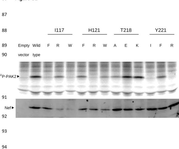
51
Figure S1. SIVmac239 Nef displays a PAK2-activating structural domain surface. 52
Autophosphorylation activity of Nef/PAK-2 complexes using the SIVmac239 Nef
53
mutants in residues I117, H121, T218 and Y221 as indicated. Substituting amino acids
54
are indicated with their letter code (top). Controls (left side of upper gel) were empty
55
vector and wild-type SIVmac239 Nef, respectively. Arrow head indicates position of the
56
complex. Lower gel shows Western blot to detect Nef proteins in lysate, arrow head
57
indicates protein bands specifically detected.
58
Figure S2. Cell surface marker modulation by SIVmac239 mutated in PAK2-59
activating structural domain surface . 60
(A) Flow cytometric analysis of HIV/SIV Nef/eGFP infected PBMC stained for CD4,
61
CD3, CD28 and MHC-I as indicated, and of transfected THP-I cells (bottom row),
62
stained for MHC class II invariant chain Ii (CD74). Top line indicates construct used,
63
resp. none (mock), Nef- construct (Nef-), wild-type SIVmac239 Nef (239) or the three
64
different mutants hereof (239 H121R, 239 Y221R and 239 H121R Y221R respectively).
65
Dot plots show GFP marker expression as a measure of Nef expression vs. the stained
66
marker. (B) Fold downregulation of the mean fluorescence intensity of surface markers
67
stained on PBMC, normalized against Nef- (=1) and measured by flow cytometry as
68
shown in (A). X-axis indicates construct used, resp. none (mock), Nef- construct (nef-),
69
wild-type SIVmac239 Nef (239nef) or the three different mutants hereof (239H121R,
70
239Y221R and 239H121RY221R respectively. Bar charts show average ± standard
71
deviation of experiments in 2 donors. (C) Fold downregulation of the mean fluorescence
intensity of surface markers stained on Jurkat cells, normalized against Nef- (=1) and
73
measured by flow cytometry. X-axis indicates construct used, resp. none (mock), Nef-
74
construct (nef-), Nef sequence unable to express due to multiple stop codons (nef*),
75
wild-type SIVmac239 Nef (239nef) or the three different mutants hereof (239H121R,
76
239Y221R and 239H121RY221R respectively. Bar charts show average ± standard
77
deviation of 2 experiments. (D) Fold upregulation of Ii and fold downregulation of MHC
78
class II (mean fluorescence intensity) stained on THP-I cells, normalized against Nef-
79
(=1) and measured by flow cytometry. X-axis indicates construct used, resp. none
80
(mock), Nef- construct (nef-), Nef sequence unable to express due to multiple stop
81
codons (nef*), wild-type SIVmac239 Nef (239nef) or the three different mutants hereof
82
(239H121R, 239Y221R and 239H121RY221R respectively. Bar charts show average ±
83
standard deviation of 2 experiments.
Figure S1 86 87 I117 H121 T218 Y221 88 Empty Wild F R W F R W A E K I F R 89 vector type 90 91 92 93 94 32 P-PAK2 Nef
Figure S2A
Figure S2B 97 98 0 1 2 3 4 5 6 mo ck n ef -239nef 239H1 21R 239Y 221R 239H1 21Y 221R X -fo ld d o w n re g u la ti o n (n e f-) CD3 down, PBMC, n=2 CD28 down, PBMC, n=2 0 1 2 3 4 5 6 7 m o c k n e f-2 3 9 n e f 2 3 9 H 1 2 1 R 2 3 9 Y2 2 1 R 2 3 9 H 1 2 1 Y2 2 1 R X-fo ld d o w n re g u la ti o n (n e f-) 0,0 0,5 1,0 1,5 2,0 2,5 mo ck n ef -239nef 239H1 21R 239Y 221R 239H1 21Y 221R X -f o ld d o w n reg u la tio n ( n ef -) CD4 down, PBMC, n=2 0,0 0,5 1,0 1,5 2,0 2,5 3,0 3,5 4,0 4,5 mo ck n ef -239nef 239H1 21R 239Y 221R 239H1 21Y 221R X -fo ld d o w n re g u la ti o n (n e f-) MHCI down, PBMC, n=2
Figure S2C 99 100 101 102 0 2 4 6 8 10 12 14 16 18 20 m ock nef -nef * 23 9ne f 23 9H 12 1R 23 9Y221 R 23 9H 12 1Y221 R X -f o ld d o w n reg u la tio n ( n ef -) CD3 down, Jurkat, n=2 0,0 0,2 0,4 0,6 0,8 1,0 1,2 1,4 1,6 m ock nef -nef * 23 9ne f 23 9H 12 1R 23 9Y221 R 23 9H 12 1Y221 X -f o ld d o w n reg u la tio n ( n ef -) CD4 down, Jurkat, n=2 0 5 10 15 20 25 30 m ock nef -nef * 23 9ne f 23 9H 12 1R 23 9Y221 R 23 9H 12 1Y221 R X -f o ld d o w n reg u la tio n ( n ef -) CD28 down, Jurkat, n=2 0 2 4 6 8 10 12 14 m ock nef -nef * 23 9ne f 23 9H 12 1R 23 9Y221 R 23 9H 12 1Y221 X -f old do w nreg ulat ion ( ne f-)
Figure S2D 103 104 105 106 0,0 0,5 1,0 1,5 2,0 2,5 m ock nef -nef * 23 9ne f 23 9H 12 1R 23 9Y221 R 23 9H 12 1Y221 R X -f old do w nreg ulat ion ( ne f-)
MHC-II down, THP-I, n=2
0,0 0,5 1,0 1,5 2,0 2,5 3,0 3,5 4,0 4,5 m ock nef -nef * 23 9ne f 23 9H 12 1R 23 9Y221 R 23 9H 12 1Y221 R X -f o ld u p reg u la tio n ( n ef -) Ii up, THP-I, n=2