DEVELOPMENT OF A METRIC TO ASSESS COMPLETENESS OF LESIONS PRODUCED BY RADIOFREQUENCY ABLATION IN THE HEART
Herman D. Himel, IV
A dissertation submitted to the faculty of the University of North Carolina at Chapel Hill in partial fulfillment of the requirements for the degree of Doctor of Philosophy in the Department of Biomedical Engineering.
Chapel Hill 2006
© 2006
ABSTRACT
HERMAN D. HIMEL, IV: Development of a Metric to Assess Completeness of Lesions Produced by Radiofrequency Ablation in the Heart
(Under the direction of Professor Stephen B. Knisley)
For approximately 25 years, ablation of cardiac tissue by radiofrequency energy has been used to halt arrhythmic conduction in the heart. Efficacy of cardiac ablation for the treatment of atrial fibrillation and other arrhythmias depends on the completeness of lesions and their ability to block arrhythmic conduction without disturbing sinus rhythm. A rapid index of lesion quality measured during surgical ablation procedures may improve the probability of long-term success in patients. Trans-lesion stimulus-excitation time delay was evaluated as an intraoperative indicator of lesion completeness. Time delay was shown to be significantly different for complete vs. incomplete lesions. Further, it was shown that activation path length corresponds with time delay, providing an explanation for post-ablation increases in time delay.
± 1.0 minutes after ablation. Recovery of the activation complex occurred at 23.7 ± 7.9 minutes after ablation.
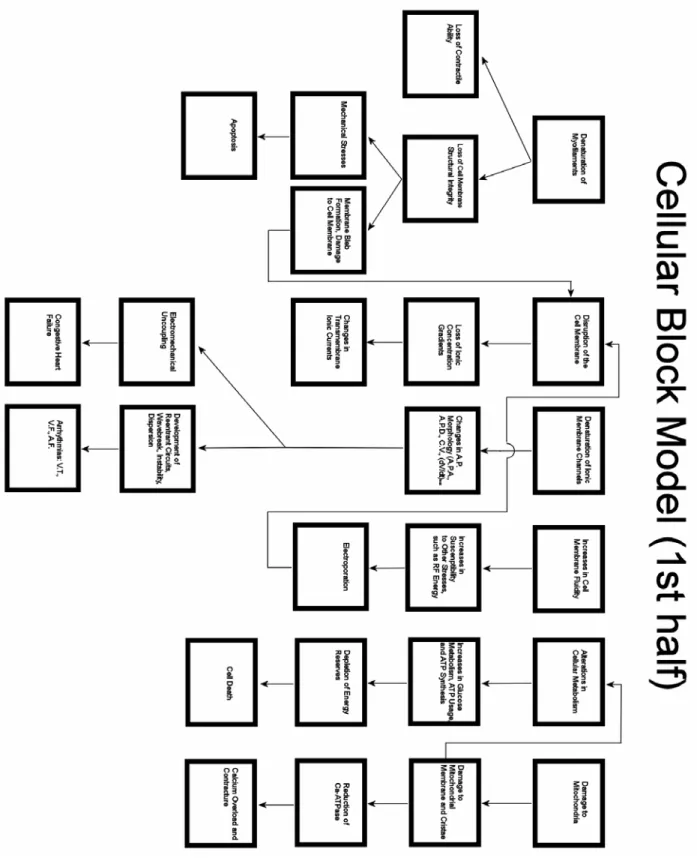
Hyperthermia affects the myocardium in a wide variety of ways, including denaturation of myofilaments, disruption of the sarcolemmal membrane and/or denaturation of membrane ion channels, alterations in cellular metabolism, and alterations in intracellular and transmembrane calcium handling. Depending on the extent of the hyperthermic damage sustained during ablation, gaps within lesions may recover following a return to physiological temperature. Conduction recovery may occur following initially successful lesion formation, and it may allow arrhythmic conduction to resume or may result in the formation of one or more new arrhythmias. Further, the temperature of tissue within a small gap of an ablation lesion may correlate with the recovery of myocytes within these gaps. Thus temperature monitoring of ablated tissue may be a useful supplement for monitoring block by measuring time delay.
DEDICATION
ACKNOWLEDGEMENTS
TABLE OF CONTENTS
LIST OF TABLES... xi
LIST OF FIGURES ... xii
ABBREVIATIONS AND SYMBOLS... xiv
I. INTRODUCTION AND BACKGROUND MATERIAL ...1
A. CELLULAR EFFECTS OF HYPERTHERMIA AND RADIOFREQUENCY ENERGY...4
B. EFFECTS OF ABLATION UPON THE MICROVASCULATURE...17
C. HEAT TRANSFER IN PERFUSED LIVING TISSUE...23
D. OPTICAL MAPPING OF CARDIAC ABLATION LESIONS...28
E. ELECTROGRAM RECORDINGS OF ABLATION LESIONS...29
F. CONDUCTION VELOCITY AND CHANGES IN NORMAL PROPAGATION...35
G. CHANGES IN CONDUCTION VELOCITY DUE TO ISCHEMIA...36
II. TRANSLESION STIMULUS-EXCITATION DELAY AS A METRIC FOR RADIOFREQUENCY ABLATION LESIONS IN HEARTS ...39
A. INTRODUCTION...39
B. METHODS...41
Rabbit Heart Preparation...41
Electrical Pacing and Recording...42
Fluorescence Recording...43
Ablation Procedures...44
Analysis of Stimulus-Excitation Time Delay...45
Statistical Analysis...46
C. RESULTS...47
Examination of TTC-stained Hearts...47
Electrograms...48
Optical Maps...49
Time Delay...53
D. DISCUSSION...56
Epicardial Approach...56
Time Delay...57
III. ABILITY OF MINIMUM ACTIVATION PATH LENGTH TO PREDICT TRANSLESION TIME DELAY...60
A. INTRODUCTION...60
B. METHODS...61
Rabbit Heart Preparation...61
Electrical Pacing and Recording...61
Ablation Procedures...62
Analysis of Stimulus-Excitation Time Delay...62
Viable Tissue Staining...63
Minimum Activation Path Length Determination...63
C. RESULTS...65
D. DISCUSSION...67
IV. RECOVERY OF TISSUE WITHIN SMALL GAPS OF LINEAR RADIOFREQUENCY CARDIAC ABLATION LESIONS ...69
A. INTRODUCTION...69
B. METHODS...72
Electrical Pacing and Recording...72
Ablation Procedure...73
Selection of Activations in Fractionated Electrograms...74
Indication of Recovery...75
Classification of Recovery...75
Rate of Change in Time Delay...76
Data Processing...76
Statistical Analysis...76
C. RESULTS...78
Control Recording...78
Recovery of the Activation Complex...79
Complete Recovery of Conduction...82
Partial Recovery of Conduction...84
Late Worsening of Conduction...86
Probability of Observing Different Lesion Types...89
D. DISCUSSION...90
V. TIME COURSE OF TEMPERATURE WITHIN STUNNED MYOCARDIUM ADJACENT TO LINEAR RADIOFREQUENCY ABLATION LESIONS ...94
A. INTRODUCTION...94
B. METHODS...95
Rabbit Heart Preparation...95
Electrical Pacing and Recording...95
Temperature Measurements...96
Temperature Time Constant...97
Time Response of Temperature Probe...98
Supernormal Conduction...99
Data Processing...100
Statistical Analysis...100
C. RESULTS...101
Summary of Temperature Results...101
Temperature During Control Recordings...102
Temperature During Complete Recovery...103
Temperature During Partial Recovery...104
Temperature During Changes in Activation Complex...105
Temperature During Supernormal Conduction...106
Correlation Between Time Delay and Temperature During Supernormal Conduction...108
Summary of Temperature Dependence of Recovery...109
D. DISCUSSION...113
APPENDIX: A GEOMETRIC MODEL FOR PREDICTING TIME DELAY AROUND A LINEAR ABLATION LESION IN A TWO-DIMENSIONAL SHEET OF MYOFIBERS...117
LIST OF TABLES
Table 2.1 Time Delay Measured by Electrogram Recordings...54 Table 2.2 Time Delay Measured by Optical Recordings...54 Table 4.1 Summary of recovery times (50% and 90%) for all lesion types except
those showing morphological changes post-ablation. ...87 Table 4.2 Times of activation complex recovery occurring post ablation...88 Table 4.3 Averaged slopes of time delay for lesions showing partial recovery and
late conduction worsening. ...88 Table 5.1 Summary of maximum temperatures (Tmax), time constants (τ), and
times for 50% recovery (t50) ...106 Table 5.2 Parameters describing supernormal conduction. tmin indicates the time of
LIST OF FIGURES
Figure 1.1 Myocardial model for cell damage following exposure to hyperthermia
(first half) ...15
Figure 1.2 Myocardial model for cell damage following exposure to hyperthermia (second half)...16
Figure 2.1 Diagram showing the arrangement of the ablation probe and electrogram recordings on the heart... 43
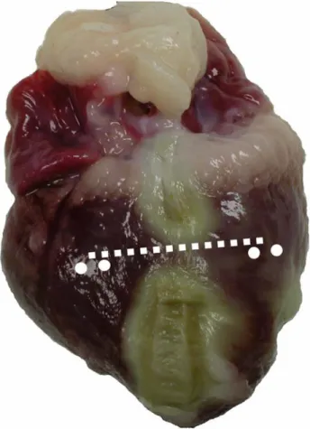
Figure 2.2 Photographs of incomplete and complete lesions ... 47
Figure 2.3 Electrograms for incomplete and complete lesions... 48
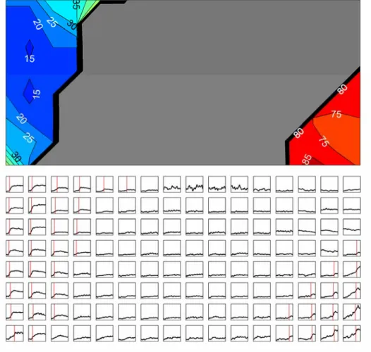
Figure 2.4 Activation contour map and action potential recordings before RF ablation... 50
Figure 2.5 Activation contour map and action potential recordings after production of an incomplete lesion. ... 51
Figure 2.6 Activation contour map and action potential recordings after production of a complete lesion. ... 52
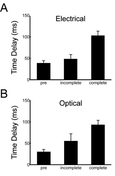
Figure 2.7 Electrical and optical time delays measured before lesion creation, and for incomplete and complete lesions... 55
Figure 3.1 Photograph showing the minimum activation path length ... 64
Figure 3.2 Stimulus to excitation time delays as a function of minimum activation path length... 66
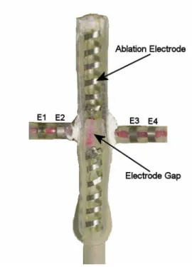
Figure 4.1 Five cm ablation probe with silicone gap... 73
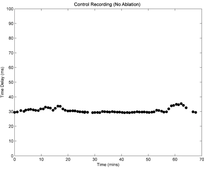
Figure 4.2 Control recording of time delay over a 1-hour period... 79
Figure 4.3 Example of activation complex recovery following an RF ablation lesion ... 81
Figure 4.4 Three-dimensional view of morphological changes following ablation indicating recovery of activation complex... 82
Figure 4.5 Complete recovery following an ablation lesion... 83
Figure 4.6 Electrograms taken during complete conduction recovery ... 84
Figure 4.8 Electrograms for partial conduction recovery in an incomplete lesion... 86
Figure 4.9 Late conduction worsening following RF ablation. Vertical dotted line indicates end of RF ablation. ... 87
Figure 5.1 Photograph of ablation probe with temperature probe attached near gap region. ... 97
Figure 5.2 Time response of temperature probe ... 98
Figure 5.3 Gap temperature following RF ablation ... 102
Figure 5.4 Control recording of time delay and temperature for a 60-minute period ... 103
Figure 5.5 Graphs illustrating the relationship between time delay and temperature for a lesion showing complete recovery. ... 104
Figure 5.6 Graphs illustrating the relationship between time delay and temperature for a lesion showing partial recovery... 105
Figure 5.7 Supernormal conduction immediately following RF ablation ... 107
Figure 5.8 Relationship between temperature and time delay during supernormal conduction... 109
Figure 5.9 Diagram for complete recovery... 110
Figure 5.10 Diagram for partial recovery ... 111
ABBREVIATIONS AND SYMBOLS AC – alternating current
AF – atrial fibrillation AgCl – silver chloride Ai – area of the ith vessel
AERP – atrial effective refractory period APA – action potential amplitude
APD – action potential duration ATP – adenosine triphosphate AV – atrioventricular
ATPase – adenosine triphosphatase
Ca-ATPase – Calcium2+ transport adenosine triphosphatase c – specific heat (heat capacity)
cb – specific heat of blood
Cys374 – cysteine residue present on F-actin DNA – deoxyribonucleic acid
(dV/dt)max – maximum derivative of an electrogram peak
(dVm/dt)max – maximum upstroke velocity of the transmembrane potential E – electric field
F-actin – filamentous polymer form of actin Hsps – heat shock proteins
G-actin – globular monomer form of actin GAG – glucosaminoglycan
J – current density
κ – thermal conductivity IVC – superior vena cava NO – nitric oxide
ρ – density
ρb – density of blood
Pi – circumference of the ith vessel PV – pulmonary vein
q – heat flux vector
Qel – heat contribution due to conductive transfer between tissue and the ablation electrode r – radius of lesion end
r - position vector
R – length of lesion minus twice the radius of the lesion end RNA – ribonucleic acid
ROS – reactive oxygen species RF – radiofrequency
SR – sarcoplasmic reticulum SVC – superior vena cava
θ – angle between the direction of propagation and the fiber direction
θi – angle between vessel and exiting of entering surface of a tissue volume
τ – time constant of temperature following ablation t50 – time of 50% recovery
t90 – time of 90% recovery
tmin – time of minimum time delay during supernormal conduction tR – time of recovery of activation complex
T – temperature
Ta – arterial temperature
Tbi – flow-weighted average temperature of blood within the ith vessel
ΔTDdecrease – maximum percent decrease in time delay during supernormal conduction TDmin – minimum time delay during supernormal conduction
TDpre – pre-lesion time delay for the 2-D linear lesion model TDpost – post-lesion time delay for the 2-D linear lesion model Tmax – maximum temperature
Ts – temperature of solid tissue Tv – venous temperature
TUNEL – terminal deoxynucleotidyl transferase biotin-dUTP nick end labeling ui – mean velocity within the ith vessel
Ui – heat transfer coefficient of the ith vessel UTP – Uridine 5’-triphosphate
vθ – velocity of activation wave as a function of theta
vc – constant component of the velocity of the activation wave
CHAPTER 1
INTRODUCTION AND BACKGROUND MATERIAL
Atrial fibrillation (AF) is the most common cardiac arrhythmia in the United States, currently affecting over 2.2 million of its citizens1. At the age of 40 years, the lifetime risk of developing AF is approximately 24.5%2. Once thought to be a benign consequence of aging, AF is now widely regarded as a dangerous arrhythmia which carries significant risks in addition to its direct effect upon cardiac output, where the atria account for approximately 25% of ventricular filling3. Some of the risks associated with AF include stroke4 and other embolic phenomena, increases in morbidity and mortality, and increased risk of congestive heart failure. Data from the Framingham study shows that the incidence of stroke increases approximately five-fold for patients who have AF5, and that the risk of death increases by a factor of 1.4-1.9 even after adjustment for preexisting cardiovascular conditions related to AF6. Atrial fibrillation has also been shown to be a progressive disease. If left untreated, AF leads to electrical and mechanical remodeling within the myocarium7-12 which leads to greater inducibility and higher stability of AF. Following electrical remodeling during AF, subsequent episodes of AF are easier to induce, and occur for a longer time period9.
invasive and requires a thoracotomy and cardiopulmonary bypass. For the Maze III procedure, lesions are formed by making surgical incisions into the myocardium, electrically disconnecting adjacent tissue, and isolating certain regions of the atria. Also known as the “cut and sew” method, the Cox-Maze III lesion set eliminates AF by isolating ectopic foci which trigger fibrillatory activity, and by breaking up reentrant circuits which sustain this activity.
Alternatively, catheter ablation is minimally invasive but is associated with relatively poor success rates, particularly among those with structural heart disease. Also, catheter procedures require that hyperthermia be applied to the myocardium while the device is within a major vessel or within the atria, exposing blood to hyperthermic environments and increasing the risk of clotting and subsequent thromboembolic complications13-15. Another weakness of catheter ablation is its inability to consistently create continuous transmural lesions which emulate those of the Cox-Maze III lesion set. Specifically, it is technically challenging to create the linear transmural (i.e., complete) lesions which are prescribed by the Cox-Maze III procedure. Nonetheless, complete linear lesions are a key building block for the creation of the Maze lesion set, and the quality of these lesions directly impacts upon the success and recurrence rates of AF ablation surgery16.
normal and abnormal conduction are examined, as well as changes in the myocardium due to ischemia, since radiofrequency ablation is known to decrease blood flow in the region of lesion creation17.
A. Cellular Effects of Hyperthermia and Radiofrequency Energy
Hyperthermia causes cellular damage by a number of different mechanisms, and has significant effects upon the sarcolemmal membrane, the cytoskeleton, cellular organelles, cellular metabolism, and specific types of protein production (heat shock proteins). This discussion will attempt to detail the effects of hyperthermia upon cardiac myocytes and indicate the relative importance of each effect.
Hyperthermia induced by radiofrequency ablation produces structural damage to sarcolemmal membranes which results in profound electrophysiological changes in cardiac myocytes. Nath et al reported that hyperthermia caused depolarization of the transmembrane potential (Vm), changes in dV/dtmax (early increase followed by a late decrease), slight changes in action potential amplitude (APA), significant changes in action potential duration (APD), as well as reversible and irreversible losses of excitability18. Most electrophysiological effects are thought to be a consequence of damage to the plasma membrane, since the plasma membrane plays a critical role in ion transport and is the first cellular component to be affected by hyperthermia19. Hyperthermia is thought to affect the plasma membrane by increasing membrane fluidity, thereby increasing vulnerability to electric fields produced by the ablation electrode. Also, hyperthermia is thought to affect ion transport directly by impairment of receptor function and thermal inactivation of ion channels19. Also, inactivation of some voltage-dependent ion channels may be a consequence of a decrease in resting membrane potential due to extracellular increases in potassium and a subsequent inability of the transmembrane potential to return to normal resting values.
death by calcium overload. In addition to their role in ATP production, mitochondria also play an important role in the regulation of calcium. Damage to the mitochondria results in a reduction in Ca-ATPase activity within the mitochondria, which leads to an inability to remove calcium from the intracellular space. This condition may then lead to calcium overload and possibly death by irreversible contracture of the myocyte. Although the hyperthermia exposure time of Quian et al was of a longer duration (40 minutes) than what is typical during RF ablation (~1-5 minutes), similar types of mitochondrial damage were observed by both Quian20 and Nath et al21, who performed RF ablation. These effects include degradation of the mitochondrial membrane and cristae, as well as mitochondrial swelling. It is possible that this damage contributes to non-acute cell death (4-6 hours) that may account for late conduction block which has been reported after RF ablation surgery. The mitochondria have also been shown to play a key role in programmed cell death, as deactivation of certain anti-apoptotic molecules within the mitochondrial membrane may lead to apoptosis22. Mitochondrial membrane permeabilization has been shown to play a key role in apoptosis for both the extrinsic death receptor and the intrinsic mitochondrial pathways23. The mitochondria have also been implicated in the acidification of the intracellular environment24,25. Intracellular acidification leads to the mitochondrial release of caspase activators24. The caspases are a class of proteases central to perhaps all types of apoptosis26,27.
Everett et al have proposed that irreversible contracture by intracellular calcium increase may be important in causing hyperthermia-induced injury to myocytes28. They reported that during moderately hyperthermic conditions (45-50 ºC), intracellular calcium concentrations were elevated but could be returned to normal levels by the calcium uptake into the SR. For temperatures above 50 ºC, however, myocytes were overwhelmed by increased calcium permeability due to hyperthermia-induced injury. It is possible that impaired function of the SR also contributes to cell death in the 50 ºC range, as studies suggest that hyperthermia exposure results in reductions in Ca2+-ATPase activity in the SR29.
authors use the fact that denatured (i.e. unfolded) actin molecules show a decrease in their DNase I-inhibiting activiy, and thus use the DNase I-inhibiting assay as a means of quantifying actin denaturation34.
It is possible that diseases such as hypertrophy which result in damage to the cytoskeleton may play a role in predisposing cardiac myocytes to damage caused by heat or ischemia-reperfusion injury. Defects causing weaknesses in the myocyte cytoskeleton have been implicated in cardiomyopathy, a disease which results in ventricular dilation and typically causes valvular disorders35.
Actin is the most abundant protein in myocytes, and comprises approximately 20% of total cell protein36. Actin exists as a globular monomer (G-actin) and as a filamentous polymer (F-actin) which is comprised of repeating units of G-actin. F-actin can form bundles of closely packed parallel filaments, and may also form loosely packed networks of filaments which may cross each other at any given angle37. Both bundles and networks are held together with actin cross-linking proteins, and may be attached to the cell membrane by membrane-microfilament binding proteins. Actin bundles are critical to the contractile function of the cardiac myocyte as well as its structural integrity. Actin has also been shown to play important roles in cell signaling38,39 and ion channel regulation40-44, which are critical to cardiac action potential conduction.
membrane directly45,46, thus hyperthermic conditions which affect membrane fluidity may affect the stability of the actin cytoskeleton.
In addition to causing cytoskeletal detachment from the cell membrane, hyperthermic conditions may also result in damage directly to actin filaments. While mild hyperthermia (41-43 ºC) typically does not induce damage to the actin filament network, higher temperatures (> 45 ºC) have been shown to destroy actin-containing contractile rings and cause damage to stress fibers47.
Protein denaturation is strongly associated with hyperthermic damage, and cell survival rates and protein denaturation rates have been shown to obey similar kinetics (i.e., decaying exponentials in time)48. Since actin is the most abundant cellular protein, and is even more abundant in myocytes, it is likely that actin denaturation plays a significant role in cell death due to hyperthermia.
al51. The degree to which cells recover will thus depend critically on the energy delivered to the actin and myosin fragments of the cytoskeleton, and whether or not sufficient energy is delivered to cause irreversible denaturation steps to occur.
The presence of ATP has been shown to have a protective effect upon F-actin52 as well as myosin53,54, while decreases in pH have sensitizing effects upon the thermal stability of actin54 and myosin55. Thus ischemic conditions and low ATP levels which occur following damage to microvasculature may affect the stability of actin and myosin bundles, predisposing them to thermal denaturation.
the myosin head is greatly reduced during hyperthermia58-60. Thus the contractility of cardiac myocytes near the border zone will likely be affected by hyperthermic exposure, and damage to the myosin head may result in permanent contractile dysfunction.
The extracellular matrix has been shown to undergo significant changes after even mild hyperthermia (~41 °C), such as that associated with the inflammatory response. Wagrowski et al have shown, in multiple cell lines, that the composition of the extracellular matrix is affected by hyperthermia61. Specifically, the production of the extracellular matrix component glucosaminoglycan (GAG) is inhibited, while the production of hyaluronan increases. Hyaluronan is typically present during inflammatory reactions, and acts to inhibit cell-to-cell adhesion and allow cell migration37. Proper myocardial conduction is dependent upon regular intercellular coupling, thus the restructuring of the extracellular matrix following hyperthermia may have significant effects upon cardiac excitation propagation, including wavebreak and slow conduction.
Although damage to the nuclear membrane has been reported for both endothelial cells in the microvasculature17 as well as cardiac myocytes21, the effects of this damage are thought to be of secondary importance in myocyte tissue death and recovery following RF ablation. While damage to DNA may be of critical importance in tumor cells where cell replication is accelerated, typically adult cardiac myocytes do not actively divide and thus DNA damage is thought to be of only minor importance. Also, damage to DNA and structured RNA are also thought to be unlikely to occur during RF ablation, since they typically do not undergo conformational changes below 85-90º C62.
addition, cells adjacent to the ischemic zone may die due to apoptosis63, which may be indicated morphologically by cell shrinkage rather than cell swelling typically found following ischemic necrosis. Other indicators of apoptosis include fragmented DNA (which may be detected by terminal UTP nick end labeling, or TUNEL), condensation/clumping of nuclear chromatin, membrane blebbing, loss of cell-cell contact, phagocytosis of the apoptotic body by adjacent cells, little inflammatory reaction in the region of apoptotic cells, intracellular acidification, and the maintenance of organelle structure and function until late in the apoptotic process22. Although the appearance of a “ladder” pattern in gel electrophoresis assay has been regarded as the most characteristic feature of apoptotic cells, investigators have shown that DNA fragmentation is neither necessary nor sufficient for events leading to apoptosis64. And while the TUNEL assay provides evidence for apoptosis, the gold standard for apoptosis is still the identification of morphological indicators using light and electron microscopy22.
Recently, apoptosis has been implicated as a possible cause of the progression of hypertensive heart disease into congestive heart failure65, and increased apoptosis has been demonstrated in rats with hypertrophied left ventricles66. Cheng et al have demonstrated that increased apoptosis occurs due to mechanical stress on papillary muscles. Electron microscopy of RF ablation lesions has revealed significant mechanical damage to cardiac gap junctions, sarcomeres, and the plasma membrane. Also, adjacent myocytes were often found in to be in varying stages of contractility21, suggesting the presence of significant mechanical stresses upon cardiac tissue adjacent to ablation lesions which may accelerate apoptosis.
irrevocably damaged, thus preventing aggregation. The Hsps probably play a minor role in regions of acute necrosis where cells are heated to temperatures above 60º C, however they may play a significant role in the recovery of tissue in the border region where heat exposure is less severe and ischemia plays a significant secondary role67. Heat shock proteins are synthesized in response not only to heat but also to ischemia and many other stresses, and therefore will be induced both by ablation-induced hyperthermia as well as late ischemia produced by damage to the microvasculature. Compelling evidence has been presented by Currie et al and others68-72 which details the protective effects of Hsps in the myocardium following exposure to heat-shock, showing that both metabolic and contractile indicators of protection following secondary exposures to stress are correlated well with the presence of Hsps. Thus Hsps may be a possible cause of late recovery of myocardial tissue following thermal exposure during ablation surgery. Ilangovan et al73 concluded one way in which Hsps increase survival rates of heat-treated cells is related to the regulation of nitric oxide. Specifically, some Hsps binding leads to the production of NO, which in turn reduces oxygen consumption in order to conserve energy during ischemic conditions. Actin depolymerization is thought to be inhibited by Hsp27, which may act as a cap-binding protein to stabilize actin filaments following hyperthermic exposure74. However a more recent report showed that while thermal denaturation of F-actin is not directly affected by Hsps, some Hsps (Hsp24 and Hsp27) will interact with denatured actin to prevent aggregation75, indicating that Hsps may act on actin filaments at different stages of the denaturation process.
inhibition may be useful in ablation therapy, as a means of achieving conduction block at lower temperature in order to prevent unwanted effects such as charring or popping which may result in thromboembolic complications (e.g., stroke).
Metabolic changes due to hyperthermia are thought to have profound effects upon the survival of the cardiac myocyte. Energy metabolism generally increases during hyperthermia, including glucose metabolism, ATP usage, and ATP synthesis78. Studies conducted in tumor cells reflect greatly increased demands for energy during heat exposure. Increases in energy metabolism may lead to a depletion of energy reservoirs, possibly contributing to late cell death following RF ablation. The synthesis of DNA, RNA, and proteins has been shown to be inhibited by hyperthermia in tumor cells. However adult myocytes do not actively replicate, thus the effects of DNA synthesis inhibition are not expected to play a large role in the physiology of post-ablation lesion healing. The degree to which protein synthesis affects the cell is highly dependent upon the type of synthesis that has been inhibited. An obvious class of proteins that may affect the survival of the cell are the heat shock proteins described above. Heat shock protein production is thought to be accelerated during mild hyperthermia, but may be inhibited during higher temperatures.
electrical energy produce a synergistic effect greater than that produced by pure electrical or thermal energy alone19,62.
B. Effects of Ablation upon the Microvasculature
The endothelial cells are thought to play an important role in the thermal injury of the microvasculature94. Gross structural alterations to microcirculatory endothelium have been reported both within and adjacent to the region of acute necrosis, including degeneration of vascular walls, irregular endothelial wall surfaces, and the presence of fibrin93. Damage to endothelial cell plasma membranes, nuclear membranes, nuclear chromatin, transport vesicles, endoplasmic reticula, and gap junctions has also been reported17. Structural damage to endothelial cells contributes to a variety of metabolic and inflammatory processes which affect the surrounding tissue. Damage to the endothelial wall may also alter the hemodynamics within the damaged vessels due to changes in vessel geometry and decreases in the diameter of the vessel lumen.
of blood cells play an important role in post-ischemic endothelial injury, and may act to promote neutrophil-induced injury99-103. Thus in addition to the affects of hyperthermia, vessel damage and subsequent ischemia, the immune response is also though to result in significant damage to the walls of the microvasculature. These later effects following RF ablation surgery may be responsible for long-term complications associated with increasing lesion size, such as complete AV block following the correction of atrioventricular nodal reentrant tachycardia104.
importance of microvascular injury in the eventual healing or death of tissue located within the border zone (~0-6 mm of the region of acute necrosis).
The larger vessels of the heart are typically avoided during the production of RF ablation lesions, and thus do not play a direct role in lesion production and late tissue death. However, damage to the larger coronary vessels may result in massive damage to the myocardium. A number of dangerous (although somewhat rare) complications are associated with damage to the larger coronary arteries which supply blood to the heart, including severe intimal hyperplasia (a result of the inflammatory response as described above for the microvasculature) and intravascular thrombus108. Both of these complications may lead to vessel blockages and subsequent myocardial infarction.
vessel diameters which must be carefully avoided, below which the vessels are nearly terminal and their destruction acts to aid in increasing lesion size and ensure uniform deactivation of myocytes, and above which the vessels are likely to be protected by the heat sink effect of convective cooling by blood flow.
A recent report by Thyer et al suggests that coronary vessels may be protected by irrigating the vessels with chilled saline111, however questions still remain regarding the clinical viability of this technique108, and ischemia-reperfusion injury following infusion with a hypothermic saline solution is a concern105,112 that must be addressed. The protective effects offered by the saline technique must be weighed against the relatively low incidence of vessel damage reported in the literature (Sosa et al reported only one instance of coronary artery occlusion in 215 procedures113). Further, over-cooling of vessels may leave viable myocytes in tissue surrounding vessels, which may allow arrhythmic conduction or act as an arrhythmogenic substrate.
Aside from the more obvious structural damage that may occur in the coronary vessels, such as coagulation necrosis and thromboembolism, endothelial tissue is also subject to the effects of inflammatory reactions that occur in response to hyperthermia and ischemia-reperfusion injury in the microvasculature. Thus in addition to avoiding direct contact with the vessels, care must also be taken to avoid heating the vessels beyond temperatures which may precipitate a potentially damaging inflammatory response.
C. Heat Transfer in Perfused Living Tissue
The Bioheat equation is a modification of the heat equation. The heat equation is based on Fourier’s law of heat conduction, which states the following:
1) qv
( )
r,t =−κ∇Twhere is the heat flux vector and corresponds to the direction of heat flow at a position r at time t, κ is the thermal conductivity of the medium (and may depend on both temperature and position within the medium), and T is the temperature of the bulk medium. Under certain conditions, κ may be considered a constant, but in general it depends on both temperature and position.
qv
By considering a volume V enclosed by a surface S containing heat sources represented by a function g
(
vr,t)
, and then applying the divergence theorem, the standard heat equation may be derived114:2) T g
( )
,t tT
c =∇⋅ ∇ + r
∂
∂ κ
ρ
3) T hm hb t
T
c =∇⋅ ∇ + +
∂
∂ κ
ρ
4) hb =V⋅cb
(
Ta −Tv)
The additional terms account for heat delivery to the tissue by way of blood circulating through the tissue (hb), and heat production within the tissue (hm) due to increases in metabolism, contraction, and other sources. Equation 4 gives an expression for the rate of heat transfer from the blood to the tissue, where V is the volume of blood flow per unit volume of tissue, cb is the specific heat of blood, and Ta and Tv are the arterial and venous blood temperatures, respectively115.
In 1980, Chen and Holmes modified the bioheat equation and provided a firm theoretical basis for the importance of the microvasculature when analyzing small-scale (~10 mm) heat distributions116 in perfused tissue. The work by Chen suggests that tissue-vessel temperature equilibrium takes place at a higher level of the vasculature than previously assumed, and occurs in the precapillary arterioles rather than the capillaries. Also, he states that larger vessels present in the region should be accounted for individually rather than be lumped together in a continuum, and gives the following expression for the blood perfusion term (hb):
5) i i
( )
ii bi b b
b c T u A
h =ρ
∑
sinθarea of the ith vessel, and θi is the angle between the vessel and the exiting or entering surface of the tissue volume.
Chen and Holmes also stressed the importance of the characteristics of the blood flow in each individual vessel of the microvasculature (velocity as well as thermal conductivity in the microvessels). He describes the governing equations as follows:
6) bi s bi
ei T T
dx dT
x = +
7) i i i b b i ei P U u c A
x ≡ ρ
Equation 6) defines how individual vessels equilibrate with the solid tissue. Ts is the temperature of the solid tissue, not including the vasculature. The tissue temperature of the bulk medium (solid tissue and vasculature), T, may be significantly different than Ts. xei represents the length over which the temperature difference between the blood and the solid temperature will be reduced by a factor of 1/e. Ui and Pi are the heat transfer coefficients and circumference of the ith vessels.
work which led to significant improvements to the existing framework (i.e., Pennes’ bioheat equation).
These models set the stage for further development of heat transfer in perfused tissues; however they were mainly developed to address problems in arising in hyperthermic therapy of cancerous tissue. Panescu et al. used finite element analysis and in vitro specimens to specifically address heat distributions occurring during RF ablation of cardiac tissue123. Their formulation is a modified version of the Pennes equation, with an additional term to address the delivery of energy by a RF current.
7) =∇⋅ ∇ + ⋅ - b −Qel
∂ ∂
h T
t T
c κ J E
ρ
D. Optical Mapping of Cardiac Ablation Lesions
Optical mapping techniques with transmembrane voltage-sensitive dyes have been used
successfully to study electrical phenomena in hearts, including the influence of defibrillation shocks,
the spatio-temporal distribution of transmembrane potentials, effects of tissue heterogeneity on
conduction, and the role of electroporation in defibrillation125-131. Optical techniques have advantages
over electrical measurements for the study of the electrical behavior of excitable tissues, such as the
ability to take simultaneous action potential recordings from many sites without requiring cellular
impalements. Also, optical recordings of activation are not subject to the effects of far-field
activations or polarization. However optical mapping also has limitations for studies of
transmembrane potentials, including the lack of a direct voltage calibration, the inability to map
three-dimensionally, the presence of artifacts produced by heart movement, and averaging effects which
affect upstroke velocities of cardiac action potentials132.
In cardiac optical mapping studies, a transmembrane voltage-sensitive dye such as
di-4-ANEPPS is typically used to map electrical activity on the surface of the epicardium. When cells in
the tissue undergo an action potential, which may be due to external pacing or sinus rhythm,
transmembrane voltage changes due to depolarization shift the emission spectrum of the dye toward
shorter wavelengths133. This causes an increase in the fluorescence intensity at short wavelengths and
a simultaneous decrease at long wavelengths.
Each of these wavelength bands is also sensitive to movement of the tissue, as is evident from
the movement artifacts that have been found during the heartbeat. Movement will cause a similar
change, either an increase or decrease, in both wavelength bands. We have found that the ratio of
these two, computed as the short wavelength fluorescence intensity divided by the long wavelength
E. Electrogram Recordings of Ablation Lesions
Extracellular potential recordings occur due to the flow of ionic currents between the intracellular and extracellular domains that occur during activation of cardiac myocytes135,136. There are numerous issues which arise while recording extracellular potentials to determine activation times, some of which are common to both unipolar and bipolar electrodes, while others are unique to only one of the two recording methods. Since our method for evaluation of conduction latency does not currently necessitate electrode mapping of the myocardium, this discussion will be limited to electrode configurations which map activation at a single tissue region.
severely damaged tissue may be hidden beneath electrical noise levels found in typical laboratory settings. Studies performed during normal propagation in non-damaged tissue suggest that timing of the negative deflection is not significantly affected by distant activations, although minima and maxima of activation waveforms may be altered137.
since distant activations which may obscure local activations tend to produce peaks with lower maxima and longer durations145, however lower frequency peaks may also be important indicators of local tissue damage.
Bipolar recordings may be susceptible to effects of activation at non-local sites, however these effects will be lessened due to the fact both the recording and the reference electrode are positioned adjacent to each other, hence their difference will reject far-field signals. The degree to which far-field effects will be lessened is a function of both the interelectrode distance as well as the distance between the activation and the recording site136,141. As the interelectrode distance in a bipolar electrode configuration increases, the effects of distant activity increase. This is logical, since increasing interelectrode distances will eventually approximate the unipolar electrode configuration.
cutoff reduces far-field effects, but does not allow reliable identification of wavefront propagation direction141.
F. Conduction Velocity and Changes in Normal Propagation
Reports by Sano et al. and others demonstrated that the speed of an activation wavefront may vary considerably, according to factors such as fiber orientation130,155-158, temperature156,159, wavefront curvature130,160, ischemia155, and depth156,161 within the myocardial wall. When predicting whether conduction block will occur across an incomplete lesion, these factors must be carefully considered.
Cabo et al. have examined the effects of various factors upon the minimum width of viable tissue necessary to support conduction (“critical gap width”)160. They showed, using both models and experiments, that increases in pacing frequency increase the critical gap width, while parallel arrangement of cardiac fibers with respect to the tissue isthmus decreases the critical gap width.
G. Changes in Conduction Velocity Due to Ischemia
Occlusion of coronary vessels and subsequent hypoxia results in immediate changes in cellular and tissue properties. Myocytes have little reserve capacity of high-energy phosphate compounds (ATP), and these are exhausted within approximately 30 seconds of normal cellular metabolism. Exhaustion of high-energy compounds leads to altered metabolic pathways, which result in ionic imbalances, acidosis, and changes in gap junctional conductance163. Using computer simulations, Jongsma and Wilders showed that halving gap junctional conductance results in a longitudinal conduction velocity decrease of only 13%, but decreases the transverse conduction velocity by 36%164. Experimental studies by Gutstein et al show that knockout mice with decreases in Cx43 (connexin 43) expression of 95% have marked reductions in both longitudinal and transverse conduction velocities. In these experiments, conduction velocity was reduced by 55% for the transverse direction and 42% in the longitudinal direction165. Others, such as Eloff et al166 and Lerner et al167, have demonstrated that significant changes in conduction velocity occur with less severe reductions in Cx43 expression (~50%). Further, Eloff also reported no change in the anisotropic ratio of longitudinal to transverse conduction velocity reduction. Although it has been recognized that gap junctions play a crucial role in the conduction of activation, the precise quantitative effect of gap junction reductions upon conduction velocity is still a subject of debate.
period (AERP) is lengthened during ischemia. In addition, the APA and dVm/dt are significantly reduced. Reductions in APA lead to a reduction of the safety factor, which causes reductions in conduction velocity and may lead to conduction block. Reductions in dVm/dt (presumably due to damage to sodium and other fast ion channels) also lead to reductions in conduction velocity.
CHAPTER 2
TRANSLESION STIMULUS-EXCITATION DELAY AS A METRIC FOR RADIOFREQUENCY ABLATION LESIONS IN HEARTS
A. INTRODUCTION
Since Dr. Melvin Scheinman performed the first catheter ablation to halt a supraventricular cardiac arrhythmia in 1981, various methods have been developed to block conduction in cardiac tissue168-176. Radiofrequency (RF) has emerged as the preferred energy source for both endocardial and epicardial tissue ablation169,177-181. Epicardial RF ablation for the treatment of atrial fibrillation (AF) has several advantages over catheter-directed endocardial RF ablation. The risk of a thromboembolic event is less with epicardial techniques182 and more complete and continuous ablation lines are obtained with surgical ablation than with catheter-directed approaches183.
B. METHODS
Rabbit Heart Preparation
Fresh hearts isolated from New Zealand rabbits (N = 11) of either gender were perfused through the aorta with Tyrode’s solution (129 mM, NaCl, 5.4 mM KCl, 1.8 mM CaCl2, 1.1 mM MgCl2, 26 mM NaHCO3, 1 mM Na2HPO4, 11 mM Dextrose, and 0.6 μM bovine serum albumin). The solution was bubbled with 95% O2 and 5% CO2, and maintained at 37 ± 0.2ºC. Hearts were stained with 50 μl injections of a 0.0021 M solution of di-4-ANEPPS, a transmembrane voltage-sensitive fluorescent dye used in detecting the action potential across membranes of nerve and cardiac muscle cells. The dye was dissolved in ethanol, and injected into the arterial perfusion tube.
Lesions (N = 29 total) were produced on the heart by RF ablation (100-1200 J/cm²). Two to three lesions were made on each heart, and approximately 60 minutes passed between the creation of each lesion. The animal was sacrificed approximately 30 minutes before the creation of the first lesion.
Electrical Pacing and Recording
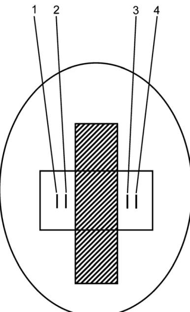
One pair of electrodes (electrodes 1-anode, and 2-cathode, Fig. 2.1) was used to deliver bipolar pacing stimuli from an isolated stimulator (Isostim A320, World Precision Instruments, Sarasota, FL). The pacing amplitude was set at twice the pacing threshold or at 10 mA if the threshold was ≥ 5 mA; pacing pulse width was 3 msec; pacing interval was chosen just above the resting heart rate and ranged from 340 to 660 msec. The other electrode pair (electrodes 3-negative and 4-positive) was used to record a bipolar electrogram. All signals (stimulus and electrogram) were conditioned with an isolation amplifier (AD210AN, Analog Devices, gain 100, low pass <16 kHz), and digitized at 10 kHz using Labview and a DAQPad 6070E data acquisition board (National Instruments, Austin TX). Inter-electrode spacing for each pair was 1-2 mm and spacing between the pairs was 10 mm. During optical mapping, stimulation and electrogram recording electrodes were Ag-AgCl (diameter 0.25 mm, length 2 mm). When electrograms were recorded without optical mapping, the pacing and recording electrodes were stainless steel (contact area with heart ~1 mm2 per electrode), and were attached to the RF probe and positioned in a line at 90 degrees from the ablation probe (Fig. 2.1).
Figure 2.1 Diagram showing the arrangement of the ablation probe and electrogram recordings on the heart. Ablation probe for continuous lesion is indicated in cross-hatched area. The open rectangle indicates the area mapped optically. Electrodes 1 and 2 were used for bipolar pacing; 3 and 4 were used for bipolar electrogram recording.
Fluorescence Recording
fluorescence (wavelengths >590 nm and 510-570 nm, respectively) were collected simultaneously with two photomultiplier tubes located in front of the heart. Each fluorescence signal was digitized at a rate of 1 kHz at each spot. Laser scanning and recording were controlled by a microprocessor system (M67, Innovative Integration, Simi Valley, CA) within a PC. To diminish effects of cardiac motion on the recordings, the green fluorescence signal was divided by the red signal133. The ratiometric signal was used to measure activation times in tissue at the laser spots.
Ablation Procedures
Analysis of Stimulus-Excitation Time Delay
The activation time for optical recordings was defined as the time of the maximum upstroke velocity (dVm/dtmax). The activation time for electrogram recordings was defined as the time at which the magnitude of the bipolar electrogram was greatest.
The stimulus-excitation time delay across the lesion was measured in two ways. For optical mapping, it was the difference between activation times at a spot near the pacing electrode and a spot near the recording electrode at the opposite side of the lesion. For electrogram recordings, the stimulus-excitation time delay was measured from the stimulus pulse to the activation time in the electrogram recorded on the opposite side of the lesion. This time delay included the time from the application of the stimulus pulse to excitation of cells near the stimulation electrode, which was measured to be 6-7 msec.
Viable Tissue Staining
Statistical Analysis
C. Results
Examination of TTC-stained Hearts
Photographs were taken of representative incomplete and complete lesions, and stained immediately post-procedure with TTC (Fig. 2.2). The incomplete lesions contained a gap between two discrete, transmural segments, or were not transmural. The complete lesions were continuous and transmural. The average length for complete lesions was 26.17 ± 1.37 mm. For all lesions, the average width was 7.1 ± 0.20 mm; depth was 3.1 ± 0.2 mm. Of 15 incomplete lesions, 12 were non-continuous, and 3 were non-transmural. Individual gap widths were 1.0, 1.5, 1.5, 1.0, 5.5, 4.0, 3.0, 7.0, 3.0, 2.0, 4.0, 3.0 mm, and had a mean of 3.0 ± 0.5 mm.
Electrograms
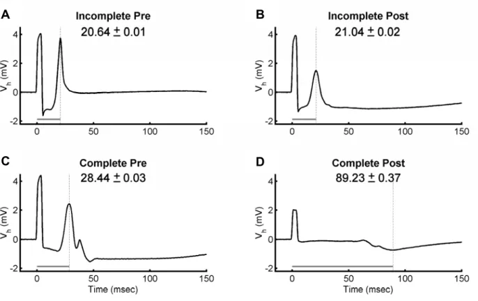
Electrograms were obtained before ablation and after production of complete and incomplete lesions (Fig. 2.3). For incomplete lesions, time delay did not change (21 msec pre-lesion and 21 msec post-lesion). After the formation of a complete lesion, time delay increased from 28 msec pre-lesion to 89 msec post-lesion.
A B
C D
Optical Maps
Time Delay
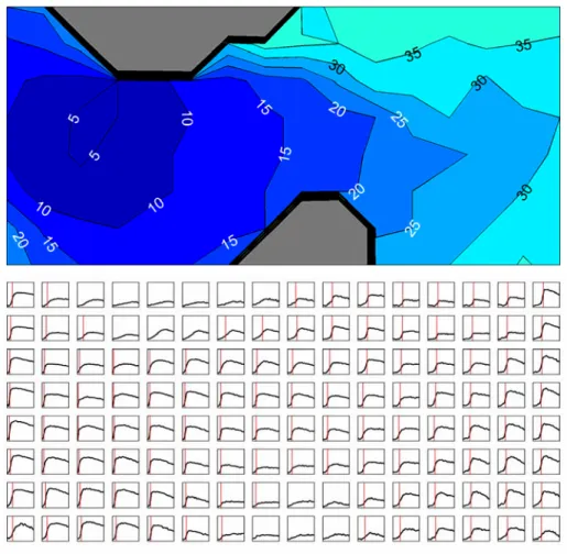
The time delays for complete and incomplete lesions as measured by electrogram and optical recordings are shown in Tables 2.1 and 2.2, and graphically in figure 2.7.
For incomplete lesions, post-lesion time delay did not differ significantly from the pre-lesion delay. The difference in post-lesion and pre-lesion mean time delay was 3 msec for electrogram measurements and 23 msec for optical recordings.
For complete lesions, the mean post-lesion time delay was significantly greater than the mean pre-lesion delay, for both electrogram (70 msec) and optical (65 msec) recordings (p<0.05). In addition, the post-lesion time delays were significantly longer for the complete lesions than they were for the incomplete lesions (p<0.05).
Electrogram measurements of the time delays produced lower SEM for incomplete lesions and a larger disparity in time delay measurements between complete and incomplete lesions than optical recordings.
Table 2.1 - Time Delay Measured by Electrogram Recordings
ELECTROGRAM Pre-lesion (msec) Post-lesion (msec) n
Incomplete 45 ± 12.20 48 ± 10.45 12
Complete 33 ± 3.97 103 ± 10.59a,b 14
Table 2.2 - Time Delay Measured by Optical Recordings
OPTICAL Pre-lesion (msec) Post-lesion (msec) n Incomplete 33 ± 10.11 56 ± 16.44 9 Complete 28 ± 8.59 93 ± 10.51a,b 9
a p<0.05 for post-lesion vs. pre-lesion
b p<0.05 for incomplete lesion vs. complete lesion
D. DISCUSSION
Methods used to determine presence of conduction block after RF ablation include P wave changes after ablation of the cavotricuspid isthmus for atrial flutter188, detailed electrogram mapping with or without electrical stimulation189,190, changes in electrogram amplitude191, and double potentials192. Many of these methods use intravascular catheter approaches and require several catheters for pacing and electrogram recordings, usually with continuous repositioning of the probe.
Endocardial ablation techniques for creating linear lesions for the treatment of atrial fibrillation using intravascular catheters have reported success rates of 20-60% despite established techniques for evaluating conduction block within the pulmonary veins193. Both surgical and catheter approaches for the treatment of atrial fibrillation have been hampered by the inability to create continuous and transmural lesions. Incomplete lesions have been associated with the recurrence of atrial fibrillation or the development of reentrant atrial tachyarrhythmias194.
Epicardial Approach
A diagnostic device incorporating bipolar pacing and recording electrodes capable of measuring time delay as described here should accurately assess ablation line continuity and transmurality, provide a metric for determining lesion completeness intraoperatively, and improve outcomes after atrial fibrillation treatment. Alternatively, diagnostic electrodes could be incorporated in the ablation device to simultaneously create the lesion and evaluate its completeness.
Time Delay
The results demonstrated recovery of time delay for lesions with gaps, whereas complete lesions had a low extent of recovery. This recovery may be due to revival of stunned tissue at the perimeter of ablated tissue188,189,192,196,197. For complete lesions, recovery may produce only a small decrease in the post-lesion path length (e.g., at the lesion ends), whereas recovery of tissue in an epicardial gap region may produce a large decrease in path length since conduction may again occur directly across the lesion. Increases in time delay were significantly larger for complete lesions and the magnitude of increase may predict stability of time delay. Complete lesions showed a greater than two-fold increase in time delay, and did not recover, indicating that the delay must exceed a threshold to avoid recovery of conduction post-ablation. A greater than two-fold increase in delay may indicate production of a complete lesion that was incapable of recovery.
anatomic structures or demonstrating the inability to induce an arrhythmia with known pacing techniques have been shown to predict long term success199.
The value of lesion completeness is a current topic of debate. It has been suggested that ablation lesions of myocardium may not need to be complete (continuous and transmural) and that over time lesions grow and outcomes improve. Results presented here suggest that incomplete lesions maintain trans-lesion conduction, which may account for the acknowledged high recurrence rates reported for catheter and surgical ablation of atrial fibrillation. This is further evidenced by Melby et al200, where electrical propagation persisted through narrow gaps in lesions created on isolated canine atria, even when conduction velocities were significantly slowed. Although atrial fibrillation required larger gaps for 1:1 propagation, their data suggest that even narrow gaps may conduct AF, and that to ensure total block, complete lines of ablation must be created. Alternatively, studies performed by Cabo et al. show that increasing pacing frequency increases the critical gap width size160, suggesting that small lesion gaps may not conduct activation of high-frequency activation which occurs during atrial fibrillation. However, lower frequency components may also be present during atrial fibrillation which may conduct through small gaps.
CHAPTER 3
ABILITY OF MINIMUM ACTIVATION PATH LENGTH TO PREDICT TRANSLESION TIME DELAY
A. INTRODUCTION
In typical lesion sets used to block and/or isolate arrhythmic conduction, activation path length is difficult to determine. However activation paths around simple linear lesions may be approximated and compared with pre-lesion pathways. These linear lesions may serve as building blocks for more complex lesion sets.
Estimation of conduction pathways around linear lesions would give an expected value of time delay based on pre-lesion conduction velocity, and may give additional confidence in time delay as an accurate predictor of lesion completeness.
Heterogeneities within the myocardium are known to affect conduction velocity 147,155-158,161,201-206, and may cause substantial deviations from expected time delays according to the nature and severity of the heterogeneity.
B. METHODS
Rabbit Heart Preparation
Fresh hearts isolated from New Zealand rabbits (N = 11) of either gender were perfused through the aorta with Tyrode’s solution (129 mM, NaCl, 5.4 mM KCl, 1.8 mM CaCl2, 1.1 mM MgCl2, 26 mM NaHCO3, 1 mM Na2HPO4, 11 mM Dextrose, and 0.6 μM bovine serum albumin). The solution was bubbled with 95% O2 and 5% CO2, and maintained at 37 ± 0.2ºC.
Lesions (N = 11) were produced on the hearts by RF ablation (100-1200 J/cm²). Two to three lesions were made on each heart, and approximately 60 minutes passed between the creation of each lesion. The animal was sacrificed approximately 30 minutes before the creation of the first lesion. Following perfusion with Tetrazolium Chloride (TTC), lesions were identified by their light gray appearance while viable tissue appeared dark red.
Electrical Pacing and Recording
TX). Inter-electrode spacing for each pair was 1-2 mm and spacing between the pairs was approximately 10 mm.
Ablation Procedures
Electrograms were taken before ablation on each heart (baseline). A prototype ablation device (nContact Surgical, Inc.) was used to create a lesion through the middle of the optical grid and between the stimulation and recording electrodes. RF energy was applied for 20-60 seconds at a power of 10-20 Watts using a 480 kHz generator. Complete lesions were created discrete and overlapping applications of RF energy using a 2-cm ablation electrode. Lesions extended from the apex to the atrioventricular annulus. Incomplete lesions were created by discrete applications of the ablation probe separated 1-7 mm apart to produce a gap, or by using a substantially lower power level to produce non-transmural lesions. Before and after each ablation, recordings were taken to examine the effects of ablation on the stimulus-excitation time delay.
Analysis of Stimulus-Excitation Time Delay
The activation time for optical recordings was defined as the time of the maximum upstroke velocity (dVm/dtmax). The activation time for electrogram recordings was defined as the time at which the magnitude of the bipolar electrogram was greatest.
pulse to the activation time in the electrogram recorded on the opposite side of the lesion. This time delay included the time from the application of the stimulus pulse to excitation of cells near the stimulation electrode, which was measured between 6-7 msec.
Viable Tissue Staining
After completion of all recordings, hearts were perfusion stained with 2,3,5-triphenyltetrazolium chloride (TTC, Sigma-Aldrich, Inc.; 2.1 g of TTC in 150 ml of 0.9% NaCl) to determine histologically the location and completeness of each lesion. After staining with TTC, viable tissue appeared red and non-viable tissue appeared grey (Fig. 3.1). Lesions were bisected to obtain photographs and to measure lesion width, depth, and length, and the minimum path length of viable tissue from the stimulation site to the recording site on the opposite side of the lesion.
Minimum Activation Path Length Determination
the annulus) and from there to the recording electrode position. Activation path length for an incomplete lesion is shown in figure 3.1. Optical time delays along with their respective activation path lengths were used to determine conduction velocities. If the optical time delay was not available for a particular lesion, the electrical time delay was used. An alternate method for predicting time delay which incorporates fiber direction is presented in an appendix following chapter 5.
C. RESULTS
D. DISCUSSION
CHAPTER 4
RECOVERY OF TISSUE WITHIN SMALL GAPS OF LINEAR RADIOFREQUENCY CARDIAC ABLATION LESIONS
A. INTRODUCTION
Trans-venous catheter ablation procedures have been shown to halt various forms of atrial fibrillation and atrial tachycardias. However these procedures are technically challenging to perform, and success rates vary widely according to operator experience, the specific nature of the arrhythmia present, and the type of procedure being attempted.
Approximately 90% of patients with paroxysmal atrial fibrillation can be cured by pulmonary vein isolation, however only a small percentage of patients with chronic atrial fibrillation may be treated with this technique183. For patients with chronic atrial fibrillation, more extensive lesion sets are desired, such as those required for the Maze III procedure. For the Maze III, a series of linear lesions are required in order to break up arrhythmic conduction in the left atrium, isolate the pulmonary veins, isolate or remove both atrial appendages, and form a corridor (“Maze”) which directs sinus activation toward the atrioventricular node170, and breaks up wavelets which sustain fibrillation.
catheter ablation lesions are not uncommon. Such gaps may recover over time following ablation, and may allow arrhythmic conduction to propagate across a lesion and sustain atrial fibrillation. Further, lesions which are incomplete may form new arrhythmic substrates for anatomically based macroreentrant circuits.
Cabo et al. performed experiments and simulations in order to characterize the minimum width of cardiac tissue slices necessary to support conduction160. They reported considerable variability in this “critical isthmus width”, which depended on pacing frequency and fiber direction. Cabo’s study addressed the importance of viable tissue width, however radiofrequency ablation lesions are complicated by the fact that partial damage to both endothelial cells within the microvasculature as well as to the myocytes may occur at distances up to 6 mm from the site of coagulation necrosis17,21. Further, a recent report by Perez et al. demonstrates the importance of gap geometry in discontinuous lesions209, and reveals that “angled gaps” (i.e., gaps which contain an approximately 90º turn in the path of conduction) are more likely to block conduction than either bifurcated gaps (~45º turn, but in two directions) or straight gaps. These factors suggest that additional complexity is present due to hyperthermic injury following radiofrequency ablation which may cause deviations from previous studies.
a small gap may acutely recover within one hour following ablation, and that after this time period has passed, acute recover is highly improbable.
B. METHODS
Rabbit Heart Preparation
Fresh hearts isolated from New Zealand rabbits (N = 7) of either gender were retrograded perfused through the aorta with Tyrode’s solution (129 mM, NaCl, 5.4 mM KCl, 1.8 mM CaCl2, 1.1 mM MgCl2, 26 mM NaHCO3, 1 mM Na2HPO4, 11 mM Dextrose, and 0.6
μM bovine serum albumin). The solution was bubbled with 95% O2 and 5% CO2, and maintained at 37 ± 0.2º C.
Lesions (N = 9) were produced on the heart by RF ablation (300-600 J/cm², 30 seconds). Two to three lesions were made on each heart, and approximately 60 minutes passed between the creation of each lesion. The animal was sacrificed approximately 30 minutes before the creation of the first lesion. The effects of ablation in epicardial regions of the anterior or posterior ventricular septum were studied. Also, control recordings were made on non-ablated epicardium to examine the stability of control recordings over time.
Electrical Pacing and Recording
at 10 kHz or 1kHz using Labview and a DAQPad 6070E data acquisition board (National Instruments, Austin TX). Inter-electrode spacing for each pair was 1-2 mm and spacing between the pairs was 10 mm. Pacing and recording electrodes were stainless steel rings (contact area with heart ~1 mm2 per electrode), and were attached to the RF probe and positioned in a line 90 degrees from the ablation probe (Fig. 4.1).
Figure 4.1 Five cm ablation probe with silicone gap. Helical stainless steel coil was used for ablation, while stainless steel ring electrodes were used to deliver bipolar pacing and record bipolar electrograms.
Ablation Procedure
Incomplete lesions were created by discrete applications of the 2-cm ablation probe separated approximately 5 mm apart to produce a gap, or by the 5-cm ablation probe as shown in figure 4.1 which contained a gap in the ablation electrode. Control recordings were taken prior to ablation, and then post-ablation recordings were taken to examine the effects of ablation on the stimulus-excitation time delay over a 60-minute period.
Selection of Activations in Fractionated Electrograms