Abstract
Introduction:
Breast cancer is a common malignancy in females in the United States and worldwide, and is associated with a high cancer-related mortality. Breast cancer is a heterogeneous disease that is diverse in natural history, histopathology, gene expression patterns, response to treatment, and patient outcomes. The spectrum of breast cancer includes several distinct biological and morphological subtypes. Clinical classification (based upon immunohistochemistry) and transcription profiling of invasive breast cancers has identified several subtypes with distinct clinical characteristics that are indicative of patient outcome and survival, including luminal A (ER+/PR+/HER2-), luminal B (ER+/PR+/HER2+), HER2+ (ER-/PR-/HER2+), and basal-like (ER-/PR-/HER2-) breast cancers (Sandhu). With the implementation of anti-estrogen therapies in combination with chemotherapy, the outcomes of ER+ breast cancer patients significantly
improved. Similarly, improved survival rates in HER2-overexpressing breast cancers reflect the use of various anti-HER2 compounds with chemotherapy in these patients. However, by virtue of lacking a targeted therapeutic approach, basal-like breast cancers represent a significant clinical challenge, requiring additional treatment options and novel treatment strategies to achieve improved results for patients.
DNMT3b in breast cancer cells sensitizes them to cell killing by cytotoxic drugs, the mechanism responsible for improved cell killing after treatment with epigenetic drugs has not been explored. In the current work, we performed a discovery-based study to identify genes associated with apoptosis that might be epigenetically regulated and silenced in breast cancer cells that exhibit aberrant DNA hypermethylation, and investigated the response of the identified genes to epigenetic treatment with 5-aza-2’-deocycytidine.
Methods
Breast Cancer Cell Lines and Cell Culture.
Human breast cancer cell lines BT20 (ATCC# HTB19), MCF7 (HTB22), MDA-MB-453 (HTB131), MDA-MB-231 (HTB26), SKBR3 (HTB30), Hs578T (HTB126) were obtained from the Tissue Culture Core Facility of the UNC Lineberger Comprehensive Cancer Center. Human breast cancer cell line SUM159 was obtained as a kind gift from the laboratory of Dr. Carolyn I. Sartor (Department of Radiation Oncology, UNC School of Medicine). All cell lines were propagated in DMEM/F12 mix medium (GIBCO/Invitrogen Life Technologies) containing 10% fetal calf serum (Hyclone) and 1% Antibiotic-Antimycotic (GIBCO/Invitrogen Life
Technologies). For some experiments, cell lines were cultured in DMEM/F12 mix medium, 10% fetal calf serum, 1% Antibiotic-Antimycotic, and 500 nM 5-aza-2’-deoxycytidine (Sigma-Aldrich) for 7 days (Sandhu et al., 2012, Breast Cancer Research and Treatment 131:385-399). RNA Isolation.
isolate were measured to ensure that there was no contamination from DNA or preparatory chemicals such as isopropanol using a NanoDrop1000.
Discovery of Candidate Genes Related to Apoptosis and Survival in Human Hs578T Breast Cancer Cells.
To identify candidate epigenetically-regulated genes in breast cancer, RNA isolates from Hs578T were utilized for gene discovery using Bio-Rad PrimePCR Pathway SYBR Green real-time PCR assays – Anti-Apoptotic TNFs/NF-kB/IAP Pathway (#100-25087), Apoptosis and Survival Tier 1 (#100-25097), Apoptosis and Survival Tier 2 (#100-25869), and Apoptosis and Survival Tier 3 (#100-25870). These assays provided information on 328 unique genes that contribute to several apoptosis-related pathways. The resulting gene expression levels were analyzed to identify genes expressed at low levels or not expressed in Hs578T cells, and to identify genes expressed at high levels in these cells.
Gene Expression Changes in Response to 5-aza Treatment.
To examine changes in gene expression following epigenetic treatment, multiple breast cancer cell lines (BT20, MCF7, MDA-MB-453, MDA-MB-231, SKBR3, Hs578T, and SUM159) were cultured in control medium or medium containing 500 nM 5-aza-2’deoxycytidine for 7 days. RNA isolates from control and 5-aza treated cells were utilized for Bio-Rad PrimePCR SYBR Green Assays for selected genes (based upon discovery results in Hs578T cells). Genes examined include FASLG (qHsaCED0003635), IGF1 (qHsaCED0038638), CD27 (qHsaCID0017180), BLK (qHsaCID0014815), HSPB1 (qHsaCED0023813), and NPM1
(qHsaCED0038211). Additional Tumor Necrosis Factor (TNF) and TNF Receptor superfamilies were examined, including TNFSF8 (qHsaCID0013499), TNFSF10 (qHsaCED0036477),
TNFRSF13B (qHsaCED0023685), TNFRSF17 (qHsaCED0020893), and TNFRSF18 (qHsaCID0011446). All real-time PCR assays were controlled using ACTB
(qHsaCED0036269).
Results
The expression of 328 genes related to apoptosis was examined in Hs578T cells to identify candidate epigenetically-regulated genes based upon expression pattern. Table 1 [Appendix] summarizes 36 genes that were found to be of interest - 27 genes that were not detected, 4 genes that were expressed at low levels, and 5 genes expressed at high levels. The majority of genes that were not expressed or expressed at low levels are inducers of apoptosis, whereas some of the genes expressed at high levels are inhibitors of apoptosis.
A subset of fourteen genes was selected for further examination in breast cancer cell lines that were treated with 500 nM 5-aza for 7 days. Several of these genes are known to be
methylation-sensitive.
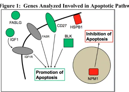
Figure 1: Genes Analyzed Involved in Apoptotic Pathways
(TNFRSF7), ligands for FASR (FASLG) and IGF1R (IGF1), and cytoplasmic tyrosine kinase (BLK). The protein products for these genes are shown in green in the schematic. Two genes that inhibit apoptosis were examined, including a chaperone of the small heat shock protein HSPB1 (HSP27) and a nucleolar phosphoprotein NPM1. The protein products for these genes are shown in red in the schematic.
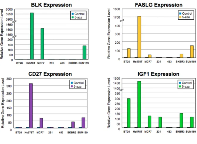
Figure 2: Expression of Genes Regulating Pro-Apoptotic Pathways in Control and 5-aza Treated Cell Lines
specific genes. BLK is known to be methylation-sensitive, consistent with these responses to 5-aza treatment. These results suggest that FASLG, CD27, and IGF1 may also be subject to methylation-dependent silencing in breast cancer. Further studies are needed to examine promoter methylation events in these genes.
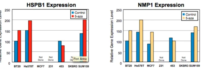
Among the genes examined that regulate anti-apoptotic pathways (and/or inhibit
apoptosis), both were found to be expressed abundantly in all breast cancer cell lines investigated as seen in Figure 3. In some cases, 5-aza treated cells expressed more of these genes, but the induction of expression in response to 5-aza was substantially less in magnitude (related to the high baseline levels of expression observed).
Figure 3: Expression of Genes Regulating Anti-Apoptotic Pathways in Control and 5-aza Treated Cell Lines
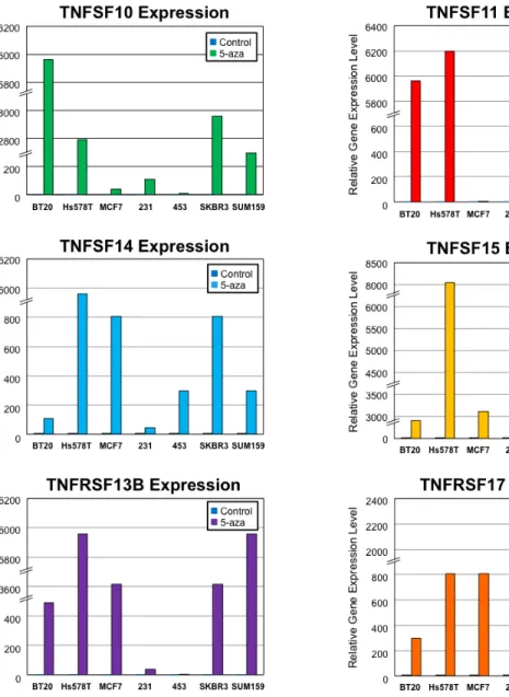
A specific subset of genes coding for tumor necrosis factor molecules and their receptors was examined as these gene regulate pro-apoptotic pathways. Figure 3 illustrates that in
conjunction with BLK, FASLG, CD27, and IGF1, TNF and TNFR genes showed lack of
in the index Hs578T cell line, and substantial induction was also seen in specific cell lines for specific genes. TNFSF10, TNFRSF1B, and TNFRSF8 are known to be methylation-sensitive, consistent with these responses to 5-aza treatment. These results suggest that TNFSF11, TNFSF14, TNFSF15, TNFRSF13B, and TNFRSF17 may also be subject to methylation-dependent silencing in breast cancer.
Discussion
Epigenetics is a course of study that is currently under wide investigation as many of these mechanisms that regulate gene expression are still unknown. Here we explored the effect of a demethylating agent on human breast cancer cells lines and the epigenetic mechanism for chemotherapeutic resistance. It has been shown that using 5-aza to inhibit DNMT3b, which encodes a DNA methyltransferase, can increase the efficacy of cytotoxic treatment (Mohandas). While we know that 5-aza can be effective, the mechanism of this drug is still unknown.
By combining epigenetic and cytotoxic treatments, we hope patients with cancer who are resistant to standard treatment may be reached. Epigenetic silencing of pro-apoptotic genes, as seen in the breast cancer cell line Hs578T, would give a survival advantage to cancer cells attempting to metastasize, as these cells would grow infinitely. If we are able to reactivate these apoptotic pathways with a demethylating agent such as 5-aza, then we could sensitize these cells to chemotherapeutic treatments to allow for programmed cell death. These treatments could also be made available as one sensitive cancerous tumor develops a resistance to treatment.
Our results could be improved by using a general control cell line. Our experiment used HS578T as a control but we need an intentional control cell line to compare the treated and non-treated gene expression. Possible error in this investigation could come from DNA or other chemical contamination in the RNA isolate samples. Additionally, we made the assumption that all the down-regulated genes we found are methylation-sensitive or significant in someway while they could just be unexpressed in all breast cells.
interest. This method would assist in focusing on the epigenetic mechanisms of cancer
development. Finally, to quantize our results we could use apoptosis detection kits consisting of an APO-tag that labels the cells of nuclei going through apoptosis. With control and 5-aza cell lines, apoptosis would be induced and the nuclei counted.
Possible goals for the future may include using varying dosages of 5-aza to see if any additional genes are affected by a more concentrated treatment. We could also explore other ‘triple-negative’ breast cancer cell lines along with some of the less aggressive forms of breast cancer. Another possibility would be to perform Western blots to study post-translational modifications of proteins in the breast cancer cells.
References
1. Centers for Disease Control and Prevention. (2013). Breast Cancer Statistics.
http://www.cdc.gov/cancer/breast/statistics/
2. Sandhu, Rupinder, Parker, Joel S., Jones, Wendell D., Livasy, Chad A., William B. Coleman. “Microarray-Based Gene Expression Profiling for Molecular Classification of Breast Sancer and Identification of New Target for Therapy” LabMedicine. 41. 6(2010): 1-10. Print.
3. Sandhu, Rupinder, Rivenbark, Ashley, and William B. Coleman. “Enhancement of chemotherapeutic efficacy in hypermethylator breast cancer cell lines through targeted and pharmacologic inhibition of DNMT3b” Springer. Large 10549. 1409 (2011): 1-15. Print.
Appendix
Table 1: Showing expression levels of genes related to apoptosis and survival in the index breast cancer cell line Hs578T