Neural Signatures of Affective Processing in PTSD Colleen J. Watson
The University of North Carolina at Chapel Hill Under the guidance of Dr. Aysenil Belger
Acknowledgments
Abstract
Emotional regulation is a fundamental aspect of adaptive behavior that is often disrupted in patients with post-traumatic stress disorder (PTSD). The goal of the present study was to
examine the neural correlates of social emotional processing in PTSD and their association with PTSD symptom severity.
Methods: 100 veterans with PTSD and 20 healthy controls without PTSD or military history underwent functional magnetic resonance imaging (fMRI) while completing an emotional face-matching task. Additionally, subjects completed the Clinician Administered PTSD Scale (CAPS), Barratt Impulsiveness Scale (BIS), and DKEFS Color-Word interference tasks. I hypothesized that the PTSD group would show greater emotional reactivity compared to control subjects in that veterans would show greater amygdala and limbic and less medial frontal cortex (mFC) activity. I further hypothesized that hyperarousal of the amygdala in the PTSD group would be associated with greater CAPS, BIS, and lower DKEFS Color-Word interference scores. Results: Veterans demonstrated greater mFC and posterior temporal fusiform cortex (ptFC) activity than controls during faces, and greater mFC, ptFC, and orbito-frontal cortex (OFC) activity than controls in the shapes block. No differences were found in amygdala activation or error rates. There were many significant correlations between regional brain activation and clinical measures, notably, between the right amygdala and total CAPS scores.
PTSD – A Disorder of Emotional Processing and Regulation
Emotion processing and regulation are fundamental aspects of adaptive behavior. When they are impaired, so are social interactions and interactions with the world. A disruption in emotional processing might lead to misattributions of threat to non-threatening stimuli. Dysfunctions of emotional regulation have further been associated with irritability, apathy, or uncontrollable anger, anxiety, or fear. Ultimately, emotional dysregulation can result in behavior that is risky, violent, or impulsive (Arciniegas & Wortzel, 2016).
Dysregulation in emotional processing and related behavioral disinhibition are frequently reported in conjunction with posttraumatic stress disorder (PTSD) (Arciniegas & Wortzel, 2016). PTSD is prevalent in veterans of Operation Enduring Freedom/Operation Iraqi Freedom
(OEF/OIF), the wars that have taken place in Iraq and Afghanistan since 2001 (Hoge et al., 2008). PTSD is an anxiety disorder that may or may not develop in individuals who have been exposed to a horrifying event (American Psychiatric Association, 2013; Frewen & Lanius, 2006; Weiss, Tull, Viana, Anestis, & Gratz, 2012). PTSD is characterized by “re-experiencing
symptoms”, a category that includes nightmares, flashbacks, and hypervigilance (Aupperle, Melrose, Stein, & Paulus, 2012). PTSD can also result in many other symptoms of emotion dysregulation, including anhedonia (inability to feel positive emotions), and persistence of negative emotions such as fear, guilt, anger, and horror (American Psychiatric Association, 2013). The CAPS is a broadly used questionnaire that clinicians utilize to diagnose and assess severity of PTSD; it will be used in this study to assess the severity of PTSD symptoms and relate them to regions of brain activation.
group, which includes irritability and aggression, recklessness and self-destructive behavior, hypervigilance, a tendency to startle easily, and difficulties concentrating and sleeping (American Psychiatric Association, 2013). These aspects of emotional dysregulation are also frequently associated with impulsivity. In the present study, we will further examine whether severity of impulsivity may be associated with aberrant emotional reactivity or dysregulation. Impulsivity will be measured using the Barratt Impulsivity Scale (BIS), a clinical questionnaire and self-report measure of impulsivity. The advantage of the BIS is that it allows the researcher to gather information not easily studied in a laboratory setting, such as risky sexual behavior.
The hyperarousal and affective dysregulation characteristics of PTSD have been associated with a dysfunctional corticolimbic circuit that causes misidentification of stimuli as threatening (Hariri, 2015). To understand the hypotheses and results of the present study, which features fMRI, it is helpful to have some background knowledge of the neural circuits that are at the focus of the study. A survey of existing knowledge is below.
Corticolimbic Neuroanatomy and Emotional Processing and Self-Regulation
The corticolimbic circuit consists of the thalamus, sensory cortices, hypothalamus, brainstem, substantia innominata, insula, hippocampal formation, and prefrontal cortex (PFC) (Hariri, 2015). Abnormalities of the circuit have been implicated in aggressive, impulsive behavior (Brown, Manuck, Flory, & Hariri, 2006; Coccaro, Sripada, Yanowitch, & Phan, 2011). The amygdala is the hub of the corticolimbic circuit (Hariri, 2015), and is strongly
is active during reminders of a traumatic event and during hyperarousal (Rauch et al., 2000), it is not activated by the cognitive portion of tasks (Morey et al., 2009); the medial PFC (mPFC) is.
One role of the mPFC is to place stimuli in context. Thus, when the function of the mPFC is disrupted, nonthreatening stimuli might be placed in a threatening context, resulting in
common PTSD symptoms such as hyperarousal (Liberzon & Martis, 2007). The ventral mPFC (vmPFC) is responsible for processing physiological and behavioral changes ordered by the amygdala. It receives input from the sensory cortices, hippocampal formation, and insula, which allow it to assign valence to basic stimuli. Input from the amygdala causes the vmPFC to attend to salient stimuli, whether sensory, mnemonic, physiological, or hedonic (Hariri, 2015).
The mPFC evaluates input from the amygdala and uses that information to modify the body’s physiological response. Then, importantly, the mPFC inhibits further signals from the amygdala, returning the body to its baseline state. Specifically, the amygdala excites the vmPFC, which in turn excites the dmPFC, which inhibits the amygdala (Hariri, 2015). An inverse
relationship exists between amygdala and prefrontal activity for fear stimuli: amygdala activity increases while PF activity decreases (Bryant et al., 2007; Shin et al., 2006). Hariri, Bookheimer, and Mazziotta (2000) found the same effect in the right amygdala only.
emotional face-matching task previously employed by Hariri (2000) and Hariri et al. (2002) during fMRI (see Figure 1).
Second, the DKEFS Color-Word Inhibition task is a neurocognitive method that will be used to measure impulsivity in terms of attention. In this case, impulsivity is the inability to inhibit responses (Moeller, Barratt, Dougherty, Schmitz, & Swann, 2001).
Given what is known about amygdala and prefrontal differences in PTSD, I hypothesize that subjects with PTSD will show greater emotional reactivity compared to control subjects during the fMRI emotional face-matching task. I predict the PTSD group will show greater amygdala and lesser mPFC activity relative to the control group during the emotional face-matching block but not the control shape-face-matching block. If confirmed, this prediction will replicate findings that the amygdala is hyperactive in PTSD (Hariri, 2015; Hariri et al., 2000; Rauch et al., 2000; Shin et al., 2006; Shvil, Rusch, Sullivan, & Neria, 2013), even when stimuli are affective but not specifically related to trauma (Hariri, 2015; Shin et al., 2006). Furthermore, it will support theories that the mPFC and amygdala have an inverse relationship (Bryant et al., 2007; Hariri, 2015; Liberzon & Martis, 2007; Shin et al., 2006; Shvil et al., 2013). I also predict that the PTSD group will commit more errors on the emotional face matching task than controls due to the predicted hypoactivation of the mPFC, which assigns valence to stimuli and aids in cognitive control (Hariri, 2015; Liberzon & Martis, 2007). This prediction can be tested independently of general attention difficulties by comparing the results on the affective face-matching task to the results on an emotionally neutral shape task, for which the amygdala should not be hyperactivated in either the control group or the PTSD group.
Clinician-Administered PTSD Scale (CAPS), Barratt Impulsivity Scale (BIS), and Delis-Kaplan Executive Function System (D-KEFS) color-word interference task. This prediction is supported by research that indicates that the degree of amygdala hyperactivation positively correlates with the severity of PTSD symptoms (Hariri, 2015; Shin et al., 2006; Rauch et al., 2000). The D-KEFS task, a task of cognitive inhibition, is included because emotion regulation requires both affective processing and cognitive control (Brown et al., 2006). I seek to build upon the
knowledge contributed by Vasterling, Verfaellie, and Sullivan (2009): veterans with comorbid mTBI/PTSD are less proficient than veterans with mTBI alone in the naming and color-word interference conditions of the D-KEFS color-color-word task, but do not differ in non-inhibition-related cognitive tasks. How do veterans with PTSD perform on these color-word tasks in comparison to controls with no history of psychiatric disorders?
The results of this study will add to the literature on the role of the amygdala and mPFC in PTSD with TBI. They will also contribute to a better understanding of how general
impulsivity correlates with regional activations in the brain during emotional arousal. Although there is existing research on emotional dysregulation in PTSD, there is a lack of research examining the relation of this disorder to impulsivity in particular. This is an important topic to explore, given that an inability to inhibit behavior or emotion can result in angry or violent outbursts, poor decision-making, and other serious consequences.
Methods Participants
subject recruitment website. In this study, a veteran is defined as someone who served in one of the branches of the military for one or more tours in Iraq or Afghanistan since October 2001. The veterans (8 female) were between the ages of 27 and 63 (M=35.9, SD=8.77). Veterans younger than 18 or older than 65 were excluded from the study. All participants were required to pass an MRI safety screening questionnaire that ensured the absence of ferromagnetic material within their bodies, as well as screening for pregnancy. PTSD symptoms were confirmed to be clinically significant if the participant scored above 48 on the Davidson Trauma Scale.
The 20 controls were recruited from flyers posted throughout the Durham-Chapel Hill area and through Join the Conquest. All participants were between the ages of 24 and 39 (M=30.35, SD=4.15). One was female, corresponding to the proportion of female veterans. Exclusionary criteria included a diagnosis of PTSD or any other psychiatric condition, including depression or anxiety. Healthy controls must not have served in the military. Like the veteran group, controls were required to pass an MRI safety screening questionnaire.
Controls were compensated $100 total; veterans were compensated $250 per day of data collection for a total of $500. All participants provided written informed consent. This study was approved by the Institutional Review Board (IRB) of the University of North Carolina at Chapel Hill (UNC-CH).
Procedure
MRI) lasted about two hours, and were scheduled over the course of two to three days, according to participants’ availability.
Clinical and Neurocognitive Assessments
A battery of clinical assessments was administered to veterans: notably, the CAPS provided information about the severity of PTSD symptoms and the BIS measured impulsivity. Controls also completed the BIS, but not the CAPS. Cognitive impulsive control was assessed in both groups using the DKEFS color-word interference task.
MRI
fMRI images were acquired on the 3T Siemens Magnetom Prisma MR B17. Participants
were greeted at the MRI 30 minutes before their scan, given an MRI safety screening form, and instructed to change into the scrubs provided for them. A urine drug test was administered to control for other factors affecting fMRI results; positive drug tests did not exclude subjects from the study. Female subjects were given urine tests to rule out pregnancy, a contraindication for MRI.
Next, the subjects practiced the emotional face-matching task outside of the scanner. In the control condition (referred to either as the “control” or “shapes” condition) of this task, two ovals of different orientations appeared in the lower half of the screen, and an oval matching one of the two appeared above. The task was to identify either the left or right bottom oval as
matching the top (Figure 1). In the experimental (also called “faces”) condition, the ovals were replaced with emotional faces displaying fear or anger. Participants identified which of the two faces on the bottom matched the top.
Results
The 20 control participants, ages 24-39 (M=30.35, SD=4.15; 1 female) were matched with 20 veterans with PTSD, ages 24-38 (M=28.75, SD=3.35; 1 female) as closely as possible based on age and race. These 20 veterans were a subset of the total sample of 100 (M=35.9, SD=8.77, 8 female). Of the 20 controls, 12 (60%) identified as “White, not Hispanic”, two (10%) as “Black, not Hispanic”, two (10%) as “Hispanic, White”, three (15%) as “Asian”, and one (5%) as “Other”. Of the 20 matched veterans, 15 (75%) identified as “White, not Hispanic”, three (15%) as “Black, not Hispanic”, and two (10%) as “Hispanic, White”.
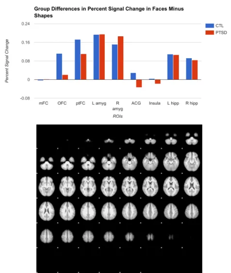
of the hypothesis, however, amygdala activation was consistent across the two groups. Although there were differences in the shape and face blocks individually, when activation during the control shape block was subtracted from activation during the face block, resulting in a
hypothetical isolation of emotion independent of attention, controls did not differ from veterans in any region, as shown in Figure 5. Because the subtractive condition does not provide all evidence of group differences, the control, experimental, and experimental-minus-control conditions are all explored to some degree throughout these analyses.
An independent samples t-test was performed on the behavioral data to determine if there were differences in response accuracy rates between veterans and controls on the emotional face-matching task. The t-test demonstrated no difference in accuracy of responses between controls (M=0.99, SD=0.02) and veterans (M=0.98, SD=0.04) during the face block; t(28.88)=1.32, p>0.05. Likewise, a t-test demonstrated no difference in performance for controls (M=0.94, SD=0.01) and veterans (M=0.94, SD=0.02) in the shape block; t(28.60)=1.78, p>0.05. For this particular test, equality of variance could not be assumed between the two groups.
Within-groups
Within the PTSD group, bivariate correlations were run to demonstrate the relationship of percent signal change in nine ROIs with scores on the CAPS, BIS, and DKEFS CW inhibition tasks, as well as a clinical measure of anger. This analysis allowed us to explore the correlation between impulsivity and symptom severity and regional brain activation. Statistics are displayed in Figures 6-8 and detailed below. Additionally, the relationships of all ROIs to each other in terms of covariance of percent signal change were explored and can be found in detail in Figures 9-11. All ROIs were significantly positively correlated with all others.
Originally, it was hypothesized that amygdala activity would correlate positively with CAPS scores. In actuality, when the shapes condition was subtracted from the faces condition, right amygdala activity was negatively correlated with total CAPS scores (Figure 12), as well as subscores B (re-experiencing) and C (avoidance and numbing). The significant relationship of subscore C, which is a combination of CAPS measures of avoidance and numbing, was driven primarily by avoidance, not numbing. The left amygdala showed no relationship with any clinical score.
In the face block alone, a negative correlation was found between right amygdala activity and total CAPS scores (Figure 12), as well as subscores C and D (hyperarousal). In the shapes block, total CAPS scores and B and C subscores increased as right amygdala activity increased (Figure 12).
CAPS – Other ROI Relationships
When the control condition was subtracted from the experimental condition, isolating a purely affective condition, total CAPS score and subscore C (driven more by avoidance than numbing) were found to decrease as right hippocampus activity increased. Higher total CAPS scores also corresponded to less ptFC activation. Avoidance was negatively correlated with left
hippocampus and mFC. Subscore B was negatively correlated with left hippocampus. An indirect relationship was demonstrated between subscore D and ptFC activation.
In the face condition, all relationships were indirect. Total CAPS scores were negatively correlated with mFC, right hippocampus, ACG, and OFC activation. Avoidance scores decreased as mFC, insula, right hippocampus, ACG, and OFC percent signal change increased, and
numbing combined form CAPS subscore C, which was negatively correlated with mFC, left and right hippocampus, ACG, OFC, and ptFC activity.
Unlike faces, shapes elicit a direct relationship between CAPS scores and regional activation. Total CAPS score and subscore C increased alongside percent signal change increases in the right hippocampus. Avoidance was directly associated with mFC, left hippocampus, and right hippocampus. A direct relationship was found between subscore B and the left and right hippocampi.
BIS – Amygdala Relationship
The hypothesis that greater amygdala activity would be related to higher BIS scores was not supported. No relationship was found between BIS scores and any ROI, whether in the face, shape, or subtractive condition.
DKEFS – Amygdala Relationship
In the face block, DKEFS scores were indirectly correlated with activity in the left amygdala. However, the hypothesis that greater amygdala activity during the subtractive condition would be related to lower DKEFS scores was not substantiated in any other way.
Anger
As part of the clinical battery, a measure of anger was administered. Scores were found to be negatively correlated with activation in the right hippocampus during the face-minus-shapes condition. During the face condition, anger was again negatively correlated with right
hippocampus activity, as well as activity in the mFC and right amygdala. In the shape condition, there was a direct relationship between anger scores and right hippocampus activation.
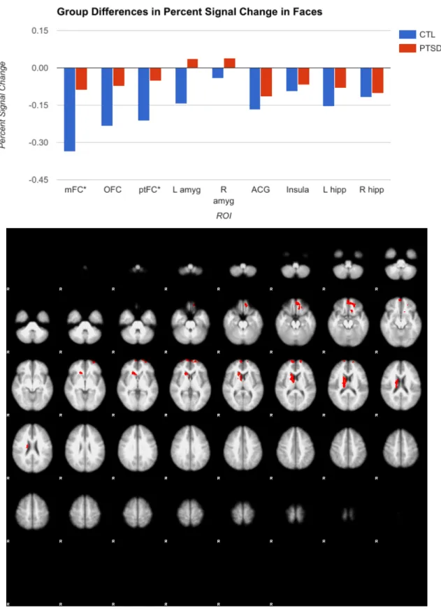
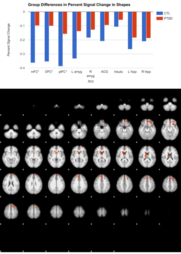
Extant research indicates that affective stimuli such as emotional faces, even stimuli that are not directly related to trauma, result in greater activation of the amygdala in PTSD compared to controls (Hariri, 2015; Shin et al., 2006). However, my research found no difference in amygdala activation between the two groups. Veterans did not differ from controls in limbic activation, contrary to hypothesis, but did have significantly more activity than controls in the mFC and OFC for both the emotionally arousing face-matching task and the non-emotionally arousing shape-matching control task. The mFC and OFC are frontal regions related to cognition and social cognition, respectively (Hariri, 2015). The fact that the mFC and OFC are hyperaroused in PTSD but not in controls might suggest that these frontal areas must work harder to regulate the amygdala in PTSD, lest it become hyperactive. This is also a possible explanation for why veterans do not show the predicted greater amygdala activity.
Veterans demonstrate greater activity in the ptFC than controls when viewing both faces and shapes, potentially indicating that PTSD is associated with face and non-face objects being processed with more salience, just as non-traumatic imagery has been shown to be processed as traumatic (Hariri, 2015; Shin et al., 2006).
attention independent of affect (instead of the reverse). In other words, the PTSD group allocates more attention at all times, not just to affective stimuli.
I predicted that veterans would commit more errors on the emotional task. In actuality, the groups did not differ in error rates, likely because face-matching is a simple, perceptual task, the purpose of which is not so much to assess working memory or executive function as to elicit an affective neural response. However, veterans required greater activation of OFC, mFC, and ptFC to achieve the same levels of accuracy as controls, which might suggest that this increased activation is a compensatory mechanism. In other words, the PTSD group must devote more energy on a neural basis to achieve the same results.
Within-PTSD Group
First, it should be noted that the data did not support the theory widely demonstrated in previous literature that mPFC and amygdala have an inverse relationship (Bryant et al., 2007; Hariri, 2015; Liberzon & Martis, 2007; Shin et al., 2006; Shvil et al., 2013); all nine regions were significantly positively correlated with one another, as shown in Figures 9-11.
Inversely, higher activation corresponds to lower CAPS scores; thus, another interpretation is that these areas regulate symptoms. For example, the negative correlation between right amygdala and total CAPS score and subscores B and C during the subtractive condition might suggest that right amygdala regulates re-experiencing and avoidance. The amygdala increases in activation, and CAPS scores decrease.
The results of the shape block are informative in a different way. Activity in several ROIs was correlated with several measures of symptoms; all relationships were direct, the opposite direction from all relationships in the face and subtractive blocks. Right amygdala activity increased alongside total CAPS scores, indicating that amygdala activity that generalizes to non-emotionally arousing tasks, where there should be low activity, may predict more severe PTSD symptomatology. Right hippocampus activity was also directly correlated with total CAPS scores, indicating that activation of other corticolimbic areas during this basic perceptual task is again correlated with higher PTSD symptomatology. Avoidance symptoms increased in direct relation to mFC, left and right hippocampus activity; re-experiencing increased with left and right hippocampus and right amygdala; and subscore C increased with right amygdala and right hippocampus. A conclusion that can be drawn is that inappropriate activation of affective processing areas is linked to more severe PTSD symptoms.
Anger is a symptom of PTSD that is not directly measured by the CAPS, but was
measured as a separate survey in the present study. The negative correlations between anger and activity in the right hippocampus, mFC, and right amygdala during the face condition might indicate that these regions are involved in regulation of anger. In the face-minus-shape condition, activity in the right hippocampus was negatively correlated with anger, while in the shape
Previous research has found that abnormalities of the corticolimbic circuit are related to impulsive behavior (Brown, Manuck, Flory, & Hariri, 2006; Coccaro, Sripada, Yanowitch, & Phan, 2011). However, my research did not substantiate this claim. The chosen measure of impulsivity, the BIS, was uncorrelated with all regions in all conditions, possibly because the complex, real-world measures of impulsivity in the BIS involve too many components of different symptoms to predict with any one region of the brain.
The lack of correlations between regional activation and DKEFS scores may signify that DKEFS is more related to prefrontal areas than corticolimbic ones, and thus, the ROIs in
question at present were not those most involved with DKEFS results. Furthermore, DKEFS and the face-matching task measure distinct outcomes (cognitive inhibition versus affective arousal to faces, respectively). It would seem that these outcomes are simply less related than originally predicted.
Future Directions & Limitations
In the future, the implications of this research could be a targeted neural circuitry-based intervention for victims of PTSD. One finding of this study was that activation across the corticolimbic circuit is highly correlated; the next step is to analyze in what way regions are connected. To do this, future research should explore diffusion tensor imaging (DTI) in PTSD and how it compares to healthy controls. DTI data was collected as a part of this study, but its analysis was outside the scope of this paper. Future research should utilize that data in order to identify differences in white matter connectivity between the two groups. Volumetric analyses should also be conducted between the two groups. Decreased amygdala volume corresponds with decreased inhibitory control (Depue et al., 2014), and decreased volumes of the
PTSD (Dolan et al., 2012; Shin et al., 2006). However, only a decrease in hippocampal volume has been well replicated (Dolan et al., 2012). Could these findings be replicated or negated in the current population? How would volume correlate with CAPS scores and measures of anger and impulsivity?
A major limitation of this study was that all veteran participants had also sustained at least one traumatic brain injury during their service. Although some data was collected on the number and severity of TBIs, measures were not detailed in terms of time since incurrence or location of the injury. Thus, the present project did not explore how severity of TBI moderates results; future studies should do so.
References
American Psychiatric Association. (2013). Trauma- and Stressor-Related Disorders. In Diagnostic and statistical manual of mental disorders (5th ed.). doi:
10.1176/appi.books.9780890425596.744053
Arciniegas, D. B., & Wortzel, H. S. (2016). Emotional and Behavioral Dyscontrol After Traumatic Brain Injury. Psychiatric Clinics of North America, 37(1): 31-53.
Aupperle, R. L., Melrose, A. J., Stein, M. B., & Paulus, M. P. (2012). Executive Function and PTSD: Disengaging from trauma. Neuropharmacology, 62: 686-694.
Brown, S. M., Manuck, S. B., Flory, J. D., & Hariri, A. R. (2006). Neural basis of individual
differences in impulsivity: Contributions of corticolimbic circuits for behavioral arousal and
control. Emotion, 6(2): 239-245.
Bryant, R. A., Kemp, A. H., Felmingham, K. L., Liddell, B., Olivieri, G., Peduto, A., … Williams, L. M. (2008). Enhanced amygdala and medial prefrontal activation during nonconscious processing of fear in posttraumatic stress disorder: An fMRI study. Human Brain Mapping, 29(5): 517-523.
Coccaro, E. F., Sripada, C. S., Yanowitch, R. N., & Phan, K. L. (2011). Corticolimbic Function in Impulsive Aggressive Behavior. Biological Psychiatry, 69(12): 1153-1159.
Defense and Veterans Brain Injury Center. (2016, Nov 8). TBI Basics. Retrieved from http://dvbic.dcoe.mil/about-traumatic-brain-injury/article/tbi-basics
Dolan, S., Martindale, S., Robinson, J., Kimbrel, N. A., Meyer, E. C., Kruse, M. I., … Gulliver, S. B. (2012). Neuropsychological Sequelae of PTSD and TBI Following War Deployment among OEF/OIF Veterans. Neuropsychology Review, 22(1): 21-34.
Frewen, P.A., & Lanius, R.A. (2006). Toward a Psychobiology of Posttraumatic Self-Dysregulation. Annals of the New York Academy of Sciences, 1071(1): 110-124.
Hariri, A.R. (2015). Looking Inside the Disordered Brain. Sunderland, MA: Sinauer Associates, Inc. Hariri, A.R., Bookheimer, S.Y., & Mazziotta, J.C. (2000). Modulating emotional responses: effects
of a neocortical network on the limbic system. NeuroReport, 11(1): 43-48.
Hariri, A.R., Tessitore, A., Mattay, V.S., Fera, F., & Weinberger D. R. (2002). The amygdala
response to emotional stimuli: a comparison of faces and scenes. Neuroimage, 17(1): 317-23. Hoge, C.W., McGurk, D., Thomas, J.L., Cox, A.L., Engel, C.C., Castro, C.A. (2008). Mild
Traumatic Brain Injury in U.S. Soldiers Returning from Iraq. The New England Journal of Medicine, 358(5): 453-463.
Liberzon, I. & Martis, B. (2006). Neuroimaging Studies of Emotional Responses in PTSD. Annals of the New York Academy of Sciences. 1071(1): 87-109.
Moeller, F.G., Barratt, E. S., Dougherty, D. M., Schmitz, J. M., & Swann, A. C. (2001). Psychiatric Aspects of Impulsivity. American Journal of Psychiatry, 158(11): 1783-1793.
Rauch, S.L., Whalen, P. J., Shin, L. M., McInerney, S. C., Macklin, M. L., Lasko, N. B. … Pitman, R. K. (2000). Exaggerated amygdala response to masked facial stimuli in posttraumatic stress disorder: a functional MRI study. Biological Psychiatry, 47(9): 769-776.
Shin, L.M., Rauch, S.L. & Pitman R.K. (2006). Amygdala, Medial Prefrontal Cortex, and
Hippocampal Function in PTSD. Annals of the New York Academy of Sciences, 1071(1): 67-79.
Shvil, E., Rusch, H. L., Sullivan, G. M., & Neria, Y. (2013). Neural, Psychophysiological, and Behavioral Markers of Fear Processing in PTSD: A Review of the Literature. Current Psychiatry Reports, 15(5): 1-10.
Simmons, A. N. & Matthews S. C. (2012). Neural circuitry of PTSD with or without mild traumatic brain injury: A meta-analysis. Neuropharmacology, 62(2): 598-606.
Swick, D., Honzel, N., Larsen, J., Ashley, V., & Justus, T. (2012). Impaired Response Inhibition in Veterans with Post-Traumatic Stress Disorder and Mild Traumatic Brain Injury. Journal of the International Neuropsychological Society, 18(5): p. 917-926.
Vanderploeg, R. D., Belanger, H. G., Curtiss, G. (2009). Mild traumatic brain injury and
posttraumatic stress disorder and their associations with health symptoms. Archives of
Physical Medicine and Rehabilitation, 90: 1084-93. doi:10.1016/j.apmr.2009.01.023
Vasterling, J. J., Verfaellie, M., & Sullivan, K. D. (2009). Mild traumatic brain injury and
posttraumatic stress disorder in returning veterans: Perspectives from cognitive neuroscience. Clinical Psychology Review, 29, 674-684. doi:10.1016/j.cpr.2009.08.004
Appendix
Figure 4: Another view of significant group differences, primarily in the mFC and OFC, from FSL, constructed in the MNI standard space. Results of the face block are in red, while results of the shape block appear in blue. In both cases, the PTSD group had greater activation than the control.
Figure 6
Figure 8
Figure 10