Microgravity induces proteomics
changes involved in endoplasmic
reticulum stress and mitochondrial
protection
Bryan J. Feger
1,*, J. Will Thompson
2,*, Laura G. Dubois
2, Reddy P. Kommaddi
3,
Matthew W. Foster
2,3, Rajashree Mishra
1, Sudha K. Shenoy
3, Yoichiro Shibata
4,
Yared H. Kidane
5,6, M. Arthur Moseley
2, Lisa S. Carnell
7& Dawn E. Bowles
1On Earth, biological systems have evolved in response to environmental stressors, interactions dictated by physical forces that include gravity. The absence of gravity is an extreme stressor and the impact of its absence on biological systems is ill-defined. Astronauts who have spent extended time under conditions of minimal gravity (microgravity) experience an array of biological alterations, including perturbations in cardiovascular function. We hypothesized that physiological perturbations in cardiac function in microgravity may be a consequence of alterations in molecular and organellar dynamics within the cellular milieu of cardiomyocytes. We used a combination of mass spectrometry-based approaches to compare the relative abundance and turnover rates of 848 and 196 proteins, respectively, in rat neonatal cardiomyocytes exposed to simulated microgravity or normal gravity. Gene functional enrichment analysis of these data suggested that the protein content and function of the mitochondria, ribosomes, and endoplasmic reticulum were differentially modulated in microgravity. We confirmed experimentally that in microgravity protein synthesis was decreased while apoptosis, cell viability, and protein degradation were largely unaffected. These data support our conclusion that in microgravity cardiomyocytes attempt to maintain mitochondrial homeostasis at the expense of protein synthesis. The overall response to this stress may culminate in cardiac muscle atrophy.
Although all life on Earth has evolved in the presence of gravity, we are only beginning to appreciate the fun-damental role that gravity plays in terrestrial biology. Because of our desire to travel off this planet, humans are beginning to place biological systems, themselves included, in environments of minimal gravity, or microgravity. Reports of decreased immune function, bone density loss, skeletal muscle atrophy, and effects on cardiac physi-ology of astronauts on off-planet missions indicate that microgravity is a cellular stressor that has the capability to detrimentally affect human health1,2. As we consider extended travel beyond Earth’s orbit, astronauts will be subjected to extreme temperature, oxidative stress, radiation exposure, and other environmental stress in addition to microgravity. Elucidating independent effects of microgravity on biological systems will help us assimilate potential additive or synergistic effects, confounded by known biological stressors. A logical extrapolation to comprehending the impact of microgravity on biological systems within an organism will require fundamental knowledge of how microgravity influences molecular processes within cellular compartments.
Our understanding of the molecular pathways involved in the response to microgravity has derived from studies of experiments performed during space flight, studies of astronauts, and modeling weightlessness through hind limb unloading of animal models or cell-based experiments in bioreactors3–6. These studies, focusing
1Department of Surgery, Duke University Medical Center, Durham, NC 27710, USA. 2Duke Proteomics and Metabolomics Shared Resource, Duke University Medical Center, Durham, NC 27710, USA. 3Department of Medicine, Duke University Medical Center, Durham, NC 27710, USA. 4Department of Genetics, the Carolina Center for Genome Sciences, and the Lineberger Comprehensive Cancer Center, University of North Carolina, Chapel Hill,
NC 27599, USA. 5Wyle Science, Technology and Engineering Group, Houston, TX 77058, USA. 6NASA Johnson Space Center, Houston, TX 77058, USA. 7NASA Langley Research Center, Hampton, VA, 23666, USA. *These authors
contributed equally to this work. Correspondence and requests for materials should be addressed to D.E.B. (email: [email protected])
received: 01 June 2016 Accepted: 05 September 2016
Published: 27 September 2016
predominantly on bone, immune, nervous, and skeletal muscle cells, have revealed pleiotropic effects rendered by microgravity, such as altering cytoskeletal content, directing cells toward apoptotic death or premature senes-cence, altering cellular division and differentiation, and influencing protein turnover7,8. These changes are medi-ated through a myriad of possible biological pathways orchestrmedi-ated by alterations of gene expression and signal transduction pathways9. Which stress-response proteins are activated in response to microgravity and the down-stream consequences of their actions is poorly understood mechanistically.
To reveal outcomes of microgravity on molecular processes within the cellular environment, we have employed a mass spectrometry-based proteomics approach. Proteomics analysis based on mass spectrometry allows for the relative quantitation of a large number of proteins concurrently, and in a relatively unbiased manner10. Mass spectrometry-based proteomics can be rendered even more informative by addition of a labeling compo-nent to understand the dynamics of the changing protein content. Use of combinations of proteomics methodol-ogies are powerful tools for examining an array of biological pathways and networks in a single experiment11,12. In this study, we utilized a combination of proteomics techniques, namely label-free quantification and dynamic stable-isotope labeling by amino acids in cell culture (Dynamic SILAC)13 to characterize the microgravity stress response in primary cardiomyocytes. Our results suggest that cardiomyocytes respond to the stress of microgravity by coordinating a reduction in protein synthesis while maintaining the integrity and function of mitochondria.
Results
Protein abundance is altered in cardiomyocytes during simulated microgravity.
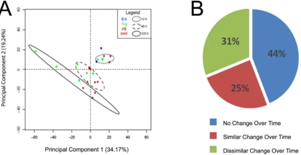
A rotating wall bioreactor engineered by NASA was used to simulate microgravity14. This bioreactor perpetually suspends cells without inducing shear, thereby creating a net gravity vector of zero3,15. Primary rat neonatal cardiomyocytes were placed into simulated microgravity (μg) or normal gravity (1xg) conditions (Fig. 1A). High-resolution nanoscale liquid chromatography tandem mass spectrometry (LC-MS/MS) was performed as outlined in Fig. 1B on cells isolated over time at 12, 48, or 120 h from μg or 1xg conditions.Intensity values for 6,174 peptides (Supplemental Table 2; peptide expression) were utilized to measure the relative expression for 848 proteins across all time points (0, 12, 48, and 120 h) and gravity conditions (μg or 1xg) (Supplemental Table 3; protein expression). Principal components analysis (PCA) demonstrated that as a whole, protein abundance between μg and 1xg samples were not different at 12 h, only slightly different at 48 h, and highly different at 120 h (Fig. 2A).
ANOVA was performed on log 2-transformed data to evaluate the relative abundance of individual proteins as a function of gravity condition at each time point, and longitudinally as a function of time under each gravity condition (Supplemental Table 3-Protein expression). From these analyses, proteins were classified into three groups (Fig. 2B): (1) no change over time under 1xg or μg (44%); (2) similar change over time or temporal protein trend under 1xg or μg (25%); and 3) dissimilar change over time between 1xg or μg (31%).
abundance (RIA) — following stable-isotope incorporation into newly-synthesized protein using Equation 2 (Supplemental Fig. 2).
Multiple scenarios exist to explain the distribution of IL and IH over time, and four major possibilities are shown in Supplemental Table 1. At the initial time point of label addition, a peptide would have no stable-isotope incorporation (IH= 0; Supplemental Fig. 1A), and thus IL would be equal to the total amount of peptide. In Supplemental Fig. 1B, there is the same theoretical quantity of total peptide (IH+ IL= 1); however, half of the peptide has been generated since the time = 0, thus the peptide RIA is 0.5. States C and D show a 50% reduction in peptide abundance compared to States A and B, and also show RIA of 0.5 and 0.8, respectively (Supplemental Fig. 1C,D). In this manner, the same dataset can be used to measure both total protein differential expression based on standard label-free proteomics by summing the intensities of all forms of all peptides and relative turnover by calculating the RIA of individual peptides using the heavy and light forms.
Estimation of protein turnover requires the measurement of RIA of the precursor amino acid pool (essentially the maximum possible protein incorporation) by utilizing a peptide with more than one labeled residue13,16. We assessed precursor amino acid RIA (r) using the peptide EATNPPIIQEEKPK in our data set (Supplemental Table 2-Peptide Expression) by the method of Doherty et al.16. Since this peptide contains two lysine (K) residues, the relative abundance of the heavy/heavy and heavy/light forms is dependent only on the ratio of the heavy and light lysine isotopes at the time of protein synthesis and is independent of protein turnover. This global measurement, defined as precursor RIA, was performed on data collected at 48 h and 120 h for both μg and 1xg (Fig. 3A). No
Figure 2. Protein abundance is altered in cardiomyocytes during simulated microgravity. (A) Principal components analysis (PCA) was performed using protein data from Supplemental Table S3, z-score normalized. The smallest variability among any sample group is among 0 h and 12 h samples, followed by 48 h and 120 h. The QC pool samples are nearly indistinguishable in the center of the PCA, showing technical variability is far less than biological variability. (B) Fraction of proteins that changed as a function of time or between groups.
significant difference in RIA was observed for the conditions (p = 0.80, Students T-test), indicating that the switch from light to heavy amino acid media was performed equally for both conditions. Additionally, since this is an intracellular measure of precursor amino acid, it indicates that there likely was no significant dysregulation in amino acid transporter function at the cell membrane between μg and 1xg. Collectively, these data suggest that incorporation of precursor amino acid (at least K and R) into proteins occurs similarly under both normal and microgravity conditions.
Next, the RIA for 359 peptides passing strict selection criteria (see Methods, Table S4-Dynamic SILAC) was determined for 0, 12, 48, and 120 h under μg or 1xg (Fig. 3B). At 12 h, there were no significant differences in protein turnover in μg versus 1xg, as measured by RIA (p = 0.32, Students T-test). However, there were large dif-ferences in protein turnover at both 48 and 120 h in μg versus 1xg (p < 1e−6 for both; Fig. 3B). These data indicate that the drastic decrease in protein turnover in simulated microgravity, despite there being no difference in the abundance of the intracellular precursor amino acid pool (Fig. 3A) and despite there being no difference in total expression levels of a majority (~70%) of the proteome during the microgravity exposure (Fig. 2B).
Microgravity differentially influences the endoplasmic reticulum and mitochondrial proteome
and stress pathways.
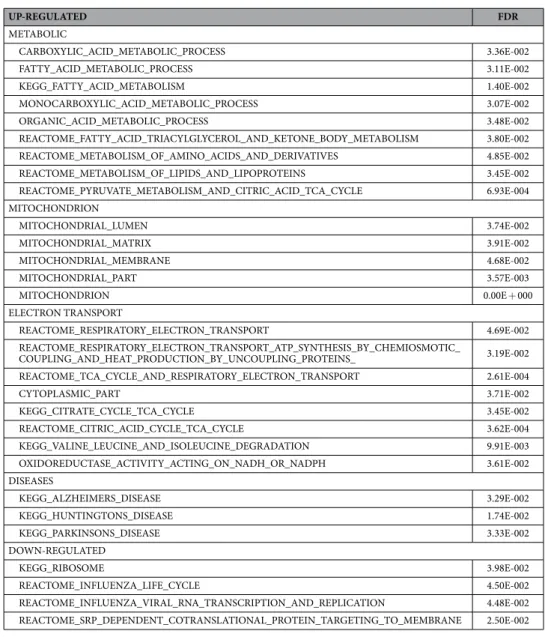
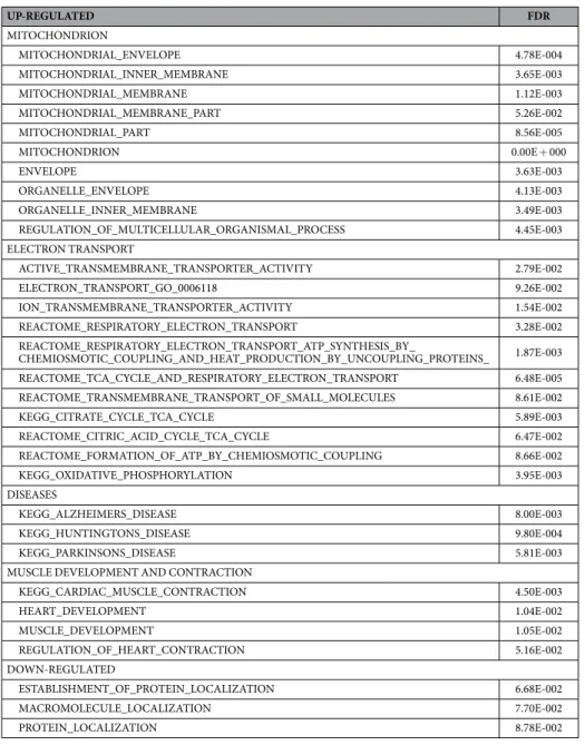
The results from both the abundance and SILAC proteomics data sets indicated that there were a number of proteins significantly changed in abundance or turnover in response to simu-lated microgravity at 120 h. Each data set (abundance and turnover) was analyzed using a bioinformatics/gene functional-enrichment approach, as outlined in the materials and methods, to map these changes to cellular loca-tion, protein funcloca-tion, and biological pathways. Following individual analysis, a comparative analysis of abun-dance and turnover was performed with the aim of elucidating any underlying commonalities and differences in quantitative and dynamic protein expression patterns for exposure of the cardiomyocytes to microgravity.Spectra from LC-MS/MS analyses (Supplemental Table 2) were assigned to peptides (and thus putative pro-tein precursors) using a search against the NCBI RefSeq database17. The protein identifiers were mapped into the corresponding genes that encode for the proteins by using Bioconductor’s MyGene package18 in order to carry out the translation from protein space to gene space; this mapping was critical since most functional annotation data sets, including the Gene Ontology19, are based on gene identifiers as opposed to protein. The differentially abundant proteins in these data sets were determined using a combination of t-test and ANOVA analysis with a p-value cut-off ≤ 0.05 after multiple hypothesis testing correction using Benjamini and Hochberg procedure. After identification of significantly impacted proteins, a functional enrichment analysis was performed with the aim of identifying biological processes, cellular components, or molecular functions that are significantly impacted by microgravity. Using the transformed proteomics data and the gene functional annotations, a path-way enrichment analysis was performed using Gene Set Enrichment Analysis (GSEA) software independently on either the protein abundance (Table 1) or SILAC turnover (Table 2) data sets at 0.05 False Discovery Rate (FDR) cutoff. Relative to normal gravity, metabolic gene sets in microgravity were exclusively up-regulated in the protein abundance data, whereas gene sets related to cardiac/heart muscle contraction and development were uniquely up-regulated in the protein turnover data. Gene sets related to mitochondrial and electron-transport processes and gene sets annotated with Alzheimer’s, Huntington’s, and Parkinson’s diseases, neurological diseases origi-nating in tissues with limited regenerative capacity similar to cardiac tissues, were up-regulated in both protein abundance and turnover data sets. Regarding down-regulated gene sets, the microgravity protein abundance data were enriched with gene sets associated with the ribosome, transcription and translation, whereas the protein turnover data were predominantly related to functions related to protein/macromolecule localization.
The analysis of the combination of abundance and turnover data revealed that the mitochondria was more impacted than other organelles in the cell (Fig. 4A). However, it appears that mitochondrial turnover was more impacted than protein abundance. A total of 40 proteins from both data sets mapped to these mitochondrial processes (Fig. 4B). Two proteins were unique to the abundance data set, 13 were unique to the turnover data, and 25 proteins were common to both data sets. We mapped these common genes to pathways using the Pathway Commons database (results not shown here). The top significant pathways include the citric acid cycle (TCA), ATP synthesis, nuclear estrogen receptor network, and metabolism of carbohydrates, amino acids, and glucose.
ER stress and the UPR are occurring while mitochondrial integrity is preserved in microgravity.
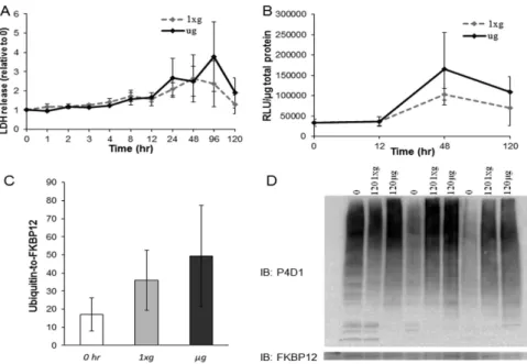
Proteomics and gene set enrichment analyses indicate that microgravity differentially influences processes in the cytoplasm and mitochondria. Nonetheless, it is possible that microgravity exposure augments cell injury, contributing to the observed differences. To investigate this possibility the media from cells placed in 1xg and μg conditions was examined for lactate dehydrogenase (LDH) levels at ten time points between 0 and 120 h (Fig. 5A). LDH is a soluble cytosolic enzyme, and its release is an indication of cell membrane permeability or damage. No statistically significant difference in LDH release from the cell was observed between μg and 1xg at any time point, although LDH was elevated at 24 and 96 h compared to at 1 h within the μg group (p < 0.05, Repeated-measures ANOVA, n = 4; Fig. 5A). These data suggest that proteomic differences are not due to enhanced physical cell injury during microgravity exposure.Another form of cell injury and death and also a surrogate measurement of mitochondrial integrity is apop-tosis, which is indicated and executed by the protease caspase-3. We surveyed caspase-3 activity at 0, 12, 48, and 120 h in μg and 1xg and did not observe a statistically significant difference in caspase-3 activity between μg and 1xg at any time point (p < 0.05, Repeated-measures ANOVA, n = 3). There was an increase at 48 h compared to 0 and 12 h under both conditions, but this was not different between μg and 1xg (Fig. 5B). This indicated that changes in the proteome between μg and 1xg were not due to increased apoptosis. It additionally suggested that mitochondrial integrity was preserved in cardiomyocytes under the stress of microgravity.
Since protein ubiquitination is associated with proteins being tagged for degradation, we assessed global ubiq-uitination of protein lysates via western blot analysis at 0 h and from each group at 120 h. Again, no difference was observed between μg and 1xg at 120 h or compared to 0 h (p = 0.21, one-way ANOVA, n = 3; Fig. 5C,D). Together, these data indicate that cardiomyocytes are not markedly damaged in response to simulated μg (LDH and Caspase-3 data), and that μg does not appear to enhance protein degradation (ubiquitination data), thus
UP-REGULATED FDR
METABOLIC
CARBOXYLIC_ACID_METABOLIC_PROCESS 3.36E-002
FATTY_ACID_METABOLIC_PROCESS 3.11E-002
KEGG_FATTY_ACID_METABOLISM 1.40E-002
MONOCARBOXYLIC_ACID_METABOLIC_PROCESS 3.07E-002
ORGANIC_ACID_METABOLIC_PROCESS 3.48E-002
REACTOME_FATTY_ACID_TRIACYLGLYCEROL_AND_KETONE_BODY_METABOLISM 3.80E-002
REACTOME_METABOLISM_OF_AMINO_ACIDS_AND_DERIVATIVES 4.85E-002
REACTOME_METABOLISM_OF_LIPIDS_AND_LIPOPROTEINS 3.45E-002
REACTOME_PYRUVATE_METABOLISM_AND_CITRIC_ACID_TCA_CYCLE 6.93E-004
MITOCHONDRION
MITOCHONDRIAL_LUMEN 3.74E-002
MITOCHONDRIAL_MATRIX 3.91E-002
MITOCHONDRIAL_MEMBRANE 4.68E-002
MITOCHONDRIAL_PART 3.57E-003
MITOCHONDRION 0.00E + 000
ELECTRON TRANSPORT
REACTOME_RESPIRATORY_ELECTRON_TRANSPORT 4.69E-002
REACTOME_RESPIRATORY_ELECTRON_TRANSPORT_ATP_SYNTHESIS_BY_CHEMIOSMOTIC_
COUPLING_AND_HEAT_PRODUCTION_BY_UNCOUPLING_PROTEINS_ 3.19E-002
REACTOME_TCA_CYCLE_AND_RESPIRATORY_ELECTRON_TRANSPORT 2.61E-004
CYTOPLASMIC_PART 3.71E-002
KEGG_CITRATE_CYCLE_TCA_CYCLE 3.45E-002
REACTOME_CITRIC_ACID_CYCLE_TCA_CYCLE 3.62E-004
KEGG_VALINE_LEUCINE_AND_ISOLEUCINE_DEGRADATION 9.91E-003
OXIDOREDUCTASE_ACTIVITY_ACTING_ON_NADH_OR_NADPH 3.61E-002
DISEASES
KEGG_ALZHEIMERS_DISEASE 3.29E-002
KEGG_HUNTINGTONS_DISEASE 1.74E-002
KEGG_PARKINSONS_DISEASE 3.33E-002
DOWN-REGULATED
KEGG_RIBOSOME 3.98E-002
REACTOME_INFLUENZA_LIFE_CYCLE 4.50E-002
REACTOME_INFLUENZA_VIRAL_RNA_TRANSCRIPTION_AND_REPLICATION 4.48E-002
REACTOME_SRP_DEPENDENT_COTRANSLATIONAL_PROTEIN_TARGETING_TO_MEMBRANE 2.50E-002
indicating that protein degradation is not playing a major role in the diminished protein turnover induced in simulated microgravity.
To evaluate the influence of microgravity on protein synthesis, orthogonal experiments were performed utiliz-ing the methionine analogue azidohomoalanine (AHA) which is incorporated into proteins via methionine tRNA and can be used to tag newly-translated proteins20. To measure protein synthesis across a small time window, cells were exchanged from Met- to AHA-containing media at 120 h, and labeling was performed for 2 h. Using the total protein lysates from each group, proteins were specifically tagged on the azido side chains with biotin and subsequently immunoblotted using HRP streptavidin (n = 3 per condition). AHA incorporation was robust after two hours of labeling, but was dramatically reduced at 120 h in μg relative to 1xg (p = 0.003; Fig. 6A,B). These data demonstrate that protein synthesis, a factor contributing to overall protein turnover, is significantly diminished after cells have been exposed to μg for 120 h, compared to the same cells under normal gravity, and they suggest that the decreased protein turnover, as measured by RIA, reflected this decrease in protein synthesis.
To evaluate the consequences of μg on gene expression and translation from a non-chromosomal gene, the luciferase marker gene on an episome was introduced into NRCMs via an Adenoviral-luciferase vector 3 h prior to separating the cells into μg and 1xg conditions. This allowed the luciferase gene to be transcribed and translated into protein using the cytoplasmic translational machinery. The adenovirus was removed when cells were placed into μg and 1xg conditions at 0 h, and at 120 h, lysates from 120 h were evaluated for luciferase enzyme activity, which is a direct correlate to luciferase protein content since no post-translational modifications are necessary to initiate luciferase enzyme function21. Luminometry revealed that the content of luciferase protein in the cell was significantly diminished 7.5 fold in μg compared to 1xg (p = 0.02, unpaired T-test, n = 4) as displayed in Fig. 6C.
UP-REGULATED FDR
MITOCHONDRION
MITOCHONDRIAL_ENVELOPE 4.78E-004
MITOCHONDRIAL_INNER_MEMBRANE 3.65E-003
MITOCHONDRIAL_MEMBRANE 1.12E-003
MITOCHONDRIAL_MEMBRANE_PART 5.26E-002
MITOCHONDRIAL_PART 8.56E-005
MITOCHONDRION 0.00E + 000
ENVELOPE 3.63E-003
ORGANELLE_ENVELOPE 4.13E-003
ORGANELLE_INNER_MEMBRANE 3.49E-003
REGULATION_OF_MULTICELLULAR_ORGANISMAL_PROCESS 4.45E-003
ELECTRON TRANSPORT
ACTIVE_TRANSMEMBRANE_TRANSPORTER_ACTIVITY 2.79E-002
ELECTRON_TRANSPORT_GO_0006118 9.26E-002
ION_TRANSMEMBRANE_TRANSPORTER_ACTIVITY 1.54E-002
REACTOME_RESPIRATORY_ELECTRON_TRANSPORT 3.28E-002
REACTOME_RESPIRATORY_ELECTRON_TRANSPORT_ATP_SYNTHESIS_BY_
CHEMIOSMOTIC_COUPLING_AND_HEAT_PRODUCTION_BY_UNCOUPLING_PROTEINS_ 1.87E-003
REACTOME_TCA_CYCLE_AND_RESPIRATORY_ELECTRON_TRANSPORT 6.48E-005
REACTOME_TRANSMEMBRANE_TRANSPORT_OF_SMALL_MOLECULES 8.61E-002
KEGG_CITRATE_CYCLE_TCA_CYCLE 5.89E-003
REACTOME_CITRIC_ACID_CYCLE_TCA_CYCLE 6.47E-002
REACTOME_FORMATION_OF_ATP_BY_CHEMIOSMOTIC_COUPLING 8.66E-002
KEGG_OXIDATIVE_PHOSPHORYLATION 3.95E-003
DISEASES
KEGG_ALZHEIMERS_DISEASE 8.00E-003
KEGG_HUNTINGTONS_DISEASE 9.80E-004
KEGG_PARKINSONS_DISEASE 5.81E-003
MUSCLE DEVELOPMENT AND CONTRACTION
KEGG_CARDIAC_MUSCLE_CONTRACTION 4.50E-003
HEART_DEVELOPMENT 1.04E-002
MUSCLE_DEVELOPMENT 1.05E-002
REGULATION_OF_HEART_CONTRACTION 5.16E-002
DOWN-REGULATED
ESTABLISHMENT_OF_PROTEIN_LOCALIZATION 6.68E-002
MACROMOLECULE_LOCALIZATION 7.70E-002
PROTEIN_LOCALIZATION 8.78E-002
This was another indication that protein synthesis was reduced under the stress of microgravity, and further, it suggests that protein synthesis is not limited by chromosomal gene transcription, but rather due to translation.
Discussion
Microgravity exposure is a known stressor that moderates mammalian physiology. For example, microgravity exposure has been reported to moderate cardiac atrophy22. Very little is known molecularly regarding the stress pathways activated or molecular processes leading to microgravity-induced cardiac atrophy. At the level of the proteome, there are three types of cellular stress responses: the classical “heat shock” response occurring in the cytoplasm; the unfolded protein response (UPR) occurring in the endoplasmic reticulum (UPRER); and the UPR occurring in the mitochondria (UPRMT)23–27. The predominant response is dependent on the type of stress encountered, the cell type, the time point post-stress, and the cellular proliferation status.
Cells capable of cell division respond to stress by focusing resources for cellular division (i.e. entry into the cell cycle) rather than global protein synthesis, thereby increasing the chance of daughter cell survival. In contrast, specialized non-dividing cells, unable to re-enter the cell cycle, respond to stressful conditions through different mechanisms in efforts to conserve their functionality. A prime example are cardiomyocytes, the fundamental working unit of the heart. Under favorable conditions, cardiomyocytes maintain function by expending energy on the maintenance of the contractile machinery and the mitochondria. This maintenance is achieved through protein regulation, i.e. the balance of protein synthesis and protein degradation of the contractile apparatus and the mitochondria.
To gain a fundamental understanding of the microgravity stress response in non-dividing cells, we under-took a study looking at global proteomic changes in non-dividing cardiomyocytes in simulated microgravity. In addition, to understand rates of protein synthesis and stability in cardiomyocytes, we utilized a modifica-tion of dynamic SILAC. Dynamic SILAC typically is utilized in the reverse manner, where dividing cells are incubated for multiple divisions in heavy isotope-labeled media, which allows cellular amino acids to become fully heavy isotope-labeled. These cells then are exchanged into media containing native, non-isotope-labeled amino acids13,16, and the rate of protein synthesis and stability is followed by monitoring the rate of heavy isotope removal from the peptide population. Because neonatal rat cardiomyocytes are non-dividing cells and have a
Primary
Protein Name Protein Description %CV FC @ 120 h (ug v 1xg) Function
392351353 Myosin-13 9.9 3.5 Molecular motor
11693154 Platelet-activating factor acetylhydrolase IB subunit beta 11.5 2.7 Activity of RhoGTPases, actin polymerization
198442897 AFG3-like protein 2 10.3 2.5 Mitochondrial protein homeostasis
9507135 Spectrin beta chain, brain 2 5.2 2.1 Actin binding
293359790 Protein FAM179A-like isoform 1 3.6 1.9 Unknown function
62945328 Protein NipSnap homolog 2 11.9 1.8 Mitochondria, neg regulation of ATP citrate synthase activity
25742739 Long-chain-fatty-acid–CoA ligase 1 2.5 1.7 Activates breakdown of complex FA
48675862 Acyl-coenzyme A thioesterase 2, mitochondrial 2.0 1.7 Catalyzes hydrolysis of acyl-CoA to free FA and co enzyme A
392347468 Poly (rC)-binding protein 1 6.2 1.6 mrna import to mitochondria
56605722 Serine hydroxymethyltransferase, mitochondrial 6.2 1.6 Conversion l-serine to glycine, provides 1 carbon units to cell
113205496 Pyruvate dehydrogenase complex, component X 2.1 1.6 Mitochondrial, tether E3 dimers to E2 core
392341350 Glyceraldehyde-3-phosphate dehydrogenase-like 1.3 1.5 Breaks down glucose for energy and carbon
13994225 3-hydroxyacyl-CoA dehydrogenase type-2 8.6 1.5 Mitochondrial tRNA maturation
11968102 Ornithine aminotransferase, mitochondrial precursor 5.2 1.5 Processes excess nitrogen during protein breakdown
392351018 Sarcalumenin 3.2 1.5 Calcium buffering in SR
410110929 Stress-70 protein, mitochondrial 3.1 1.5 Mitochondrial protein homeostasis
209954804 Plastin-3 9.5 − 24.3 EF-hand protein, bone?
293346882 Mitochondrial aspartate aminotransferase-like 18.0 − 22.2 metabolite exchange between mitochondria and cytosol, uptake of long chain free fatty acids
8394079 Proteasome subunit beta type-2 11.8 − 15.4 ATP dependent proteolytic activity
157818179 Elongation factor 1-beta 5.3 − 13.4 Translation elongation, gdp to gtp exchange
157787127 40 S ribosomal protein S28 8.2 − 12.6 Ribosomal protein, translation
392348740 Laminin subunit beta-1 4.2 − 12.2 ECM structural component
77993298 Translocon-associated protein subunit alpha precursor 5.3 − 12.0 ER protein, ER UPR
12018252 Transketolase 14.2 − 8.9 Pentose phosphate pathway
56744249 Reticulocalbin 3, EF-hand calcium binding domain precursor 4.9 − 7.6 ER Calcium binding
40254781 Rab GDP dissociation inhibitor beta 15.7 − 6.8 Regulates gdp/gtp exchange of most rab proteins, poly A RNA binding
392342369 60 S ribosomal protein L30-like 3.9 − 6.7 Ribosomal protein, translation
6981326 Protein S100-A4 7.0 − 6.5 Ca binding, polyA RNA binding
61556832 Adenine phosphoribosyltransferase 13.5 − 6.3 AMP biosynthesis salvage pathway
6981574 SPARC precursor 15.7 − 6.2 Regulates cell growth, thru ECM and binds calcium
27665858 40 S ribosomal protein S9-like 12.2 − 5.9 Ribosomal structural protein
392346755 Ribosome-binding protein 1 14.7 − 5.6 Mediates interaction between ribosome and ER
392347136 Collagen alpha-2(I) chain-like isoform 1 7.2 − 5.5 Extracellular matrix
13592133 Actin, cytoplasmic 1 3.7 − 5.3 motility
203097140 Myosin regulatory light chain RLC-A 9.0 − 5.2 Muscle contraction
392348865 Histone H2A. V-like 4.5 − 4.7 Chromosomal binding
77404180 Ras-related protein Rab-4A 12.5 − 4.6 ATPase activator activity
157819753 Reticulocalbin-1 precursor 13.3 − 4.5 ER calcium binding
61556967 Elongation factor 1-delta 9.3 − 4.1 Transfer of aminoacyl-tRNA to ribosome
6981672 Tropomyosin alpha-4 chain 13.0 − 3.7 Muscle contraction
6978589 Non-muscle caldesmon 5.7 − 3.6 Muscle contraction
157822227 60 S ribosomal protein L12 7.8 − 3.4 translation
148747365 Heat shock protein HSP 90-beta 2.3 − 3.4 Classic heat shock protein
392356007 Actin, cytoplasmic 1-like 5.0 − 3.3 Cell motility
9506845 Rab GTPase-binding effector protein 1 15.1 − 3.2 Membrane trafficking, protein localization
limited lifespan, they are incapable of being fully conditioned in heavy isotope-containing media. Thus, we per-formed dynamic SILAC in the forward direction28,29, starting with native isotope-labeled cells and switching the cells into heavy isotope-containing media. To our knowledge, this method of calculating protein turnover and simultaneous changes in overall protein expression by label-free quantitation has not been utilized13,16.
In non-dividing cardiomyocytes exposed to 120 h of simulated microgravity, levels of cytoplasmic heat shock proteins were diminished and protein ubiquitination was not different compared to normal gravity, suggesting that the classical heat shock response is not a predominant stress response to microgravity. In contrast, indicators of the UPRER and UPRMT were increased. Overall, from quantitative and SILAC proteomics data and bioinfor-matics analysis, we observed that the content and processes of two cellular organelles were differentially affected by simulated microgravity, namely the mitochondria (up-regulated) and the cytoplasmic translational machinery, including the ribosomes and endoplasmic reticulum (down-regulated).
Mitochondria are the energy-producing powerhouses of the cell and compose > 30% of cardiac cell volume, but they are exquisitely sensitive to stress-related damage30. They are constantly turning over through fusion and fission processes, being replaced with newer, more efficient mitochondria under optimal conditions31. Further, mitochondria may enhance their import machinery in response to stress32. Our data revealed that in response to simulated microgravity, two proteins directly involved in mitochondrial protein import were increased in
Primary
Protein Name Protein Description %CV FC @ 120 h (ug v 1xg) Function
157786744 Dihydropyrimidinase-related protein 2 4.5 − 3.0 Cytoskeletal organization
6981240 Myosin light chain 3 4.3 − 2.5 Muscle contraction
124107592 Unconventional myosin-Ic 6.1 − 2.3 Associated with transcriptionally active ribosomal genes
12083607 40 S ribosomal protein S14-like 2.5 − 2.2 translation
56090293 Pyruvate dehydrogenase E1 component subunit beta, mitochondrial precursor 4.1 − 2.1 metabolism 219275589 Asparaginyl-tRNA synthetase, cytoplasmic isoform 2 3.5 − 2.1 translation
293350511 M2 pyruvate kinase-like isoform 1 2.1 − 2.0 metabolism
155369650 Myosin light polypeptide 6 6.2 − 1.8 Muscle contraction
11968086 Ribosomal protein L4 5.1 − 1.7 translation
66730475 Tropomyosin beta chain 7.2 − 1.6 Muscle contraction
6981236 Myosin-9 4.8 − 1.6 Cytoskeletal organization
6981666 Troponin T, cardiac muscle 3.5 − 1.5 Cardiac structure
Table 3. Individual protein abundance alterations in microgravity.
abundance – mortalin and AFG3L2, which suggests there may be an increase in mitochondrial protein transla-tion or a decrease in the degradatransla-tion of these proteins under conditransla-tions of microgravity33, although we did not directly measure sub-compartmental, mitochondrial protein translation or degradation.
Mortalin, also known as mitochondrial stress 70 protein (mitoHsp-70) or grp75, is a mitochondrial chaperone protein located in the mitochondrial matrix and considered a major mediator of protein homeostasis contrib-uting to mitochondrial biogenesis and energetics (reviewed in refs 34 and 35). Mortalin cooperates with Hsp60, another major chaperone protein in the mitochondrial matrix, to import unfolded proteins and subsequently fold the majority of the nascent synthesized proteins translocated into the mitochondria35. Mortalin is also found in several extra-mitochondrial sites, including the ER36, cytoplasmic vesicles, and the cytoplasm (reviewed in refs 34 and 35). In this extra-mitochondrial role, mortalin is a “pro-survival chaperone” that is anti-apoptotic. Moreover, it is one of the most important anti-apoptotic genes in that it protects the cell from many different stressors, including arsenite, glucose starvation, and ischemia-reperfusion.
AFG3L2 is involved in the same mitochondrial homeostasis pathway as mortalin34,35,37,38. However, as a mem-ber of the AAA ATPase family, AFG3L2 removes permanently damaged proteins that fail to refold and can only be rendered safe by proteolysis. In particular, AFG3L2 helps maintain mitochondrial protein quality control and degrades proteins of the respiratory chain that fail to assemble into the respiratory chain enzyme complexes39. Together, the up-regulation of mortalin and AFG3L2 suggest the mitochondria of cardiomyocytes respond to microgravity by supporting mitochondrial protein maintenance.
In contrast to the up-regulation of protein content and processes related to mitochondrial protein trans-lation, proteins and processes mapping to translation occurring in the rough ER (RER) and ribosomes were down-regulated after 120 h of simulated microgravity exposure. Specifically, six ribosomal proteins were dimin-ished in abundance − 40 S components (S28, S9-like, S14-like), 60 S components (L30 like, L12), and ribosomal protein L4. Decreases in ribosomal proteins and tRNAs have been observed in skeletal muscle atrophy40,41. Also significantly decreased was asparaginyl-tRNA synthetase, an important component in translation occurring at the RER. Moreover, a mediator of RER and ribosome interaction, ribosome binding protein 1, was diminished in microgravity. Coupled to RER translation is import into and protein folding in the ER. Specific folding proteins that diminished in abundance in microgravity included translocon-associated protein subunit alpha precursor, reticulocalbin 3, reticulocalbin-1 precursor, and proteasome subunit beta type-2. Together, the down-regulation of these proteins suggests that protein translation via ribosomal translational machinery is reduced.
Both the ER and mitochondria are presently accepted as dynamic organelles capable of modifying their struc-ture and function in response to changing environmental conditions. The ER and mitochondria interact both physiologically and functionally, and one of the most well-known and critical aspects of this interaction is calcium signaling between the two organelles24. However, another interaction between these organelles in cardiomyocytes appears to be a mechanism that biases mitochondrial protein translation over RER translation, most likely to maintain cellular energy production for global cell viability. Mortalin seems to be in two locations – between the ER and mitochondria and in the mitochondrial matrix – and may be the key molecule mediating this preferred translation.
proteins, the SILAC and AHA data further support that global cellular protein turnover and synthesis is signif-icantly reduced in primary cardiac cells exposed to simulated microgravity. The SILAC data revealed decreased amino acid isotope incorporation, and that this reduction was not due to different amino acid availability or amino acid transport capability between gravity conditions. The AHA data depicted reductions at the transla-tional level, since AHA is incorporated into protein via tRNA.
While there appears to be a global down-regulation in protein turnover as indicated by reduced RER transla-tion and protein synthesis, protein turnover is also influenced by protein degradatransla-tion; however, our data suggest there not to be an increase in protein degradation. Global ubiquitination was assessed, and although there was a qualitative increase in ubiquitination at 120 h compared to 0 h, no difference existed between μg and 1xg at 120 h. Overall, the lack of change in LDH release, caspase-3 activity, or ubiquitination at 120 h between groups suggests there was minimal-to-no enhancement of cell damage and protein degradation between μg and 1xg. Although, other contributors to degradation exist that were not evaluated, such as lysosomal proteases (e.g. cathepsins) and calpain activation, the surrogates used here provide evidence that simulated microgravity does not severely activate damage and degradation pathways in the cardiomyocyte. Lack of apoptosis induction observed in micro-gravity is consistent with other studies42,43. Further, mortalin, which was up-regulated, has been reported to be directly anti-apoptotic44.
Cytoarchitecture is linked to mitochondrial localization and energetics30, whereby the cytoarchitecture com-prising structural and contractile proteins may moderate energetic micro domains and energetic cross talk. The abundance of seven structural and/or contractile proteins was significantly diminished in microgravity. Notably, myosin regulatory light chain and tropomyosin (Table 1) diminishment is a hallmark characteristic of atrophy41. How potential microgravity-moderated cytoarchitectural changes may mediate mitochondrial localization and energetics deserves further research.
Cardiac atrophy is a phenomenon that can lead to orthostatic intolerance and has been observed in spaceflight studies with rats, human bed rest studies, and is identified as a major challenge for crew returning from long dura-tion missions45–47. However, the mechanisms leading to this phenotype are unclear. Our data suggest that under exposure to microgravity, cardiac cells respond by upregulating mitochondrial proteins to preserve energy pro-duction and promote short-term survival, but are not able to overcome apparent disruption of protein translation at the RER because of decreases in the levels of specific ribosomal proteins. This disruption at the RER ultimately decreases overall protein translation and contributes to the development of cardiac atrophy.
Our data suggest that a certain degree of protein misfolding, possibly compartmentalized misfolding is occur-ring in microgravity. The trigger for this remains unclear. Stress responses generally originate at the cell surface, trigger signal transduction cascades which induce induction of nuclear gene transcription; however, microgravity affects the entire cell equally and at once, so this may represent an entirely new type of cellular stress response. Mortalin may possibly be a key regulator. The upregulation of mortalin may be a means of selectively allow-ing continuallow-ing mitochondrial translation at the expense of RER translation so that the energetics of the cell are maintained. Understanding the interplay of mortalin in pathways and biological processes will provide a better understanding of the coordinated cellular response to microgravity, and further studies to improve understanding of the cellular responses to microgravity will help us prepare for extended travel beyond Earth’s orbit.
Materials and Methods
Neonatal Rat Cardiomyocyte (NRCM) isolation.
Animal experiments were approved by Duke University Medical Center Institutional Animal Care and Use Committee (IACUC) and are in accordance with United States federal and North Carolina state regulations. NRCMs were isolated from ventricles of 2-day-old Sprague-Dawley rats (Charles River Laboratories) as previously reported48.Experimental design including dynamic SILAC labeling of NRCM.
Following isolation, NRCMs were plated on 100 mm tissue culture dishes. Cells were collected by trypsinization, exchanged into DMEM:F12 SILAC media (Thermo Scientific; prepared as previously described49, and seeded onto Cytodex-3 microcarrier beads (Sigma). Normal gravity (1xg) cells were plated on tissue culture dishes containing 10 ml SILAC media. Simulated μg cells were transferred to rotating wall vessels (RWV; Synthecon, Inc., Houston, TX) containing 10 ml SILAC media. Initially, the cell-bead mixture was allowed to sit in the RWV for 2 h to enhance cell attach-ment prior to rotation. Both the 1xg and μg cells were cultured for 12, 48, and 120 h, with fresh media added every 48 h. NRCMs were collected by trypsinization, washed three times in 50 mM ammonium bicarbonate (pH 8.0), pelleted (300 xg for 2 min), flash frozen in liquid N2, and stored at − 80 °C until analysis.Sequencing grade porcine trypsin was added at 1:50 enzyme: protein ratio and samples were incubated at 37 °C overnight while shaking. Following overnight digestion, RapiGest surfactant was hydrolyzed by addition of 1% TFA/2% ACN final. All samples were also spiked with ADH1_YEAST digest (Massprep standard, Waters Corp.) as a surrogate standard (50 fmol ADH per μ g total protein). All samples were heated at 60 °C for 2 h and centri-fuged at 15,000 rpm for 10 min before pipetting all supernatant into individual TotalRecovery LC vials (Waters, Corp.). 3 μ L was pipetted from each and combined to make a pool for QC purposes.
Data collection for LC-MS/MS proteomics.
All samples were randomized within a biological replicate group for acquisition order. Run order for the raw data collection is displayed in S1. Before each replicate group queue, 1 μ g from the pooled sample was injected, and was also injected 4× at the end of the queue, for a total of 7 QC pool runs. One of the seven pooled analyses was run in data-dependent acquisition mode (DDA) for comple-mentary peptide identifications whereas the others were performed as follows.Quantitative LC/MS/MS was performed on 1 μ g of protein digest per sample, using a nanoAcquity UPLC system (Waters Corp) coupled to a Synapt G2 HDMS high resolution accurate mass tandem mass spectrometer (Waters Corp.) via a nanoelectrospray ionization source. Briefly, the sample was first trapped on a Symmetry C18 300 mm × 180 mm trapping column (5 μ l/min at 99.9/0.1 v/v water/acetonitrile), after which the analyti-cal separation was performed using a 1.7 μ m Acquity BEH130 C18 75 mm × 250 mm column (Waters Corp.) using a 90-min gradient of 5 to 40% acetonitrile with 0.1% formic acid at a flow rate of 400 nanoliters/minute (nL/min) with a column temperature of 55 °C. Data collection on the Synapt G2 mass spectrometer was per-formed in ion-mobility assisted data-independent acquisition (HDDIA or HDMSE) mode, using 0.6 s alternating cycle time between low (6 V) and high (27–50 V) collision energy (CE). Scans performed at low CE measure peptide accurate mass and intensity (abundance), while scans at elevated CE allow for qualitative identification of the resulting peptide fragments via database searching. The total analysis cycle time for each sample injection was approximately 1.5 h.
Following the 28 analyses (with the additional QC standard injections), data was imported into Rosetta Elucidator v3.3 (Rosetta Biosoftware, Inc.), and all LC-MS files were aligned based on the accurate mass and retention time of detected ions (“features”) using PeakTeller algorithm (Elucidator). The relative peptide abun-dance was calculated based on area-under-the-curve (AUC) of aligned features across all runs. The dataset had 280,291 quantified features and 24,433 annotated spectra. This MS/MS data was searched against a NCBI RefSeq database with rattus norvegicus taxonomy (25,485 forward entries) appended with a decoy reverse-sequence of each forward entry for false positive rate determination. The sequences for ADH_yeast, CASA1_bovin, and CASA2_bovin were also appended to the database representing internal standards used. Static mass modifi-cation corresponding to carbamidomethylation (from alkylation protocol) was required on Cys residues, whereas dynamic mass modifications corresponding to oxidation was allowed on Met residues, deamidation was allowed on Asn and Gln residues, and heavy labeled (13C15N) Arg and Lys. After individual peptide scoring using PeptideProphet algorithm (Elucidator), the data was annotated at a < 1% peptide false discovery rate. This analysis yielded identifications for 6,339 peptides and 864 proteins across samples, including 493 proteins with two or more peptides quantified. For quantitative processing, only 25 of the 28 runs were used (only the 4 pooled analyses performed in the exact same way were used whereas the last 3 were excluded from quantitation). The data was first curated to contain only high-quality peptides with appropriate chromatographic peak shape, and the dataset was intensity scaled to the robust median across all samples analyzed; the final quantitative dataset was based on 6,174 peptides and contained 848 proteins.
Proteomic statistical analyses.
For analysis of relative protein expression between conditions (time or microgravity) both heavy- and native isotope-labeled peptides were quantified by accurate mass and retention time alignment, and the heavy and light forms of each peptides were summed to yield a total expression value for each peptide. Peptide intensities were summed to the protein level to obtain values for each protein in each sample analyzed, and protein intensities were log 2 transformed prior to statistical analysis. The criteria for signif-icance are noted in Results. Protein expression data were z-score normalized. Protein-level expression data were analyzed after log 2 transformation followed by Error-Weighted Analysis of Variance (ANOVA) with Bonferroni correction to control for multiple-hypothesis testing. Tables for peptide expression (S2 and for protein expression (S3) are displayed in Supplemental Information. PCA was performed using the mass spec protein expression intensity values from Table S3. The PCA plot was made using FactoMineR50. Ellipses represent 99.999% confi-dence intervals around each of the time point groups. The smallest variability among any sample group is among 0 h (in blue) and 12 h (short dashed) samples, followed by 48 h (long dashed) and 120 h (solid line). The QC pool samples (in red) are nearly indistinguishable in the center of the PCA, showing technical variability is far less than biological variability.Bioinformatics Analysis.
Data Preparation. For the gene enrichment/pathways analysis 478 peptides were chosen from the protein abundance data set using the criterion of matched peptides ≥ 2, and pooled quality control Coefficient of Variation < 20%. In addition, there were 359 peptides for which turnover information was available from the SILAC component of the experiment. The proteomics data was transformed into gene-space to facilitate functional enrichment analysis. To transform this data, protein RefSeq identifiers were mapped to gene symbols using Bioconductor’s MyGene package18. For the protein abundance data set, the 478 RefSeq identifiers were mapped to 474 distinct gene symbols. Similarly, the 359 RefSeq identifiers in the SILAC data were mapped to 195 gene symbols.Approximately 3,000 gene functional annotations were collected, comprising Gene Ontology (GO) biolog-ical processes, molecular functions, cellular components and canonbiolog-ical pathways from KEGG, BIOCARTA, REACTOME, and Pathway Interaction Database (PID). We used a Gene Set Enrichment Analysis technique51 to identify statistically up- and down-regulated gene set in the protein abundance and turnover data sets (Tables 1 and 2).
Assays of cell death and protein ubiquitination.
LDH in culture media was detected using the CytoTox-ONE Homogenous Membrane Integrity Assay (Promega). Caspase-3 activity was performed on cell lysates using Caspase-GLO 3/7 assay (Promega) as previously described48. Protein ubiquitination was quantified by western blotting using P4D1 (Santa Cruz sc-8017).Assays for protein turnover.
Cells were starved of Met and treated with azidohomoalanine (AHA) as previously described52. AHA labeling was performed for 2 h at 118–120 h. AHA incorporation in cell lysates was assessed by reaction with sulfo-dibenzocyclooctyne-biotin (Sigma) followed by western blotting. Coomassie staining was used to confirm protein loading. Membranes were stripped and reprobed for FKBP12 (ab108420, abcam, Cambridge, MA) as a loading control. AHA incorporation was quantified by densitometry using Image J (NIH, Bethesda, MD) after normalization to FKPB12. Adenovirus was used to express luciferase in NRCMs. Cells were treated with Ad-luciferase (multiplicity of infection of 100 particles per cell) for 3 h prior to group separa-tion. At 120 h, cell pellets were assayed for luciferase activity as described previously48.Assay statistical analyses.
All analyses independent from open platform proteomics were performed using JMP v11 (SAS, Cary, NC). Statistical significance was set at p = 0.05.References
1. Bungo, M. W., Goldwater, D. J., Popp, R. L. & Sandler, H. Echocardiographic evaluation of space shuttle crewmembers. J Appl Physiol
(1985) 62, 278–283 (1987).
2. Williams, D., Kuipers, A., Mukai, C. & Thirsk, R. Acclimation during space flight: effects on human physiology. CMAJ180,
1317–1323 (2009).
3. Akins, R. E., Schroedl, N. A., Gonda, S. R. & Hartzell, C. R. Neonatal rat heart cells cultured in simulated microgravity. In Vitro Cell Dev Biol Anim33, 337–343 (1997).
4. Barrila, J. et al. Organotypic 3D cell culture models: using the rotating wall vessel to study host-pathogen interactions. Nature reviews. Microbiology8, 791–801 (2010).
5. Morey-Holton, E. R. & Globus, R. K. Hindlimb unloading rodent model: technical aspects. J Appl Physiol (1985) 92, 1367–1377 (2002).
6. Pietsch, J. et al. The effects of weightlessness on the human organism and mammalian cells. Curr Mol Med11, 350–364 (2011). 7. Lescale, C. et al. Hind limb unloading, a model of spaceflight conditions, leads to decreased B lymphopoiesis similar to aging. FASEB
J29, 455–463 (2015).
8. Nichols, H. L., Zhang, N. & Wen, X. Proteomics and genomics of microgravity. Physiol Genomics26, 163–171 (2006).
9. Liu, Y. & Wang, E. Transcriptional analysis of normal human fibroblast responses to microgravity stress. Genomics Proteomics Bioinformatics6, 29–41 (2008).
10. Van Riper, S. K., de Jong, E. P., Carlis, J. V. & Griffin, T. J. Mass spectrometry-based proteomics: basic principles and emerging technologies and directions. Advances in experimental medicine and biology990, 1–35 (2013).
11. Sabido, E., Selevsek, N. & Aebersold, R. Mass spectrometry-based proteomics for systems biology. Current opinion in biotechnology 23, 591–597 (2012).
12. Bensimon, A., Heck, A. J. & Aebersold, R. Mass spectrometry-based proteomics and network biology. Annual review of biochemistry 81, 379–405 (2012).
13. Claydon, A. J. & Beynon, R. J. Protein turnover methods in single-celled organisms: dynamic SILAC. Methods Mol Biol759, 179–195 (2011).
14. Schwarz, R. P., Goodwin, T. J. & Wolf, D. A. Cell culture for three-dimensional modeling in rotating-wall vessels: an application of simulated microgravity. J Tissue Cult Methods14, 51–57 (1992).
15. Hammond, T. G. & Hammond, J. M. Optimized suspension culture: the rotating-wall vessel. Am J Physiol Renal Physiol281, F12–F25 (2001).
16. Doherty, M. K., Hammond, D. E., Clague, M. J., Gaskell, S. J. & Beynon, R. J. Turnover of the human proteome: determination of protein intracellular stability by dynamic SILAC. J Proteome Res8, 104–112 (2009).
17. Pruitt, K. D. et al. RefSeq: an update on mammalian reference sequences. Nucleic acids research42, D756–D763 (2014).
18. Wu, C., Macleod, I. & Su, A. I. BioGPS and MyGene.info: organizing online, gene-centric information. Nucleic acids research41,
D561–D565 (2013).
19. Ashburner, M. et al. Gene ontology: tool for the unification of biology. The Gene Ontology Consortium. Nat Genet25, 25–29 (2000). 20. Kiick, K. L., Saxon, E., Tirrell, D. A. & Bertozzi, C. R. Incorporation of azides into recombinant proteins for chemoselective
modification by the Staudinger ligation. Proc Natl Acad Sci USA99, 19–24 (2002).
21. Hawkins, E. M. et al. ONE-GloTM Luciferase Assay System: New Substrate, Better Reagent. In Promega Notes 30–32 (Promega Cooperation, www.promega.com, 2007).
22. Schluter, K. D. & Piper, H. M. Regulation of growth in the adult cardiomyocytes. FASEB J13 Suppl, S17–S22 (1999). 23. Haynes, C. M. & Ron, D. The mitochondrial UPR - protecting organelle protein homeostasis. J Cell Sci123, 3849–3855 (2010). 24. Malhotra, J. D. & Kaufman, R. J. ER stress and its functional link to mitochondria: role in cell survival and death. Cold Spring Harb
Perspect Biol3, a004424 (2011).
25. Paschen, W. Shutdown of translation: lethal or protective? Unfolded protein response versus apoptosis. J Cereb Blood Flow Metab23,
26. Reid, D. W., Chen, Q., Tay, A. S., Shenolikar, S. & Nicchitta, C. V. The unfolded protein response triggers selective mRNA release from the endoplasmic reticulum. Cell158, 1362–1374 (2014).
27. Vabulas, R. M., Raychaudhuri, S., Hayer-Hartl, M. & Hartl, F. U. Protein folding in the cytoplasm and the heat shock response. Cold Spring Harb Perspect Biol2, a004390 (2010).
28. Kim, T. Y. et al. Metabolic labeling reveals proteome dynamics of mouse mitochondria. Mol Cell Proteomics11, 1586–1594 (2012). 29. Lam, M. P. et al. Protein kinetic signatures of the remodeling heart following isoproterenol stimulation. J Clin Invest (2014). 30. Ventura-Clapier, R., Garnier, A., Veksler, V. & Joubert, F. Bioenergetics of the failing heart. Biochimica et biophysica acta1813,
1360–1372 (2011).
31. Gottlieb, R. A. & Gustafsson, A. B. Mitochondrial turnover in the heart. Biochimica et biophysica acta1813, 1295–1301 (2011). 32. Harbauer, A. B., Zahedi, R. P., Sickmann, A., Pfanner, N. & Meisinger, C. The protein import machinery of mitochondria-a
regulatory hub in metabolism, stress, and disease. Cell metabolism19, 357–372 (2014).
33. Baker, M. J., Tatsuta, T. & Langer, T. Quality control of mitochondrial proteostasis. Cold Spring Harb Perspect Biol3 (2011). 34. Kaul, S. C., Deocaris, C. C. & Wadhwa, R. Three faces of mortalin: a housekeeper, guardian and killer. Experimental gerontology42,
263–274 (2007).
35. Voos, W. Chaperone-protease networks in mitochondrial protein homeostasis. Biochimica et biophysica acta1833, 388–399 (2013). 36. Ran, Q. et al. Extramitochondrial localization of mortalin/mthsp70/PBP74/GRP75. Biochem Biophys Res Commun275, 174–179
(2000).
37. Tatsuta, T. & Langer, T. AAA proteases in mitochondria: diverse functions of membrane-bound proteolytic machines. Research in microbiology160, 711–717 (2009).
38. Wadhwa, R. et al. Upregulation of mortalin/mthsp70/Grp75 contributes to human carcinogenesis. International journal of cancer. Journal international du cancer118, 2973–2980 (2006).
39. Banfi, S. et al. Identification and characterization of AFG3L2, a novel paraplegin-related gene. Genomics59, 51–58 (1999). 40. Cros, N. et al. Analysis of altered gene expression in rat soleus muscle atrophied by disuse. J Cell Biochem83, 508–519 (2001). 41. Stein, T. P. & Wade, C. E. Protein turnover in atrophying muscle: from nutritional intervention to microarray expression analysis.
Curr Opin Clin Nutr Metab Care6, 95–102 (2003).
42. Chang, H. et al. Nuclear translocation of calpain-2 regulates propensity toward apoptosis in cardiomyocytes of tail-suspended rats.
J Cell Biochem112, 571–580 (2011).
43. Ulbrich, C. et al. Effects of basic fibroblast growth factor on endothelial cells under conditions of simulated microgravity. J Cell Biochem104, 1324–1341 (2008).
44. Deocaris, C. C., Kaul, S. C. & Wadhwa, R. On the brotherhood of the mitochondrial chaperones mortalin and heat shock protein 60.
Cell stress & chaperones11, 116–128 (2006).
45. Perhonen, M. A. et al. Cardiac atrophy after bed rest and spaceflight. J Appl Physiol (1985) 91, 645–653 (2001).
46. Philpott, D. E. et al. Morphological and biochemical examination of Cosmos 1887 rat heart tissue: Part I–Ultrastructure. FASEB J4,
73–78 (1990).
47. Stenger, M. et al.Risk of Orthostatic Intolerance During Re-exposure to Gravity (HHC, Houston, Texas, 2015).
48. Piacentino, V. 3rd et al. X-linked inhibitor of apoptosis protein-mediated attenuation of apoptosis, using a novel cardiac-enhanced adeno-associated viral vector. Human gene therapy23, 635–646 (2012).
49. Foster, M. W. et al. Proteomic characterization of the cellular response to nitrosative stress mediated by s-nitrosoglutathione reductase inhibition. J Proteome Res11, 2480–2491 (2012).
50. Le, S., Josse, J. & Husson, F. FactoMineR: An R Package for Multivariate Analysis. Journal of Statistical Software25, 1–18 (2008). 51. Subramanian, A. et al. Gene set enrichment analysis: a knowledge-based approach for interpreting genome-wide expression profiles.
Proc Natl Acad Sci USA102, 15545–15550 (2005).
52. Dieterich, D. C. et al. Labeling, detection and identification of newly synthesized proteomes with bioorthogonal non-canonical amino-acid tagging. Nat Protoc2, 532–540 (2007).
Acknowledgements
This work was supported by the National Aeronautics and Space Association grant NNX12AK76G to Dawn E. Bowles.
Author Contributions
B.J.F., J.W.T. and D.E.B. designed research; B.J.F., J.W.T., L.G.D., R.P.K. and R.M. performed research; M.W.F., S.K.S., M.A.M. and L.S.C. contributed reagents, analytic tools, and valuable advice; B.J.F., J.W.T., L.G.D., R.P.K., M.W.F., M.A.M., L.S.C., Y.H.K., Y.S. and D.E.B. analyzed data; B.J.F., J.W.T. and D.E.B. wrote the paper.
Additional Information
Supplementary information accompanies this paper at http://www.nature.com/srep
Competing financial interests: The authors declare no competing financial interests.
How to cite this article: Feger, B. J. et al. Microgravity induces proteomics changes involved in endoplasmic reticulum stress and mitochondrial protection. Sci. Rep.6, 34091; doi: 10.1038/srep34091 (2016).
This work is licensed under a Creative Commons Attribution 4.0 International License. The images or other third party material in this article are included in the article’s Creative Commons license, unless indicated otherwise in the credit line; if the material is not included under the Creative Commons license, users will need to obtain permission from the license holder to reproduce the material. To view a copy of this license, visit http://creativecommons.org/licenses/by/4.0/