ATOMIC FORCE MICROSCOPY STUDIES OF EUKARYOTIC CLAMP/CLAMP LOADER COMPLEX AND MISMATCH REPAIR INITIATION COMPLEX
Na Zhang
A dissertation submitted to the faculty of the University of North Carolina at Chapel Hill in partial fulfillment of the requirements for the degree Doctorate of Philosophy of the
Department of Chemistry
Chapel Hill 2009
Approved by: Sergei Sheiko
Mathew Redinbo
Linda Spremulli
Thomas A. Kunkel
ii
ABSTRACT
Na Zhang
ATOMIC FORCE MICROSCOPY STUDIES OF EUKARYOTIC CLAMP-CLAMP LOADER COMPLEX AND MISMATCH REPAIR INITIATION COMPLEX
(Under the direction of Dr. Dorothy A. Erie)
As an advanced single molecule technique, atomic force microscopy (AFM) is a powerful and versatile tool for high resolution surface imaging and probing physical properties of soft, nonconductive bio-materials in vitro. Imaging of protein-protein and protein-DNA complexes provides structural and conformational information about the interactions of these biomolecular assemblies. In this study, we have used AFM to examine two different protein complexes: the eukaryotic RFC complex function in loading PCNA clamp onto different DNA substrate and eukaryotic MutS homologs function in the initiation of DNA mismatch repair (MMR).
iii
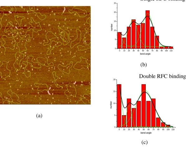
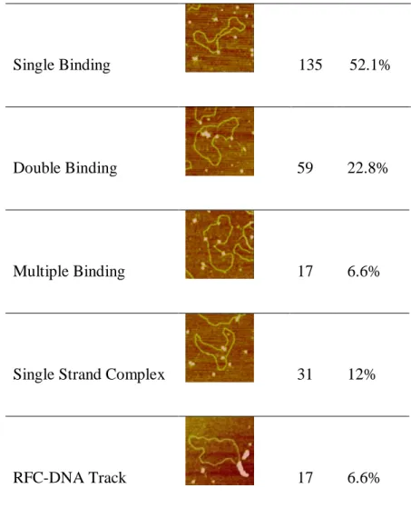
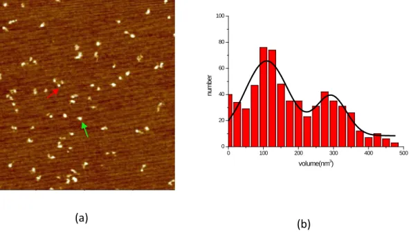
subcomplex bound to the DNA, and it appears that these subcomplexes form stable interaction with PCNA on the DNA. We proposed that this DNA-bound RFC subcomplex tethers PCNA ring at the single strand/double strand junction of primer-template DNA or nick DNA. We further suggest that dissociation of RFC subcomplex from PCNA and DNA substrate is promoted by downstream PCNA-interacting proteins, such as DNA polymerase. In addition to these insights into the complicated potential loading mechanism of PCNA, we observed other DNA complexes such as RFC-DNA filaments with nicked RFC-DNA without nucleotide cofactor and RFC-RFC-DNA spider-like complexes containing multiple RFCs and DNAs in the presence of ATP. Although we do not know the physiological role, if any, of such RFC-DNA complexes, these complexes suggest RFC can possess other functions besides as clamp loader, such as helicase.
In the study of MMR initiation complexes, eukaryotic MutS homologs (MutSα and
MutSβ), we found, unlike their prokaryotic homologs, eukaryiotic MutS homologs bind
different DNA substrates with similar conformation. MutSα and MutSβ both exhibits
weak binding specificity to their specific DNA substrates, which makes it more complicated to analyze their specific complexes. However, it appears that eukaryotic MutS homologs do not recognize mismatched bases simply depending on the formation of unbent complexes as seen in the prokaryotic MutS. It is possible they employ other high class mechanism in which the event of recognition of different mismatched DNA substrates happens downstream of mismatch binding.
iv
ACKNOWLEDGMENT
I would like to acknowledge my supervisor, Erie A. Dorothy, who has given me an incredible amount of help and guidance; not only in science but also as a mentor. Under her direction I have become a dedicated scientist. I appreciate all the time she took to instruct and guide me.
I am truly grateful for the help and encouragement of my previous lab mates: Dr. Ingrid Tessmer, Dr. Elizabeth Sacho and Susan Doyle. They taught me their precious expert knowledge about atomic force microscopy. I especially appreciate Dr. Yu Xue, Dr. Scott Kennedy, and Dr. Erika Pearson who led me into the field of molecular biology.
I would like thank to my current lab mates: Vanessa Van Vranken, Cherie Lanyi, and Dan Burke. I appreciate their encouragement and advice during my writing of this dissertation.
During the past six years, I cooperated with the people from Manju M Hingorani lab. Without their continued supply of protein, the project would not be complete.
v
TABLE OF CONTENTS
Page
LIST OF TABLES---vii
LIST OF FIGURES---viii
LIST OF ABBREVIATIONS---xi
Chapter 1. Introduction of DNA Mismatch Repair and Clamp-Clamp Loader Complex 1.1Introduction of DNA Mismatch Repair (MMR)---1
1.2Introduction of Clamps and Clamp Loaders---15
2. AFM Study of Eukaryotic RFC Loading PCNA Process 2.1 Introduction---68
2. 2Materials and Methods---72
2.3 Results---73
2.4Discussion---87
vi
3.1Introduction---133
3.2Materials and Methods---137
3.3Results---140
3.4Discussion---146
vii LIST OF TABLES
Table 1.1 Identity and function of E. coli and eukaryotic proteins involved in MMR-- 36
Table2.1. Relationship of S.cerevisiae RFC’MW with AFM volume---101
Table2.2 Statistical conformation analysis of RFC on 1077Nick DNA substrate in
absence of nucleotide---113
Table 2.3 Statistical conformation analysis of RFC on PUC18Nick DNA substrate in absence of nucleotide---121
viii
LIST OF FIGURES
Figure1.1 Mechanism of E.coli methyl-directed mismatch repair---35
Figure 1.2 Reconstitute eukaryotic mismatch-provoked excision systems---37
Figure 1.3 Crystal structure of E. coli MutS binding to G·T mismatch---49
Figure 1.4 Crystal structure of Mutα binding with G·T mispair complex ---41
Figure1.5. Models for signaling downstream MMR events following mismatch recognition and mismatch recognition mechanism of E. coli MutS---43
Figure 1.6 Crystal structure of E.coli γ-complex and schematic view of the mechanism of opening β-clamp---46
Figure 1.7 Crystal structure of RFC clamp loader complex and schematic of the clamp loading process---58
Figure 1.8 Schematic view of AAA+ ATPase family ---52
Figure2.1 AFM images of RFC in the presence of different nucleotide cofactors---99
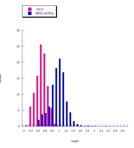
Figure2.2 Histograms of RFC volume distributions with different nucleotide cofactors---102
Figure 2.3 Nucleotide binding introduces RFC conformational change---104
Figure 2.4 AFM images of RFC·PCNA complex in the presence of ATP and ATPγS respectively---105
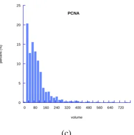
Figure 2.5 Histograms of volume analysis of RFC-PCNA complex in the presence of ATP and ATPγS and histogram of volume analsysis of PCNA---106
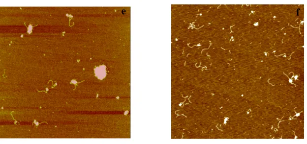
Figure 2.6 AFM images and histograms of volume distribution of RFC–primer template DNA complex with ATPγS in the presence or absence of PCNA---108
Figure 2.7 Schematic view of DNA substrates used in AFM study---110
ix
Figure 2.9 AFM images of RFC and DNA complex in presence of ATP or ADP---114 Figure 2.10 Representative AFM image and the bending angle and binding position analysis of RFC in the presence of ATPγS---116 Figure 2.11 Schematic view of RFC binding to the 1077Nick dsDNA in the presence of ATPγS and histogram of volume analysis results---118 Figure 2.12 Representative AFM image and histogram of bent angle analysis of RFC on pUC18Nick DNA substrate in the absence of nucleotide---120 Figure 2.13 AFM image RFC with pUC18Nick DNA complex in the presence of
ATP in high salt buffer. Histograms of bend angle and volume analysis of RF pUC18Nick DNA complex in the presence of ATP---122 Figure2.14 AFM image RFC+PCNA with pUC18Nick DNA complex in the presence of ATP in high salt buffer. Histograms of bend angle and volume analysis of RFC –PCNA-pUC18Nick DNA complex in the presence of ATP---124 Figure 2.15 Schematic view of the hypothesis mechanism of RFC loading PCNA
onto primer-template DNA substrate---126 Figure 3.1 Representative AFM image of Msh2Msh3 and histogram of volume
distribution of Msh2Msh3---151 Figure 3.2: Schematic view of the DNA substrate used in AFM study---152 Figure 3.3 Representative AFM image of Msh2Msh3 in the presence homoduplex
dsDNA substrate, the distribution of position Msh2Msh6 on homoduplex DNA and histogram of DNA bend angle introduced by Msh2Msh3
binding---153 Figure 3.4 Representative AFM image of Msh2Msh3 in the presence of T-bulge
DNAsubstrate, position distribution of Msh2Msh3 on T-bulge
heteroduplex DNA and histogram of DNA bend angle introduced by Msh2Msh3 binding---155
x
DNA substrate, position distribution of human Msh2Msh3 on TT-bulge DNA and histogram of bend angle introduce by human Msh2Msh3 bound at double T-bulge site and homoduplex site on 1125TTbulge---159 Figure 3.7 Representative AFM image of Msh2Msh6 in presence of homoduplex
DNA position distribution of Msh2Msh6 on homoduplex DNA and
histogram of DNA bend angles introduced by Msh2Msh6 binding---161 Figure 3.8 Representative AFM image of Msh2Msh6 in the presence of G/T mismatch DNA, position distribution of Msh2MSh6 on 982GT heteroduplex DNA and histogram of DNA bend angles introduced by Msh2Msh6 bound at
G/T mismatch site and homogenous site on 982GT DNA substrate---163 Figure 3.9 Representative AFM image of Msh2Msh6 in the presence of T-bulge DNA substrate, position distribution of Msh2Msh6 on T-bulge DNA and
xi
LIST OF ABBREVIATION
% percent
α alpha
β beta
γ gamma
δ Delta
Ɵ theta
χ phi
χ chi
µL microliter
~ approximately
°С degrees Celsius
3’ three prime end
5’ five prime end
A adenine
xii ADP adenosine diphosphate
AFM atomic force microscopy
Arg argnine
ASCE additional strand conserved E faminly
ATP adenosine-5’-triphosphate
ATPase adenosine triphosphatase
C cytosine
cal calorie
DNA deoxyribonucleic acid
dsDNA double stranded deoxyribonucleic acids
ssDNA single stranded deoxyribonucleic acids
E.coli Escherichia coli
EM electron microscopy
et. al and others
G guanine
xiii IDL nsertion-deletion loop
in vitro outside a living system
in vivo inside a living system
IRC initial recognition complex
URC ultimate recognition complex
Kd dissociation constant
kDa kilo Dalton
m meter
MMR mismatch repair
mol molar
nM nanomolar
PCNA proliferating cell nuclear antigen
PCR polymerase chain reaction
Phe phenylalanine
RFC replication factor C
xiv SV40 simian virus40
T thymine
Taq Thermus aquaticus
CHAPTER ONE: INTRODUCTION OF DNA MISMATCH REPAIR AND CLAMP-CLAMP LOADER COMPLEX
Mismatched nucleotides arise from polymerase misincorporation errors, recombination between heteroallelic parental DNAs, and chemical or physical DNA damage. Mismatch repair (MMR) corrects DNA biosynthetic errors, increasing
replication fidelity ~1000 fold, ensures the fidelity of recombination, and participates in the earliest steps of checkpoint, and apoptotic responses to several classes of DNA damage (Iyer, Pluciennik, et al, 2006; Kunkel, and Erie, 2005; Li, 2008; Modrich, 2006). Inactivation of MMR in humans is associated with hereditary nonpolyposis colon cancer and 15-25% sporadic tumors that occur in a number of tissues (Peltomaki, 2003;
Peltomaki, 2005).
1.1 Introduction of DNA mismatch repair (MMR)
The E.coli methyl-directed Mismatch Repair Reaction
- 2 -
facilitates the assembly of a functional MMR complex (Dao, and Modrich, 1998; Guarne, Ramon-Maiques, et al, 2004). Both MutS and MutL function as homodimers and possess ATPase activity (Ban, and Yang, 1998a). In E. coli, DNA is methylated at the N6
position of adenine in dGATC sequences. In replicating DNA, the daughter stand is transiently unmethlylated, and it is the presence of hemimenthylated dGATC sequence that molecularly distinguishes the newly synthesized daughter strand from the parental DNA strand. In MMR, MutH recognizes hemimethlyated dGATC sequences and functions as monomer. Upon its activation by MutS and MutL in the presence of ATP and a mismatch, MutH specially incises the unmethlyated daughter strand of hemi-methlyated dGATC, and the strand specific nick is a starting point for mismatch-provoked excision (Ban, and Yang, 1998b; Lee, Chang, et al, 2005; Ramilo, Gu, et al, 2002). In the presence of MutL, helicase II (UvrD) loads at the nick and unwinds the duplex from the nick towards the mismatch, generating single-strand DNA. The parent strand is rapidly bound by single-stranded DNA-binding protein (SSB) and protected from nuclease attack (Dao, and Modrich, 1998; Ramilo, Gu, et al, 2002). Depending on the position of the strand break relative to the mismatch, ExoI or ExoX(3’-5’
exonuclease), or ExoVII or RecJ (5’-3’ exonuclease) excises the nicked strand from the nicked site (the dGATC site ) up to and slightly past the mismatch. The resulting single gap undergoes DNA resynethsis and ligation by DNA polymerase III holoenzyme, SSB and DNA ligase (Modrich, and Lahue, 1996).
- 3 -
Several eukaryotic MMR proteins have been identified based on their homology to E.coli MMR proteins. These include eukaryotic homologs MutS, MutL, EXOI, single strand DNA-binding protein RPA, proliferation cellular nuclear antigen (PCNA), DNA
polymerase δ or ε and DNA ligase I (Table 1.1).
-MutS homologs-
Eukaryotic cells possess two MutS homologs that function as heterodimers and
share Msh2 as common subunit: MutSα (Msh2·Msh6 heterodimer) and MutSβ
(Msh2·Msh3 heterodimer). MutSα, which represents 80-90% of the cellular Msh2,
preferentially recognizes base-base mismatches and insertion/ deletion (IDL) mispairs in which one strand contains 1 or 2 unpaired nucleotides, but it is also capable of
recognizing of large IDL heterologies with reduced affinity (Drummond, Li, et al, 1995; Genschel, Littman, et al, 1998; Palombo, Gallinari, et al, 1995; Palombo, Iaccarino, et al, 1996). MutSβ recognizes IDL mismatches of 2-to ~10 nucleotides, weakly recognizes single-nucleotide IDL mispairs, and is essentially inert on base-base mismatches (Genschel, Littman, et al, 1998; Palombo, Gallinari, et al, 1995).
-MutL homologs-
Three eukaryotic MutL homologs have been identified and like eukaryotic MutS homologs, they function as heterodimeric complexes with MLH1 as a common subunit.
MutLα, a heterodimer of MLH1 and PMS2 (PMS1 in yeast), is the primary MutL
- 4 -
MutSβ (Li, and Modrich, 1995). In a reconstituted human MMR system, human MutLα
regulates termination of mismatch-provoked excision (Zhang, Yuan, et al, 2005). Recent
studies show that MutLα possesses a PCNA/RFC-dependent endonuclease activity that
plays a critical role in 3’ nick –directed MMR involving EXO1 (Kadyrov, Dzantiev, et al, 2006). Although MutLα accounts for ~90% of the MLH1 in human cells, two low
abundance complexes involving MLH1 have also been identified. Human heterodimer MLH1·PMS1 (MutLβ) has been isolated, but its molecular activity have not been ascertained (Raschle, Marra, et al, 1999). MLH1 heterodimerizes with MLH3 to form
MutLγ, and the MutLγ complex has been reported to support modest levels of base-base
and single nucleotide IDL mismatch repair in vitro, events that are presumably initiated by MutS homologs in vitro (Cannavo, Marra, et al, 2005).
-PCNA and RFC-
PCNA plays multiple roles in MMR. PCNA interacts with Msh2 and MLH1 and is thought to play roles in the initiation steps of MMR (Clark, Valle, et al, 2000; Gu, Hong, et al, 1998; Umar, Buermeyer, et al, 1996). PCNA also interacts with Msh2·Msh3 and Msh2·Msh6 via a conserved PCNA interation motifs in Msh3 and Msh6, termed the PIP box (Clark, Valle, et al, 2000; Warbrick, 2000). PCNA increases the mismatch –binding specificity of Msh2-Msh6, and it can assist in delivering of Msh2-Msh6 to mismatched DNA (Flores-Rozas, Clark, and Kolodner, 2000; Lau, and Kolodner, 2003). PCNA and
polymerase δ also have been implicated in repair DNA resynthesis, as PCNA confers
- 5 -
Umar, Buermeyer, et al, 1996). PCNA is also required for mismatch-provoked excision. The most compelling evidence for PCNA involvement in the excision step of MMR has been provided by p21 inhibition study. p21 forms a stable complex with DNA-bound PCNA so that p21 interferes with downstream PCNA-dependent events after PCNA loading in MMR. p21 is found to abolish 3’-directed mismatch-provoked excision in Hela cell extracts, however only 40-50% of 5’-directed excision events are subjected to p21 inhibition, implying occurrence of at least two hydrolytic modes of 5’-derected excision (Genschel, and Modrich, 2003; Guo, Presnell, et al, 2004; Umar, Buermeyer, et al, 1996).
RFC provides at least two functions in the reconstituted bidirectional excision system: loading of PCNA onto helix and suppression of the 5’ to 3’ hydrolysis from a 3’ strand nick. In vital, the amino terminal ligase homology domain of the large RFC subunit, which is not required for PCNA loading on the helix, is important for
suppression of 5’ to 3’ hydrolysis from a 3’-strand break but is not required for activation of 3’ directed excision. Domain II of the large RFC subunit (Figure 1.6, a), which
functions in PCNA loading, is not necessary for suppression of 5’ to 3’ hydrolysis from the 3’-nick but it is required for activation of 3’-directed excision. There results suggest a mechanism in which face of PCNA would be oriented toward the mismatch in 3’ and 5’heteroduplexes and the interaction of PCNA with ExoI will determine the direction of excision (Dzantiev, Constantin, et al, 2004).
- 6 -
ExoI is a 5’-3’ exonuclease, and is involved in both 5’ and 3’ directed MMR. ExoI can readily carry out 5’ directed mismatch excision in the presence of MutSα or MutSβ and RPA (Genschel, and Modrich, 2003; Zhang, Yuan, et al, 2005); however, its role in catalyzing 3’ nick-directed excision requires the MutLα endonulcease activity, which is activated by PCNA and RFC. After recognition of the 3’ nick and the MutS- mismatch
complex, MutLα endonuclease makes incisions 5’ and 3’ to the mismatch in a manner
dependent on PCNA and RFC. Exo I performs 5’ to 3’ excision from the MutLα incision
site through and beyond the site of the mismatch (Dzantiev, Constantin, et al, 2004; Kadyrov, Dzantiev, et al, 2006).
-RPA-
RPA seems to be involved in all stages of MMR. It appears to bind to nicked
heteroduplex DNA before MutSα and MutLα and stimulates mismatched-provoked
excision. In addition, it protects the ssDNA gapped region generated during excision and facilitates DNA resynthesis (Dzantiev, Constantin, et al, 2004; Ramilo, Gu, et al, 2002; Zhang, Yuan, et al, 2005). Furthermore, RPA is phosphorylated after polyrmerase δ is recruited to the gapped DNA substrate. Recent studies indicate that (i) phosphorylation reduces the affinity of RPA to the DNA, (ii) Unphosphorylated RPA stimulates
- 7 -
bound MutSα/MutLα, while a lower affinity RPA-DNA complex might facilitate DNA
resynthesis by Pol δ (Guo, Zhang, et al, 2006).
-Polymerase δand ε-
The editing exonuclease functions of DNA polymerase δ and ε have been postulated to provide hydrolytic functions in mismatch repair (Tran, Gordenin, and Resnick, 1999; Wang, and Hays, 2002), however this suggestion has been questioned (Datta, Schmeits, et al, 2000). The evidence for polymerase δ being involved in MMR resynthesis comes from the depleted in DNA polymerase extracts system that sustains mismatch-provoked excision but fails to support complete repair reaction is restored repair with highly purified fraction of polymerase δ devoid of datable pol α and pol ε. Thus polymerase δ is
required for eukaryotic mismatch correction, but supporting roles for polymerase α and ε
have not been ruled out (Longley, Pierce, and Modrich, 1997).
-HMGB1-
HMGB1, a non-histone chromatin protein, binds to certain type of DNA damage,
Interacts with MutSα and may play an important role in early steps of the reaction prior
to excision (Zhang, Yuan, et al, 2005).
Reconstituted Eukaryotic Mismatch Excision/Repair System
Several systems have been reconstituted to support mismatch-provoked excision by purified human proteins (Figure, 1.2). The simplest one is comprised of only four
- 8 -
hydrolysis from a 5’ strand break. MutSα activates ExoI hydrolysis on a 5’-heteroduplex
in a mismatch- and ATP- dependent manner. In the absence of RPA, MutSα renders the ExoI highly processive, resulting removal of ~2000 nucleotides prior to dissociation, an
effect attributed to formation of a MuSα·ExoI complex. However, RPA modulates the
behavior of this complex, reducing the processivity of ExoI to ~250 nucleotides. A single strand gap coated with RPA is an extremely poor substrate for ExoI, but MutSα promotes ExoI initiation at such sites provided that the DNA contains a mismatched base pair. It has been suggested that hydrolysis is dramatically attenuated upon mismatch removal because of the absence of MutSα. MutLα also plays an important role of acting with
MutSα to suppress ExoI hydrolysis on DNA that lacks a mispair (Genschel, and Modrich,
2003; Zhang, Yuan, et al, 2005).
This four protein system also supports mismatch-provoked excision on 3’-heteroduplex. However, the hydrolysis precedes 5’ to 3’ from 3’ strand break, which is the wrong polarity for mismatch removal. Adding PCNA and RFC to the reaction yields a system that supports mismatch removal from both 5’ and 3’ heteroduplex. The
nonspecific 5’to 3’ hydrolysis activity initiating at the 3’ nick is largely suppressed by RFC, and excision occurs with apparent 3’ to 5’ polarity resulting in mismatch removal (Dzantiev, Constantin, et al, 2004). Furthermore, other research shows that MutSα, RFC,
and PCNA activate a latent endonuclease activity in MutLα in an ATP and mismatch
-dependent manner. Incision by activated MutLα endonuclease occurs both 3’and 5’ to the
- 9 -
removal by the 5’to 3’ action of MutSα·ExoI complex. The PCNA-dependent endonuleolyic system also incises 5’-heteroduplex strand (Kadyrov, Dzantiev, et al, 2006). This model posits that the nick serves as a strand signal but not as site for
excision initiation, which actually occurs at strand break produced by mismatch-activated
MutLα endonuclease. This result is consistent with the result found in Xenopus egg
extracts, which demonstrated a significantly higher specific radioactivity in the vicinity of the mismatch relatively near the strand break, through analyzing the repair products incorporated with radiolabeled nucleotides (Varlet, Canard, et al, 1996).
The established systems to date are minimal systems. The fact the excision products in purified systems are more disperse than those observed in nuclear extracts indicates the probable existence of other excision activities that may function in a redundant manner with respect to ExoI (Genschel, Bazemore, and Modrich, 2002). Furthermore, the mismatch dependence of reconstituted 5’- directed excision is not as dramatic as that observed in nuclear extracts indicating that one or more specificity
factors may be lacking (Dzantiev, Constantin, et al, 2004; Genschel, and Modrich, 2003) .
Coupling of Mismatch Recognition and Strand Discrimination
mismatch-- 10 mismatch--
provoked excision, human MMR is presumed to be nick directed in vital, it is generally agreed the strand discrimination signal is a strand specific nick in both prokaryotic and eukaryotic systems, but how the nick is generated in eukaryotes remains unknown.
Previous studies have proposed several alternative models for the mismatch and strand discrimination signal process, which can be classified into a “stationary” model, a “translocation” model and a “sliding clamp” model (Figure 1.5, a). The stationary model proposes that interaction among MMR proteins induces DNA bending or looping that brings these two distant sites together, while MutS remains bound at or near mismatch. In this model, the MutS ATPase activity acts in a proof-reading role to verify mismatch binding and authorize the downstream excision (Guarne, Ramon-Maiques, et al, 2004; Junop, Obmolova, et al, 2001). In the translocation model, ATP reduces the mismatch binding affinity of MutS or MSH heterodimer, and ATP hydrolysis drives bidirectional translocation of MutS proteins along the DNA helix. DNA is threaded through the protein complex until the latter reaches a strand discrimination signal in either orientation,
- 11 -
Structure and Function of E.coli MutS and Human MutSα
- 12 -
mispaired base. The glutamic acid hydrogen bounds to the same base (to N7 if the base is a purine and to N3 if it is pyrimidine). Most contacts from two subunits are to the DNA backbone and are, therefore, DNA sequence nonspecific, as expected given the need to repair replication errors in a variety of different sequence contexts.
The crystal structure of human MutSα, with Msh6 lacking the first 340 amino acids, in complex with G: T mismatch DNA shares common domain architecture with prokaryotic MutS. Each protein can be divided into five domains, referred as mismatch binding domain, connector, levers, clamps and ATP binding domains (Figure 1.4). Msh2·Msh6 heterodimer forms an asymmetric oval disc, pierced by two channel, like the letter “Ɵ”. A DNA helix containing a single mispair is bent by ~45° and bound in the lower channel. Only Msh6 makes specific contacts with mispair through conserved Phe-X-Glu motif. Furthermore, the N-terminus of Msh6 (residues 360-398) contains
additional nonspecific contacts with the DNA backbone. By contrast, the DNA binding domain of Msh2 is rotated up and away from backbone and makes only one contact with the DNA, which is different from the prokaryotic homodimer. Interactions between Msh6 and the DNA substrate bury 1142 Å2 (with domain I contributing 856 Å2), which is probably sufficient to bend the DNA without any contribution from Msh2. The upper channel is surrounded by disordered loops and is too small to contain a second DNA double strand as been proposed in prokaryotic analog (Warren, Pohlhaus, et al, 2007).
- 13 -
- 14 -
Allosteric coupling between the ATPase sites and the DNA binding sites plays a central role in the MutS homologs mechanism. DNA substrate binding is known to modulate the ATPase activity: both hetero and homoduplex DNA stimulate ADP-ATP exchange, but only heteroduplex DNA appears to change the rate limiting step for turnover of ATP. In the presence of ATP alone or ATP and homoduplex DNA, there is burst of hydrolysis of one ATP equivalent per dimer; whereas, in the presence of mismatch, the burst of hydrolysis is suppressed (Acharya, Foster, et al, 2003; Antony, and Hingorani, 2003; Bjornson, Allen, and Modrich, 2000). Similarly, ATP binding modulates DNA substrate binding. ADP has little effect on the affinity of MutS proteins for mismatch DNA. However, ATP and ATP analogs decrease mismatched DNA-binding affinity, although substantial mismatch specificity is retained under conditions that
support ATP hydrolysis (Blackwell, Martik, et al, 1998; Blackwell, Bjornson, et al, 2001; Gradia, Acharya, and Fishel, 1997; Hess, Gupta, and Kolodner, 2002; Joshi, Sen, and Rao, 2000; Martik, Baitinger, and Modrich, 2004).
Like other DNA repair enzymes, MutS homologs must locate a subtle base pair anomaly within a vast excess of nonsubstrate, correctly paired DNA. Insights into repair specificity come from recent AFM studies that directly visualize a MutS bound to
- 15 -
state. These data led to the proposal that MutS binds to DNA nonspecifically and bends in search of mismatch (Figure 1.5, b), and upon specific recognition of a mismatch, MutS undergoes a conformational change to an initial recognition complex (IRC) in which the DNA is kinked, with interactions similar to those in the crystal structures. MutS then undergoes further conformational changes to the ultimate recognition complex (URC) in which the DNA is unbent with the mismatched base possibly being flipped out.
Similar AFM studies have not been conducted on the eukaryotic homologs.
Strikingly, the crystal structure of human MutSα reveals that it recognizes different DNA substrates in a similar manner (Warren, Pohlhaus, et al, 2007) . MutSα recognizes the G·T mispair or a single base T insertion /deletion loop in the DNA MMR , as well as an O6-methyl guanine·T mispair in a similar manner although mismatch initiates repair and O6-methyl initiates apoptosis. MutSα also recognizes a G·dU mispair, a putative
immediate in somatic hypermutation. All the crystal structures of MutSα-DNA
complexes are virtually identical. This result is surprising given the significant
differences in DNA base paring, conformation, and thermal stability of these mismatches (Kramer, Kramer, and Fritz, 1984; Kramer, Kramer, et al, 1989). This observation led to
the suggestion that diversity of MutSα dependent responses to DNA lesions is generated
in events downstream of the lesion recognition step or they all crystallize in the same conformation.
- 16 -
Chromosome replication requires a DNA polymerase that can rapidly duplicate thousands of nucleotides. In all cells, the replicative polymerase is tethered to DNA by a ring- shaped clamp that encircles DNA and slides freely along it. This interaction confers a high degree of processivity to the replicative polymerase. Ring-shaped sliding clamps are constructed from either two or three identical crescent-shaped protomers and their closed circular structure requires an active mechanism to open them for assembly onto DNA. This loading process is carried out by multiprotein machines known as clamp loaders, which crack open the ring and place it around the primed DNA in an ATP driven reaction.
Brief introduction of AAA+ proteins family
The defining feature of AAA+ proteins is a structurally conserved ATP-binding module that oligomerizes into active arrays. ATP binding and hydrolysis events at the face of neighboring subunits drive conformational changes within the AAA+ assembly that direct translocation or remodeling of target substrates (Erzberger, and Berger, 2006). AAA+ proteins are involved in a myriad of biological process, including ATP-dependent proteolysis, membrane fusion, protein trafficking, and DNA replication, recombination, and repair.
As a subfamily of additional strand conserved E family (ASCE), AAA+ proteins have conserved ASCE core nucleotide-binding pocket lying at the apex of three adjacent, parallel β-strands in a compact αβα-fold (Figure,1.8 a). The primary distinguishing
- 17 -
helical bundle fused to the C terminus of the central αβα-fold. Several other features are
characteristic of this family (Figure 1.8, b). For example, the conserved argnine residues (argnine finger) of homology SHC or SRC motif interact with nucleotide-binding pocket of neighboring subunit. In addition, there are two other nucleotide-interaction motifs termed sensor I and sensor II elements. The sensor I motif, which resides at the top of
strand β4, is typically an Asn, although other polar residues, such as Ser, Thr, or Asp are
also found. This residue is believed to act in concert with the second acidic residue of the Walker-B motif to properly orient a water molecule for nucleophilic attack on the γ
-phosphate of ATP. Sensor II usually contains an arginine at the base of helix α7 that
interacts with the γ-phosphate of ATP (Guenther, Onrust, et al, 1997).
Seven major clades of AAA+ proteins have been defined on the basis of sequence alignments and structural information (Figure 1.8,c). The differences between clades arise from the insertion of secondary structural elements at defined places within and around the core AAA+ fold (Iyer, Leipe, et al, 2004).
Clade 1 is represented by the clamp loader family. The pentameric complex recognizes the primer-template junctions and promots the opening and loading of the polymerase processivity clamp onto DNA.
Clade 2, the initiator clade, includes all cellular origin-processing proteins, as well as the bacterial, archaeal, and eukaryal helicase-loading proteins. This group is defined by
the presence of an extra α-helix inserted between the second and third strands of the
- 18 -
replication origins. The initiators are responsible for properly recognizing replication origins and for enabling the subsequent assembly of the replication machinery (Messer, 2002). Most of AAA+ initiators are monomeric in solution and assemble into
oligomeric structures when bound to specific target DNA sequences at origins
(Cunningham, and Berger, 2005). Remarkably, DnaA has been revealed to form a helical filament on DNA in presence of ATP. The filament arrangement places DNA-binding domains appended to the C-terminus of the AAA+ module on the outside of the filament. This orientation suggests that the interaction with origin DNA sequences may lead to the introduction of right-handed helical wrap to this region, destabilizing adjacent DNA elements as a prelude to origin melting and replisome assembly. In addition, the
assembly at the boundaries of the filament, exposing an arginine finger at one end and a nucleotide-binding pocket at the other end, may serve as a docking sites for auxiliary replication factors that also contain AAA+ domains (Erzberger, Mott, and Berger, 2006).
Clade 3 is a group of proteins that form closed hexameric assemblies. These classic clade proteins are involved in processes such as protein degradation, microtubule serving, membrane fusion, and peroxisome biogensis. Classic AAA+ modules share a small
helical insertion before helix α2 of the ASCE fold. The classic clade is unique for
possessing two argnine residues instead of a single residue commonly see in other AAA+ proteins (Erzberger, and Berger, 2006).
- 19 -
insertion appears to link ATP turnover to DNA translocation. Clade5, composed of the HslU, ClpAB-CTD, LonAB, and RuvB families (HCLR clade), represents the central group of the PS-I superclade. Clade 4 differs from clade 5 with an unusual bundle formed by elements N- and C- terminal to the ASCE core instead of AAA+ lid. Clade 6 and Clade 7 contain an additional β-hairpin insertion that disrupts helix α2.
There is little correlation between AAA+ protein subtypes and a specific remodeling activity. Nevertheless, Clade 1 and Clade 2 are characterized by open-ring assemblies that differ significantly from other AAA+ proteins, most of which form closed hexameric rings. These kinds of assemblies appear to function on as a single turnover allosteric switches, rather than processive molecular machines. ATP binding appears to flip these proteins into an activated state by influencing the conformation of the AAA+ modules and enabling them to remodel respective proteins.
E.coli and bacteriophage T4 clamp loader
In E. coli, the β- clamp is a ring-shaped homodimer that encircles the double-stranded DNA and tethers polymerase (Pol III) to the template for high processivity (Kong, Onrust, et al, 1992; Stukenberg, Studwell-Vaughan, and O'Donnell, 1991). The
clamp loader is required to open the closed ring of the β clamp and load it onto the
template in an ATP-driven process.
In E. coli, the clamp loader is known as γ-complex, which comprises five subunits: δ,
- 20 -
not required for clamp loading in vitro (Onrust, and O'Donnell, 1993). However, the ψ·χ complex provides the linkage between the clamp loader and single strand binding
proteins (SSB), and this linkage increases the replication activity. The ψ protein binds
tightly to the collar domain of the γ complex and the χ tucks onto the C-terminal tail
segment of SSB, thus bridging the clamp loader and SSB (Glover, and McHenry, 1998; Kelman, Yuzhakov, et al, 1998; Olson, Dallmann, and McHenry, 1995; Simonetta, Kazmirski, et al, 2009; Witte, Urbanke, and Curth, 2003).
The crystal structure of the γ-complex revealed that the five subunits are arranged in
a spiral (Figure 1.6, a), with δ and δ’ bracketing the three γ subunits. The C-terminal
domain of each subunit mediates oligomerization into a spiral heteropentamer. The primer-template DNA is thought to get out of the gap between δ and δ’ subunit. The three
γ subunits are the only subunits of the clamp loader that bind ATP, and therefore
comprise the ‘motor’ that drives the clamp loading reaction. The δ subunit crackes open
the β-dimer, even in the absence of γ and δ’, at one interface, and thus is termed the
‘wrench’. The δ’ seems to be a rigid protein that lacks the flexible motions that occur in
the δ and γ subunits. The rigidity allows δ’ to act as a stator or a backboard, directing
ATP-induced conformational changes, propagating from γ1 through γ2, γ3 and finally
through δ, pulling δ away from δ’ (Jeruzalmi, Yurieva, et al, 2001).
The γ complex belongs to AAA+ family of ATPases, although only the γ subunits have functional ATPase sites. The ATP-binding sites are at the interface of the pairs of
- 21 -
argnine finger extends toward to the ATP binding site in the adjacent subunit and senses
ATP binding and hydrolysis. Crystal structures of the nucleotide free and ATPγS bound
states revealed that only two of three ATP binding sites (γ1 and γ3) are occupied by
ATPγS, with the γ2 subunit binding site being physically blocked by the γ1 subunit
(Jeruzalmi, O'Donnell, and Kuriyan, 2001; Kazmirski, Podobnik, et al, 2004). The sites that bind ATP do so with equal affinity and there appears to be little or no cooperativity
in ATP binding. The conformation of γ complex with two ATP-bound is likely to
represent a stable, but inactive state of the clamp loader. Modeling the kinetics of ATP hydrolysis in the absence of the clamp suggest that the clamp loader binds and hydrolyzes three molecules of ATP (Williams, Snyder, et al, 2004).
On binding ATP, the γ complex undergoes conformational changes that increase the affinity of the clamp loader for the clamp and DNA (Turner, Hingorani, et al, 1999) (Hingorani, and O'Donnell, 1998; Naktinis, Onrust, et al, 1995; Turner, Hingorani, et al, 1999) (Bertram, Bloom, et al, 1998; Hingorani, and O'Donnell, 1998). Within the γ
complex, δ is blocked from interaction with β by δ’. However, ATP binding introduces a
conformational change in the γ complex, exposing δ for interaction with β and
subsequent ring opening (Naktinis, Onrust, et al, 1995).The δ subunit opens the β clamp using the energy from protein-protein interactions and does not need ATP. The binding of
δ subunit relaxes the spring tension of α helix at the interface with the β clamp (Figure
- 22 -
ATP binding is sufficient for the formation of a ternary clamp loader–clamp- DNA complex; nevertheless, ATP hydrolysis is required for releasing the clamp onto the DNA (Bertram, Bloom, et al, 1998; Bloom, 2006; Turner, Hingorani, et al, 1999). Template DNA triggers three ATP sites in γ complex to hydrolyze ATP at the same global stage of reaction, releasing the clamp on DNA (Bertram, Bloom, et al, 2000). Although the β
clamp does not induce the γ complex to hydrolyze ATP in the absence of DNA, the β
clamp does increase the overall rate of hydrolysis in the presence of DNA. In the absence
of the β clamp, the γ complex hydrolyzes two molecules of ATP more rapidly than the
third one. In the presence of the β clamp, all three molecules of ATP hydrolyze as the
same rapid rate as the two molecules of ATP hydrolyzed in the absence of the β clamp
(Williams, Snyder, et al, 2004). The results from studies of mutation of arginine finger of the clamp loader suggest that ATP binding at one subset, γ2 and γ3, is largely responsible
for increasing the affinity of the complex for DNA, and ATP binding at γ1 is largely
responsible for increasing the affinity of complex for β clamp (Bloom, 2006; Snyder,
Williams, et al, 2004).
The recently published crystal structure of E.coli γ complex with primer template DNA provides insight into clamp loader and DNA interactions (Figure 1.6, c). In the
crystal structure, the AAA+ modules of the γ and δ’ subunits form a right-handed spiral
around the double-stranded portion of the DNA with a uniform rise and rotation around a common helical axis (Figure 1.6, c). The symmetry in the quaternary arrangement of the
ATPase domains of the γ complex results in the configuration of each interfacial ATP
- 23 -
formation of catalytically competent ATP-binding sites facilitates release the clamp loader upon recognition of primer-template DNA. The positive ends of the helix dipoles
of helices α4 and α5 within each subunit are positioned close to negatively charged
phosphate groups, and the tips of these two helices are bisected by the backbone of the
template strand. Domain I of the δ subunit is disengaged from DNA. Domain III of the δ
subunit positions the side chain containing Tyr 316 so that it stacks on the nucleotide base at the 3’ end of the primer strand, resulting in termination of the primer strand and a sharp bend in the template strand as it exits the clamp loader chamber. The collar
domains encircle a tunnel that leads into the site within the central chamber where the last nucleotide of 3’ primer strand is bound. The blocking function of the Tyr 316 side chain is reminiscent of the role of an aromatic side chain in the UvrD helicase that serves as a “separation pin” by splitting the path of DNA (Lee, and Yang, 2006). The
single-stranded 5’ overhang of the template strand exits the central chamber of the clamp loader
and binds to the exterior surface of the domain III of δ subunit (Simonetta, Kazmirski, et
al, 2009).
- 24 -
the complete gp44/62 loader complex. This result suggests that the gp62 subunit may be
analogous to the δ subunit of E.coli and this serves as the wrench of the T4 clamp loader complex (Rush, Lin, et al, 1989).
The T4 gp44/gp62 clamp loader initially binds four ATPs with equal affinity (Young, Weitzel, and von Hippel, 1996). On binding gp45, two of four molecules are hydrolyzed to open the clamp wide enough so that it could be loaded on dsDNA. The remaining two molecules of ATP are hydrolyzed to close the clamp on the DNA. The clamp loader gp44/gp62 plays an additional chaperone-like role in the formation of the DNA polymerase holoenzyme. Gp44/gp62 mediates the interaction between the gp45-DNA complex and gp43 (T4 bacteriophage gp45-DNA polymerase). The transient formation of a multiple proteins complex of gp45·gp44/gp62·DNA·gp43 is observed before release of gp44/gp62 (Sexton, Kaboord, et al, 1998; Trakselis, Berdis, and Benkovic, 2003).
Gp45 is partially open in solution with a separation distance of 42 Å at one subunit interface and 17 Å at the other two interfaces (Alley, Shier, et al, 1999). Moreover, the
T4 clamp is much less stable than γ complex with a Kd of 250 nM for the dimer
compared to a Kd of < 60 pM for the β clamp (Yao, Turner, et al, 1996). It is intriguing
- 25 -
complete its loading onto the primer-template DNA (Pietroni, Young, et al, 2001; Pietroni, and von Hippel, 2008).
The result that gp44/gp62 forms relatively stable complex with DNA in the presence of ATP is remarkably contrasted to the results from E.coli γ complex and eukaryotic RFC , in which ATP binding and hydrolysis destabilizes the clamp loader-DNA complex. In addition, this gp44/gp62-DNA binary complex is active in loading gp45 onto DNA. These results suggest that gp44/gp62 binds the clamp after binding DNA, whereas, it’s generally believed that for E.coli and eukaryotic RFC that the clamp must bind the clamp loader before binding DNA. The binding of ATP to gp44/gp62 induces a conformational change in gp44/gp62 that configures the clamp loader for effective DNA binding. A gp44/gp62-DNA complex is formed with the hydrolysis of one equiv of ATP to interact with the opened form of gp45 on primer-template DNA and orient the clamp properly (Zhuang, Berdis, and Benkovic, 2006).
RFC Structure
RFC was shown to be essential for simian virus40 (SV40) DNA replication in vitro
(Tsurimoto, Stillman, 1989). It is involved in polymerase switching from Polα to Pol δ
- 26 -
The eukaryotic clamp PCNA is a trimer of three identical subunits arranged head to tail to generate a ring with a large central cavity for encircling DNA (Gulbis, Kelman, et al, 1996; Krishna, Kong, et al, 1994). Both DNA polymerase and clamp loader bind to the C-terminal face of PCNA in a hydrophobic pocket between two domains (Figure 1.7 f). Studies in the E. coli system have shown that clamp loader and DNA polymerases compete for binding the clamp, and therefore the clamp loader must eject from the clamp for DNA polymerase to use it (Naktinis, Turner, and O'Donnell, 1996). Likewise, polδ also competes for binding to PCNA in eukaryotes (Mossi, Jonsson, et al, 1997; Oku, Ikeda, et al, 1998). Therefore after PCNA is linked to DNA, RFC is thought to eject from the clamp to allow the polymerase access to the clamp (Podust, Tiwari, et al, 1998).
- 27 -
- 28 -
Resent crystal structure results revealed a stabled RFC complex (Figure 1.7, d) with
a closed PCNA clamp in the presence of ATPγS (Bowman, O'Donnell, and Kuriyan,
2004). Because ATP hydrolysis weakens the interaction between clamp and clamp loader, they replaced the arginine finger residues that are present in the highly conserved Ser-Arg-Cys (SRC) motifs of four RFC subunits (RFC B, C, D, and E) with glutamine, which resulted in a significant reduction in ATPase activity. In the structure of RFC: PCNA complex, the five AAA+ modules of RFC assemble into a right- handed spiral, leaving a gap between RFC-A and RFC-E. This gap serves the purpose of allowing DNA to enter the central chamber in the clamp loader and thereby positions DNA into the open clamp, which is docked underneath the clamp loader. The right-handed spiral arrangement of the five AAA+ domains of RFC displays roughly the same pitch as that of double-stranded B-form DNA, which suggests a mechanism of DNA recognition. Only three of the RFC subunits (RFC-A, RFC-B and RFC-C) make contact with closed PCNA clamp. RFC-A and RFC-C interact with two of PCNA conserved hydrophobic grooves. RFC-B which is located between RFC-A and RFC-C, makes limited and primarily polar interactions with the intersubunit region of the clamp. All three RFC subunits interact with PCNA
primarily through the C-terminal end of the clamp-interaction helix (α4).
The PCNA ring is closed in this crystal structure, yet ATPγS enables the RFC: PCNA complex to bind DNA and therefore presumably promotes PCNA opening. An EM structure of RFC-PCNA-primer DNA ternary complex revealed an open spiral form
of the RFC: PCNA: ATPγS complex and it is likely all the subunits of RFC have contacts
- 29 -
the PCNA ring could open out of plane and have a tendency to form a right handed spiral (Kazmirski, Zhao, et al, 2005). A fluorescence resonance energy transfer study indicates that PCNA is held open by RFC in response to bound nucleotide (Figure 1.7d) (Zhuang, Yoder, et al, 2006). Therefore this closed conformation of PCNA may be due to the specific point mutation that prevents nucleotide hydrolysis during crystal growth.
In this crystal structure, only two of three PCNA hydrophobic pockets interact with RFC subunits. RFC-D and RFC-E are suspended above PCNA with RFC-E positioned above the third hydrophobic pocket of PCNA. However, a surface plasmon study demonstrated that RFC-E and RFC-D both interact with PCNA, and a PCNA unloading experiment suggests that the RFC-(D,E) subcomplex is primarily responsible for the PCNA-clamp-opening function of RFC (Yao, Johnson, et al, 2006). Therefore, the crystal structure of RFC: PCNA (closed): ATPγS is suspected to represent the
intermediate conformation after RFC loads PCNA onto DNA. Ring closure is presumed to lead to loss of RFC affinity for PCNA and dissociation of RFC from the PCNA-DNA complex.
ATP utilization by yeast RFC
RFC belongs to the AAA+ ATPase family. This family includes a wide variety of factors that couple ATP binding and hydrolysis to remodel protein and substrates
- 30 -
chain is referred to as an “arginine finger” by analogy to an arginine residue inserted at the active site of Ras protein by the GTPase-activating proteins. Interactions between AAA+ modules of RFC subunits create four interfacial ATP sites that are competent for hydrolysis (Bowman, O'Donnell, and Kuriyan, 2004; Yao, Coryell, et al, 2003). The four ATP sites are referred to as ATP sites A-D, with site A at the RFC-A/B interface and site D at the RFC-D/E interface. The RFC-B arginine finger functions in ATP site A, and the arginine finger of RFC-C functions in ATP site B and so on. RFC-E (RFC5) also binds nucleotide, but the nucleotide-binding pocked of RFC-E is not competent for hydrolysis nor is it needed for RFC activity (Bowman, O'Donnell, and Kuriyan, 2004; Podust, Tiwari, et al, 1998).
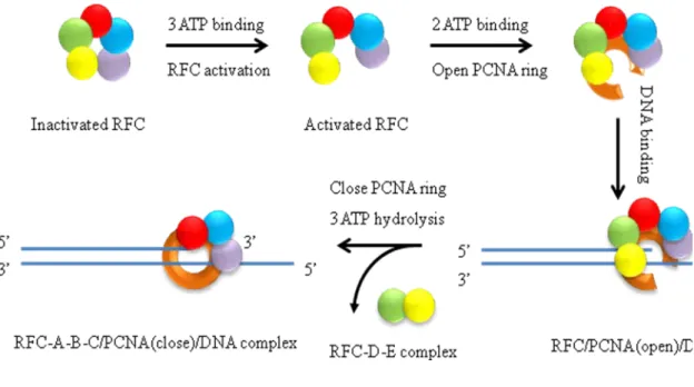
The process of RFC loading PCNA is an ATP driven pathway. RFC can load and release PCNA onto a primer-template site at a rate of 1-2 s-1, which is compatible with the estimated rate of Okazaki fragment synthesis in vital (0.5-1 s-1). There are four key steps of clamp loading, which are manipulated by ATP binding and hydrolysis, PCNA and primer DNA template binding (Figure 1.7, e).
I. ATP binding induces activation of RFC such that it can bind PCNA with high affinity. RFC has five nucleotide binding sites and shows weak ATPase activity (0.024s-1). Full length RFC alone can bind three molecules of ATPγS, and when PCNA or primer DNA or both are present in the reaction, full length RFC can
- 31 -
change in RFC, enabling it to bind and open PCNA, and subsequently bind DNA (Chen, Levin, et al, 2009).
II. PCNA opening locks RFC into an active state, and the resulting RFC: ATP: PCNA (open) intermediate is ready for DNA entry into the clamp (Chen, Levin, et al, 2009; Gomes, Schmidt, and Burgers, 2001; Zhuang, Yoder, et al, 2006). Activated RFC has high affinity for both PCNA and primer-template DNA. However, RFC forms a stable complex with primer-template DNA only in the
presence of ATPγS. Interaction with primer-template DNA leads to rapid ATP
hydrolysis and disassembly of RFC from the DNA (~25s-1). However, RFC and PCNA can form a relatively stable complex which slowly disassembles (~1.7s-1). Inclusion of PCNA results in a faster rate of RFC activation (1.5s-1-4.6s-1), and PCNA stimulates RFC-DNA-dependent ATPase activity downstream of DNA binding (~11s-1 to ~53s-1). The conformation of PCNA docked underneath RFC is predicted to open into a spiral conformation complementary to that adopted by the RFC subunits. Conversely, the open conformation of PCNA locks RFC in an activated state (Chen, Levin, et al, 2009; Kazmirski, Zhao, et al, 2005; Miyata, Suzuki, et al, 2005; Zhuang, Yoder, et al, 2006).
- 32 -
confers RFC nonspecific DNA binding properties (Gomes, Gary, and Burgers, 2000; Schmidt, Gomes, and Burgers, 2001; Uhlmann, Cai, et al, 1997). Deletion of the ligase homolog domain increases the clamp loading activity. The DNA binding domain at the C-terminal site of RFC-A and other RFC subunits may function as coordinate units that require ATP for binding with DNA. The RFC A-D subunits contain several conserved positively charged and polar side chains that are oriented toward the central chamber and may function to bind DNA (Bowman, O'Donnell, and Kuriyan, 2004; Yao, Johnson, et al, 2006). Mutation of conserved basic residues in RFC subunit B, C, D, or all three subunits together dramatically decreases affinity to DNA (Johnson, Yao, et al, 2006; Yao, Johnson, et al, 2006). In addition, mutation analysis in yeast RFC has shown that ATP binding to site C and D of RFC is essential for DNA binding and the argining finger of RFC-D is likely needed for the ATP in site C to promote the
conformation change in RFC required for DNA binding. RFC with an argining finger mutation of RFC-D subunit loses the affinity to primer-template DNA (Johnson, Yao, et al, 2006; Schmidt, Gomes, and Burgers, 2001)
IV. ATP hydrolysis triggered by recognition of a DNA primer-template junction results in the dissociation of RFC from the clamp and PCNA closure (Chen, Levin, et al, 2009; Gomes, and Burgers, 2001; Yao, Johnson, et al, 2006). RFC alone shows a slow ATPase activity (~0.024s-1). PCNA has a small but
- 33 -
~11s-1 in the absence of PCNA. RFC hydrolyzes three molecules of ATP in the presence of PCNA, and two molecules of ATP in the absence of PCNA. Studies of the argnine finger mutation suggest that DNA triggers ATP hydrolysis in site C which in turn, may drive hydrolysis in additional ATP sites. ATP hydrolysis in site D is specially triggered by PCNA, and it has been proposed that this site leads to closure of PCNA around DNA It is not clear how the clamp loader is released from DNA for another cycle of clamp loading. (Chen, Levin, et al, 2009; Johnson, Yao, et al, 2006; Schmidt, Gomes, and Burgers, 2001).
Alternative clamp loaders
There are three additional clamp loaders or putative clamp loaders and one clamp identified in eukaryotic cells. These three clamp loaders consist of four RFC small subunits in common and one pathway-specific large subunit instead of RFC-A (Majka, and Burgers, 2004).
Rad24-RFC clamp loader functions in DNA damage check-point pathway, loading Rad17-Mec3-Ddc1 PCNA- like clamp onto DNA (Kondo, Matsumoto, and Sugimoto, 1999; Majka, and Burgers, 2003). Ctf18-RFC clamp loader is involved in establishment of sister chromatid cohesion (Hanna, Kroll, et al, 2001; Mayer, Gygi, et al, 2001). Elg1-RFC is essential for maintainance of chromosome stability. Yeast Elg1 mutants show elevated levels of recombination, chromosome loss and gross chromosome
- 34 -
chromatin cohesion too. However, Elg1-RFC cohesion activity is distinctive from that of Ctf18-RFC (Maradeo, and Skibbens, 2009; Parnas, Zipin-Roitman, et al, 2009).
Although it is well established from genetic studies that Rad24, Ctf18, and Elg1 RFC-like clamps function in separate pathways, there is also cross-talk between these pathways. All three RFC-like clamp loaders interact with PCNA. Ctf18-RFC can load and unload PCNA onto a primer-template DNA substrate (Bermudez, Maniwa, et al, 2003). A physical interaction between Elg and PCNA suggest Elg1-RFC may also open and thus load PCNA onto DNA (Kanellis, Agyei, and Durocher, 2003). Although Rad24-RFC could not load PCNA, it could open PCNA and unload it from DNA (Yao, Johnson, et al, 2006). PCNA is crucially involved in establishment of sister chromatid cohesion in S phase (Moldovan, Pfander, and Jentsch, 2006). Sister chromatid cohesion is essential for the equal segregation of replicated chromosomes to the daughter cell. Appropriate cohesion involves identifying the products of chromosome replication as sisters, depositing cohesions onto each sister and then modifying those cohesions to form structural bridges that tether together sister chromatids until anaphase onset. Ctf7/ Eco1 is an acetyltransferase that activates cohesion during S-phase and is essential for sister chromatid paring (Skibbens, Corson, et al, 1999). Ctf7/Eco1 is found associated with all four clamp loading complexes, RFC-A, Rad24, Ctf18 and Elg1 (Kenna, and Skibbens, 2003; Maradeo, and Skibbens, 2009). In addition, RFCs often compensate for one
- 35 -
Figure1.1 Mechanism of E.coli methyl-directed mismatch repair (reproduced from G. Li, Cell Research, 2008). E. coli mismatch repair is initiated when MutS specifically recognizes mismatched DNA. MutS interaction with MutL activates the latent
endonulease activity of MutH in an ATP-dependent manner, which cleaves the newly synthesized daughter strand at hemimethlated GATC sites. The resulting nick, which can either 3’ or 5’ to the mismatch, is the entry point for MutL-dependent loading of DNA helicase II and binding of single-strand DNA binding protein. Depending on the location of the nick to the mismatch, the generated single strand is digested by a 3’ or 5’
exonuclease. The excision removes the error and allows highly accurate DNA
- 36 -
Table 1.1 Identity and function of E. coli and eukaryotic proteins involved in MMR
E coli Function Homologs Function
MutS Binds mismatch Msh2·Msh6 (MutSα)
Msh2·Msh3 (MutSβ)
Repairs single base-base and 1-2 base IDL mismatches
Repairs mismatches of 2 to ~10 nucleotides and some small single IDLs
MutL Matchmaker that coordinates multisteps in MMR MLH1·PMS2 (yPms1) (MutLα) MLH1·MLH2 (hPMS1) (MutLβ) MLH1·MLH3(MutLγ) Matchmaker, endonuclease, termination of mismatch-provoked excision
Unknown function in MMR
Supports modest levels of base-base and single IDL
MutH Strand discrimination, endonuclease
None
UvrD DNA helicase None SSB Participates in
excision and DNA synthesis
RPA ssDNA binding/protection, stimulates mismatch excision, termination of DNA excision, promoting DNA synthesis
β-clam May recruit MutS to mismatch/ replication foci, enhances processivity of pol III
PCNA Recruits MMR proteins to mismatch, participates in excision, activation of MutLα endonuclease, DNA
resynthesis
γ-complex β-clamp loader RFC PCNA clamp loader, ,modulates excision polarity, activation of MutLα endonuclease
ExoI or ExoX
RecJ or ExoVII
3’ to 5’ excision of ssDNA
5’ to 3’ excision of ssDNA
ExoI 5’ to 3’ excision of ssDNA
DNA pol III
DNA resynthesis DNA pol δ and pol ε DNA resynthesis DNA
ligase
- 37 -
a
- 38 -
Figure 1.2 Reconstituted eukaryotic mismatch-provoked excision systems (reproduced from P.Mordrich, JBC,2006). a. 5’-heteroduplex excision depending on
MutSα, MutLα, RPA and ExoI. MutSα actives ExoI 5’ to3’ hydrolysis on 5’
heteroduplex and renders ExoI highly processive about ~2000 nucleotides, an effect
attributed formation of MutSα·ExoI complex. RPA reduces the high processivity of
MutSα·ExoI complex to ~250 nucleotides. MutLα acts in concert with MuSα to suppress
ExoI hydrolysis on DNA that lacks mismatch. MutSα also activates ExoI hydrolysis on 3’-heteroduplex, but in this case hydrolysis proceeds in the wrong direction. b.
Reconstituted bidirection eukaryotic mismatch-provoked excision system is comprised of
MutSα, MutLα, RPA, ExoI, PCNA and RFC. In this case, PCNA and RFC activate
MutLα latent endonuclease. MutLα incises 5’ or 3’- heteroduplex strand in an
ATP-dependent manner, generating a strand break as the entry site of MutSα activated ExoI,
- 39 -
a
b
c
d
Domain I (A)
- 40 -
Fig 1.3 Crystal structure of E. coli MutS binding to G·T mismatch (reproduce from M. H. Lamers, et. al, Nature, 2000). a. Overview of MutS-DNA complex. The
- 41 -
a
b
- 42 -
Figure 1.4 Crystal structure of Mutα binding with G·T mispair complex (reproduced
from J.J. Warren, et.al, Molecular Cell, 2007). a. Ribbon diagram of the structure of
MutSα-DNA complex. Msh6 is colored blue, Msh2 red, DNA green ribbon, and ADP
green sphere. Long α helices connecting clamp and ATPase domains in Msh2 and Msh6
- 43 -
a
- 44 -
Figure1.5. Models for signaling downstream MMR events following mismatch recognition and mismatch recognition mechanism of E. coli MutS. a. A schematic diagram for signaling between the mismatch and the strand discrimination signal is shown (reproduced from G. Li, Cell Research, 2008). Here, a 5' nick is the strand discrimination signal. Similar models apply for 3' nick-directed MMR. The "stationary" or "trans" model (right) emphasizes that MutS or its homologous (MSH) proteins remain bound at the mismatch. It is the protein-protein interactions that induce DNA bending or looping that brings the two distant sites together. The two DNA sites can cooperate in a "trans" configuration. In two "cis" or "moving" models, one called the "translocation" model (left) and the other called the "molecular switch" or "sliding clamp" model (middle), it is hypothesized that the MSH proteins bind to the mismatch and then move away from the site to search for the strand discrimination signal. The translocation model suggests that ATP hydrolysis drives bidirectional movement of the MSH proteins,
resulting in the formation of an -like loop. In the molecular switch model (center), binding of an MSH protein (in its ADP-bound state) to the mismatch triggers an ADP to ATP exchange that promotes bidirectional sliding of the protein away from the mismatch, thereby emptying the mismatch site for another incoming MSH protein. Mismatch
- 45 -
- 46 -
a
b
- 47 -
Figure 1.6 Crystal structure of E.coli γ-complex and schematic view of the
mechanism of opening β-clamp. a. The individual subunits from thecrystal structure of
γ-complex (reproduced from M.J. Davey, J. Kuriyan, M. O’Donnell, Nature Review,
2002).The three domains (I, II and III) are indicated. The amino and carboxyl termini are marked (N and C respectively). The colour of each subunit corresponds to the colour of the subunit in the structure of γ-complex below. The crystal structure reveals that the five subunits are arranged in a circle, with δ and δ’ bracketing the three γ subunits. b. The β clamp is composed of two crescent-shaped promoters (reproduced from M.J. Davey, J. Kuriyan, M. O’Donnell, Nature Review, 2002)., each consisting of three domains (each domain is indicated by a different color). In the dimer form (left) the promoters must adopt a bent shape to form the dimeric interfaces of the clamp, therefore placing the promoters under spring tension. When one interface of the clamp is disrupted by the δ wrench, the promoters relax (right), resulting in an opening at one interface of the clamp for DNA strand passage. c. . The structure of clamp loader with primer-template DNA is shown (refer to K. R. Simonetta, J. Kuriyan, M. O’Donnell, Cell, 2009)., with the δ’ subunit removed to reveal a tunnel leading through the collar, indicated by red spheres. In
the expanded view on the right, side chains presented by the collar domain of the δ
subunit and that interact with DNA are shown. Two side chains that line the collar
- 48 -
a
b
c
- 49 -
e
- 50 -
Figure 1.7 Crystal structure of RFC clamp loader complex and schematic of the clamp loading process. a. A schematic view of RFC complex viewed from the PCNA interacting face. The five subunits of the RFC complex are referred to as RFC-A, RFC-B, RFC-C, RFC-D, and RFC-E, respectively (reproduced from G.D. Bowman, M.
O’Donnell, J. Kuriyan, Nature, 2004). b. Left: schematic diagram depicting ATPase site C from the crystal structure. Right: Schematic of the arrangement of ATP sites in the AAA+ modules of RFC heteropentamer. Each ATPase site is at a subunit interface. The neighboring subunit contains an arginine finger in a conserved SRC motif that interacts
with the γ phosphate of the ATP bound to the adjacent subunit (reproduced from A.
Jonson, J. Kuriyan, M, O’Donnell, JBC, 2006). c. The small subunits align to the middle part of the large subunit of RFC complex. There are eight conserved RFC boxes
numbered consecutively from N-terminus to C-terminus. BoxI is the DNA ligase
- 51 -
subsequently binds to primer-template DNA, and the recognition of the double strand/ single strand junction stimulates ATP hydrolysis by clamp loader. This hydrolysis results in the dissociation of RFC from the clamp and DNA, leaving PCNA encircling DNA (reproduced from G.D. Bowman, M. O’Donnell, J. Kuriyan, Nature, 2004). f. A proposed structural representation of the polymerase competing with clamp loader at the same clamp face. A clamp-clamp loader-polymerase media complex may be formed during the process that polymerase takes place of clamp loader after it completes loading the clamp onto DNA. Clamp is colored green, clamp loader blue and orange, and polymerase purple. DNA is demonstrated as black helix (adapted from M. A. Trakselis, S.J. Bencovic, JMB, 2003).
- 52 -
a
b
- 54 -
Figure 1.8 Schematic view of AAA+ ATPase family (Taken from J.P. Erzberger and J.M. Berger, Annu. Rev. Biophys. Biomol. Struct., 2006). a. Topology diagram of AAA+
ATPase. The core ASCE fold is colored green, additional β strands colored grey and C
-terminal helical bundle colored yellow. b. Detail of the active site of ATP-DnaA showing the position of nucleotide-interacting motifs and ATP. The coloring reflects subunit contribution. c. Basic AAA+clades. The first three AAA+ clades show few structural changes relative to the basic ASCE fold. The last four AAA+ clades are
Pre-sensor I insert superclade members share a common β- hairpin insertion but are also
distinguished by additional clade-specific features. Basic AAA+ secondary structure elements (blue and yellow) as well as clade-specific structure feature (red) are depicted.
- 55 - Reference List
Acharya, S., Foster, P.L., Brooks, P., and Fishel, R. (2003). The coordinated functions of the E. coli MutS and MutL proteins in mismatch repair. Mol. Cell 12, 233-246.
Allen, D.J., Makhov, A., Grilley, M., Taylor, J., Thresher, R., Modrich, P., and Griffith, J.D. (1997). MutS mediates heteroduplex loop formation by a translocation mechanism. EMBO J. 16, 4467-4476.
Alley, S.C., Shier, V.K., Abel-Santos, E., Sexton, D.J., Soumillion, P., and Benkovic, S.J. (1999). Sliding clamp of the bacteriophage T4 polymerase has open and closed subunit interfaces in solution. Biochemistry 38, 7696-7709.
Antony, E., and Hingorani, M.M. (2004). Asymmetric ATP binding and hydrolysis activity of the Thermus aquaticus MutS dimer is key to modulation of its interactions with mismatched DNA. Biochemistry 43, 13115-13128.
Antony, E., and Hingorani, M.M. (2003). Mismatch recognition-coupled stabilization of Msh2-Msh6 in an ATP-bound state at the initiation of DNA repair. Biochemistry 42, 7682-7693.
Ban, C., and Yang, W. (1998a). Crystal structure and ATPase activity of MutL: implications for DNA repair and mutagenesis. Cell 95, 541-552.
Ban, C., and Yang, W. (1998b). Structural basis for MutH activation in E.coli mismatch repair and relationship of MutH to restriction endonucleases. EMBO J. 17, 1526-1534. Bellaoui, M., Chang, M., Ou, J., Xu, H., Boone, C., and Brown, G.W. (2003). Elg1 forms an alternative RFC complex important for DNA replication and genome integrity. EMBO J. 22, 4304-4313.
Bermudez, V.P., Maniwa, Y., Tappin, I., Ozato, K., Yokomori, K., and Hurwitz, J. (2003). The alternative Ctf18-Dcc1-Ctf8-replication factor C complex required for sister chromatid cohesion loads proliferating cell nuclear antigen onto DNA. Proc. Natl. Acad. Sci. U. S. A. 100, 10237-10242.
Bertram, J.G., Bloom, L.B., Hingorani, M.M., Beechem, J.M., O'Donnell, M., and Goodman, M.F. (2000). Molecular mechanism and energetics of clamp assembly in Escherichia coli. The role of ATP hydrolysis when gamma complex loads beta on DNA. J. Biol. Chem. 275, 28413-28420.