Allosteric
“
beta-blocker
”
isolated from a DNA-encoded
small molecule library
Seungkirl Ahna,1, Alem W. Kahsaia,1, Biswaranjan Pania, Qin-Ting Wangb, Shuai Zhaob, Alissa L. Walla,2,
Ryan T. Strachanc, Dean P. Stausa,d, Laura M. Winglera,d, Lillian D. Suna,3, Justine Sinnaevea,4, Minjung Choie, Ted Chof,5, Thomas T. Xua, Gwenn M. Hanseng,6, Michael B. Burnettg,7, Jane E. Lamerdinh, Daniel L. Bassonih, Bryant J. Gavinoh, Gitte Husemoeni, Eva K. Olseni, Thomas Franchi, Stefano Costanzij, Xin Chenb,8, and Robert J. Lefkowitza,d,e,8
aDepartment of Medicine, Duke University Medical Center, Durham, NC 27710;bDepartment of Medicinal Chemistry, School of Pharmaceutical Engineering
and Life Science, Changzhou University, Changzhou 213164, Jiangsu, China;cDepartment of Pharmacology, University of North Carolina, Chapel Hill, NC
27599;dHoward Hughes Medical Institute, Duke University Medical Center, Durham, NC 27710;eDepartment of Biochemistry, Duke University Medical
Center, Durham, NC 27710;fDepartment of Biology, Duke University Medical Center, Durham, NC 27710;gLexicon Pharmaceuticals, Inc., The Woodlands,
TX 77381;hDiscoverX Co., Fremont, CA 94538;iNuevolution A/S, 2100 Copenhagen, Denmark; andjDepartment of Chemistry, American University,
Washington, DC 20016
Contributed by Robert J. Lefkowitz, December 19, 2016 (sent for review November 22, 2016; reviewed by Stéphane A. Laporte and Jürgen Wess)
Theβ2-adrenergic receptor (β2AR) has been a model system for
under-standing regulatory mechanisms of G-protein–coupled receptor (GPCR) actions and plays a significant role in cardiovascular and pulmonary diseases. Because all known β-adrenergic receptor drugs target the orthosteric binding site of the receptor, we set out to isolate allosteric ligands for this receptor by panning DNA-encoded small-molecule li-braries comprising 190 million distinct compounds against purified hu-manβ2AR. Here, we report the discovery of a small-molecule negative
allosteric modulator (antagonist), compound 15 [([4-((2S)-3-(((S )-3- (3-bromophenyl)-1-(methylamino)-1-oxopropan-2-yl)amino)-2-(2-cyclohexyl-2-phenylacetamido)-3-oxopropyl)benzamide], exhibiting a unique chemotype and low micromolar affinity for theβ2AR. Binding
of 15 to the receptor cooperatively enhances orthosteric inverse agonist binding while negatively modulating binding of orthosteric agonists. Studies with a specific antibody that binds to an intracellular region of theβ2AR suggest that 15 binds in proximity to the G-protein binding
site on the cytosolic surface of theβ2AR. In cell-signaling studies, 15
inhibits cAMP production through theβ2AR, but not that mediated by
other Gs-coupled receptors. Compound 15 also similarly inhibits
β-arrestin recruitment to the activatedβ2AR. This study presents an
allosteric small-molecule ligand for theβ2AR and introduces a broadly
applicable method for screening DNA-encoded small-molecule libraries against purified GPCR targets. Importantly, such an approach could facilitate the discovery of GPCR drugs with tailored allosteric effects.
G-protein–coupled receptor
|
β2-adrenergic receptor|
allostericmodulator
|
DNA-encoded small-molecule library|
drug discoveryG
-protein–coupled receptors (GPCRs), also known as seventransmembrane receptors, represent the largest family of cellular receptors and the most common therapeutic drug tar-gets. Accordingly, GPCRs are the subject of intensive research, both in academia and the pharmaceutical industry, aimed at elucidating their structures, detailed mechanisms of action, and
discovery of novel ligands with therapeutic potential (1–3). To
date, the overwhelming majority of GPCR drugs target the orthosteric site on the receptors. This is the binding site of endog-enous ligands, which generally faces the extracellular surface of the receptor (4, 5). However, in recent years, functionally active allo-steric ligands, which bind outside the orthoallo-steric site, have also been discovered. Allosteric ligands that augment or reduce the binding affinity and/or functional responses of orthosteric ligands are re-ferred to as positive or negative allosteric modulators (PAMs or NAMs), respectively (5). Such allosteric ligands hold great thera-peutic promise due to their enhanced selectivity among receptor subtypes compared with orthosteric drugs targeting the same sub-type. The first approved allosteric drugs for GPCRs target che-mokine CCR5 (6) and calcium-sensing receptors (7) to treat HIV infections and hyperparathyroidism, respectively, with many more modulators in preclinical development. Allosteric modulators also
have great utility as tool compounds in biophysical studies as they are able to lock receptors into specific conformations by virtue of their cooperative interactions with orthosteric ligands (4, 5).
In the past, screening for GPCR ligands, either allosteric or orthosteric, has been cumbersome and labor-intensive. Such screens have generally been based on the functional ability of compounds to either stimulate or block receptor-mediated activ-ities in whole-cell–based settings (8). A more rapid and efficient approach is to use interaction-based methods for initial screening wherein large libraries of molecules are panned against the target receptor. However, until recently, such libraries have consisted only of macromolecules such as phage-displayed antibodies (9, 10)
Significance
The present study reports the discovery of a small-molecule negative allosteric modulator for the β2-adrenergic receptor (β2AR) via in vitro affinity-based iterative selection of highly diverse DNA-encoded small-molecule libraries. Characteriza-tion of the compound demonstrates its selectivity for theβ2AR and that it negatively modulates a wide range of receptor functions. More importantly, our findings establish a generally applicable, proof-of-concept strategy for screening DNA-enco-ded small-molecule libraries against purified G-protein–coupled receptors (GPCRs), which holds great potential for discovering therapeutic molecules.
Author contributions: S.A., A.W.K., B.P., R.T.S., D.P.S., L.M.W., M.C., G.M.H., G.H., E.K.O., T.F., S.C., X.C., and R.J.L. designed research; S.A., A.W.K., B.P., Q.-T.W., S.Z., A.L.W., R.T.S., D.P.S., L.D.S., J.S., M.C., T.C., T.T.X., G.M.H., M.B.B., G.H., E.K.O., S.C., and X.C. performed research; S.A., A.W.K., Q.-T.W., S.Z., G.M.H., J.E.L., D.L.B., B.J.G., G.H., E.K.O., T.F., and X.C. contrib-uted new reagents/analytic tools; S.A., A.W.K., B.P., A.L.W., R.T.S., L.D.S., J.S., M.C., T.C., T.T.X., G.M.H., M.B.B., S.C., X.C., and R.J.L. analyzed data; and S.A., A.W.K., B.P., A.L.W., R.T.S., L.M.W., G.M.H., X.C., and R.J.L. wrote the paper.
Reviewers: S.A.L., McGill University; and J.W., NIH.
The authors declare no conflict of interest.
Freely available online through the PNAS open access option.
1S.A. and A.W.K. contributed equally to this work.
2Present address: Department of Internal Medicine, University of Michigan, Ann Arbor, MI
48109.
3Present address: Cleveland Clinic Lerner College of Medicine, Case Western Reserve
University, Cleveland, OH 44195.
4Present address: Department of Cell and Developmental Biology, Vanderbilt University
School of Medicine, Nashville, TN 37240.
5Present address: School of Medicine, Georgetown University, Washington, DC 20007. 6Present address: Library Discovery Team, Nurix, Inc., San Francisco, CA 94158. 7Present address: Bethyl Laboratories, Inc., Montgomery, TX 77356.
8To whom correspondence may be addressed. Email: [email protected] or
or RNA aptamers (11). Another approach, which has not yet been widely applied, is screening of DNA-encoded small-molecule li-braries (DELs). In this approach, remarkably large combinatorial libraries consisting of up to billions of small-molecule compounds are displayed on DNA fragments that serve as barcodes for their subsequent identification (12, 13). In the past few years, application of the DEL screening technology for soluble proteins has produced inhibitors against cancer and immune disorders and against thera-peutic targets. These are protein kinases such as Src, MK2, Akt3, Pim1, Aurora A kinase, p38 mitogen-activated protein (MAP) ki-nase, and antiapoptotic protein Bcl-xL (14, 15). Expanding such technology to GPCRs has the potential to yield both orthosteric and allosteric ligands. However, its application to GPCR screening has been challenging, largely because of difficulties associated with preparing appropriate receptor targets as well as the membrane-bound nature of the receptors, which can lead to nonspecific in-teractions. To date, identification of a compound that inhibits the NK3tachykinin receptor by screening against the receptor expressed in whole cells represents the only successful application of DEL technology against GPCRs (16).
The β2-adrenergic receptor (β2AR) has served as the model system for molecular studies of ligand-binding GPCRs for over 40 y (1) and plays a significant role in cardiovascular and pulmonary diseases. So-called“β-blockers,”which are orthosteric antagonists of the receptor, are mainstays of cardiovascular medicines used
to treat a wide variety of illnesses (17–19). On the other hand,
β-agonists have proven very effective against asthma (20).
How-ever, all knownβ-adrenergic ligands act orthosterically; thus, it is possible that allosteric modulators would possess enhanced ther-apeutic efficacy, selectivity, or even unique therther-apeutic properties such as signaling bias. Such ligands would also facilitate the iso-lation and characterization of specific receptor conformations for biophysical studies. Accordingly, here we set out to isolate
allo-steric ligands for theβ2AR using DELs. We report isolation of a
small-molecule negative allosteric modulator (antagonist) for the β2AR and provide a detailed characterization of its pharmaco-logical properties and interaction with the receptor.
Results
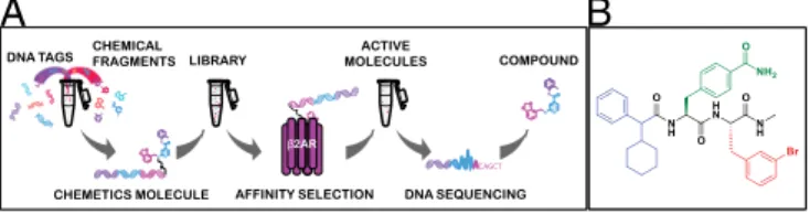
Isolation of Compound 15 from DELs.To identify unique chemotypes that bind at structurally relevant sites on the surface ofβ2AR, we
screened DELs against purified, unligandedβ2AR maintained in
the detergentn-dodecyl-β-D-maltoside (DDM) (Fig. 1AandFig. S1). A similar beads-only selection was performed in parallel as a control. In total, we screened three libraries together containing approximately a total of 190 million unique compounds synthe-sized using a DNA-tagged, split-and-pool combinatorial chemical synthesis approach (Chemetics; Nuevolution). In each library, ∼5×10¹³molecules in total were used as input, and 1–7×10⁷ molecules remained after screening. Relative quantification of the
recovered compounds was achieved by a combination of PCR amplification and next-generation sequencing of eluted DNA barcodes, followed by computational decoding approaches. To refine the out-put and eliminate potential nonspecific binders, compounds that displayed less than a 260- to 470-fold increase in frequency from baseline as well as those that were observed in bead-only con-trol selections were filtered from the dataset, leaving a total of 394 potentialβ2AR binders for further analysis (Table S1). These com-pounds were then clustered based on their structural similarity, and 16 putative hits were selected as representatives for these clusters. DNA-tagged versions of these 16 hits were resynthesized and screened individually to evaluate their influence on the binding affinity of orthosteric agonists in radioligand binding assays with membranes obtained fromβ2AR-overexpressing cells. One compound [4-((2S )-3-(((S )-3-(3-bromophenyl)-1-(methylamino)-1-oxopropan-2-yl)amino)-2-(2-cyclohexyl-2-phenylacetamido)-3-oxopropyl)benzamide], desig-nated as compound“15”(Fig. 1B), markedly decreased orthosteric agonist binding to the receptor and was thus selected for further characterization.
Characterization of Compound 15 for Its Binding to theβ2AR.To further
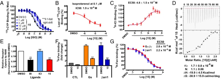
characterize the pharmacological properties of15, we
synthe-sized it in its DNA-free form (Fig. 1B). We first performed
competition binding experiments with the radioiodinated
an-tagonist cyanopindolol (125I-CYP) to evaluate the influence of
15on the binding ability of orthosteric ligands to theβ2AR recon-stituted into high-density lipoprotein (HDL) (also known as nanodisc) particles (21). We found that, although15had little to no influence on binding of125I-CYP, which is a neutral orthosteric antagonist of the β2AR, it robustly decreased the binding affinity of the agonist iso-proterenol for the receptor in a dose-dependent fashion (Fig. 2A).
Compound15caused the isoproterenol competition curve to shift to
the right by close to one log (ninefold) at the maximal concentration
tested. The half-maximal concentration of 15 for this shift was
∼1.9μM (Fig. 2B). On the other hand, when the inverse agonist [3 H]-ICI-118,551 was used as an orthosteric ligand tracer,15significantly increased (18±1% max) its binding to theβ2AR in a dose-dependent manner (Fig. 2C) with an EC50value of 0.48μM, which is close to that obtained in Fig. 2B(1.9μM). This reflects positive cooperativity be-tween15and the orthosteric antagonist [3H]-ICI-118,551 for binding to the receptor. To validate the direct binding of15to theβ2AR, we performed isothermal titration calorimetry (ITC). By this technique, we found that the equilibrium dissociation constant (Kd) of15for the receptor is 1.7±0.8μM, and the stoichiometry of the interaction is 1, suggesting that15binds to one site on theβ2AR (Fig. 2D). TheKd value obtained by ITC is in good agreement with the half-maximal concentration of the shift obtained in the radioligand competition binding experiment (Fig. 2B) as well as the EC50value obtained from the15titration curve for [3H]-ICI-118,551 binding (Fig. 2C).
To further confirm that15favors an inactive conformational state of the receptor, we assessed the extent of binding of an inactive
conformation-specificβ2AR single domain antibody [nanobody-60
(Nb60)] that binds to an intracellular region of the receptor (10, 22). Contrary to the positive cooperativity predicted to occur between a negative allosteric modulator and Nb60 (22), we found a decrease in Nb60 binding to the receptor in the presence of15(Fig. 2E). This
suggests that15competes with Nb60 for binding to the receptor,
and thus that their binding sites at least partially overlap, indicating that15binds to an intracellular region of theβ2AR.
It has been well appreciated that allosteric transducers such as
heterotrimeric G proteins or β-arrestins promote high-affinity
agonist binding (10, 23, 24). We also observed that15inhibited the high-affinity binding of a radiolabeledβ2AR-agonist, [3H](R,R′
)-4-methoxyfenoterol (3H-Fen) (25) that is promoted by either
het-erotrimeric Gs protein orβ-arrestin1 (Fig. 2F). This finding also
suggests that 15negatively modulates activation of theβ2AR by
agonists. Furthermore, the dose-dependent effects of15in3H-Fen
binding assays conducted in the presence of either G protein or
A
B
Fig. 1. Screening of the DEL and the chemical structure of15. (A) Schematic illustration of the screening of a encoded compound library. DNA-encoded library molecules, synthesized using a DNA-tagged, split-and-pool combinatorial chemical synthesis approach, were mixed with a target (pu-rifiedβ2AR) immobilized on a matrix. Target binding (active) molecules were
collected through affinity-based selection, and the encoding DNA tags were sequenced to identify the binding molecules. (B) Compound15is composed of three building blocks: methybenzamide (green), bromo-benzyl (red), and cyclohexylmethyl-benzene (blue). The amide backbone is shown in black.
P
β-arrestin were similar to each other (Fig. 2G). We used Fab30, which stabilizes an active conformation ofβ-arrestin1 (26, 27), to
enhance the weak high-affinity3H-Fen binding signal induced by
β-arrestin to levels comparable to those observed with G protein.
The EC50values obtained from the15dose–response curves for
the two transducer-promoted signals were comparable (2.8 vs. 2.2μM, respectively, for G protein andβ-arrestin; Fig. 2G). This suggests that15 displays no strong“bias”between its inhibitory
activities in the two transducer-induced high-affinity 3H-Fen
binding signals. The EC50values of15obtained here (Fig. 2G) are
also in good agreement with itsKd measured by ITC (Fig. 2D).
Taken all together, the results in Fig. 2 show that15behaves as a
negative allosteric modulator for the β2AR, and suggest that it
binds at the cytoplasmic surface of the receptor.
Functional Modulation ofβ2AR Activity by Compound 15.Next, we
investigated the effects of 15 on β2AR function in cells by
measuring G-protein–mediated cAMP production (28, 29) and
β-arrestin recruitment to the receptor (29, 30). Due to the high
signal amplification of the G-protein activation assay compared
with β-arrestin recruitment, to achieve comparable signaling
outputs between the two, we used endogenously expressedβ2AR
in the reporter cells to measure cAMP production, but we used
stably overexpressed β2V2R for monitoring β-arrestin
recruit-ment. Theβ2V2R, a chimeric receptor with a V2R tail at the C
terminus, displays stronger and more stable agonist-promoted
β-arrestin binding than the native β2AR while retaining the
pharmacological properties of the nativeβ2AR (31). Compound
15 decreased the isoproterenol-stimulated responses in both
assays (Fig. 3AandB). We observed rightward shifts of the EC50
values, as well as decreases in the maximal level of the stimulated
responses in the presence of increasing concentrations of15,
indi-cating that15inhibitsβ2AR agonist-induced functional responses.
Additionally, the extent of rightward shift of isoproterenol potency
promoted by 15 was similar for both G-protein activation and
β-arrestin recruitment to activated receptor. On the other hand, the
decreases in the maximal response induced by15were more robust
inβ-arrestin recruitment than in cAMP production (Fig. 3AandB). This likely is attributed to the differences in the sensitivity between
-8 -7 -6 -5 -4
95 100 105 110 115 120 125 DMSO
EC50: 4.8± 1.5 x 10⁻⁷M
Log [15] (M)
3H-I C I Bi n d in g (%)
Isoproterenol at 0.1μM
-6 -5 -4 0
20 40 60 80
IC50: 1.9 x 10⁻⁶M
DMSO
Log [15] (M)
Los t of 125 I-C Y P Bi n d in g (% ) 0.00 0.25 0.50 0.75 1.00 1.25 ** **
DMSO BI ICI 15
Ligands Re la ti v e Re c e p to r Ca pt ur e by Nb6 0 (N o rm a li z e d to N b 6 0 -D M S O )
B
E
G
-10 -9 -8 -7 -6 -5 -4
0 20 40 60 80 100 120
D M S O
1μM
3μM
1 0μM
3 2μM
Log [ISO] (M)
125 I-CY P Bi n d in g (% M ax) ***9.1-fold right shift 15
}
A
C
0 10 2030 40 506070 8090 0 -2 -4 0 -5 -10 -15 -20 -250.0 0.5 1.0 1.5 2.0
Molar Ratio, [15]/[β22AR]
Heat (
μ
cal/sec)
Kcal mol of 15
-1
Kd: 1.7 0.8 x 10 M N: 1.1 0.06
ΔH:-19.9 4.5Kcal/mol
ΔS:-38.9 15.9cal/mol/deg
-6
D
F
-7 -6 -5 -4
0 20 40 60 80 100 120 DM SO
Log [15] (M)
3H -Fe n bi ndi n g ( % ) DMSO 15 DMSO 15 DMSO 15 0 2 4 6 8 10 *** *** 3H -Fe n bi ndi n g (fo ld ch an g e )
CTL Gs βarr1
βarr1
Gα βγ 2.8 x 10⁻⁶ M
2.2 x 10⁻⁶ M
EC50
Fig. 2. Characterization of15for its binding to theβ2AR. (A) Dose–response curves of isoproterenol (ISO) competition binding to theβ2AR reconstituted
in nanodiscs with125I-CYP were obtained in the presence of various concentrations of15as indicated. Values were expressed as percentages of the maximal
125I-CYP binding level obtained from a one-site competition binding-log IC
50curve fit of the vehicle [0.9% dimethyl sulfoxide (DMSO)] control data. Points on
curves represent mean±SEM obtained from at least three independent experiments done in duplicate. (B) The half-maximal concentration of15in the changes of isoproterenol competition binding was obtained from a dose–response curve replotted with the data set at 0.1μM isoproterenol with various doses of15inA. (C) Dose-dependent increases in inverse agonist3H-ICI-118,551 (ICI) binding to theβ
2AR in nanodiscs. Points on the curve represent
nor-malized values as percentages of the3H-ICI-118,551 binding amount in the absence of15and mean±SEM obtained from at least four independent
ex-periments done in duplicate. (D) Characteristics of15for its physical interaction with theβ2AR were determined by the isothermal titration calorimetry (ITC)
analysis with the detergent-solubilized, purified receptor. The thermogram (Top) and binding isotherm with the best titration curve fit (Bottom) shown are representatives of three independent experiments. Values represent mean±SEM. (E) Extent of nanobody-60 (Nb60) binding to theβ2AR determined by ELISA
in the presence of different ligands including15. Values were expressed as ratios of the level of Nb60 binding in the vehicle (0.5% DMSO) control sample and represent mean±SEM obtained from three independent experiments done in duplicate. BI, BI-167107. (F) The levels of3H-Fen binding to theβ
2AR upon
treatment with the vehicle control (0.5% DMSO) or15at 50μM in the absence or presence of transducers, either trimeric Gαβγprotein orβ-arrestin1 (β-arr1)
together with Fab30. Values were expressed as fold changes of the level of3H-Fen binding in the vehicle (DMSO) control sample without the transducer and
represent mean±SEM obtained from three independent experiments done in duplicate. (G) Compound15dose-dependent decreases in the level of3H-Fen
high-affinity binding to theβ2AR promoted by either Gαβγprotein orβ-arr1 together with Fab30. Values were expressed as percentages of the maximal 3H-Fen binding level promoted by each transducer in the vehicle control (0.5% DMSO) and represent mean±SEM obtained from at least three independent
experiments done in duplicate. All of the statistical analyses in the figure were performed, as described inMaterials and Methods.
-11-10 -9 -8 -7 -6 -5
0 20 40 60 80 100 120 DMSO 15 (-ISO
)
5μM
2 0μM
5 0μM
D M S O
1 .2 5μM
β-arrestin Recruitment
Log [ISO] (M)
β-a rr e s tin ( % ISO Ma x )
-11 -10 -9 -8 -7 -6 -5
0 20 40 60 80 100 120 DMSO 15 (-ISO )
5μM
2 0μM
5 0μM
D M S O
1 .2 5μM
cAMP Production
Log [ISO] (M)
c A MP (% I S O Ma x ) 15 15 ***7.1-fold right shift ***7.4-fold right shift
A
B
}
}
Fig. 3. The effect of15onβ2AR-mediated functional activities. After
pre-treatment with15for 20 min at various concentrations as indicated, the β2AR-mediated activities in cells were measured upon stimulation with
iso-proterenol (ISO) in a dose-dependent manner: (A) cAMP production by the endogenously expressedβ2AR and (B)β-arrestin recruitment to the
exoge-nously expressedβ2V2R. Values were expressed as percentages of the
the assays, as described before (29), rather than to any biased
ac-tivity of 15. Such unbiased inhibition of β2AR activity by 15 is
consistent with the finding that 15 similarly inhibits the high-affinity
binding of3H-Fen promoted by either transducer (G protein or
β-arrestin) (Fig. 2G). These results confirm that15modulatesβ2AR by antagonizing its agonist-induced activity.
We further validated that the inhibitory effect of15in functional assays was a result of specific inhibition at the receptor level and not due to nonspecific effects by testing15in other cell-based assays that have distinct readouts from the luminescence-based assays
shown in Fig. 3. We monitored the effect of15on cAMP
pro-duction upon stimulation of the overexpressed β2AR using a
fluorescence resonance energy transfer (FRET)-based biosensor,
ICUE2 (32). In this experiment,15caused a rightward shift in the
EC50for isoproterenol-stimulated cAMP production (Fig. S2A),
consistent with that shown in Fig. 3A, albeit without changing the maximal response. This may be attributed to the increased am-plification of the signal due to overexpression of theβ2AR in this assay compared with the signal by the receptor expressed at
en-dogenous levels in Fig. 3A. Similarly, we also confirmed the
in-hibitory effect of15on agonist-stimulatedβ-arrestin recruitment
to theβ2AR by assessing FRET signals betweenβ2AR-YFP and
CFP-β-arrestin2 (Fig. S2B). We further confirmed the inhibitory effect of15onβ2AR activation using an in vitro GTPase activity
assay with theβ2AR reconstituted into HDL particles together
with purified Gs protein (Fig. S2C). The15-induced shift in the EC50for isoproterenol obtained in this in vitro assay is comparable
to that observed in the cell-based assays, suggesting that15must
readily penetrate cell membranes to bind to an intracellular region of theβ2AR, which is suggested by the result in Fig. 2E.
Selective Inhibition ofβ2AR-Mediated Activities by Compound 15.To
confirm the selectivity of15 for the β2AR, we performed
func-tional assays to evaluate whether15inhibited activation of other
members of the GPCR family closely or distantly related to the β2AR. First, we compared the extent of15 blockade of
agonist-stimulated activities of β2AR with two other endogenously
expressed Gs-coupled receptors in HEK-293 cells, the prosta-glandin E2 (PGE2) and vasoactive intestinal peptide (VIP)
re-ceptors. Notably,15had no effect on cAMP production following
stimulation of these receptors (Fig. S3AandB). Additionally, we
looked at the specificity of 15for theβ2AR by assessing its
in-hibitory effect onβ-arrestin recruitment to other receptors besides
the chimericβ2AR (β2V2R). Here, we used the parental cell line
stably expressing β-arrestin2 alone, and transiently expressed
re-ceptors indicated in Fig. 4. First, we confirmed that the extent of
15-mediated inhibition of agonist-stimulatedβ-arrestin recruitment to
the wild-typeβ2AR (Fig. 4A) is comparable to that obtained in the
transiently expressed chimeric receptor, β2V2R (Fig. 4B). On the
other hand, following stimulation of the β1AR, a receptor closely
related to theβ2AR,15substantially inhibited the maximal response as well as the basal activity in a concentration-dependent manner, whereas no significant changes were observed in the EC50value (Fig. 4C). We also observed significant, but much reduced, inhibitory ef-fects of15onβ-arrestin recruitment to the vasopressin V2receptor (V2R), which is also a Gs-coupled receptor (Fig. 4D). In contrast to this,15 only minimally inhibited agonist-inducedβ-arrestin recruit-ment to the VIPR, another Gs-coupled receptor. We observed only minimal decreases in the maximal response to stimulation with VIP without any change in the EC50value induced by15(Fig. 4E), con-sistent with the result obtained in cAMP accumulation (Fig. S3B). Furthermore, no significant inhibition by15was detected inβ-arrestin recruitment to the Gq-coupled angiotensin II type 1 receptor (AT1R) (Fig. 4F). To further assess the extent of the15inhibitory activities
among different receptors, we quantified the level of15-mediated
decreases in the maximal response as well as shifts of the EC50value exhibited as fold shifts (Table S2). These results demonstrate that the inhibitory effect of15on agonist-stimulated responses is greatest for theβ2AR and is substantially diminished in even closely related re-ceptors such asβ1AR.
To obtain further insights into the specificity of 15 for the
β2AR, we examined its inhibitory activity on agonist-induced
β-arrestin internalization. Unlike“class B” receptors, including
the V2R and the AT1R whose tight interactions withβ-arrestin
allow for their cointernalization,“class A”receptors such as the β2AR have weakerβ-arrestin interactions and are not
cointer-nalized withβ-arrestin (33). Therefore, we examined the effect of
15 on this functional activity with the transiently expressed
β2V2R (Fig. S4A), V2R (Fig. S4B), and AT1R (Fig. S4C). The
extent ofβ-arrestin internalization was monitored by measuring
the amount of β-arrestin targeted to endosomes (34). Results
obtained in this assay are consistent with the inhibitory effects of
-11 -10 -9 -8 -7 -6 -5
40 60 80 100 120
DMSO 15
(-An gII)
5μM 2 0μM 5 0μM D M S O
AT1R
Log [AngII] (M)
β-a
rr
e
s
ti
n
(% An
g
II
Ma
x
)
-11 -10 -9 -8 -7 -6 -5
20 40 60 80 100 120
DMSO 15 (-AVP)
5μM 2 0μM 5 0μM D M S O 1 .2 5μM
V2R
Log [AVP] (M)
β-a
rr
e
s
tin
(%
A
VP Ma
x
)
-11-10 -9 -8 -7 -6 -5
40 60 80 100 120
DMSO 15 (-I SO)
5μM 2 0μM 5 0μM D M S O 1 .2 5μM
β1AR
Log [ISO] (M)
β-a
rr
e
s
ti
n
(% I
S
O
Ma
x
)
-11-10 -9 -8 -7 -6 -5
0 20 40 60 80 100 120
DMSO 15 ( -ISO)
5μM 2 0μM 5 0μM D M S O 1 .2 5μM
β2V2R
Log [ISO] (M)
β-a
rr
e
s
ti
n
(% I
S
O
Ma
x
)
-11-10 -9 -8 -7 -6 -5
0 20 40 60 80 100 120
DMSO 15 (-ISO
) 5μM 2 0μM 5 0μM D M S O 1 .2 5μM
β2AR
Log [ISO] (M)
β-a
rr
e
s
ti
n
(% I
S
O
Ma
x
)
A
B
C
D
F
15
}
}
15}
1515
}
}
15-11-10 -9 -8 -7 -6 -5
20 40 60 80 100 120
DMSO 15 (
-VIP)
5μM 2 0μM 5 0μM D M S O
VIPR
Log [VIP] (M)
β-a
rr
e
s
tin
(%
VI
P Ma
x
)
E
}
15Fig. 4. Specificity of15inhibition forβ2AR-mediated activity. Various receptors were transiently expressed to monitorβ-arrestin recruitment, including (A) β2AR, (B)β2V2R, (C)β1AR, (D) V2R, (E) VIPR, and (F) AT1R. After pretreatment with15at different concentrations as indicated for 20 min, the extent of
agonist-inducedβ-arrestin recruitment to these receptors was determined in a dose-dependent manner. Values were expressed as percentages of the maximal level of the activity induced by the agonist of each receptor in the vehicle (0.5% DMSO) control. Points on graphs represent mean±SEM obtained from at least three independent experiments done in duplicate. AngII, angiotensin II; AVP, arginine vasopressin; ISO, isoproterenol; VIP, vasoactive intestinal peptide.
P
15 onβ-arrestin recruitment to activated receptors as shown in
Fig. 4, which are summarized inTable S2, further confirming the
specificity of the modulating activity of15for theβ2AR.
We also investigated whether the NAM activity of15 at the
β2AR was dependent on the presence of a specific agonist at the orthosteric site (i.e., probe dependence). We performed this by
monitoring Gs-mediated cAMP production and β-arrestin
re-cruitment to the receptor in the presence of orthosteric probes ranging from full to weak partial agonists, including epinephrine,
fenoterol, and clenbuterol (Fig. S5) as done with isoproterenol
(Fig. 3). We also compared competition binding of these agonists
to the β2AR with 125I-CYP in the presence of different
con-centrations of15(Fig. 2AandFig. S5), which allowed us to
as-sess the probe dependence of 15 among the agonists in the
absence of transducer coupling. Table S3shows the summary of
quantified values in each assay, including the extent of15-mediated
decreases in the maximal response and shifts of the EC50value
exhibited as fold shifts. Overall, 15appears to display no
sig-nificant probe dependence among the tested agonists. We
ob-served that the extent of the EC50value shift by15, which is
consistent among the tested assays, follows the efficacy of the
tested agonists. On the other hand, the magnitude of 15
in-hibition of the maximal response is negatively correlated with the efficacy of these agonists.
Structure–Activity Relationships of Compound 15 Analogs at theβ2AR. To discern the structure–activity relationship (SAR) pattern for the
allosteric modulation of15at theβ2AR, we designed and
synthe-sized a series of15derivatives (Table S4). We assessed the ability of these derivatives to modulateβ2AR functions in two different types of experimental settings. These were cell-based activity assays,
in-cluding G-protein–mediated cAMP production and β-arrestin
re-cruitment to the activatedβ2AR, as well as high-affinity binding of
the agonist3H-Fen to the receptor induced by transducers, Gs or
β-arrestin. To assist our SAR analyses,15was divided into three
structural subunits, the methylbenzamide (region I), bromo-benzyl (region II), and cyclohexylmethyl-benzene (region III) regions, into each of which we introduced modifications. We found that the formamide group in region I (methylbenzamide) is an important
determinant of functional properties of15. Removal of this group
on the phenyl ring (A1) led to a dramatic decrease in the inhibitory activity of15down to about 20% or less of its original activity. The same, but less severe, trend was observed when the position of this
formamide group was changed from its originalpara-position to a
meta-position (A2), which resulted in a ∼60% reduction of its
original activity. In the case of region II (bromo-benzyl), removal of the electronegative atom bromine (A3) also caused variable but substantial attenuation of the inhibitory activity of15down to about
10–55%. Two other modifications in this region, replacement of
bromine with fluorine, an atom of comparable electronegativity but smaller radius (A4), and introduction of additional bromine at the
meta-position of the phenyl ring (A5) modestly decreased the
functional effects of15. Next, we evaluated the activity of
deriv-atives with modifications on the aromatic ring in region III (cyclohexylmethyl-benzene). Interestingly, addition of a hydroxyl group to this ring at thepara-position (A6) led to dramatic loss of inhibitory activity, while replacing the hydroxyl group at this po-sition with a slightly hydrophobic methoxy group (A7) partially restored the inhibitory activity. This strongly suggests that the hydrophobic nature of this region is another important
determi-nant for efficient interaction of 15 with the presumably
hydro-phobic portion of the putative binding site of15on theβ2AR.
Discussion
We report here the discovery and characterization of a small
molecule, compound 15, as an allosteric β-blocker. Compound
15was derived from an in vitro affinity-based screening of DELs
against the purified human β2AR. Compound 15 shares no
structural or chemical similarities with knownβ2AR orthosteric
ligands, and it does not compete with radiolabeledβ2AR ligands
for binding at the orthosteric site. On the other hand, it binds
allosterically to theβ2AR with low micromolar affinity. The
compound negatively modulates the binding of agonists to the
β2AR while it clearly displays positive cooperativity with an
orthosteric inverse agonist. In addition, in cell-based functional
assays,15 displays robust inhibition ofβ2AR agonist-promoted,
Gs-mediated cAMP generation as well asβ-arrestin recruitment
to the receptor. Together, these characteristics demonstrate that
15allosterically binds to and stabilizes an inactive conformation
of theβ2AR, which are the classic hallmarks of a negative
allo-steric modulator (5, 35).
A large number of orthosteric ligands for theβ2AR have been
developed, whereas before this study, no allosteric small-molecule β2AR ligand had been identified. The affinity-based screening strategy is an ideal way to identify allosteric ligands for a receptor, and DEL screening is an innovative strategy to perform affinity-based selections against targets that are isolated or expressed on whole cells (12). Although this technique enables an unprecedented increase in the size of libraries that can be screened compared with conventional activity-based screening formats, its use has mostly been limited to soluble protein targets (12, 13). Due to the inherent difficulty in isolating functional membrane proteins, this technique has been only rarely used to obtain ligands for GPCRs. To date, there has been only one report describing the discovery of a ligand
for a GPCR from a DEL (16). There, the recombinant NK3
tachykinin receptor expressed on HEK-293 cells was used as a target in a whole-cell selection format to identify an inhibitor for the receptor (16). Here, we have demonstrated that the DEL screening strategy can be successfully applied to the isolation of small-molecule ligands using a purified GPCR. Although our study was focused on isolating and characterizingβ2AR allosteric ligands, this strat-egy could be used as well to isolate ligands that target orthosteric sites of GPCRs. Despite the power of this approach, predicting the functional outcomes of the isolated compounds remains empirical. However, in our in vitro purified receptor target system, it is highly feasible to bias the selections through differential display of the receptor in unique conformations (e.g., agonist vs. antagonist vs. no ligand in the orthosteric site) or in complex with signaling
partners such as G proteins orβ-arrestins. This should provide
allosteric modulators with distinct properties (e.g., NAMs, PAMs, or even biased molecules for coupling to transducers, leading to signaling bias).
To date, pharmacological studies of GPCR allosteric modu-lation have been restricted to a few receptor families including muscarinic acetylcholine, adenosine, chemokine, and metabo-tropic glutamate receptors (5, 36). Compared with orthosteric ligands, drugs targeting allosteric sites often display greater re-ceptor subtype selectivity and therefore potentially reduce ad-verse side effects (4, 5). This is presumably due to decreased evolutionary pressure at allosteric sites than at the orthosteric site of GPCRs (35), leading to their greater divergence within a family. Moreover, multiple allosteric sites can exist on a given receptor (37). In addition, allosteric GPCR modulators may have greater potential than orthosteric ligands to engender biased signaling through selective modulation of specific signaling
path-ways, for example, G-protein versusβ-arrestin pathways (4).
Our SAR studies provide insights into key regions of15that must engage in contacts with the allosteric binding site on theβ2AR to allow its functional modulation. We found some alterations, including complete deletions of the formamide group in region I and bromine in region II, lead to dramatic decreases in the functional activities of
the parent compound,15. We also observed a positive association
between increased polarity of region III and loss of functional activity. This suggests that this region of the molecule might interface deep
within theβ2AR allosteric site to establish contacts with core
As with other GPCRs, several putative allosteric sites on the β2AR have been recently proposed based on crystal structures (37). Some are located at the intracellular face of the receptor; these are relatively large and can accommodate a wide range of
compound sizes. Interestingly, most of the currently reported non–
small-moleculeβ2AR allosteric modulators, such as nanobodies
(10, 22, 38) and RNA aptamers (11), bind to intracellular cavities that overlap with the G-protein binding site (39). This appears to be true as well for15because it competes forβ2AR binding with a nanobody (Nb60) that favors an inactive conformation and that binds to this intracellular region of theβ2AR (22). Although our
findings suggest that15 binds to the intracellular region of the
β2AR, further SAR and structural studies at atomic-level resolu-tion will be required to precisely define the site and mechanism of
action, by which15acts as a NAM.
In summary, our study reports the discovery via in vitro affinity selection of a DEL against purified receptors and functional
characterization of aβ2AR-selective negative allosteric
modu-lator. Our findings suggest that targeting GPCR allosteric sites with such combinatorial small-molecule libraries provides a powerful and efficient approach for developing highly selective
ligands that can modulate a wide range of receptor’s functional
activities. Furthermore, our findings establish a proof-of-concept strategy using the DEL screening technique, which can be broadly applied to discover small molecules for other GPCRs.
Materials and Methods
Complete details and descriptions of the materials used; cell culture and transfections; expression and purification of theβ2AR; purifiedβ2AR-based DEL
selection, quantitative PCR, and next-generation sequencing analysis; recon-stitution of the β2AR into HDL particles; radioligand binding; ITC; ELISA;
measurement of cAMP accumulation and in vitro GTPase activity;β-arrestin recruitment andβ-arrestin endocytosis assays; data analyses; and synthesis and characterization of compounds are provided inSI Materials and Methods.
ACKNOWLEDGMENTS.We are grateful to Dr. Li-Yin Huang, Dr. Helen Yao, Xinrong Jiang, and Paul Shim (Duke University) for technical assistance; Dr. Sudarshan Rajagopal (Duke University) for discussing data analysis; and Donna Addison and Quivetta Lennon for secretarial assistance. We also thank Dr. Irving Wainer (Laboratory of Clinical Investigation, National Institute on Aging Intramural Research Program) for providing [3H](R,R′)-4-methoxyfenoterol
(40–43). This work was supported in part by National Institutes of Health Grant HL16037 (to R.J.L.), National Science Foundation of China Grant #21272029, Priority Academic Program Development of Jiangsu Higher Education Institu-tion (X.C.), and NIH Training Grant T32HL007101 (to A.W.K.). R.J.L. is an in-vestigator with the Howard Hughes Medical Institute.
1. Lefkowitz RJ (2007) Seven transmembrane receptors: Something old, something new.
Acta Physiol (Oxf)190(1):9–19.
2. Whalen EJ, Rajagopal S, Lefkowitz RJ (2011) Therapeutic potential ofβ-arrestin- and G protein-biased agonists.Trends Mol Med17(3):126–139.
3. Kobilka BK (2011) Structural insights into adrenergic receptor function and phar-macology.Trends Pharmacol Sci32(4):213–218.
4. Wootten D, Christopoulos A, Sexton PM (2013) Emerging paradigms in GPCR allo-stery: Implications for drug discovery.Nat Rev Drug Discov12(8):630–644. 5. Christopoulos A, et al. (2014) International Union of Basic and Clinical Pharmacology.
XC. Multisite pharmacology: Recommendations for the nomenclature of receptor allosterism and allosteric ligands.Pharmacol Rev66(4):918–947.
6. Dorr P, et al. (2005) Maraviroc (UK-427,857), a potent, orally bioavailable, and se-lective small-molecule inhibitor of chemokine receptor CCR5 with broad-spectrum anti-human immunodeficiency virus type 1 activity.Antimicrob Agents Chemother 49(11):4721–4732.
7. Lindberg JS, et al. (2005) Cinacalcet HCl, an oral calcimimetic agent for the treatment of secondary hyperparathyroidism in hemodialysis and peritoneal dialysis: A ran-domized, double-blind, multicenter study.J Am Soc Nephrol16(3):800–807. 8. Kumari P, Ghosh E, Shukla AK (2015) Emerging approaches to GPCR ligand screening
for drug discovery.Trends Mol Med21(11):687–701.
9. Day PW, et al. (2007) A monoclonal antibody for G protein-coupled receptor crys-tallography.Nat Methods4(11):927–929.
10. Staus DP, et al. (2014) Regulation ofβ2-adrenergic receptor function by conformationally selective single-domain intrabodies.Mol Pharmacol85(3):472–481.
11. Kahsai AW, et al. (2016) Conformationally selective RNA aptamers allosterically modulate theβ2-adrenoceptor.Nat Chem Biol12(9):709–716.
12. Mannocci L, Leimbacher M, Wichert M, Scheuermann J, Neri D (2011) 20 years of DNA-encoded chemical libraries.Chem Commun (Camb)47(48):12747–12753. 13. Franzini RM, Randolph C (2016) Chemical space of DNA-encoded libraries.J Med
Chem59(14):6629–6644.
14. Kleiner RE, Dumelin CE, Tiu GC, Sakurai K, Liu DR (2010) In vitro selection of a DNA-templated small-molecule library reveals a class of macrocyclic kinase inhibitors.J Am
Chem Soc132(33):11779–11791.
15. Clark MA, et al. (2009) Design, synthesis and selection of DNA-encoded small-molecule libraries.Nat Chem Biol5(9):647–654.
16. Wu Z, et al. (2015) Cell-based selection expands the utility of DNA-encoded small-molecule library technology to cell surface drug targets: Identification of novel an-tagonists of the NK3 tachykinin receptor.ACS Comb Sci17(12):722–731.
17. Tomiyama H, Yamashina A (2014) Beta-blockers in the management of hypertension and/or chronic kidney disease.Int J Hypertens2014:919256.
18. Thanawala VJ, et al. (2014) Ligand bias prevents class equality among beta-blockers.
Curr Opin Pharmacol16:50–57.
19. Weberpals J, Jansen L, Carr PR, Hoffmeister M, Brenner H (2016) Beta blockers and cancer prognosis—the role of immortal time bias: A systematic review and meta-analysis.Cancer Treat Rev47:1–11.
20. National Asthma Education and Prevention Program (2007) Expert Panel Report 3 (EPR-3): Guidelines for the Diagnosis and Management of Asthma-Summary Report 2007.J Allergy Clin Immunol120(5, Suppl):S94–S138.
21. Whorton MR, et al. (2007) A monomeric G protein-coupled receptor isolated in a high-density lipoprotein particle efficiently activates its G protein.Proc Natl Acad Sci USA104(18):7682–7687.
22. Staus DP, et al. (2016) Allosteric nanobodies reveal the dynamic range and diverse mechanisms of G-protein-coupled receptor activation.Nature535(7612):448–452. 23. De Lean A, Stadel JM, Lefkowitz RJ (1980) A ternary complex model explains the
agonist-specific binding properties of the adenylate cyclase-coupled beta-adrenergic receptor.J Biol Chem255(15):7108–7117.
24. Strachan RT, et al. (2014) Divergent transducer-specific molecular efficacies generate biased agonism at a G protein-coupled receptor (GPCR).J Biol Chem289(20):14211–14224. 25. Toll L, et al. (2012) Thermodynamics and docking of agonists to theβ2-adrenoceptor
determined using [3H](R,R′)-4-methoxyfenoterol as the marker ligand.Mol Pharmacol
81(6):846–854.
26. Shukla AK, et al. (2013) Structure of activeβ-arrestin-1 bound to a G-protein-coupled receptor phosphopeptide.Nature497(7447):137–141.
27. Shukla AK, et al. (2014) Visualization of arrestin recruitment by a G-protein-coupled receptor.Nature512(7513):218–222.
28. Binkowski BF, Fan F, Wood KV (2011) Luminescent biosensors for real-time moni-toring of intracellular cAMP.Methods Mol Biol756:263–271.
29. Rajagopal S, et al. (2011) Quantifying ligand bias at seven-transmembrane receptors.
Mol Pharmacol80(3):367–377.
30. Bassoni DL, Raab WJ, Achacoso PL, Loh CY, Wehrman TS (2012) Measurements of
β-arrestin recruitment to activated seven transmembrane receptors using enzyme complementation.Methods Mol Biol897:181–203.
31. Tohgo A, et al. (2003) The stability of the G protein-coupled receptor-beta-arrestin interaction determines the mechanism and functional consequence of ERK activation.
J Biol Chem278(8):6258–6267.
32. Violin JD, et al. (2008)β2-Adrenergic receptor signaling and desensitization elucidated
by quantitative modeling of real time cAMP dynamics.J Biol Chem283(5):2949–2961. 33. Oakley RH, Laporte SA, Holt JA, Caron MG, Barak LS (2000) Differential affinities of visual arrestin, beta arrestin1, and beta arrestin2 for G protein-coupled receptors delineate two major classes of receptors.J Biol Chem275(22):17201–17210. 34. Eglen RM (2005) Functional G protein-coupled receptor assays for primary and
sec-ondary screening.Comb Chem High Throughput Screen8(4):311–318.
35. Langmead CJ (2011) Determining allosteric modulator mechanism of action: Inte-gration of radioligand binding and functional assay data.Methods Mol Biol746: 195–209.
36. Christopoulos A (2014) Advances in G protein-coupled receptor allostery: From function to structure.Mol Pharmacol86(5):463–478.
37. Ivetac A, McCammon JA (2010) Mapping the druggable allosteric space of G-protein coupled receptors: A fragment-based molecular dynamics approach.Chem Biol Drug Des76(3):201–217.
38. Rasmussen SG, et al. (2011) Structure of a nanobody-stabilized active state of theβ2
adrenoceptor.Nature469(7329):175–180.
39. Rasmussen SG, et al. (2011) Crystal structure of theβ2adrenergic receptor-Gs protein
complex.Nature477(7366):549–555.
40. Wang JB, et al. (2013) Synthesis ofβ2-AR agonist BI-167107.Youji Huaxue33(3):634–639.
41. Kobilka BK (1995) Amino and carboxyl terminal modifications to facilitate the produc-tion and purificaproduc-tion of a G protein-coupled receptor.Anal Biochem231(1):269–271. 42. Nobles KN, et al. (2011) Distinct phosphorylation sites on theβ2-adrenergic receptor
es-tablish a barcode that encodes differential functions ofβ-arrestin.Sci Signal4(185):ra51. 43. Violin JD, Ren XR, Lefkowitz RJ (2006) G-protein-coupled receptor kinase specificity
forβ-arrestin recruitment to theβ2-adrenergic receptor revealed by fluorescence
resonance energy transfer.J Biol Chem281(29):20577–20588.
P