The Biological Significance of BRG1 Mutations
Chris Bartlett
A dissertation submitted to the faculty of the University of North Carolina at Chapel Hill in partial fulfillment of the requirements for the degree of Doctor of Philosophy in the
Curriculum of Toxicology.
Chapel Hill 2006
ABSTRACT Chris Bartlett
The Biological Significance of BRG1 Mutations “Under the direction of Dr. Bernard Weissman”
Eukaryotic organisms package DNA into chromatin for compact storage in the cell nucleus, but this packaging process results in transcriptional repression of genes. Chromatin remodeling complexes have evolved to overcome the transcriptional repression caused by chromatin packaging of DNA into nucleosomes by histones. One example of a chromatin remodeling complex is the SWI/SNF complex in yeast which uses ATP to drive the
chromatin apart and make DNA accessible to transcription factors. The yeast SWI2 protein was discovered as the catalytic subunit of the yeast SWI/SNF chromatin remodeling complex and is required for the complex to counteract the repressive nature of chromatin. BRG1 and BRM, SWI2 homologs, are part of human chromatin remodeling complexes and have been shown to play a redundant role in the regulation of certain cell cycle and cellular adhesion genes, as well as cellular pathways. Recent studies showing loss of BRG1 in human tumor cell lines and primary tissue samples, BRG1 mutations in human tumor cell lines, a
TABLE OF CONTENTS
Page
List of Tables ……….viii
List of Figures………. ………...ix
Abbreviations...x
Chapter I. Introduction A. Significance……….1
B. Chromatin ………...1
C. Chromatin Remodeling………...2
D. Epigenetics………..7
a. DNA Methylation………...7
b. Other Forms of Epigenetics ………...9
c. Chromatin and DNA methylation………10
E. Epigenetics and Human Disease………...….10
F. SWI/SNF Chromatin Remodeling Complex……….13
a. SWI/SNF Gene Regulation in Development and ……...14
Differentiation b. SWI/SNF Gene Regulation in Cell Adhesion and ……...16
Proliferation c. Other Roles of SWI/SNF ……….17
G. BRG1 ………...18
a. Identification……….18
H. BRG1 is implicated in development of human cancer ………19
I. Specific Aims ………...23
J. References………25
II. BRG1 Mutations found in Human Cancer Cell Lines Inactivate Rb-mediated Cell Cycle Arrest A. Introduction ………36
B. Material and Methods ………38
C. Results a. Characterization of BRG1 Mutant cell lines ………..43
b. Complex Formation of BRG1 Mutants………..45
c. Promoter Targeting of BRG1 mutants………...48
d. Analysis of HCT116 and Hs578t RNAi stable …...49
knockdowns e. Rb Sensitivity. ………..52
f. Protein Expression in RNAi stable knockdowns ……….52
D. Discussion ………54
E. References ………60
III. SWI/SNF Chromatin Remodeling Factors Induce Changes in DNA Methylation to promote Transcriptional Activation A. Abstract ………...65
B. Introduction ……….66
C. Material and Methods ……….66
E. References ………..82
IV. Identification of Potential Targets of SWI/SNF complex Transcriptional Activation A. Introduction……….84
B. Material and Methods ……….86
C. Results a. Protein expression analysis of BRG1/BRM deficient cells ………….88
b. Transfection and Treatment of BRG1/BRM deficient cells …………88
c. Global Methylation Analysis of BRG1/BRM deficient cells ………..90
D. Discussion ………..93
E. References………97
V. Summary and Perspectives A. Summary………99
B. Perspective………100
C. References……….105
LIST OF TABLES
1.1 Histone Modifications………..5
1.2 Human Chromatin Remodeling Complexes………....6
1.3 Chromatin Remodeling and Human Disease……….12
A.1 Cell Lines………107
LIST OF FIGURES
1.1 Compaction of DNA ……….………. ………...3
1.2 Structure of Catalytic Subunits of hSWI/SNF Complexes……….20
2.1 Location of BRG1 Mutations …….………...44
2.2 Western-blot analysis of BRG1 mutant cell lines……….………..46
2.3 Immunoprecipitation of BRG1 mutant cell lines ………. ………..47
2.4 Mutant BRG1 protein is at the promoter of E-cadherin and CD44………..…..50
2.5 Analysis of RNAi stable clones by immunoblot ………..…..51
2.6 RB sensitivity of HCT116 and Hs578t RNAi stable clones ……….….53
2.7 Western-blot analysis of Hs578t and HCT116 RNAi stable clones……...…………55
3.1 Promoters are hypermethylated in cell lines that lack Brg1 and Brm...72
3.2 Brg1 and Brm induce CD44 and E-cadherin expression ……….…….74
3.3 CD44 and E-cadherin promoter methylation analysis ………. .76
3.4 Brg1 and Brm interact with the CD44 and E-cadherin promoters …………...…….80
4.1 Western blot analysis of BRG1/BRM deficient cells ...89
4.2 Western-blot analysis of A427 cells and H522 cells ………....……….91
4.3 Western blot analysis of SW13 cells ………... ……...92
LIST OF ABBREVIATIONS DNA - deoxyribonucleic acid
bp -basepairs nm – nanometers
ATP – adenosine triphosphate HAT – histone acetyltransferase HDAC – histone deacetylase
SWI/SNF – Switching/Sucrose Non-fermenting BRM - Brahma
BRG1 – Brahma-related gene BAF – BRG1 associated factors DNMT – DNA methyltransferases LOI – Loss of Imprinting
5-azaC – 5-azacytidine RNA – ribonucleic acid AR – androgen receptor ER – estrogen receptor
EMT – epithelial-mesenchymal transition ChIP – Chromatin Immunoprecipitation DNBRG1 – dominant-negative BRG1 RB – retinoblastoma gene
Rb – retinoblastoma protein
NSCLC – Non-small cell lung cancer TMA – tissue microarray
CHAPTER 1
INTRODUCTION
A. Significance
Cancer accounts for 557,271 deaths a year, nearly one quarter of all deaths in the United States (1). Since 1950, the death rate for other major diseases such as, heart disease, cerebrovascular disease, and pneumonia/influenza, have decreased while the death rate for cancer has remained the same (1). Progress in diagnosis and treatment of cancer has been slower because of the many types of cancer and the multiple factors involved in development of cancer. Therefore, research on the mechanisms involved in cancer initiation and
progression can lead to breakthroughs in the diagnosis of cancer and potential therapeutic targets for treatment of cancer.
B. Chromatin
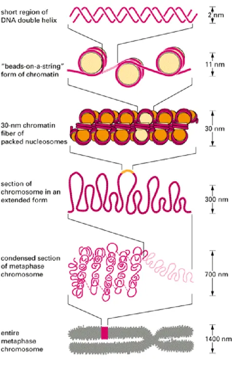
To fit cellular deoxyribonucleic acid (DNA) into a cell, DNA must be compacted by
compaction of the chromatin structure since proteolytic cleavage of the tails abolishes this process (3). Compaction of chromatin into a solenoid arrangement is stabilized by “linker” histone H1, which along with 22bp of DNA is responsible for connecting the nucleosome cores to each other (2). The solenoid structure contains six nucleosomes per turn creating a condensed chromatin fiber approximately 30nm in diameter. According to the radial-loop model, the solenoid structure is further compacted by the formation of looped domains of DNA by non-histone proteins attached to a chromosome scaffold (3). In non-dividing cells, chromosomes are not visible even with the aid of DNA stains or electron microscopy. Chromosome condensation due to helical folding of looped DNA attached to a protein scaffold occurs during mitosis and meiosis in dividing cells to create a visible structure (3) (Figure 1).
C. Chromatin Remodeling
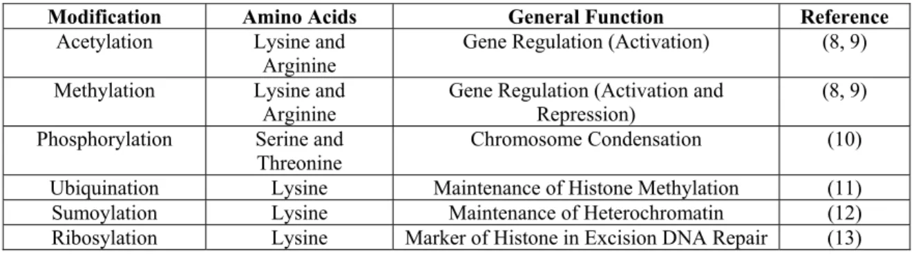
modifications represent distinct chromatin states (Table 1). A common enzymatic alteration of histones is by histone acetyltransferase (HAT) complexes, which decrease the affinity of histones for DNA upon acetylation of lysines on histone tails, and histone deacetylase (HDAC) complexes, which reverse the effects of HATs (9). Hyperacetylation of histones is indicative of transcriptionally active genes, while hypoacetylation of histones is characteristic of inactive regions of transcription.
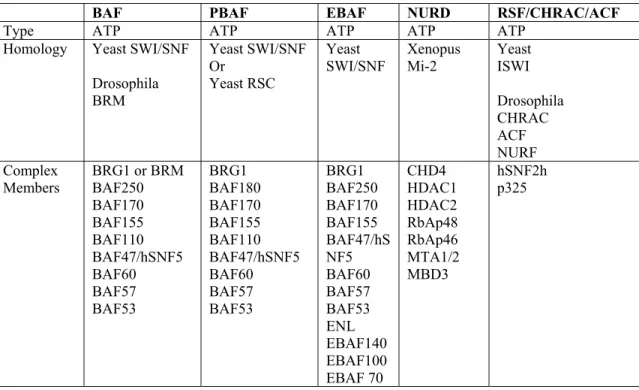
The other class of chromatin remodeling enzymes, ATP-dependent chromatin remodeling enzymes, are multi-protein complexes that use the energy of adenosine triphosphate (ATP) to remodel nucleosomes. The ATP-dependent movement of nucleosomes in cis along a DNA fragment result in enhanced accessibility of nucleosomal DNA (2). ATP-dependent
chromatin remodeling complexes are conserved in species ranging from yeast to humans (Table 2) (6, 7). In each ATP-dependent chromatin remodeling complex there is a helicase-like subunit of the switching/sucrose non-fermenting (SWI2/SNF2) family of SF2 helicases (14). Three major subfamilies of ATP-dependent chromatin remodeling complexes exist based on sequence homology within the catalytic subunit: SWI2/SNF2, Mi-2/CHD, and ISWI families (14). The unique domains characteristic of the SWI/SNF complex catalytic subunits are the bromodomain and an AT-hook region (14). Bromodomains interact with acetylated lysines and the AT-hook region binds to AT-rich regions of DNA. These domains may help target the complex to histones and DNA helping the SWI/SNF complex in its main role of transcriptional regulation. The Mi-2 complex catalytic subunits contain
Table 1.1: Histone Modifications
Modification Amino Acids General Function Reference
Acetylation Lysine and Arginine
Gene Regulation (Activation) (8, 9)
Methylation Lysine and
Arginine Gene Regulation (Activation and Repression) (8, 9) Phosphorylation Serine and
Table 1.2: Human ATP-dependent Chromatin Remodeling Complexes
BAF PBAF EBAF NURD RSF/CHRAC/ACF
Type ATP ATP ATP ATP ATP
Homology Yeast SWI/SNF
Drosophila BRM Yeast SWI/SNF Or Yeast RSC Yeast
SWI/SNF Xenopus Mi-2 Yeast ISWI
Drosophila CHRAC ACF NURF Complex
deacetylases. The ISWI complex catalytic subunits also contain unique domains that interact with histone tails called SANT domains, and SLIDE domains, that interact with nucleosomal DNA (14). ISWI complexes are involved in transcriptional regulation, chromatin assembly, and nucleosome spacing (14). These unique domains may indicate a functional specificity of different classes of SWI2/SNF2 ATP-dependent chromatin remodelers.
D. Epigenetics
The function of chromatin remodeling complexes is an essential part of the epigenetic machinery responsible for maintaining proper genome regulation. Each cell in an organism has basically identical genomes, but each cell has a distinct structure and function. In a single cell, the majority of genes are inactivated and the structure and function of the cell is defined by a few selectively activated genes. The difference in gene expression is
established in a cell by the epigenetic machinery composed of DNA methyltransferases, methyl-CpG binding proteins, histone modifying enzymes, chromatin remodeling factors, and transcriptional factors (15). The unique gene expression of cells is maintained through replication by epigenetics, which is defined as “heritable changes in gene expression that occur without a change in DNA sequence” (15).
DNA Methylation
genes, and subsequent expression of oncogenes in cancer cells. HRAS is an example of an oncogene activated by hypomethylation in human cancer (18). The mechanism by which demethylation occurs is still unknown. Two possible mechanisms are either a passive mechanism whereby methylation patterns are not maintained during DNA replication, or by an active mechanism which would be catalyzed by an unidentified DNA demethylase (15). Research into how global hypomethylation in human cancer occurs has revealed two possible links between chromatin remodeling and maintenance of DNA methylation. Patients with the developmental disorder ATRX have mutations in the ATRX gene, a SNF2 DNA helicase involved in chromatin remodeling (19). These patients have hypomethylation of ribosomal DNA repeats (19). Lsh, a SNF2 family member, was found to be required for maintenance of normal methylation, since gene knockout in mice of Lsh leads to a global defect in methylation (20, 21).
In contrast to hypomethylation, hypermethylation or the covalent addition of
Other Forms of Epigenetics
Another form of heritable epigenetics is genomic imprinting, which is the silencing of one parent allele, partially regulated by methylation (28). Imprinting results in silencing of a specific parental allele and loss of imprinting (LOI) can lead to increased expression of a gene and subsequent genomic effects. IGF2 is an imprinted gene commonly associated with LOI (29). LOI of IGF2, may lead to the development of Wilms tumors. This observation along with other LOI studies shows aberrations in imprinting contribute to human disease (30).
Chromatin and DNA Methylation
The link between chromatin and DNA methylation was discovered when DNA templates pre-methylated in vivo only became transcriptionally silenced after packaging into repressive chromatin states (35). Repressive chromatin states are passed along by histone modifications, such as histone methylation, which is critical to a gene’s repressed state and is catalyzed by SUV39H1 (36). As mentioned before, methylation of H3 lysine 9 is critical to cytosine methylation-independent resilencing of the CDKN2A gene (32). Studies showing histone modifications leading to gene silencing independent of DNA methylation, and the involvement of ATRX and Lsh in the maintenance of DNA methylation, establish an indirect link between chromatin modification and DNA methylation. Chromatin remodeling and subsequent DNA methylation may occur in two ways. DNA methylation may occur on a gene promoter after chromatin remodeling of nucleosomes increased accessibility to the promoter. Alternatively, chromatin remodeling and DNA methylation may be linked by a chromatin remodeling enzyme directly associating with a DNMT. The complex may first remodel and then methylate the DNA. Interestingly, a direct interaction has been shown between SNF2H and DNMT3B (37). DNMTs and other proteins that associate with
methylated cytosines, such as MBD and MeCP2, have been found to associate with HDACs a common member in some chromatin remodeling complexes (38). MBD and MeCP2 associate with methylated DNA and mediate dynamic repression of gene expression (39). Research into epigenetics has revealed a complex system of gene activation and repression mediated by the cooperative efforts of DNA methylation and chromatin modification.
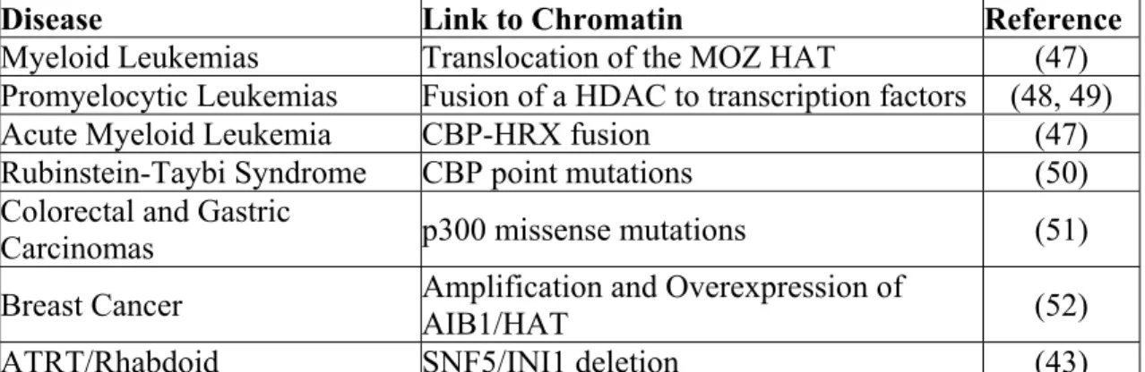
Aberrations in epigenetic machinery result in altered gene regulation and the development of human disease. Mutations in methyl-CpG binding protein, MeCP2, leads to altered gene expression by aberrant activation of BDNF in Rett Syndrome (40). BDNF plays a crucial role in neuronal survival, development, and plasticity, and dysregulation of it accounts for the neuropathology observed in Rett Syndrome. Mutations in SNF2 family member, ATR-X, leads to changes in methylation patterns and ATR-X syndrome, a severe X-linked form of mental retardation (41). One would expect the alteration of gene expression from aberrations in epigenetics to lead to silencing of tumor suppressor genes and activation of oncogenes leading to neoplastic disease. In fact, cancer development is a common result of aberrations in epigenetic machinery (Table 3). As discussed before, altered DNA methylation patterns can lead to altered gene expression and cancer development (16). Alterations in histone modifications through changes in HDAC expression lead to cancer by increased gene repression in gastrointestinal cancers, and several forms of leukemia (42). Aberrations in ATPase dependent chromatin remodeling complexes have also been implicated in the development of human disease. Findings that loss of SNF5, a member of the human SWI/SNF complex, occurs in 99% of rhabdoid tumor cases, identifies SNF5 as a bona fide tumor suppressor gene (43). Increasing evidence also indicates a role for inactivation of other members of the SWI/SNF complex BRG1, BRM, BAF155, and BAF57 in cancer development and/or cancer progression (44-46). Loss of function of members of the
Table 1.3: Chromatin Remodeling and Human Disease
Disease Link to Chromatin Reference
Myeloid Leukemias Translocation of the MOZ HAT (47) Promyelocytic Leukemias Fusion of a HDAC to transcription factors (48, 49)
Acute Myeloid Leukemia CBP-HRX fusion (47)
Rubinstein-Taybi Syndrome CBP point mutations (50) Colorectal and Gastric
Carcinomas p300 missense mutations (51)
Breast Cancer Amplification and Overexpression of AIB1/HAT (52)
increased investigation into the mechanism by which chromatin remodeling complexes work, in hopes of discovering more about the pathogenesis of these diseases.
F. SWI/SNF Chromatin Remodeling Complex
chromatin remodeling (60-62). These findings led to the belief that the main function of the SWI/SNF complex is gene regulation by relieving compaction of DNA from histones. The SWI/SNF complex has been shown to associate with ~5% of yeast genes, but the mechanism of SWI/SNF promoter specificity is not completely understood (63). Several theories exist to explain SWI/SNF promoter specificity; 1) The “Catalytic Model” explains that SWI/SNF causes transient changes in chromatin structure in a random manner. 2) The “Holoenzyme Model” states SWI/SNF is recruited to the promoter of target genes by ribonucleic acid (RNA) Polymerase II. 3) The most established theory is the “Activator Model” in which gene-specific transcriptional activators recruit SWI/SNF to the promoter. Once at the promoter, SWI/SNF, possibly in conjunction with HATs, remodels nucleosomes in a cis (sliding), or trans (displacement of nucleosome to another DNA strand) manner, to allow transcription of DNA (7, 64).
SWI/SNF and Gene Regulation in Development and Differentiation
Further studies of the SWI/SNF complex have shown a requirement for the complex in the regulation of a variety of genes. SWI/SNF was initially found to be involved in
the complex in early development (65-69). The SWI/SNF complex has also been shown to be required for transcriptional activation of nuclear receptors, which are critical for
homestasis and development. BAF57 has been shown to interact with both the estrogen receptor (ER) and androgen receptor (AR) (70-72). This interaction recruits the SWI/SNF complex to AR and ER target promoters. BRG1 is also required for expression of
glucocorticoid receptor target genes (73). CSF-1 was one of 80 genes found to be activated by the mammalian BAF complex by DNA microarray assay (74). It was found prior binding of NFI/CTF transcription factor was required for recruitment of the complex. CSF1 is
implicated in proliferation and differentiation of macrophages. CIITA binds SWI/SNF and is responsible for transcriptional activation of MHC class II genes involved in antigen
presentation (75). The SWI/SNF complex is also shown to be required for neuronal differentiation (76), thymocyte differentiation (77, 78), vasculogenesis and heart chamber maturation (79-81), osteoblast differentiation (82), and muscle differentiation (83). The SWI/SNF complex is responsible for altering chromatin at promoters of differentiation-specific loci.
The Wnt signaling pathway is essential to a number of development processes and is commonly altered in cancer development (84). B-catenin, a molecule that docks TCF transcription factors to target promoters, was found to interact with BRG1 (85).
transition (EMT) (87). BRG1/BRM deficient cell lines and tumors appear to be
dedifferentiated and may be undergoing EMT, due to a loss of regulation of genes involved in development and differentiation.
SWI/SNF and Gene Regulation in Cellular Adhesion and Proliferation
The SWI/SNF complex has also been shown to be involved in the regulation of other genes that may play a role in cellular adhesion. Reexpression of BRG1 in SW13 cells enhances MMP2 expression (88). Chromatin immunoprecipitation (ChIP) demonstrates a requirement of BRG1 in recruitment of transcription factors to the MMP2 promoter. MMP2 is involved in maintaining the extracellular matrix and loss of MMP2 leads to invasion and metastasis. Reexpression of BRG1 or BRM in deficient cells upregulates expression of CD44, a protein involved in cellular adhesion and cellular metastasis (89).
The complex is also involved in controlling cellular proliferation. BRG1 or BRM chromatin remodeling activity has shown to be required for endogenous stress response by hsp70 (90). Expression of BRG1 in SW13 cells was found to affect the RHOA pathway by increasing expression of Rock1 (91). Increased expression of RHOA leads to stress fiber formation in cells. Additional studies have shown that expression of dominant-negative BRG1 (DNBRG1) increases the cell volume, area of attachment, and nuclear size of the cell indicating altered growth. These changes correlate with over-expression of two integrin proteins. The SWI/SNF complex is also found to be involved in the pathway responsible for activation of p53-dependent promoters (92). p53 is commonly mutated in human cancer. Several subunits of the complex have been shown to bind to p53 and chromatin
p53-mediated growth suppression and apoptosis. Loss of SWI/SNF function may lead to cancer by altering these important tumor suppressor pathways in cellular adhesion and cellular proliferation.
The SWI/SNF complex has also been found to be involved in transcriptional repression of several genes including c-fos and cyclin E (93, 94). This repression may be carried out by an interaction with MeCP2 and/or mSin3a HDAC complex (95, 96). SWI/SNF also associates with several proteins involved in human disease, BRCA1 (97), FANCA (98), LKB1 (99), and TACC2 (100). These proteins may recruit the complex to target genes, which are critical for normal cellular function. The role of the complex in transcriptional regulation has led to its participation in multiple cellular activities.
Other Roles of SWI/SNF
SWI/SNF modification of mononucleosomes in concert with histone acetylases enhances RSS cleavage in vitro indicating a role for SWI/SNF in recombination (101).
Mutations in two homologs of the SWI/SNF complex in C. elegans disrupts asymmetric T cell division (102). PBAF is found to localize at kinetochores of mitotic chromosomes during mitosis and may be involved in cell division (103).
repression of cyclin E, allowing progression through G1 arrest (94). The association of SWI/SNF and Rb maintains repression of cyclin A, inhibiting exit from S phase until the cell is ready to progress. BRG1 has been shown to upregulate p21 leading to Rb
hypophosphorylation and RB-mediated growth arrest (106). Loss of BRG1 and BRM impairs RB-mediated growth arrest and allows the cell to proliferate. The role of the SWI/SNF complex in gene regulation and cellular proliferation makes it important in maintaining proper gene expression and cellular growth.
G. BRG1
Identification:
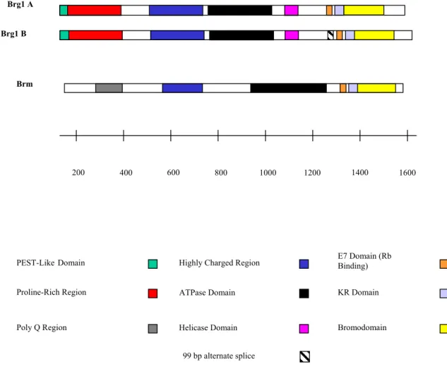
SWI2 was identified in yeast as part of a protein complex required for activation of messenger RNA in eukaryotes. SWI2 was one of a group of activators distinct from transcription machinery and transcription factors (57). SWI2 is known to assist binding of DNA-binding regulatory factors, and suppression of SWI2 mutations by certain histone genes suggest SWI2 may aid in overcoming the repressive effects of chromatin. SWI2 is a DNA dependent ATPase that functions as a helicase and interacts with a transcription factor to go to specific DNA sequences and remodel chromatin structure. Genes homologous to SWI2 have been found in Drosophila BRM and higher order eukaryotes (59). Screening of a human HeLa cDNA library with a Drosophila BRM isolated BRG1( BRM-related gene 1), a 1,613 amino acid protein highly related to Drosophila BRM (52% identity) (59). Another homolog to Drosophila BRM has been identified as, hBRM, which is 76% homologous to BRG1 (Figure 2). The catalytic subunit is required for proper functioning of the complex.
Little is known about the structure and function of BRG1. Four domains appear to be conserved from Drosophila to humans (59, 107). The proline rich domain is conserved and the function is unknown. Recent studies have discovered the highly charged domain II interacts with transcription factors like B-catenin. The ATPase domain is the third domain and is responsible for the catalytic ability of the subunit. The fourth conserved domain is the bromodomain found to bind acetylated lysines, which may help target the complex to
histones (108). BRG1 also contains a Pest-like domain, which can be involved in ubiquination, at the N-terminal end of the protein where most sequence differences with BRM exists. BRG1 also contains a helicase domain that plays a role in binding DNA and helping catalyze ATP. BRM, which has a similar structure to BRG1, has been shown to have a redundant function in terms of gene regulation and RB- mediated arrest (109). Differences in BRG1 and BRM must exist since BRG1 knockout mice are embryonic lethal, while BRM knockout mice survive and are slightly larger with no significant phenotype (66, 110). In fact, differences in the transcription factors BRG1 and BRM associate with, and the genes that they regulate have been identified (111). More research is still needed to help explain how loss of either member contributes to cancer development and cancer progression.
H. BRG1 is implicated in development of human cancer
As mentioned previously, recent studies implicate a role for BRG1 in the development of human cancer. These studies include:
Figure 1.2: Structure of Catalytic Subunits of hSWI/SNF Complexes. The two catalytic subunits of the human SWI/SNF complex are BRG1 and BRM. The structure of BRG1 and BRM is 76% homologous. BRG1 has two splice forms, the most common form of BRG1 is Brg1A.
Brg1 B
Brm
PEST-LikeDomain
ATPase Domain
200 400 600 800 1000 1200 1400 1600
Proline-Rich Region
Bromodomain Highly Charged Region
Poly Q Region
KR Domain E7 Domain (Rb Binding)
Helicase Domain
99 bp alternate splice
additional cell lines with reduced or absent expression of BRG1 (45, 104), making the total 10/85. All 10 cell lines were derived from adenocarcinoma of the respective tissue (lung, pancreas, adrenal). Immunhistochemistry on lung adenocarcinoma primary tissue revealed 4/40 lacked expression of BRG1 (46). This may indicate a role for the loss of BRG1 in progression of adenocarcinoma.
2) The presence of BRG1 mutations in human tumor cell lines. Wong et al. screened a panel of tumor cell lines to determine if BRG1 is targeted for mutation (44). They identified 16/180 human tumor cell lines that possessed mutations in BRG1. Of these 16 cell lines, 15 were adenocarcinomas, again drawing a correlation between loss of BRG1 function and the development of adenocarcinomas. Characteristic of tumor suppressor genes, reintroduction of functional BRG1 into the tumor cell lines resulted in senescence (44). A screen of primary Non-small cell lung cancer (NSCLC) tumors found two mutations in BRG1 (112). They are in domain V of the ATPase in a region of BRG1 that also has the point mutation in BRG1 found in HCT116 cells.
RB-mediated cell cycle arrest (104). These results showed a way in which loss of BRG1 function may cause loss of RB tumor suppressor activity.
4) BRG1 heterozygous mice are predisposed to tumor development. BRG1 null mice die early in development during the peri-implantation stage. BRG1 heterozygote mice are predisposed to exencephaly (5/36 mice) and tumors (66). Some BRG1 heterozygote mice (3/20) were found to develop large subcutaneous tumors of the neck and inguinal regions, compared to 0/15 wildtype mice that developed tumors (66). These tumors were epithelial and formed glandular structures (66), similar to adenocarcinomas. These tumors did not appear to have LOH of BRG1 and tumor formation appeared to be due to haploinsufficiency of BRG1. This finding indicated a potential role of BRG1 in mammalian tumor
development.
5) BRG1 loss is a poor prognostic marker in NSCLC. NSCLC BRG1/BRM-negative tumor patients have a shorter survival time then NSCLC BRG1/BRM-positive tumor patients (115). NSCLC BRG1/BRM-negative tumor patients of all stages have a shorter survival time then NSCLC BRG1/BRM-positive tumor patients in stage 3 (115). BRG1/BRM loss is a poor prognostic marker for NSCLC. Immunohistochemical examination of 12 core proteins involved in chromatin remodeling on a tissue microarray (TMA) of 150 lung
I. Specific Aims
Since all members of the SWI/SNF complex are required for proper functioning in yeast, we wanted to see how loss of other complex members besides SNF5 lead to cancer
development and/or cancer progression. A logical subunit to investigate is one of the
catalytic subunits, BRG1, since loss of the catalytic ability of the complex would most likely result in loss of function of the complex. The specific aims for this research are:
Specific Aim 1. To determine the effects of known BRG1 mutations on BRG1's
biological activity. To test the effects of BRG1 mutations from known human tumor cell lines we made stable clones of BRG1 mutant cell lines expressing BRM RNAi and tested them in four ways to determine the functional effects of the BRG1 mutations. 1) We tested the ability of BRG1 to growth arrest cells in the presence of PSM-RB or p16 by bromo-deoxyuridine (BRDU) incorporation assays. 2) We tested if the BRG1 mutants retain the ability to regulate CD44 and E-cadherin protein expression. 3) We tested if BRG1 mutants remained in a complex with other SWI/SNF proteins by immunoprecipitation. 4) We tested if the BRG1 mutants were able to target promoters by chromatin immunoprecipitation
Specific Aim 2. To determine if loss of BRG1 alters promoter methylation patterns.
5-azacytidine led to their re-expression. We also examined if cytosine methylation status of target gene promoters changed in the presence or absence of BRG1 by bisulfite sequencing. To determine if loss of BRG1 altered promoter methylation globally we ran assays to
REFERENCES
1. ACS Cancer Statistics 2005. American Cancer Society, 2005.
2. Workman, J. L. and Kingston, R. E. Alteration of nucleosome structure as a mechanism of transcriptional regulation. Annu Rev Biochem, 67: 545-579, 1998. 3. Nemeth, A. and Langst, G. Chromatin higher order structure: opening up chromatin
for transcription. Brief Funct Genomic Proteomic, 2: 334-343, 2004.
4. Luo, R. X. and Dean, D. C. Chromatin remodeling and transcriptional regulation. J Natl Cancer Inst, 91: 1288-1294, 1999.
5. Tsukiyama, T. and Wu, C. Chromatin remodeling and transcription. Curr Opin Genet Dev, 7: 182-191, 1997.
6. Armstrong, J. A. and Emerson, B. M. Transcription of chromatin: these are complex times. Curr Opin Genet Dev, 8: 165-172, 1998.
7. Vignali, M., Hassan, A. H., Neely, K. E., and Workman, J. L. ATP-dependent chromatin-remodeling complexes. Mol Cell Biol, 20: 1899-1910, 2000.
8. Strahl, B. D. and Allis, C. D. The language of covalent histone modifications. Nature, 403: 41-45, 2000.
9. Peterson, C. L. and Laniel, M. A. Histones and histone modifications. Curr Biol, 14: R546-551, 2004.
10. Nowak, S. J. and Corces, V. G. Phosphorylation of histone H3: a balancing act between chromosome condensation and transcriptional activation. Trends Genet, 20: 214-220, 2004.
11. Osley, M. A. H2B ubiquitylation: the end is in sight. Biochim Biophys Acta, 1677: 74-78, 2004.
12. Shin, J. A., Choi, E. S., Kim, H. S., Ho, J. C., Watts, F. Z., Park, S. D., and Jang, Y. K. SUMO modification is involved in the maintenance of heterochromatin stability in fission yeast. Mol Cell, 19: 817-828, 2005.
13. Althaus, F. R. Poly ADP-ribosylation: a histone shuttle mechanism in DNA excision repair. J Cell Sci, 102 ( Pt 4): 663-670, 1992.
15. Nakao, M. Epigenetics: interaction of DNA methylation and chromatin. Gene, 278: 25-31, 2001.
16. Feinberg, A. P. and Tycko, B. The history of cancer epigenetics. Nat Rev Cancer, 4: 143-153, 2004.
17. Feinberg, A. P. and Vogelstein, B. Hypomethylation distinguishes genes of some human cancers from their normal counterparts. Nature, 301: 89-92, 1983.
18. Feinberg, A. P. and Vogelstein, B. Hypomethylation of ras oncogenes in primary human cancers. Biochem Biophys Res Commun, 111: 47-54, 1983.
19. Gibbons, R. J., McDowell, T. L., Raman, S., O'Rourke, D. M., Garrick, D., Ayyub, H., and Higgs, D. R. Mutations in ATRX, encoding a SWI/SNF-like protein, cause diverse changes in the pattern of DNA methylation. Nat Genet, 24: 368-371, 2000. 20. Fan, T., Yan, Q., Huang, J., Austin, S., Cho, E., Ferris, D., and Muegge, K.
Lsh-deficient murine embryonal fibroblasts show reduced proliferation with signs of abnormal mitosis. Cancer Res, 63: 4677-4683, 2003.
21. Dennis, K., Fan, T., Geiman, T., Yan, Q., and Muegge, K. Lsh, a member of the SNF2 family, is required for genome-wide methylation. Genes Dev, 15: 2940-2944, 2001.
22. Baylin, S. B., Hoppener, J. W., de Bustros, A., Steenbergh, P. H., Lips, C. J., and Nelkin, B. D. DNA methylation patterns of the calcitonin gene in human lung cancers and lymphomas. Cancer Res, 46: 2917-2922, 1986.
23. Robertson, K. D. DNA methylation and chromatin - unraveling the tangled web. Oncogene, 21: 5361-5379, 2002.
24. Gonzalez-Zulueta, M., Bender, C. M., Yang, A. S., Nguyen, T., Beart, R. W., Van Tornout, J. M., and Jones, P. A. Methylation of the 5' CpG island of the p16/CDKN2 tumor suppressor gene in normal and transformed human tissues correlates with gene silencing. Cancer Res, 55: 4531-4535, 1995.
25. Kane, M. F., Loda, M., Gaida, G. M., Lipman, J., Mishra, R., Goldman, H., Jessup, J. M., and Kolodner, R. Methylation of the hMLH1 promoter correlates with lack of expression of hMLH1 in sporadic colon tumors and mismatch repair-defective human tumor cell lines. Cancer Res, 57: 808-811, 1997.
26. Herman, J. G., Latif, F., Weng, Y., Lerman, M. I., Zbar, B., Liu, S., Samid, D., Duan, D. S., Gnarra, J. R., Linehan, W. M., and et al. Silencing of the VHL
27. Graff, J. R., Herman, J. G., Lapidus, R. G., Chopra, H., Xu, R., Jarrard, D. F., Isaacs, W. B., Pitha, P. M., Davidson, N. E., and Baylin, S. B. E-cadherin expression is silenced by DNA hypermethylation in human breast and prostate carcinomas. Cancer Res, 55: 5195-5199, 1995.
28. Tycko, B. and Efstratiadis, A. Genomic imprinting: piece of cake. Nature, 417: 913-914, 2002.
29. DeChiara, T. M., Robertson, E. J., and Efstratiadis, A. Parental imprinting of the mouse insulin-like growth factor II gene. Cell, 64: 849-859, 1991.
30. Henry, I., Puech, A., Riesewijk, A., Ahnine, L., Mannens, M., Beldjord, C., Bitoun, P., Tournade, M. F., Landrieu, P., and Junien, C. Somatic mosaicism for partial paternal isodisomy in Wiedemann-Beckwith syndrome: a post-fertilization event. Eur J Hum Genet, 1: 19-29, 1993.
31. Veigl, M. L., Kasturi, L., Olechnowicz, J., Ma, A. H., Lutterbaugh, J. D., Periyasamy, S., Li, G. M., Drummond, J., Modrich, P. L., Sedwick, W. D., and Markowitz, S. D. Biallelic inactivation of hMLH1 by epigenetic gene silencing, a novel mechanism causing human MSI cancers. Proc Natl Acad Sci U S A, 95: 8698-8702, 1998. 32. Bachman, K. E., Park, B. H., Rhee, I., Rajagopalan, H., Herman, J. G., Baylin, S. B.,
Kinzler, K. W., and Vogelstein, B. Histone modifications and silencing prior to DNA methylation of a tumor suppressor gene. Cancer Cell, 3: 89-95, 2003.
33. Clark, S. J. and Warnecke, P. M. DNA methylation analysis in mammalian cells. Methods, 27: 99-100, 2002.
34. Stirzaker, C., Song, J. Z., Davidson, B., and Clark, S. J. Transcriptional gene
silencing promotes DNA hypermethylation through a sequential change in chromatin modifications in cancer cells. Cancer Res, 64: 3871-3877, 2004.
35. Buschhausen, G., Wittig, B., Graessmann, M., and Graessmann, A. Chromatin structure is required to block transcription of the methylated herpes simplex virus thymidine kinase gene. Proc Natl Acad Sci U S A, 84: 1177-1181, 1987.
36. Rea, S., Eisenhaber, F., O'Carroll, D., Strahl, B. D., Sun, Z. W., Schmid, M., Opravil, S., Mechtler, K., Ponting, C. P., Allis, C. D., and Jenuwein, T. Regulation of
chromatin structure by site-specific histone H3 methyltransferases. Nature, 406: 593-599, 2000.
38. Jones, P. L., Veenstra, G. J., Wade, P. A., Vermaak, D., Kass, S. U., Landsberger, N., Strouboulis, J., and Wolffe, A. P. Methylated DNA and MeCP2 recruit histone deacetylase to repress transcription. Nat Genet, 19: 187-191, 1998.
39. Nan, X., Cross, S., and Bird, A. Gene silencing by methyl-CpG-binding proteins. Novartis Found Symp, 214: 6-16; discussion 16-21, 46-50, 1998.
40. Chen, W. G., Chang, Q., Lin, Y., Meissner, A., West, A. E., Griffith, E. C., Jaenisch, R., and Greenberg, M. E. Derepression of BDNF transcription involves calcium-dependent phosphorylation of MeCP2. Science, 302: 885-889, 2003.
41. Gibbons, R. J., Picketts, D. J., Villard, L., and Higgs, D. R. Mutations in a putative global transcriptional regulator cause X-linked mental retardation with alpha-thalassemia (ATR-X syndrome). Cell, 80: 837-845, 1995.
42. Roux-Rouquie, M., Chauvet, M. L., Munnich, A., and Frezal, J. Human genes involved in chromatin remodeling in transcription initiation, and associated diseases: An overview using the GENATLAS database. Mol Genet Metab, 67: 261-277, 1999. 43. Versteege, I., Sevenet, N., Lange, J., Rousseau-Merck, M. F., Ambros, P.,
Handgretinger, R., Aurias, A., and Delattre, O. Truncating mutations of hSNF5/INI1 in aggressive paediatric cancer. Nature, 394: 203-206, 1998.
44. Wong, A. K., Shanahan, F., Chen, Y., Lian, L., Ha, P., Hendricks, K., Ghaffari, S., Iliev, D., Penn, B., Woodland, A. M., Smith, R., Salada, G., Carillo, A., Laity, K., Gupte, J., Swedlund, B., Tavtigian, S. V., Teng, D. H., and Lees, E. BRG1, a
component of the SWI-SNF complex, is mutated in multiple human tumor cell lines. Cancer Res, 60: 6171-6177, 2000.
45. Decristofaro, M. F., Betz, B. L., Rorie, C. J., Reisman, D. N., Wang, W., and Weissman, B. E. Characterization of SWI/SNF protein expression in human breast cancer cell lines and other malignancies. J Cell Physiol, 186: 136-145, 2001. 46. Reisman, D. N., Strobeck, M. W., Betz, B. L., Sciariotta, J., Funkhouser, W., Jr.,
Murchardt, C., Yaniv, M., Sherman, L. S., Knudsen, E. S., and Weissman, B. E. Concomitant down-regulation of BRM and BRG1 in human tumor cell lines:
differential effects on RB-mediated growth arrest vs CD44 expression. Oncogene, 21: 1196-1207, 2002.
48. Grignani, F., De Matteis, S., Nervi, C., Tomassoni, L., Gelmetti, V., Cioce, M., Fanelli, M., Ruthardt, M., Ferrara, F. F., Zamir, I., Seiser, C., Lazar, M. A., Minucci, S., and Pelicci, P. G. Fusion proteins of the retinoic acid receptor-alpha recruit histone deacetylase in promyelocytic leukaemia. Nature, 391: 815-818, 1998.
49. Lin, R. J., Nagy, L., Inoue, S., Shao, W., Miller, W. H., Jr., and Evans, R. M. Role of the histone deacetylase complex in acute promyelocytic leukaemia. Nature, 391: 811-814, 1998.
50. Petrij, F., Giles, R. H., Dauwerse, H. G., Saris, J. J., Hennekam, R. C., Masuno, M., Tommerup, N., van Ommen, G. J., Goodman, R. H., Peters, D. J., and et al.
Rubinstein-Taybi syndrome caused by mutations in the transcriptional co-activator CBP. Nature, 376: 348-351, 1995.
51. Muraoka, M., Konishi, M., Kikuchi-Yanoshita, R., Tanaka, K., Shitara, N., Chong, J. M., Iwama, T., and Miyaki, M. p300 gene alterations in colorectal and gastric
carcinomas. Oncogene, 12: 1565-1569, 1996.
52. Anzick, S. L., Kononen, J., Walker, R. L., Azorsa, D. O., Tanner, M. M., Guan, X. Y., Sauter, G., Kallioniemi, O. P., Trent, J. M., and Meltzer, P. S. AIB1, a steroid receptor coactivator amplified in breast and ovarian cancer. Science, 277: 965-968, 1997.
53. Stern, M., Jensen, R., and Herskowitz, I. Five SWI genes are required for expression of the HO gene in yeast. J Mol Biol, 178: 853-868, 1984.
54. Neigeborn, L. and Carlson, M. Genes affecting the regulation of SUC2 gene expression by glucose repression in Saccharomyces cerevisiae. Genetics, 108: 845-858, 1984.
55. Hirschhorn, J. N., Brown, S. A., Clark, C. D., and Winston, F. Evidence that
SNF2/SWI2 and SNF5 activate transcription in yeast by altering chromatin structure. Genes Dev, 6: 2288-2298, 1992.
56. Kruger, W., Peterson, C. L., Sil, A., Coburn, C., Arents, G., Moudrianakis, E. N., and Herskowitz, I. Amino acid substitutions in the structured domains of histones H3 and H4 partially relieve the requirement of the yeast SWI/SNF complex for transcription. Genes Dev, 9: 2770-2779, 1995.
57. Peterson, C. L. and Herskowitz, I. Characterization of the yeast SWI1, SWI2, and SWI3 genes, which encode a global activator of transcription. Cell, 68: 573-583, 1992.
583-59. Khavari, P. A., Peterson, C. L., Tamkun, J. W., Mendel, D. B., and Crabtree, G. R. BRG1 contains a conserved domain of the SWI2/SNF2 family necessary for normal mitotic growth and transcription. Nature, 366: 170-174, 1993.
60. Wang, W., Cote, J., Xue, Y., Zhou, S., Khavari, P. A., Biggar, S. R., Muchardt, C., Kalpana, G. V., Goff, S. P., Yaniv, M., Workman, J. L., and Crabtree, G. R. Purification and biochemical heterogeneity of the mammalian SWI-SNF complex. Embo J, 15: 5370-5382, 1996.
61. Cote, J., Quinn, J., Workman, J. L., and Peterson, C. L. Stimulation of GAL4 derivative binding to nucleosomal DNA by the yeast SWI/SNF complex. Science, 265: 53-60, 1994.
62. Kwon, H., Imbalzano, A. N., Khavari, P. A., Kingston, R. E., and Green, M. R. Nucleosome disruption and enhancement of activator binding by a human SW1/SNF complex. Nature, 370: 477-481, 1994.
63. Peterson, C. L. and Workman, J. L. Promoter targeting and chromatin remodeling by the SWI/SNF complex. Curr Opin Genet Dev, 10: 187-192, 2000.
64. Sudarsanam, P. and Winston, F. The Swi/Snf family nucleosome-remodeling complexes and transcriptional control. Trends Genet, 16: 345-351, 2000. 65. Kadam, S., McAlpine, G. S., Phelan, M. L., Kingston, R. E., Jones, K. A., and
Emerson, B. M. Functional selectivity of recombinant mammalian SWI/SNF subunits. Genes Dev, 14: 2441-2451, 2000.
66. Bultman, S., Gebuhr, T., Yee, D., La Mantia, C., Nicholson, J., Gilliam, A.,
Randazzo, F., Metzger, D., Chambon, P., Crabtree, G., and Magnuson, T. A Brg1 null mutation in the mouse reveals functional differences among mammalian SWI/SNF complexes. Mol Cell, 6: 1287-1295, 2000.
67. Kim, J. K., Huh, S. O., Choi, H., Lee, K. S., Shin, D., Lee, C., Nam, J. S., Kim, H., Chung, H., Lee, H. W., Park, S. D., and Seong, R. H. Srg3, a mouse homolog of yeast SWI3, is essential for early embryogenesis and involved in brain development. Mol Cell Biol, 21: 7787-7795, 2001.
68. Roberts, C. W., Galusha, S. A., McMenamin, M. E., Fletcher, C. D., and Orkin, S. H. Haploinsufficiency of Snf5 (integrase interactor 1) predisposes to malignant rhabdoid tumors in mice. Proc Natl Acad Sci U S A, 97: 13796-13800, 2000.
70. DiRenzo, J., Shang, Y., Phelan, M., Sif, S., Myers, M., Kingston, R., and Brown, M. BRG-1 is recruited to estrogen-responsive promoters and cooperates with factors involved in histone acetylation. Mol Cell Biol, 20: 7541-7549, 2000.
71. Belandia, B., Orford, R. L., Hurst, H. C., and Parker, M. G. Targeting of SWI/SNF chromatin remodelling complexes to estrogen-responsive genes. Embo J, 21: 4094-4103, 2002.
72. Link, K. A., Burd, C. J., Williams, E., Marshall, T., Rosson, G., Henry, E., Weissman, B., and Knudsen, K. E. BAF57 governs androgen receptor action and androgen-dependent proliferation through SWI/SNF. Mol Cell Biol, 25: 2200-2215, 2005.
73. Fryer, C. J. and Archer, T. K. Chromatin remodelling by the glucocorticoid receptor requires the BRG1 complex. Nature, 393: 88-91, 1998.
74. Liu, R., Liu, H., Chen, X., Kirby, M., Brown, P. O., and Zhao, K. Regulation of CSF1 promoter by the SWI/SNF-like BAF complex. Cell, 106: 309-318, 2001.
75. Mudhasani, R. and Fontes, J. D. Multiple interactions between BRG1 and MHC class II promoter binding proteins. Mol Immunol, 42: 673-682, 2005.
76. Seo, S., Richardson, G. A., and Kroll, K. L. The SWI/SNF chromatin remodeling protein Brg1 is required for vertebrate neurogenesis and mediates transactivation of Ngn and NeuroD. Development, 132: 105-115, 2005.
77. Chi, T. H., Wan, M., Lee, P. P., Akashi, K., Metzger, D., Chambon, P., Wilson, C. B., and Crabtree, G. R. Sequential roles of Brg, the ATPase subunit of BAF chromatin remodeling complexes, in thymocyte development. Immunity, 19: 169-182, 2003. 78. Gebuhr, T. C., Kovalev, G. I., Bultman, S., Godfrey, V., Su, L., and Magnuson, T.
The role of Brg1, a catalytic subunit of mammalian chromatin-remodeling complexes, in T cell development. J Exp Med, 198: 1937-1949, 2003.
79. Dauvillier, S., Ott, M. O., Renard, J. P., and Legouy, E. BRM (SNF2alpha) expression is concomitant to the onset of vasculogenesis in early mouse postimplantation development. Mech Dev, 101: 221-225, 2001.
80. Lickert, H., Takeuchi, J. K., Von Both, I., Walls, J. R., McAuliffe, F., Adamson, S. L., Henkelman, R. M., Wrana, J. L., Rossant, J., and Bruneau, B. G. Baf60c is essential for function of BAF chromatin remodelling complexes in heart development. Nature, 432: 107-112, 2004.
82. Young, D. W., Pratap, J., Javed, A., Weiner, B., Ohkawa, Y., van Wijnen, A., Montecino, M., Stein, G. S., Stein, J. L., Imbalzano, A. N., and Lian, J. B. SWI/SNF chromatin remodeling complex is obligatory for BMP2-induced, Runx2-dependent skeletal gene expression that controls osteoblast differentiation. J Cell Biochem, 94: 720-730, 2005.
83. de la Serna, I. L., Carlson, K. A., and Imbalzano, A. N. Mammalian SWI/SNF complexes promote MyoD-mediated muscle differentiation. Nat Genet, 27: 187-190, 2001.
84. Akiyama, T. Wnt/beta-catenin signaling. Cytokine Growth Factor Rev, 11: 273-282, 2000.
85. Barker, N., Hurlstone, A., Musisi, H., Miles, A., Bienz, M., and Clevers, H. The chromatin remodelling factor Brg-1 interacts with beta-catenin to promote target gene activation. Embo J, 20: 4935-4943, 2001.
86. Huber, O., Korn, R., McLaughlin, J., Ohsugi, M., Herrmann, B. G., and Kemler, R. Nuclear localization of beta-catenin by interaction with transcription factor LEF-1. Mech Dev, 59: 3-10, 1996.
87. Savagner, P. Leaving the neighborhood: molecular mechanisms involved during epithelial-mesenchymal transition. Bioessays, 23: 912-923, 2001.
88. Ma, Z., Chang, M. J., Shah, R., Adamski, J., Zhao, X., and Benveniste, E. N. Brg-1 is required for maximal transcription of the human matrix metalloproteinase-2 gene. J Biol Chem, 279: 46326-46334, 2004.
89. Strobeck, M. W., DeCristofaro, M. F., Banine, F., Weissman, B. E., Sherman, L. S., and Knudsen, E. S. The BRG-1 subunit of the SWI/SNF complex regulates CD44 expression. J Biol Chem, 276: 9273-9278, 2001.
90. de La Serna, I. L., Carlson, K. A., Hill, D. A., Guidi, C. J., Stephenson, R. O., Sif, S., Kingston, R. E., and Imbalzano, A. N. Mammalian SWI-SNF complexes contribute to activation of the hsp70 gene. Mol Cell Biol, 20: 2839-2851, 2000.
91. Asp, P., Wihlborg, M., Karlen, M., and Farrants, A. K. Expression of BRG1, a human SWI/SNF component, affects the organisation of actin filaments through the RhoA signalling pathway. J Cell Sci, 115: 2735-2746, 2002.
92. Lee, D., Kim, J. W., Seo, T., Hwang, S. G., Choi, E. J., and Choe, J. SWI/SNF complex interacts with tumor suppressor p53 and is necessary for the activation of p53-mediated transcription. J Biol Chem, 277: 22330-22337, 2002.
94. Zhang, H. S., Gavin, M., Dahiya, A., Postigo, A. A., Ma, D., Luo, R. X., Harbour, J. W., and Dean, D. C. Exit from G1 and S phase of the cell cycle is regulated by repressor complexes containing HDAC-Rb-hSWI/SNF and Rb-hSWI/SNF. Cell, 101: 79-89, 2000.
95. Harikrishnan, K. N., Chow, M. Z., Baker, E. K., Pal, S., Bassal, S., Brasacchio, D., Wang, L., Craig, J. M., Jones, P. L., Sif, S., and El-Osta, A. Brahma links the SWI/SNF chromatin-remodeling complex with MeCP2-dependent transcriptional silencing. Nat Genet, 37: 254-264, 2005.
96. Sif, S., Saurin, A. J., Imbalzano, A. N., and Kingston, R. E. Purification and characterization of mSin3A-containing Brg1 and hBrm chromatin remodeling complexes. Genes Dev, 15: 603-618, 2001.
97. Bochar, D. A., Wang, L., Beniya, H., Kinev, A., Xue, Y., Lane, W. S., Wang, W., Kashanchi, F., and Shiekhattar, R. BRCA1 is associated with a human SWI/SNF-related complex: linking chromatin remodeling to breast cancer. Cell, 102: 257-265, 2000.
98. Otsuki, T., Furukawa, Y., Ikeda, K., Endo, H., Yamashita, T., Shinohara, A.,
Iwamatsu, A., Ozawa, K., and Liu, J. M. Fanconi anemia protein, FANCA, associates with BRG1, a component of the human SWI/SNF complex. Hum Mol Genet, 10: 2651-2660, 2001.
99. Marignani, P. A., Kanai, F., and Carpenter, C. L. LKB1 associates with Brg1 and is necessary for Brg1-induced growth arrest. J Biol Chem, 276: 32415-32418, 2001. 100. Lauffart, B., Gangisetty, O., and Still, I. H. Molecular cloning, genomic structure and
interactions of the putative breast tumor suppressor TACC2. Genomics, 81: 192-201, 2003.
101. Roth, D. B. and Roth, S. Y. Unequal access: regulating V(D)J recombination through chromatin remodeling. Cell, 103: 699-702, 2000.
102. Sawa, H., Kouike, H., and Okano, H. Components of the SWI/SNF complex are required for asymmetric cell division in C. elegans. Mol Cell, 6: 617-624, 2000. 103. Xue, Y., Canman, J. C., Lee, C. S., Nie, Z., Yang, D., Moreno, G. T., Young, M. K.,
Salmon, E. D., and Wang, W. The human SWI/SNF-B chromatin-remodeling
complex is related to yeast rsc and localizes at kinetochores of mitotic chromosomes. Proc Natl Acad Sci U S A, 97: 13015-13020, 2000.
105. Dahiya, A., Gavin, M. R., Luo, R. X., and Dean, D. C. Role of the LXCXE binding site in Rb function. Mol Cell Biol, 20: 6799-6805, 2000.
106. Kang, H., Cui, K., and Zhao, K. BRG1 controls the activity of the retinoblastoma protein via regulation of p21CIP1/WAF1/SDI. Mol Cell Biol, 24: 1188-1199, 2004. 107. Hurlstone, A. F., Olave, I. A., Barker, N., van Noort, M., and Clevers, H. Cloning and
characterization of hELD/OSA1, a novel BRG1 interacting protein. Biochem J, 364: 255-264, 2002.
108. Zeng, L. and Zhou, M. M. Bromodomain: an acetyl-lysine binding domain. FEBS Lett, 513: 124-128, 2002.
109. Strobeck, M. W., Reisman, D. N., Gunawardena, R. W., Betz, B. L., Angus, S. P., Knudsen, K. E., Kowalik, T. F., Weissman, B. E., and Knudsen, E. S. Compensation of BRG-1 function by Brm: insight into the role of the core SWI-SNF subunits in retinoblastoma tumor suppressor signaling. J Biol Chem, 277: 4782-4789, 2002. 110. Reyes, J. C., Barra, J., Muchardt, C., Camus, A., Babinet, C., and Yaniv, M. Altered
control of cellular proliferation in the absence of mammalian brahma (SNF2alpha). Embo J, 17: 6979-6991, 1998.
111. Kadam, S. and Emerson, B. M. Transcriptional specificity of human SWI/SNF BRG1 and BRM chromatin remodeling complexes. Mol Cell, 11: 377-389, 2003.
112. Medina, P. P., Carretero, J., Fraga, M. F., Esteller, M., Sidransky, D., and Sanchez-Cespedes, M. Genetic and epigenetic screening for gene alterations of the chromatin-remodeling factor, SMARCA4/BRG1, in lung tumors. Genes Chromosomes Cancer, 41: 170-177, 2004.
113. Knudsen, K. E., Weber, E., Arden, K. C., Cavenee, W. K., Feramisco, J. R., and Knudsen, E. S. The retinoblastoma tumor suppressor inhibits cellular proliferation through two distinct mechanisms: inhibition of cell cycle progression and induction of cell death. Oncogene, 18: 5239-5245, 1999.
114. Strober, B. E., Dunaief, J. L., Guha, and Goff, S. P. Functional interactions between the hBRM/hBRG1 transcriptional activators and the pRB family of proteins. Mol Cell Biol, 16: 1576-1583, 1996.
115. Reisman, D. N., Sciarrotta, J., Wang, W., Funkhouser, W. K., and Weissman, B. E. Loss of BRG1/BRM in human lung cancer cell lines and primary lung cancers: correlation with poor prognosis. Cancer Res, 63: 560-566, 2003.
CHAPTER 2
BRG1 Mutations found in Human Cancer Cell Lines Inactivate Rb-mediated Cell Cycle Arrest
A. Introduction
Eukaryotic organisms package DNA into condensed solenoid chromatin for compact storage in the cell nucleus. This packaging results in transcriptional repression of genes. Therefore, mechanisms of chromatin remodeling have evolved to overcome this repressive nature of chromatin and make DNA accessible to sequence-specific transcription factors and transcription machinery (1, 2). Chromatin remodeling results in an alteration of nucleosomes to allow the binding of transcription factors and initiation of transcription (1, 2). Two classes of chromatin remodeling enzymes exist, histone modifying enzymes and ATP-dependent chromatin remodeling enzymes.
The SWI/SNF complex is an ATP-dependent chromatin remodeling complex that is conserved from humans to yeast (3). The complex is approximately 2-Mda in size and consists of 9-12 members in a tissue specific manner (4). To date, six SWI/SNF complexes have been isolated with each complex consisting of one catalytic subunit, either BRG1 or BRM, plus 8-11 BAFs (BRG1 associated factors). A minimum catalytic core of BRG1 or BRM, BAF155 or BAF170, and BAF47 is required for disruption of nucleosome arrays in vitro, but all members of the complex are required for proper functioning in yeast.
adhesion such as CD44 (5) and E-cadherin (6), and genes involved in cellular proliferation such as cyclin A (7) and cyclin E (7), CSF1 (8), and p53-dependent target promoters (9). SWI/SNF has also been shown to be required for RB-mediated arrest by upregulating p21, which inhibits cyclin-dependent kinases and leads to RB hypophosphorylation (10). Due to the role of the complex in cellular adhesion and growth arrest, it is not surprising evidence has linked loss of SWI/SNF complex members with human disease.
In a previous study, tumor cell lines lacking both BRG1 and BRM expression were analyzed for impaired RB-mediated growth arrest and for expression of CD44 (18). It was found that either BRM or BRG1 was sufficient to restore RB-mediated growth arrest and CD44 expression, indicating that BRM can compensate for BRG1 loss. As a further characteristic of tumor suppressor genes, the stable reintroduction of BRG1 into the tumor cell lines resulted in replicative senescence.
In this study we analyzed if the point mutations in BRG1 found in human tumor cell lines resulted in altered functions. Therefore, we suppressed BRM expression in each cell line by stable expression of shRNA and assessed them for impaired RB-mediated arrest, CD44 expression, and E-cadherin expression. We found that 2 of the BRG1 mutations have lost the ability to promote RB-mediated cell cycle arrest and CD44 expression. We also showed that these mutant BRG1 proteins still form a complex with other members by co-immunoprecipitation and were present at target promoters by Chromatin
immunoprecipitation. Our results indicate that the point mutations found in these cell lines impair the function of BRG1 by altering the ability of the complex to remodel chromatin. The sites of these point mutations should provide insight into the structure and function of this important cellular regulatory protein.
B. Material and Methods
Cell Lines:
Immunoblotting:
Protein was collected from cells grown in 100 mm tissue-culture dishes. Detergent-based extraction buffer (6) was added to the dishes and cells were collected with a scraper into an Eppendorf tube. The tube was rocked in the cold room for 10 min and then spun at 14,000 rpm for 10 min. Supernatant was collected and protein concentration was determined by Bradford protein assay. Equal amounts of protein lysate (30 ug) were separated by SDS-polyacrylamide gel electrophoresis (either 7.5% or 4-20% gradient) for 1 hr at 100 V. Protein was then transferred to Immobilon-P membranes (Millipore) for 16-18 hr at 80 mA. Membranes were blocked for 1 hr in 5% non-fat dry milk/0.2% Tween 20 in PBS.
Membranes were then incubated for 2 hr in primary antibody at room temperature. Primary antibodies included BRG1 (G-7) (Santa Cruz), BRM (Dr.Yaniv Moshe), E-cadherin
(Transduction Labs), Cytokeratin 18 (DC-10) (Santa Cruz), Fhit (Zymed), CD44 (H3) (Dr. Larry Sherman), VHL (G-7) (Santa Cruz), vimentin (Biomeda), p16 (BD PharMingen). Membranes were then washed 3 times for 10 min each in 200 ml of 0.2% Tween 20 in PBS. Membranes were then incubated in a 1:2000 dilution of either anti-mouse or anti-rabbit secondary antibody for 1 hr. Membranes were then washed once again, 3 times for 10 min each time. Bands were detected with ECL chemiluminescence reagent (Amersham) on Biomax ML film (Kodak).
Nuclear Extraction:
pellet volume with hypotonic buffer for 10 min. The pellet was Dounce homogenized with a size A pestel. The nuclei were centrifuged in an SS34 Sorvall rotor at 4K for 10 min. The supernatant was removed, the tubes were rebalanced and centrifuged again at 18K for 20 min. The pellet was resuspended in Nuclear Extract Buffer (20 mM Hepes ph 7.9/420 mM NaCl/1.5mM MgCl2/2.0 mM DTT/0.2 mM EDTA/25% glycerol) using 3 ml per 1×10⁹ cells. Nuclei were sonicated on Bronson Digital 1 sonicator at a setting of 20 4 times for 15
seconds each time, then dounce homogenized to release nuclear proteins. The homogenate was placed into a prechilled plastic beaker and stirred in an ice water bath for 30 minutes. Nuclear proteins were collected by centrifugation at 14K for 30 min in an Eppendorf microfuge. The supernatant was then dialyzed against three changes of dialysis buffer (20 mM Hepes pH 7.9/50 mM KCl/1.0 mM DTT/0.2 mM EDTA/10% glycerol) and clarified by an additional centrifugation in an Eppendorf microfuge at 14,000 rpm for 30 min. Protein concentrations were determined by Bradford assay.
Immunoprecipitation:
Protein A and G sepharose beads (Amersham) were washed 5X in RIPA buffer (150mM NaCL/1% NP40/0.5% deoxycholate/0.1% SDS/50 mM Tris pH8/5 mM EDTA/protease inhibitor cocktail (Sigma)). Forty microliters of beads were added to 300 ug of nuclear extract per antibody and diluted to 1 ml in RIPA buffer. Five percent of the protein sample was removed for input control. Samples were rocked for 2 hr at 4°C. At the same time 40 ul of beads per IP reaction were saturated in 1 mg/ml BSA and 0.3 mg/ml salmon sperm (SS). Supernatant was collected and 300 ug of protein was diluted to 1 ml in PBS. Primary antibodies were added (10 ul for commercial, 2 ul for BRG1 (J1)) and rocked at 4°C
then washed once in RIPA buffer, once in IP wash buffer (100 mM Tris HCl pH 8.5/500 mM LiCl/1% NP40/1% deoxycholate), and once in PBS. Beads were diluted in 36 ul PBS and 9 ul of western loading buffer. After heating beads at 95°C for 5 min, 20 ul was then loaded onto a SDS-polyacrylamide gel for immunoblot analysis.
QT-PCR ChIP:
Six 150 mm dishes of each cell line were fixed with Paro-Fixation Solution (PBS with 50 mM Hepes pH 8/1 mM EDTA/0.5 mM EGTA/100 mM NaCl/11% formaldehyde) at 32°C for 20 min. To stop fixation, glycine was added to the dishes to a final concentration of 0.125 M. Plates were washed with PBS and then cells were collected with a scraper into 1X TE. Samples were freeze-thawed in liquid nitrogen and then sonicated with 15 pulses for 15 sec at 30% setting. Sonicated sample was spun in a SW55 rotor for 10 min at 12,000 rpm. Protein was then quantified by microbradford protein assay.
For Chromatin immunoprecipitation, Protein A/Protein G sepharose beads
sample was added to a fresh tube along with 10 ul 5M NaCl and incubated for 5 hr at 65°C to reverse crosslink. Next, protein was degraded with the addition of 2 ug of Proteinase K and incubation for 30 min at 45°C. DNA was phenol-chloroform extracted and ethanol
precipitated and resuspended in 150 ul 0.1X TE. Five microliters of DNA was used in 25 ul QPCR reaction with 12.5 ul SYBER Green (Applied Biosystems), 7 ul ddH20, and 0.5 ul of 50 uM primer for either CD44 (forward TCCAGCCGGATTCAGAGAAA-3’ reverse TTCAGCCTTTGGCCTCTCCT-3’) or E-cadherin (forward
5’-TCGAACCCAGTGGAATCAGAA-3’ reverse 5’-GGGTCTAGGTGGGTTATGGGAC-3’). The fluorescent signal (fluorescent staining by SYBER green of replicated DNA) was
detected at each cycle. The fluorescent signal (in the exponential stage of replication) for the samples were subtracted from PBS alone samples to give a signal intensity above
background to allow for comparison across samples. Generation of Stable Clones:
The BRM and BRG1 shRNA expression vectors were previously generated by Dr. Gary Rosson (19, 20). Cell lines were transfected with these shRNAi vectors using Lipofectamine 2000 (Invitrogen) following the manufacturers instructions. Clones were selected using puromycin selection media based on death curves established for each cell line; 0.4ug/ml puromycin in RPMI for Hs578t cells, 1 ug/ml puromycin in RPMI for OVCAR3, and 2 ug/ml puromycin in DMEM for HCT116 cells. Protein was extracted from clones using a urea extraction buffer. Protein levels from the clones were analyzed by immunoblotting. Brdu Incorporation Assay:
using lipofectamine 2000 (Invitrogen), following the manufacturers instructions. After 36 hr 1 ug/ml Brdu was added for an additional 12 hr. The cells were then fixed in 10% formalin for 15-20 min. Brdu incorporation was detected using mouse anti-Brdu (Amersham) primary antibody followed by alexa fluor 546 anti-mouse IgG secondary antibody (Molecular
Probes). Fifty to one hundred GFP positive cells were counted for Brdu incorporation by fluorescent microscopy. The average of three experiments per sample was normalized to the parent cell line transfected with pcDNA3.
Transient Transfection:
Cell lines were transfected with shRNAi vectors using Lipofectamine 2000 (Invitrogen), following the manufacturers instructions. Transfected cells were harvested for protein 48 hours after transfection with detergent based extraction buffer (6).
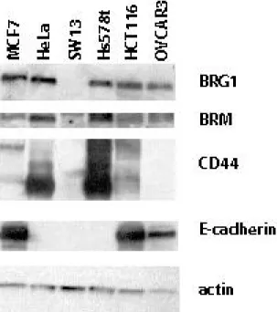
C. Characterization of BRG1 mutant cell lines
To determine whether point mutations in BRG1 affected its normal functions, we chose 3 human tumor cell lines with known mutations (Figure 1). These cell lines were chosen because the mutations are in three different domains of BRG1. The Hs578t mutation is in the proline rich domain I, near an area shown to be required for B-catenin signaling (22). The OVCAR3 mutation is hemizygous, and in the highly charged domain II, whose function remains unknown (13). The HCT116 mutation is in the ATPase domain, which is responsible for hydrolyzing ATP (13). Previously, our lab determined a functional copy of either BRG1 or BRM was enough for CD44 expression and RB-mediated arrest. We have also shown transfection of BRG1/BRM-deficient cells with either BRG1 or BRM
Pest domain
Proline rich domain I – amino acids 1-336
Highly charged domain II – amino acids 350-571
ATPase domain III – amino acids 728-1388
LXCXE RB-binding domain
Bromodomain domain IV – amino acids 1447-1523
1 1613 P196S L1149P E402G Hs578t NIH-OVCAR3 HCT-116 sensitive insensitive sensitive Rb Sensitive positive positive negative E-cadheirn Expression positive negative positive CD44 Expression
Figure 2.1: Location of BRG1 Mutations. The location of the three point mutations in human cancer cell lines Hs578t, OVCAR3, and HCT116 are
Interestingly, a western-blot screen of our BRG1 mutant cell lines shows OVCAR3 lacks CD44 expression and Hs578t lacks E-cadherin expression, even though they presumably contain a functional copy of BRM (Figure 2).
D. Mutant BRG1 proteins still form SWI/SNF complexes
Point mutations in proteins can affect their folding or their binding properties. We wanted to determine if the mutations in BRG1 in HCT116, Hs578t, and OVCAR3 inhibited formation of the SWI/SNF complex. To see if the mutant BRG1s still associated with other members in the complex, we performed immunoprecipitation of nuclear extracts with antibodies against BRG1 and BAF155. Immunoprecipitated protein was analyzed by western-blot analysis (Figure 3a-e). Protein from HeLa, our positive control for an intact SWI/SNF complex, precipitated with either anti-BAF 155 or anti-BRG1, contained other members of the complex including BRG1 or BAF155, and BAF47 (Figure 3b). Protein from SW13, our negative control that lacks BRG1 and BRM, protein, precipitated with BRG1 showed a small amount of BRG1 present by western blot (due to a small sub-population in the cell line) (23). Therefore, the other members of the complex, BAF155, and BAF47 were observed by immunoblotting (Figure 3a). In HCT116, the nuclear extract precipitated with anti-BAF155 showed less BRG1 but more BAF47 than extract precipitated with anti- BRG1 (Figure 3d). Similarly, HCT116 extract precipitated for BRG1 showed low levels of
BAF155 (Figure 3d). These results may indicate that the mutant BRG1 is not as efficient at forming a complex. This may also be true for Hs578t, and OVCAR3 since
immunoprecipitation with anti-BAF155 resulted in low levels of BRG1 while
Figure 2.2: Western-blot analysis of BRG1 mutant cell lines. Protein from BRG1 mutant cell lines was separated by SDS-PAGE and
than the BAF155 pull-down lane may indicate interference with wtBRG1 by the BRG1 mutant protein. These results demonstrate that the mutant BRG1 proteins in the HCT116, OVCAR3, and Hs578t cell lines might associate with other members of the complex, but probably in a less efficient manner.
E. Promoter Targeting
lines as in HeLa positive control cell. In contrast, the signal for BRG1 was reduced in the BRG1 deficient-SW13 cell line but was completely reduced either due to background from the assay or from background levels of BRG1 in SW13 due to a small subpopulation (23) (Figure 4). These data show that the BRG1 mutations in HCT116 and Hs578t do not prevent targeting of the complex to the CD44 and E-cadherin promoters.
F. Analysis of BRM-deficient HCT116 and Hs578t cell lines
Since the BRG1 mutant protein appeared to be in a complex with other members of the SWI/SNF complex and at the promoter of target genes, we wanted to determine if the
G. Rb sensitivity
Normally, cells transfected with p16INK4A or a constitutively active form of Rb, PSM-RB, undergo growth arrest (18). However, in previous studies, we and others have shown that cell lines without a functional BRM and BRG1 were insensitive to RB-mediated cell cycle arrest (18). Previously, Hs578t was found to be sensitive to RB-mediated arrest due to the presence of functional BRM. In a similar fashion, the p16-deficient HCT116 cell line undergoes cell-cycle arrest when transfected with PSM-RB. To determine if the BRG1 mutations affected RB sensitivity, we analyzed the RNAi stable clones of HCT116 and Hs578t for incorporation of Brdu during the S phase of the cell cycle. In parent cell lines, stable vector clones, and BRG1 RNAi stable clones, transfection of Hs578t with p16, and transfection of HCT116 cells with PSM-RB caused reduction of Brdu incorporation due to growth arrest of the cells (Figure 6). In contrast, the BRM RNAi stable clones became resistant to Rb-mediated growth arrest and continued to incorporate Brdu (Figure 6). These data indicate that the mutations in BRG1 abrogated its ability to regulate the cell cycle.
H. Expression of Target Genes
Since the BRG1 mutant proteins appeared to be in a complex with other members of the SWI/SNF complex and at the promoter of target genes, we assessed if they were still able to regulate gene expression. Western blotting showed that CD44 expression was
CD44 expression (Figure 7b). The BRG1 mutation in HCT116 may impair CD44 transcriptional regulation.
E-cadherin is another gene regulated by the SWI/SNF complex. Transfection of deficient cell lines with BRG1 or BRM upregulates expression of E-cadherin. The Hs578t cell line doesn’t express E-cadherin , possibly due to the mutation near the B-catenin binding domain of the BRG1 protein (22). We therefore determined levels of E-cadherin protein in the HCT116 BRM RNAi cell lines. Interestingly, E-cadherin levels were unaffected in the HCT116 BRM RNAi stable cell lines (Figure 7a). In addition to the genes known to be regulated by BRG1 (CD44 and E-cadherin), we tested other commonly methylated genes (FHIT, MGMT, vimentin, cytokeratin 18), since CD44 and E-cadherin are commonly methylated (6), to see if their expression was affected in the BRM RNAi cell lines. We observed no consistent effects on the other proteins expression levels in the BRM RNAi stable cell lines.
I. Discussion