Chapter 9
© McGraw-Hill Education.
9-2
Genetics and Genes
• Genetics – the study of heredity
• The science of genetics explores:
– Transmission of biological traits from parent to
offspring
– Expression and variation of those traits
© McGraw-Hill Education.
9-4
Microbial Genomes
Genome – sum total of genetic material (DNA)
in a cell
• Most exists as chromosomes
• Some appear in non-chromosomal sites:
– Mitochondria
– Chloroplasts – Plasmids
• Genome of cells – DNA
Chromosomes (1 of 2)
• Chromosome - discrete cellular structure
composed of a neatly packaged DNA molecule
– Eukaryotic chromosomes are located in the
nucleus and are multiple and linear
© McGraw-Hill Education.
9-6
Genotypes and Phenotypes (1 of 2)
• A chromosome is subdivided into genes, the fundamental unit of heredity responsible for a given trait
– Site on the chromosome that provides
information for a certain cell function
– Segment of DNA that contains the necessary code to make a protein or RNA molecule
• Three basic categories of genes:
© McGraw-Hill Education.
9-8
Genotypes and Phenotypes (2 of 2)
• All types of genes constitute the genetic makeup – genotype
Size and Packaging of Genomes
• Smallest virus – 4-5 genes
• E. coli – single chromosome containing 4,288
genes; 1 mm; 1,000X longer than cell
• Human cell – 46 chromosomes containing
© McGraw-Hill Education.
9-10
The Packaging of DNA (1 of 2)
• The DNA molecule is compacted in the cell by supercoils, or superhelices:
– In prokaryotes, by the action of the enzyme DNA gyrase, which coils the chromosome into a tight bundle by reversible series of twists
into the DNA molecule
© McGraw-Hill Education.
9-12
The Structure of DNA: Double
Helix (1 of 6)
• Basic unit of DNA structure is the nucleotide:
– A deoxyribose sugar
– A phosphate group
– A nitrogenous base: adenine (A), guanine (G), thymine (T), cytosine (C)
© McGraw-Hill Education.
9-14
The Structure of DNA: Double
Helix (3 of 6)
• Nitrogenous bases covalently bond to the 1′ carbon of each sugar and span the center of the molecule to pair with a complementary base on the other strand:
– Adenine (A) to thymine (T) with 2 hydrogen bonds
– Guanine (G) to cytosine (C) with 3 hydrogen
© McGraw-Hill Education.
9-16
The Structure of DNA: Double
Helix (5 of 6)
• Antiparallel arrangement: in one strand, the helix runs in a 5′ to 3′ direction and the other side is oriented from 3′ to 5′
• Each strand provides a template for the exact copying of a new strand
© McGraw-Hill Education.
9-18
The Significance of DNA Structure
1. Maintenance of code during reproduction
- Constancy of base pairing guarantees that the code will be retained. When strands are separated, each strand serves as a template for replication of the molecule into an exact copy.
2. Providing variety - order of bases
The Overall Replication Process
(1 of 2)
• Replication occurs on both strands simultaneously
• Semiconservative process:
1. The parent DNA molecule is uncoiled
2. The two strands are separated exposing the nucleotide sequence to serve as templates 3. Two new complementary strands are
© McGraw-Hill Education.
9-20
Events in DNA Replication (1 of 9)
• All chromosomes have a specific origin of
replication site as the place where
replication will be initiated
• The origin of replication is AT-rich, thus less energy is required to separate the two
strands
© McGraw-Hill Education.
9-22
Events in DNA Replication
© McGraw-Hill Education.
9-24
Events in DNA Replication (8 of 9)
• When replication forks meet, ligases link the DNA fragments along the lagging strand
• As replication proceeds, one newly synthesized strand loops down
• When the forks have gone full circle, a termination site shuts replication down
• The two circular daughter molecules remain connected briefly but are nicked and
© McGraw-Hill Education.
9-26
Enzymes Involved in DNA
Replication
TABLE 9.1 Some Enzymes Involved in DNA Replication and Their Functions
Enzyme Function
Helicase Unzipping the DNA helix Primase Synthesizing an RNA primer
DNA polymerase III Adding bases to new DNA chain; proofreading the chain for mistakes
DNA polymerase I Removing RNA primers, replacing gaps between Okazaki fragments with correct nucleotides, repairing mismatched bases
Applications of the DNA Code (1 of 2)
• Genetic information in DNA molecules is conveyed to RNA through the process of
transcription
• The information contained in the RNA
molecule is then used to produce proteins in the process of translation
© McGraw-Hill Education.
9-28
Gene-Protein Connection
• Each structural gene is an ordered sequence of nucleotides that codes for a protein’s
primary structure
• Groups of three consecutive bases, triplets
or codons, on one DNA strand are
transcribed into RNA sequence triplets
• Each triplet of nucleotides on the RNA specifies a particular amino acid
• A protein’s primary structure (chain of amino acids) determines its shape and function
RNAs: Major Participants in
Transcription and Translation
The general structure of the ribonucleic acid
(RNA) is different than that of the DNA molecule in several ways:
1. RNA is a single-stranded molecule that can assume secondary and tertiary levels of
complexity, leading to specialized forms of RNA (mRNA, tRNA, and rRNA)
© McGraw-Hill Education.
9-30
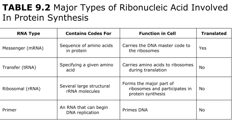
Major Types of RNA
TABLE 9.2 Major Types of Ribonucleic Acid Involved In Protein Synthesis
RNA Type Contains Codes For Function in Cell Translated
Messenger (mRNA) Sequence of amino acids in protein Carries the DNA master code to the ribosomes Yes
Transfer (tRNA) Specifying a given amino acid Carries amino acids to ribosomes during translation No
Ribosomal (rRNA) Several large structural rRNA molecules Forms the major part of ribosomes and participates in protein synthesis No
Messenger RNA (mRNA) (1 of 2)
• Transcribed version of a structural gene or genes in DNA
• Synthesized following complementary-base pairing by a process similar to synthesis of the leading strand during DNA replication
© McGraw-Hill Education.
9-32
Messenger RNA (mRNA) (2 of 2)
a) Messenger RNA (mRNA)
A short piece of messenger RNA (mRNA)
Transfer RNA: tRNA (1 of 3)
• Acts as a translator of the mRNA code into protein
• 75 - 95 nucleotides in length bent into
hairpin loops to form a cloverleaf structure further packed into a complex helix
• Bottom loop of the cloverleaf exposes the
tRNA specific anticodon complementary to a mRNA codon
© McGraw-Hill Education.
9-34
Transfer RNA: tRNA (2 of 3)
The figure of “Transfer RNA.”
b) Transfer RNA (tRNA)
Left: The tRNA strand loops back on itself to form intrachain hydrogen bonds. The result is a
cloverleaf structure, shown here in simplified
Transfer RNA: tRNA (3 of 3)
© McGraw-Hill Education.
9-36
Ribosomal RNA: rRNA
• The prokaryotic (70S) ribosome is a particle composed of tightly packaged ribosomal RNA (rRNA) and protein
• Forms complex three-dimensional figures that
Major Events in Transcription (3 of 3)
© McGraw-Hill Education.
9-38
Translation: The Second Stage of
Gene Expression
• All the elements needed to synthesize protein (mRNA, tRNA, amino acids) are brought together on the
ribosomes
• The process occurs in five stages: initiation,
The Master Genetic Code (1 of 2)
• Represented by mRNA codons and their specific amino acids
© McGraw-Hill Education.
9-40
The Master Genetic Code (2 of 2)
*This codon initiates translation.
Interpreting the DNA Code
• Transcription produces mRNA complementary to the DNA gene
• During translation, tRNAs use their anticodon to
© McGraw-Hill Education.
9-42
Translation
• Ribosomes assemble on the 5′ end of an mRNA transcript
• Ribosome scans the mRNA until it reaches the start codon, usually AUG
• A tRNA molecule with the complementary
Translation Termination
• Termination codons – UAA, UAG, and UGA –
are codons for which there is no corresponding tRNA
• When this codon is reached, the ribosome falls off and the last tRNA is removed from the
© McGraw-Hill Education.
9-44
Polyribosomal Complex
Eukaryotic Transcription and
Translation
1. Do not occur simultaneously – transcription
occurs in the nucleus and translation occurs in the cytoplasm
2. Eukaryotic start codon is AUG, but it does not use formyl-methionine
3. Eukaryotic mRNA encodes a single protein, unlike bacterial mRNA which encodes many
4. Eukaryotic DNA contains introns– intervening
© McGraw-Hill Education.
9-46
Splicing of Eukaryotic pre-mRNA
(1 of 2)
• Eukaryotes gene coding sequences, or
exons, are interrupted by segments called
introns
• Introns are transcribed but not translated, they are removed before translation
© McGraw-Hill Education.
9-48
Regulation of Protein Synthesis and
Metabolism
• Genes are regulated to be active only when their products are required
• In prokaryotes this regulation is coordinated
by operons, a set of genes, all of which are
Mutations: Changes in the Genetic
Code
• A change in phenotype due to a change in
genotype (nitrogen base sequence of DNA) is called a mutation
• A natural, nonmutated characteristic is known as a wild type (wild strain)
• An organism that has a mutation is a mutant
strain, showing variance in morphology,
© McGraw-Hill Education.
9-50
Causes of Mutations (1 of 2)
• Spontaneous mutations – random change
in the DNA due to errors in replication that occur without known cause
• Induced mutations – result from exposure
to known mutagens, physical (primarily
Causes of Mutations (2 of 2)
TABLE 9.3 Selected Mutagenic Agents and Their Effects
Agent Effect
Chemical
Nitrous acid, bisulfite Remove an amino group from some nitrogen bases Ethidium bromide Inserts between the paired bases
Acridine dyes Cause frameshifts due to insertion between base pairs
Nitrogen base analogs Compete with natural bases for sites on replicating DNA
© McGraw-Hill Education.
9-52
Categories of Mutations
• Point mutation – addition, deletion, or substitution of a few bases
• Missense mutation – causes change in a single
amino acid
• Nonsense mutation – changes a normal codon into a stop codon
• Silent mutation – alters a base but does not change the amino acid
• Back-mutation – when a mutated gene
reverses to its original base composition
Effect of Major Types of Mutations
(1 of 5)
TABLE 9.4 Classification of Major Types of
Mutations.
I. Wild-type (nonmutated) sequence
Example: THE BIG BAD CAT ATE THE FAT RED BUD
The wild-type sequence of a gene is the
DNA sequence found in most organisms and is generally considered the “normal”
© McGraw-Hill Education.
9-54
Effect of Major Types of Mutations
(2 of 5)
The “TABLE 9.4” continues on this slide.
II. Categories of mutations based on type of DNA alteration
A. Substitution mutations
1. Missense:
Example: THE BIG MAD CAT ATE THE FAT RED BUG
A missense mutation causes a different amino acid to be incorporated into a
Effect of Major Types of Mutations
(3 of 5)
The “TABLE 9.4” continues on this slide.
2. Nonsense
Example: THE BIG BAD XXX (stop)
A nonsense mutation converts a codon to a stop codon, resulting in premature
© McGraw-Hill Education.
9-56
Effect of Major Types of Mutations
(4 of 5)
The “TABLE 9.4” continues on this slide.
B. Inversion mutations Example:
THE BIG ABD CAT ATE THE FAT RED BUG THE BIB GAD CAT ATE THE FAT RED BUG Inversion arise when adjacent letters
Effect of Major Types of Mutations
(5 of 5)
The “TABLE 9.4” continues on this slide.
C. Frameshift Mutations
1. Insertion: Example: THE BIG BAB DCA TAT ETH EFA TRE DBU G
2. Deletion: Example: THE BIG * ADC ATA TET HEF ATR EDB UG
Insertion (addition of letter) and deletion (removal of letter) mutations cause a
© McGraw-Hill Education.
9-58
Repair of Mutations
• Since mutations can be potentially fatal, the cell has several enzymatic repair mechanisms in place to find and repair damaged DNA
– DNA polymerase – proofreads nucleotides
during DNA replication
– Mismatch repair – locates and repairs
mismatched nitrogen bases that were not repaired by DNA polymerase
– Light repair – for UV light damage
The Ames Test (1 of 2)
• Any chemical capable of mutating bacterial DNA can similarly mutate mammalian DNA
• Agricultural, industrial, and medicinal
compounds are screened using the Ames test
• Indicator organism is a mutant strain of Salmonella typhimurium that has lost the ability to synthesize histidine
back-© McGraw-Hill Education.
9-60
The Ames Test (2 of 2)
Conjugation (1 of 5)
Transfer of a plasmid or chromosomal fragment from a donor cell to a recipient cell via direct
contact
• Gram-negative cell donor has a fertility
plasmid (F factor) that allows the synthesis of a conjugative pilus
• Donor (F+ cell) transfers fertility plasmid through pilus to recipient (F- cell), which becomes F+ cell
© McGraw-Hill Education.
9-62
Conjugation (2 of 5)
Physical Conjugation
(1) The pilus of donor cell
Conjugation (3 of 5)
• High-frequency recombination – donor’s fertility plasmid is integrated into the
bacterial chromosome
© McGraw-Hill Education.
9-64
Conjugation (4 of 5)
F Factor Transfer