ARTIGO ORIGINAL
Subtelomeric Rearrangements: Presentation of
21 Probands with Emphasis on Familial Cases
Rearranjos Subteloméricos: Apresentação de
21 Probandos, com Ênfase nos Casos Familiares
Ana Rita SOARES1, Gabriela SOARES1, Manuela MOTA-FREITAS2,3, Natália OLIVA-TELES2,3,
Ana Maria FORTUNA1,2
Acta Med Port 2019 Jul–Aug;32(7–8):529–535 ▪ https://doi.org/10.20344/amp.11466
1. Unidade de Genética Médica. Centro de Genética Médica Jacinto Magalhães. Centro Hospitalar Universitário do Porto. Porto. Portugal. 2. Unidade de Citogenética. Centro de Genética Médica Jacinto Magalhães. Centro Hospitalar Universitário do Porto. Porto. Portugal. 3. Unidade Multidisciplinar de Investigação Biomédica. Instituto de Ciências Biomédicas Abel Salazar. Universidade do Porto. Porto. Portugal. Autor correspondente: Ana Rita Soares. [email protected]
Recebido: 22 de outubro de 2018 – Aceite: 08 de março de 2019 | Copyright © Ordem dos Médicos 2019 ABSTRACT
Introduction: Intellectual disability affects 2% – 3% of the general population, with a chromosomal abnormality being found in 4% – 28% of these patients and a cryptic subtelomeric abnormality in 3% – 16%. In most cases, these subtelomeric rearrangements are submicro-scopic, requiring techniques other than conventional karyotype for detection. They may be de novo or inherited from an affected parent or from a healthy carrier of a balanced chromosomal abnormality. The aim of this study was to characterize patients from our medical genetics center, in whom both a deletion and duplication in subtelomeric regions were found.
Material and Methods: Clinical and cytogenetic characterization of 21 probands followed at our center, from 1998 until 2017, with subtelomeric rearrangements.
Results: There were 21 probands from 19 families presenting with intellectual disability and facial dysmorphisms. Seven had behavior changes, five had epilepsy and 14 presented with some other sign or symptom. Four had chromosomal abnormalities detected by conventional karyotype and four were diagnosed by array-comparative genomic hybridization. In four cases, parental studies were not possible. The online mendelian inheritance in man classification was provided whenever any of the phenotypes (deletion or duplication syndrome) was dominant.
Discussion: Patients and relevant family members were clinically and cytogenetically characterized. Although rare, subtelomeric changes are a substantial cause of syndromic intellectual disability with important familial repercussions. It is essential to remember that a normal array-comparative genomic hybridization result does not exclude a balanced rearrangement in the parents.
Conclusion: Parental genetic studies are essential not only for a complete characterization of the rearrangement, but also for accurate genetic counselling and screening of family members at risk for recurrence.
Keywords: Intellectual Disability/genetics; Subtelomeric Rearrangements Gene Rearrangement/genetics; Telomere/genetics RESUMO
Introdução: O défice intelectual afeta 2% – 3% da população geral, sendo encontrada uma alteração cromossómica em 4% – 28% dos casos e uma alteração subtelomérica em 3% – 16%. Estas alterações subteloméricas são, na maioria dos casos, submicroscópi-cas, não sendo detetadas no cariótipo convencional. Podem ser de novo ou herdadas de um progenitor afetado ou de um progenitor saudável portador de um rearranjo equilibrado. O objetivo deste estudo foi caracterizar os doentes seguidos no nosso centro de gené-tica médica com uma deleção e uma duplicação nas regiões subteloméricas.
Material e Métodos: Caracterização clínica e citogenética de 21 probandos com alterações subteloméricas seguidos no nosso centro entre 1998 e 2017.
Resultados: Foram caracterizados 21 probandos que apresentavam défice intelectual e dismorfia facial, pertencentes a 19 famílias. Sete tinham alterações do comportamento, cinco epilepsia e 14 outro sinal ou sintoma. Quatro tinham alterações no cariótipo e quatro foram diagnosticados por array-comparative genomic hybridization. Em quatro famílias não foi possível o estudo dos progenitores. Quando um dos fenótipos era dominante (síndrome de deleção ou duplicação), foi atribuída a classificação online mendelian inheri-tance in man.
Discussão: Foi realizada classificação dos doentes e das famílias. As alterações nas regiões subteloméricas são, apesar de raras, uma causa substancial para défice intelectual sindrómico com repercussões familiares importantes. É essencial lembrar que um array-comparative genomic hybridization normal não exclui um rearranjo equilibrado familiar.
Conclusão: O estudo dos progenitores é essencial não só para caracterização completa do rearranjo mas também para um aconse-lhamento genético preciso e identificação de familiares em risco de recorrência.
Palavras-chave: Deficiência Intelectual/genética; Rearranjo Génico/genética; Telómero/genética
INTRODUCTION
Intellectual disability, previously known as mental retar-dation, affects 2% – 3% of the general population.1–3 The Diagnostic and Statistical Manual of Mental Disorders (DSM)-5 defines intellectual disability as a disorder with
ARTIGO ORIGINAL and includes both genetic and environmental causes, 2 and
sometimes it is multifactorial. Most patients, particularly those with moderate/severe ID and with dysmorphic fea-tures/congenital abnormalities, have a genetic aetiology, which can be a chromosomal imbalance or a single gene pathogenic variant. A chromosomal abnormality is found in 4% – 28% of patients with ID.2,5 Subtelomeric regions are gene-rich chromosomal regions with a highly repetitive structure, frequently involved in chromosomal rearrange-ments.6,7 Cryptic subtelomeric abnormalities, syndromic or isolated, are well known causes of ID, and are responsible for 3% – 16% of diagnoses.
These subtelomeric rearrangements are predominant-ly submicroscopic [i.e. smaller than 5 megabases (Mb)], requiring techniques other than conventional karyotype for detection. Karyotype with fluorescent in situ hybridization (FISH) was the first diagnostic approach for these subtel-omeric abnormalities. Multiplex ligation-dependent probe amplification (MLPA), a semi-quantitative methodology, allowed a better characterization of duplications or dele-tions. Currently, array-comparative genomic hybridization (aCGH) is used as the first tier-test for these patients.8,9 All the above techniques have been used as routine tests for identifying these patients.2,3,8,10
An accurate family history may show important clues for a possible inherited rearrangement, mainly more than one family member affected or history of recurrent pregnancy losses. In fact, these subtelomeric abnormalities may be de
novo or inherited. An unbalanced rearrangement may be
inherited from an affected parent or from a healthy carrier of a balanced chromosomal abnormality. Parental genetic studies are thus essential not only for a complete character-ization of the rearrangement (de novo or inherited) but also, and more importantly, for accurate genetic counselling and screening of family members at risk for recurrence.
Following the identification of a patient (case 1) in whom aCGH showed the presence of a deletion and a duplication in subtelomeric regions, and complementary studies in par-ents confirmed that the mother was a carrier of a balanced subtelomeric cryptic translocation, we decided to review and characterize the patients from our medical genetics center, from clinical and cytogenetic perspectives, in whom both deletion and duplication on subtelomeric regions were found. With this work, our main objective was to raise awareness for these rare genetic syndromes as well as to the importance of familial studies.
MATERIAL AND METHODS
Our study included 21 probands followed at our medical genetics center, from 1998 until 2017, who have simultane-ously a deletion and a duplication in chromosomal subtel-omeric regions. We describe gender, age at first consulta-tion, somatometry, craniofacial and other dysmorphisms or congenital abnormalities, family history, as well as cytoge-netic findings in patients and parents.
Written informed consent was obtained for all published photos.
RESULTS
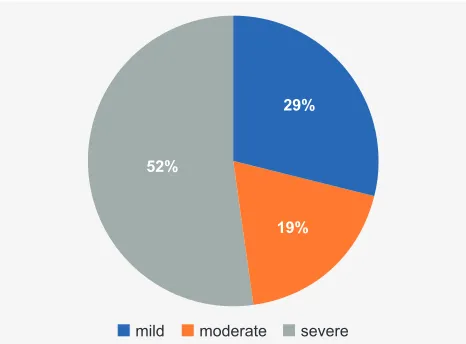
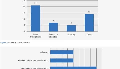
Results are summarized in Table 1, Table 2 and Figs. 1 to 3. Twenty one probands (eight males and 13 females) from 19 families presented with developmental delay/intel-lectual disability (11 cases with severe, four with moderate and six with mild ID) and facial dysmorphisms (Table 1A and Fig. 4). Seven had behaviour changes, five had epi-lepsy and 14 presented with some other sign or symptom. Cases 4 to 6 represent a brother, a sister and a mater-nal cousin from the same family. Four cases presented with visible G-bands by trypsin using Leishman (GTL) banding chromosomal abnormalities: three of them with subtelomeric abnormalities [case 18: 46,XY,der(6)t(6;9) (q27;p12); case 19: 46,XX,der(8)t(4;8)(p16,1;p23,1); case 21: 46,XX,der(3)t(3;11)(p26.2;p15.4)pat], and case 15 had a balanced and non-pathogenic robertsonian translocation [45,XX,rob(13;14)(q10;q10)], in addition to the subtelomeric submicroscopic abnormalities. Four cases were diagnosed by aCGH and the others by MLPA/FISH. In four cases, the parental clinical and cytogenetic studies were not possi-ble. Of the cases in which family studies were possible, six were de novo events, seven were inherited from a balanced cryptic rearrangement and three probands inherited the abnormality from a similarly affected parent. In case 21 the translocation was shown to have been inherited from the paternal grandfather. Most cases presented with phenotypic characteristics of both deletion and duplication syndromes involved in the rearrangement. Therefore, the OMIM classi-fication was provided whenever any of the phenotypes was dominant (Tables 1A and 1B).
DISCUSSION
Subtelomeric regions are gene-rich chromosomal regions responsible for a significant number of patients with syndromic ID. At Centro Hospitalar Universitário do Porto, 21 cases with a subtelomeric chromosomal rearrangement have been diagnosed and characterized, and accurate genetic counselling was offered.
Case 1 presented with a phenotype more compatible with chromosome 16p13.3 duplication syndrome. This
19% 29%
52%
mild moderate severe
ARTIGO ORIGINAL
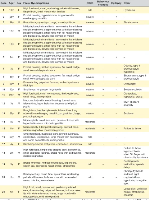
Table 1A – Clinical and cytogenetic characterization of our patients’ cohort
Case Age* Sex Facial Dysmorphisms DD/ID Behaviour changes Epilepsy Other
1 13m F High forehead, small, upslanting palpebral fissures, flat philtrum, small mouth with thin lips mild – – Hypotonia
2 25y F Frontal bossing, hypertelorism, long nose with overhanging nasal tip severe – + –
3 29y M Round face, synophrys, large, smooth philtrum severe + + Short stature
4 12m M
Mild plagiocephaly and facial asymmetry, flat midface, straight eyebrows, deeply set eyes with downslanting palpebral fissures, small nose with flat nasal bridge and bulbous tip, downturned corners of mouth
severe + + –
5 11m F
Mild plagiocephaly and facial asymmetry, flat midface, straight eyebrows, deeply set eyes with downslanting palpebral fissures, small nose with flat nasal bridge and bulbous tip, downturned corners of mouth
severe + + –
6 7m F
Mild plagiocephaly and facial asymmetry, flat midface, straight eyebrows, deeply set eyes with downslanting palpebral fissures, small nose with flat nasal bridge and bulbous tip, downturned corners of mouth
severe + + –
7 3y F Frontal bossing, arched eyebrows, flat nasal bridge, small low-set dysplastic ears severe – – Obesity, type 4 brachydactyly, hypotonia
8 10y F Frontal bossing, arched eyebrows, flat nasal bridge, small low-set dysplastic ears severe + – Short stature, type 4 brachydactyly
9 39y F Downslanting palpebral fissures, arched eyebrows, thick lips severe – – Overweight
10 12y F Small eyes, long nose, large teeth severe + – Severe scoliosis
11 22m M High forehead, small low-set ears, thick eyebrows, small nose, micrognathia severe – – Cleft palate, hypotonia, ataxia
12 3y M Macrocephaly with frontal bossing, low-set ears, telecanthus, hyperthelorism, decentered elliptical
pupils mild – –
MVP, Rieger’s anomaly
13 39y F Rough face, blepharophimosis, telecanthus, long nose with overhanging nasal tip, prognathism, large,
protruding tongue severe – – Scoliosis
14 8y M Microcephaly, small forehead, prominent nose with hypoplastic nares, microretrognathia moderate – – –
15 11y F Microcephaly, bitemporal narrowing, pointed nose, microretrognathia, mentonian groove moderate + – Failure to thrive
16 23y M Small forehead, dysplastic ears, arched eyebrows, epicanthus, telecanthus, large mouth with microdontia
and some conical teeth, micrognathia mild – – –
17 4y F Blepharophimosis, left ptosis, epicanthus, strabismus mild – – –
18 3m M High forehead, simple cup-shaped ears, epicanthus, small palpebral fissures, broad nose with bulbous tip,
microretrognathia moderate – –
Failure to thrive, kyphoscoliosis, short 5th finger with clinodactily, hypotonia
19 3y F Broad forehead, midface hypoplasia, big cheeks, spoon ear, depressed nasal bridge, strabismus mild – – Foetal growth restriction, speech delay
20 11m M Brachycephaly, round face, epicanthus, upslanting palpebral fissures, bulbous nose with anteverted
nares, thin lips, low-set ears mild – –
Short puffy hands and feet, right cryptorchidism, hypotonia, mongolian spot
21 1m F
High front, small, low-set and posteriorly rotated ears, downslanting palpebral fissures, bulbous nose tip with wide anteverted nares, large mouth with macroglossia, mild micrognathia
moderate – – Loose skin, umbilical hernia, strabismus, scoliosis
ARTIGO ORIGINAL
Table 1B – Prevalence of children from 0 to 9 years old exposed to SHS at home according to parents’ tobacco consumption, by region
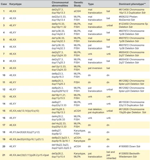
Case Karyotype Chromosomal abnormalities Genetic Study Type Dominant phenotype**
1 46,XX del2q37.3,dup16p13.3 aCGH mat translocation bal #613458 Chromosome 16p13.3 Duplication Sdr
2 46,XX del22q13.33,dup19q13.4 MLPA, FISH mat translocation bal #606232 Phelan-McDermid Sdr
3 46,XY del5p15.33,dup18p11.32 MLPA, FISH mat translocation unbal #123450 Chromosome 5p Deletion Sdr
4 46,XY del1p36.33,dup14q32.3 MLPA, FISH mat translocation bal #607872 Chromosome 1p36 Deletion Sdr
5 46,XX del1p36.33,dup14q32.3 MLPA, FISH mat translocation bal #607872 Chromosome 1p36 Deletion Sdr
6 46,XX del1p36.33,dup14q32.3 MLPA, FISH pat translocation bal #607872 Chromosome 1p36 Deletion Sdr
7 46,XX del2q37.3,dup12q24.33 MLPA, FISH unk unk #600430 Chromosome 2q37 Deletion Sdr
8 46,XX del2q37.3,dup17q25.3 MLPA, FISH mat translocation bal #600430 Chromosome 2q37 Deletion Sdr
9 46,XX del12p13.33,dup12q24.33 MLPA, FISH pat inversion bal
10 46,XX del8p23.3,dup4p16.3 MLPA, FISH dn dn
11 46,XY del6p25.3,dup17q25.3 FISH dn dn #612582 Chromosome 6pter-p24 Deletion Sdr
12 46,XY del6p25.3,dupXq28/Yq12 MLPA, FISH pat translocation unbal #612582 Chromosome 6pter-p24 Deletion Sdr
13 46,XX del18p11.21,dup11p15.5 MLPA, FISH unk unk
14 46,XY del6q27,dup22q13.33 MLPA, FISH unk unk #615538 Chromosome 22q13 Duplication Sdr
15 45,XX,rob(13;14)(q10;q10) del15q26.3,dup19p13.3 aCGH mat deletion,pat duplication unbal #612626 Chromosome 15q26-qter Deletion Sdr
16 46,XY del4q35.2,dup1p36.33 MLPA, FISH unk unk
17 46,XX del14q32.33,dup20q13.33 MLPA, FISH dn dn
18 46,XY,der(6)t(6;9)(q27;p12) del6q27,dup9p12 Karyotype, FISH dn dn
19 46,XX,der(8)(4;8)(p16,1;p23,1) del8p23.3p23.1,dup4p16.3p16.1 aCGH, Karyotype dn dn
20 46,XY del18q22.3q23,dup21q22.2q22.3 aCGH dn dn #190685 Down Sdr
21 46,XX,der(3)t(3;11)(p26.2;p15.4)pat del3p26.2, dup11p15.4 Karyotype, MLPA pat translocation bal and pat inherited
#130650 Beckwith-Wiedemann Sdr
*age at first consultation; **OMIM dominant phenotoype when it was possible to characterize; y: years-old; m: months-old; M/F: male/ female; DD/ID: developmental delay/intellectual disability; mat/pat: maternal/paternal; dn: de novo; unk: unknown; del/dup: deletion/dupli-cation; (un)bal: (un)balanced; Sdr: syndrome.
syndrome is characterized by ID, facial dysmorphisms (high forehead, sparse eyebrows, blepharophimosis with palpebral ptosis, short nose, everted upper lip, high-arched palate and cupped ears), pre and postnatal growth defi-ciency, clef palate, congenital heart defects and urogenital abnormalities.11
Case 2 presented mainly with a phenotype consistent with Phelan-McDermid syndrome, characterized by neo-natal hypotonia, global developmental delay, moderate to severe ID, absent or severely impaired speech, normal to
accelerated growth, large fleshy hands, dysplastic toenails, decreased sudoresis with tendency to overheat and behav-iour changes (chewing, decreased perception of pain, autis-tic-like features).12
ARTIGO ORIGINAL
Figura 3 – Cytogenetic characteristics
Figure 2 – Clinical characteristics
micrognathia, hypertelorism and low-set ears), cleft lip and palate, congenital heart disease, kidney, genital and skel-etal abnormalities. However, different sized duplications have shown variable phenotypic severity and inconsistent phenotype even in affected members of the same family.13
Case 3 presented with severe developmental delay, behaviour changes, epilepsy, short stature and facial dys-morphisms (round face, micrognathia, epicanthic folds, hypertelorism). This phenotype is mainly caused by the 5p15.33 deletion, compatible with chromosome 5p deletion syndrome.14
Cases 4, 5 and 6 are three affected individuals (a boy and two girls) within the same family. They all presented with a phenotype characteristic of chromosome 1p36 dele-tion syndrome: this is a well known syndrome comprising a characteristic facial appearance (microcephaly, brachy-cephaly, prominent forehead, midface hypoplasia, deep-set eyes with straight eyebrows, thick ear helices, flat nose and nasal bridge and pointed chin), hypotonia, developmental delay, growth retardation, seizures, hearing impairment, visual problems, cardiovascular and limb abnormalities.15,16
Both cases 7 and 8 presented with some phenotypic features of chromosome 2q37 deletion syndrome, which is characterized by facial dysmorphisms (prominent forehead, round face, midface hypoplasia, sparse arched eyebrows,
deep-set eyes, depressed nasal bridge, thin upper lip and dysmorphic ears), short stature, obesity, brachydacty-ly type E, mild to moderate ID and behavioural problems. Other major malformations may occur, including congenital heart disease, central nervous system malformations, and gastrointestinal or genitourinary abnormalities.17,18
Vaglio et al have described a case of a boy present-ing with partial monosomy 12p and trisomy 12q and com-pared him with other cases described in the literature. As in our case 9, patients with this chromosomal abnor-mality show clinical features that are characteristic of both deletion and duplication syndromes. It was not possible to detect predominance of either. All of these cases occurred as a consequence of a balanced inversion present in a healthy parent.19
The chromosomal study of cases 10 and 19 revealed in both cases a deletion in 8p23.3 and a duplication in 4p16.3.
Epilepsy Other
Facial
dymorphisms Behaviouralteration 0
5 10 15 20
21
7
5
14 25
7 8 9
6 5 4 3 2 1 0 unknown
inherited unbalanced translocation
inherited balanced translocation
de novo
Table 2 – Demographic data
< 2y 2 – 18y > 18y
M 4 2 2
F 4 6 3
Total 8 8 5
ARTIGO ORIGINAL
In fact, the translocation between the subtelomeric regions of chromosomes 4 and 8 is considered the second most common in humans. The 8p23 duplication has been asso-ciated with ID, autism and psychiatric manifestations, as well as dysmorphisms (such as high forehead, epicanthic folds, hypertelorism, small eyes, strabismus, long philtrum, micrognathia, low-set malformed ears) and cardiac or renal malformations. The 4p16.3 duplication causes mild devel-opmental delay/ID, speech delay, growth delay and mild dysmorphic features (hypertelorism, epicanthic folds and abnormal ears). Both cases have phenotypic manifesta-tions that are very different, and it is hard to categorize their characteristics into a specific syndrome. Patient 19 seems to fit better into 4p16.3 duplication characteristics.20–22
Both cases 11 and 12 presented with chromosome 6pter-p24 deletion syndrome, characterized by ID, oph-thalmologic abnormalities, craniofacial dysmorphisms (macrocephaly, prominent forehead, down-slanting palpe-bral fissures, hypertelorism and depressed nasal bridge), Dandy-Walker malformation, congenital heart defects, hypotonia, hearing loss, and others, with high phenotypic variability.23,24 The father of case 12 presented the same phenotype as his son.
Case 15 was more compatible with chromosome 15q26-qter deletion syndrome, characterized by pre and postnatal growth retardation, variable ID, mild non-specific facial dys-morphisms and other congenital abnormalities.25
Case 18’s manifestations seem to be caused mainly by 9p duplication as patients with this syndrome present with
peculiar facial dysmorphisms and digital abnormalities, as well as variable developmental delay/ID, cardiac and skel-etal abnormalities.27
Case 20 could be misclassified as classic Down syn-drome, since failure to thrive and other malformations (such as cardiac abnormalities) were missing. The 21q22 duplica-tion was the dominant phenotype.28
Case 21 was clinically diagnosed as Beckwith-Wiedemann syndrome. This syndrome is mainly charac-terized by macrosomia, macroglossia, hemihyperplasia, omphalocele, visceromegaly, embryonal tumours and neo-natal hypoglicemia.29
Cases 13, 16 and 17 presented with characteristics from both deletion and duplication and it was not possible to detect predominance of either (Table 1).
Focusing on genetic screening and counselling of the family, it is important to remember the examples of cases 4, 5 and 6 and case 21. The first family is a good illustration of how balanced translocations may originate unbalanced gametes and affect different members of the same fami-ly. Thus, it is essential to study those at risk. The second example shows an apparently innocuous family history but in whom the translocation is present and transmitted throughout three generations.
In the aCGH era, it is tempting to perform only this tech-nique in both children and parents to confirm a de novo occur-rence; however, a normal aCGH result does not exclude a balanced translocation in parents, leaving the family at risk for chromosomal imbalances in future pregnancies of
ARTIGO ORIGINAL
healthy carriers. It is essential to study parents, by perform-ing karyotype and FISH techniques when a deletion or dupli-cation is found in a subtelomeric region, particularly when both a deletion and duplication are found in the proband, because the probability that a balanced rearrangement is present in one of the parents is very high in this situation.
CONCLUSION
This study shows that only after complete genetic char-acterization is it possible to identify other relatives at risk and offer accurate genetic counselling and reproductive options, particularly invasive prenatal testing (either by cho-rionic villous sampling or amniocentesis) or preimplantation genetic diagnosis.
The authors wish to emphasize that, although rare, these subtelomeric changes constitute a substantial cause of syndromic ID with important familial repercussions. It is essential to be aware of these cases and refer them to a Medical Genetics consultation, so that familial studies and management can be carried out accurately.
PROTECTION OF HUMANS AND ANIMALS
The authors declare that the procedures were followed according to the regulations established by the Clinical Research and Ethics Committee and to the Helsinki Declaration of the World Medical Association.
DATA CONFIDENTIALITY
The authors declare having followed the protocols in use at their working center regarding patients’ data publica-tion. Patient consent obtained.
CONFLICTS OF INTEREST
All authors report no conflict of interest.
FUNDING SOURCES
This research received no specific grant from any funding agency in the public, commercial, or not-for-profit sectors.
REFERENCES
1. Miller DT, Adam MP, Aradhya S, LG Biesecker, AR Brothman, NP Carter, et al. Consensus statement: chromosomal microarray is a first-tier clinical diagnostic test for individuals with developmental disabilities or congenital anomalies. Am J Hum Genet. 2010;86,749–64.
2. Freitas MM, Silva ML, Ribeiro J, Oliva-Teles N, Candeias C, Bronze-da-Rocha E. Análise das regiões subteloméricas em 1180 doentes com atraso mental por FISH e MLPA. Arq Med. 2013;27:10–4.
3. de Vries BB, White SM, Regan R, Homfray T, Young ID, Super M, et al. Clinical studies on submicroscopic subtelomeric rearrangements: a checklist. J Med Genet. 2001;38:145–50.
4. Manning M, Hudgins L. Use of array-based technology in the practice of medical genetics. Genet Med. 2007;9:650–3.
5. Diagnostic and Statistical Manual of Mental Disorders (DSM–5). Washington: American Psychiatric Publishing, Inc; 2013.
6. Esparza-Garrido RR, Velázquez-Wong AC, Araujo-Solís MA, Huicochea-Montiel JC, Velázquez-Flores MÁ, Salamanca-Gómez F, et al. Duplication of the Miller-Dicker critical region in a patient with a subtelomeric unbalanced translocation t(10;17)(p15.3;p13.3). Mol Syndromol. 2012;3:82–8.
7. Knight SJ, Flint J. Perfect endings: a review of subtelomeric probes and their use in clinical diagnosis. J Med Genet. 2000;37:401–9.
8. Manning M, Hudgins L, Professional Practice and Guidelines Committee. Array-based technology and recommendations for utilization in medical genetics practice for detection of chromosomal abnormalities. Genet Med. 2010;12:742–5.
9. Darilek S, Ward P, Pursley A, Plunkett K, Furman P, Magoulas P, et al. Pre- and postnatal genetics testing by array-comparative genomic hybridization: genetic counselling perspectives. Genet Med. 2008;10:13–8.
10. Ravnan JB, Tepperberg JH, Papenhausen P, Lamb AN, Hedrick J, Eash D, et al. Subtelomere FISH analysis of 11 688 cases: an evaluation of the frequency and pattern of subtelomere rearrangements in individuals with developmental disabilities. J Med Genet. 2006;43:478–89. 11. Digilio MG, Bernardini L, Capalbo A, Capolino R, Gagliardi MG,
Marino B, et al. 16p subtelomeric duplication: a clinically recognizable syndrome. Eur J Hum Genet. 2009;17:1135–40.
12. Phelan K, McDermid HE. The 22q13.3 deletion syndrome (Phelan-McDermid syndrome). Mol Syndromol. 2012;2:186–201.
13. Samanich J, Montagna C, Morrow BE, Babcock M. Interstitial duplication of 22q13.2 in a girl with short stature, impaired speech and language, and dysmorphism. J Pediatr Genet. 2012;1:47–53.
14. Mainardi PC, Perfumo C, Calì A, Coucourde G, Pastore G, Cavani S, et al. Clinical and molecular characterisation of 80 patients with 5p deletion: genotype-phenotype correlation. J Med Genet. 2001;38:151–8. 15. Rudnik-Schoneborn S, Zerres K, HAusler M, Lott A, Krings T, Schuler
HM. A new case of proximal monosomy 1p36, extending the phenotype. Research letter. Am J Med Genet A. 2008;146A:2018–22.
16. Watanabe M, Hayabuchi Y, Ono A, Naruto T, Horikawa H, Kohmoto T, et al. Detection of 1p36 deletion by clinical exome-first diagnostic approach. Hum Genome Var. 2016;3:16006
17. Cho EK, Kim J, Yang A, Cho SY, Jin DK. 2q37 Deletion syndrome confirmed by high-resolution cytogenetic analysis. Ann Pediatr Endocrinol Metab. 2017;22:129–32.
18. Mehraein Y, Pfob M, Steinlein O, Aichinger E, Eggert M, Bubendorff V, et al. 2q37.3 deletion syndrome: two cases with highly distinctive facial phenotype, discordant association with schizophrenic psychosis, and shared deletion breakpoint region on 2q37.3. Cytogenet Genome Res. 2015;146:33–8.
19. Vaglio A, Milunsky A, Huang XL, Quadrelli A, Mechoso B, Quadrelli R. A fourteen years follow-up of a case of partial trisomy 12q and monosomy 12p recombinants of a familial pericentric inversion of chromosome 12: clinical, cytogenetic and molecular observations. Eur J Med Genet. 2007;50:224–32.
20. Sagar A, Pinto D, Najjar F, Guter SJ, Macmillan C, Cook EH. De novo unbalanced translocation (4p duplication/8p deletion) in a patient with autism, OCD, and overgrowth syndrome. Am J Med Genet A. 2017;173:1656–62.
21. Skrlec I, Wagner J, Pubeljić S, Heffer M, Stipoljev F. De novo case of a partial trisomy 4p and a partial monosomy 8p. Coll Antropol. 2014;38:319–23.
22. Carmany EP, Bawle EV. Microduplication of 4p16.3 due to an unbalanced translocation resulting in a mild phenotype. Am J Med Genet A. 2011;155A:819–24.
23. Lin RJ, Cherry AM, Chen KC, Lyons M, Hoyme HE, Hudgins L. Terminal deletion of 6p results in a recognizable phenotype. Am J Med Genet A. 2005;136:162–8.
24. Descipio C, Schneider L, Young TL, Wasserman N, Yaeger D, Lu F, et al. Subtelomeric deletions of chromosome 6p: molecular and cytogenetic characterization of three new cases with phenotypic overlap with Ritscher-Schinzel (3C) syndrome. Am J Med Genet A. 2005;134A:3–11. 25. Reiss R, Ahern D, Sandstrom M, Wilkins-Haug L. Recurrent enlarged nuchal translucency: First trimester presentation of a familial 15q26→qter deletion. Am J Med Genet A. 2014;167A:612–6.
26. Guilherme RS, Meloni VA, Perez AB, Pilla AL, Ramos MA, Dantas AG, et al. Duplication 9p and their implication to phenotype. BMC Med Genet. 2014;15:142.
27. Tonnies H, Schulze I, Hennies H, Neumann LM, Keitzer R, Neitzel H. De novo terminal deletion of chromosome 15q26.1 characterised by comparative genomic hybridisation and FISH with locus specific probes. J Med Genet. 2001;38:617–21.
28. Asim A, Kumar A, Muthuswamy S, Jain S, Agarwal S. Down syndrome: an insight of the disease. J Biomed Sci. 2015;22:41.