R E S E A R C H A R T I C L E
Open Access
Evaluation of emotion processing in HIV-infected
patients and correlation with cognitive
performance
Eleonora Baldonero
1,2*, Nicoletta Ciccarelli
1,2, Massimiliano Fabbiani
1, Manuela Colafigli
1, Erika Improta
1,2,
Alessandro D
’
Avino
1, Annalisa Mondi
1, Roberto Cauda
1, Simona Di Giambenedetto
1and Maria Caterina Silveri
2Abstract
Background:Facial emotion recognition depends on cortical and subcortical networks. HIV infection of the central nervous system can damage these networks, leading to impaired facial emotion recognition.
Methods:We performed a cross-sectional single cohort study consecutively enrolling HIV + subjects during routine outpatient visits. Age, gender and education-matched HIV-negative healthy individuals were also selected. Subjects were submitted to a Facial Emotion Recognition Test, which assesses the ability to recognize six basic emotions (disgust, anger, fear, happiness, surprise, sadness). The score for each emotion and a global score (obtained by summing scores for each emotion) were analyzed. General cognitive status of patients was also assessed.
Results:A total of 49 HIV + and 20 HIV−subjects were enrolled. On the Facial Emotion Recognition Test, ANOVA revealed a significantly lower performance of HIV + subjects than healthy controls in recognizing fear. Moreover, fear facial emotion recognition was directly correlated with Immediate Recall of Rey Words. The lower the patients’ neurocognitive performance the less accurate they were in recognizing happiness. AIDS-defining events were negatively related to the correct recognition of happiness.
Conclusions:Fear recognition deficit in HIV + patients might be related to the impaired function of neural networks in the frontostriatal system. AIDS events, including non-neurological ones, may have a negative effect on this system. Inclusion of an emotion recognition test in the neuropsychological test battery could help clinicians during the long term management of HIV-infected patients, to better understand the cognitive mechanisms involved in the reduction of emotion recognition ability and the impact of this impairment on daily life.
Keywords:Emotions, HIV, Neurocogntive correlates of emotions, HIV-related factors and emotions
Background
Facial emotion recognition depends on a large number of different cortical and subcortical structures which par-ticipate in recognizing emotions shown on the face. When an emotionally meaningful stimulus is presented, informa-tion is first scanned along the occipital and temporal neo-cortex, where perceptual information is extracted from the face. Then, after≈100 ms, the stimulus is categorized as expressing an emotion or not, based on the structural properties of the image. The amygdala and orbitofrontal
cortices might participate in the facial emotion recog-nition process in at least three different ways. First, they might modulate perceptual representations via feedback. This mechanism could contribute to fine-tuning the ca-tegorization of the facial expression and allocating atten-tion to some of its features. Second, the amygdala and the orbitofrontal cortices might trigger associated knowledge, projecting to other regions of the neocortex and the hip-pocampal formation. This mechanism could contribute specifically to the retrieval of conceptual knowledge about the emotion. Third, these structures might generate an emotional response in the subject through connections to motor structures, the hypothalamus and brainstem nuclei, where the components of an emotional response to facial
* Correspondence:[email protected] 1
Institute of Clinical and Infectious Diseases, Catholic University of the Sacred Heart, Rome, Italy
2
Memory Clinic, Catholic University of the Sacred Heart, Rome, Italy
expression can be activated. This mechanism could con-tribute to generating knowledge about another person’s emotional state via the process of simulation, drawing on the somatosensory-related cortices in the right hemis-phere to represent the emotional changes in the perceiver (see [1] for a review).
Several studies have demonstrated disrupted facial emo-tion recogniemo-tion abilities in patients with Parkinson’s disease [2-4], Huntington’s disease [5,6], and obsessive compulsive disorder [7], consistently with dysfunction of the frontostriatal pathway and amygdala [8,9].
We wanted to analyze whether facial emotion recogni-tion is also impaired in HIV + patients, as this pathology primarily involves the frontostriatal connections [10] and the temporal limbic structures [11].
The severity of HIV-associated neurocognitive disor-ders has been significantly reduced thanks to combination antiretroviral therapy (cART) [12-14], even if milder forms of neurocognitive disorders still persist. This can be as-cribed to a possible neurotoxicity of antiretrovirals on cog-nitive functions [15], to cardiovascular risk factors [16,17], or to the natural effect of aging [18].
Notably, some features of the neuropsychological im-pairment observed in HIV-infected populations have been associated with HIV-related frontostriatal abnormalities [10], suggesting that these difficulties are caused by the neuropathological process of HIV infection of the central nervous system (CNS) [19]. As mentioned above, these neural structures, which are involved in the recogni-tion of basic facial emorecogni-tions, interact within a larger cortico-limbic system [1]. Although atrophy of the tem-poral and limbic lobes has also been described in HIV-infected patients [20] this finding has not been confirmed in other study [21].
If impairment of the frontostriatal connections and temporal limbic structures is a typical expression of HIV pathology, we would expect that HIV infected patients, compared to an healthy population of subjects, perfor-med worse on emotion recognition tasks, as described in a recent paper [22]. Moreover, we hypothesize that the severity of the HIV pathology (quantified by HIV-related variables such as CD4 cell counts or past AIDS-defining events) could be related to the severity of the emotion recognition impairment, consistently with pre-vious evidence [23].
Methods Subjects
We performed a cross-sectional single-cohort study. HIV-infected neurologically asymptomatic patients were con-secutively enrolled during routine outpatient visits from April 2010 to May 2011. Exclusion criteria were age below 18 years, active or known past CNS opportunistic infec-tions, history of neurological disorders, active psychiatric
disorders, alcoholism or drug abuse, and linguistic difficul-ties for non-native patients.
Demographic, clinical and laboratory variables were col-lected for each subject at the time of the neuropsycho-logical examination.
CNS penetration-effectiveness (CPE) rank was calcu-lated for each antiretroviral regimen according to the CHARTER group criteria revised in 2010 [24].
We also selected an age, gender and education-matched HIV-negative healthy control population (HC), which in-cluded 20 subjects. HC had no history or risk factor for neurological impairment and were not taking any medica-tion deemed to affect cognitive abilities. They had no his-tory for HCV infection and were not past injecting drug users. Moreover, they had no clinical or anamnestic evi-dences of depression. They were recruited among students above 18 years of age, hospital personnel, or patients’ care-givers or relatives. All subjects were volunteers. They received no financial remuneration for participating.
Standard protocol approvals, registrations, and patient consents
The research design and protocol received ethical appro-val from University of Sacred Heart-Rome Ethics Commit-tee. Informed consent was obtained from all participants according to the Helsinki Declaration.
Neuropsychological examination
All patients were administered a comprehensive neuro-psychological battery to assess general cognitive status. The following areas were investigated: memory by means of the Immediate and Delayed recall of Rey’s words [25], Digit and Spatial Span [26]; attention and executive abil-ities by means of the Stroop test [27], Trail-making test B [28], Drawings [29], and Multiple Features Target Cancel-lation (MFTC) [30]; language by means of the Phonolo-gical Fluency test [25]; and speed of mental processing by means of the Wechsler Adult Intelligence Scale (WAIS) digit symbol [31] and the Grooved Pegboard Test [32]. Patients’scores on each test of the neuropsychological bat-tery were adjusted for age, gender and education based on normative data available for the Italian population. Pa-tients were diagnosed with Asymptomatic Neurocognitive Impairment (ANI) if they scored 1SD below the normative cut-off in two or more domains according to standard cri-teria [33]. To obtain an evaluation of global cognitive performance, the total number of pathological tests was calculated for each patient.
The Zung Self-Rating Depression Scale [34] and the Instrumental Activities of Daily Living (IADL) scale [35] were also administered.
Facial Emotion Recognition Test, photographs of the faces of ten people (six female, four male) corresponding to each of six basic emotions (disgust, anger, fear, happiness, surprise, sadness) were given. Participants were asked to select the emotion represented on the face from six emotion labels displayed below each face. Responses were given orally and recorded by the examiner. Parti-cipants could view the facial stimuli until they gave the re-sponse with no time limitations.
A score of 1 was assigned for each correct response. The scores for each emotion category and a global score (obtained by summing scores for each emotion) were calculated.
Data analysis
Inspection of emotion global score distribution with the Kolmogorov–Smirnov Test of Normality revealed that data were normally distributed (p = 0.34). Therefore, sta-tistical analyses were conducted using parametric tests on the raw emotion scores.
Performance of patients and controls on the Facial Emo-tion RecogniEmo-tion Test was assessed by carrying out a mixed design ANOVA with group (HIV, HC) and emotion (disgust, anger, fear, happiness, surprise, sadness) as cate-gorical factors, and each emotion score as dependent va-riable. According to the recommendations for exploratory analyses [37], effect sizes were computed in addition to p values to determine meaningful effects for the emotion processing data.
The correlation between facial emotion recognition ac-curacy and cognitive performance was assessed using the Pearson product–moment correlation coefficient. To control for the probability of committing a type I error in multiple comparisons, the Bonferroni correction was adopted by setting the p value at≤0.004.
For HIV-infected patients, standard linear regression analyses were used to determine the extent to which HIV-related variables or the total number of pathological neuropsychological tests affected emotion recognition accuracy for each emotion and the emotion global score. Variables showing a p value < 0.1 associated with the outcome in the univariate analysis were then investi-gated in a multivariate model.
Several studies showed that age [38] and depression levels [39] have an impact on emotion recognition abilities; according to these studies, we included this demographic and clinical variables as covariates in multivariate analysis.
All analyses were performed using the SPSS version 13.0 software package (SPSS Inc., Chicago, IL).
Results
Characteristics of patients and controls
A total of 49 HIV-infected patients were enrolled; their main demographic and clinical characteristics are reported
in Table 1. At the time of the neuropsychological examin-ation, 46 (93.9%) participants were on cART. IADL score was at ceiling for all patients (8/8).
Overall, 28.6% of the patients showed an ANI, as assessed by the neuropsychological battery; the others 65.3% showed no cognitive impairment; 3 patients (6.1%) did not perform the neuropsychological battery. On the Zung Self-Rating Depression Scale, 10.2% of the patients obtained a pathological score.
Patients and controls were matched for age, gender and education (p value of t-test and chi square reported in Table 1).
Performance on the facial emotion recognition test Analyses of HIV and HC groups’ performances on the Facial Emotion Recognition Test revealed a significant main effect of group [F (1, 402) = 6.44, p = 0.011], a significant main effect of emotion [F (5, 402) = 21.35, p < 0.001] and a significant group by emotion inter-action [F (5, 402) = 6.40, p < 0.001]. To examine the interaction effect, we conducted a post hoc Fischer LSD test, which showed that HIV-infected patients were significantly less accurate than HC in identi-fying fearful expressions (p < 0.001). Moreover, HIV infected patients [β=−2.37, 95% confidence interval (CI) -3.42 to −1.32, p < 0.001] confirmed to have a worse performance on fear recognition after adjusting for age and education in a multivariate linear regres-sion model.
The groups did not significantly differ in their ability to recognize any other emotion (Figure 1).
Mean raw scores of the HIV and HC groups and the Effect Size values for each emotion category are shown in Table 2.
To determine whether the fear recognition deficit was an expression of the cognitive deficit, we divided the HIV + group into two subgroups: ANI group and pa-tients who showed no cognitive impairment. T-test for independent samples showed that both subgroups per-formed significantly worse than the HC group in fear re-cognition [ANI vs. HC: 5.38 (2.3) vs. 8.05 (1.35), p = 0.001; not cognitively impaired HIV + vs. HC: 5.9 (2.42) vs. 8.05 (1.35), p < 0.001].
Correlation between fear facial emotion recognition score and performance on neuropsychological tests
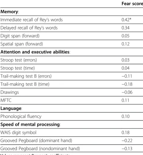
For HIV-infected patients, we conducted a correlation analysis between their Fear Facial Emotion Recognition score and scores obtained on each test of the neuro-psychological battery (see Table 3).
Relationship between emotion recognition and HIV-related factors
The potential influence of HIV-related factors on emotion recognition abilities was investigated by linear regression analysis: in particular, we explored the relationship be-tween HIV-related variables, the Facial Emotion Recogni-tion global score and each emoRecogni-tion category score. The number of pathological scores on the neuropsychological examination was considered an index of HIV-related neu-rocognitive impairment. In the multivariate analysis, only lower education level was independently associated with a worse facial emotion recognition global score (β= 0.53, 95% CI 0.15 to 0.91; p = 0.007) after adjusting for age, total number of pathological scores and CD4 cell counts at
Figure 1HIV and HC groups’accuracy on each emotion of the facial emotion recognition test.
Table 1 Personal and clinical data of patients (n = 49) and controls (n = 20)
Patients Controls p
No. (%) or median (IQR)*
No. (%) or median (IQR)*
Male 40 (81.6) 16 (80) 0.87
Age, y* 49 (43–55) 48.5 (44–54) 0.88 Education, y* 13 (8.5–17) 13 (11.3–14.6) 0.85 Pathological Zung
depression scale
5 (10.3)
Transmission risk factor:
Heterosexual 15 (30.6) Injecting drug users 7 (14.3)
Homosexual 20 (40.8)
Unknown 7 (14.3)
Time from HIV diagnosis, y* 14 (3.5–18.1) HCV co-infection 9 (18.4) Past AIDS-defining events 9 (18.4) Past suboptimal therapy 19 (38.8)
Off Therapy 3 (6.1)
Time from starting last cART regimen, y*
1.6 (1.1–3.1)
Time from starting first cART regimen, y*
10.2 (3.1–14.6)
CPE rank* 6 (4–7)
HIV-RNA < 50 copies/mL 44 (89.8) CD4 cell count, cells/μL* 570 (437–734) CD4 cell count nadir, cells/μL* 211 (115–314)
Abbreviations: No: number; IQR: interquartile range; y: years; cART: combined antiretroviral therapy; CPE: penetration effectiveness score; HCV: hepatitis C virus.
Table 2 Performance of HIV (n = 49) and HC (n = 20) groups on the facial emotion recognition test
HIV HC p EFFECT SIZE
Mean (sd)
Mean (sd)
Partial Eta squares values
Emotion Global Score 50.0 (3.8) 48.7 (5.2) 0.32 0.010 Disgust score 8.2 (1.5) 8.4 (1.1) 0.57 0.005 Anger score 7.6 (1.8) 8.0 (1.1) 0.32 0.012 Fear score 5.6 (2.3) 8.0 (1.3) < 0.001 0.208 Happiness score 9.7 (0.9) 9.6 (0.6) 0.81 0.003 Surprise score 9.1 (1.3) 8.5 (1.1) 0.11 0.052 Sadness score 8.2 (1.5) 8.5 (1.4) 0.51 0.006
Table 3 Correlation among the Fear Facial Emotion Recognition and the Neuropsychological tasks in the HIV group
Fear score
Memory:
Immediate recall of Rey’s words 0.42* Delayed recall of Rey’s words 0.34
Digit span (forward) 0.05
Spatial span (forward) 0.12
Attention and executive abilities:
Stroop test (errors) 0.03
Stroop test (time) 0.04
Trail-making test B (errors) −0.11 Trail-making test B (time) −0.18
Drawings −0.06
MFTC 0.11
Language:
Phonological fluency 0.10
Speed of mental processing:
WAIS digit symbol 0.18
Grooved Pegboard (dominant hand) −0.22 Grooved Pegboard (nondominant hand) −0.13
Values represent Pearson’s coefficients.
nadir. Patients with past AIDS-defining events (β=−0.75, 95% CI-1.45 to−0.06; p = 0.035) and a higher total num-ber of pathological scores on the neuropsychological test battery (β=−0.24, 95% CI−0.41 to−0.06, p = 0.008) sho-wed poorer ability to recognize happiness, when scores were adjusted for age and depression levels.
No other variable was associated with any other emo-tion category when adjusted for age and depression levels.
Discussion
Similar to previous studies [22,23], we found that HIV-infected patients performed worse than HC in recogniz-ing the facial emotion of fear. This deficit does not seem related to severity of the cognitive impairment; in fact, patients with ANI performed as accurately as patients who had no documented cognitive deficit.
It has been suggested that specific deficits in the re-cognition of different categories of facial emotions may reflect task difficulty factors [40]. This is also supported by cross-cultural studies in healthy subjects showing that accuracy in recognizing happiness is high (94% correct responses on emotion recognition tasks) and fear is low (70%) [41,42]. On the other hand, there are considerable evidence that the correct recognition of facial expres-sion of fear depends on specific neural structures, that seems to have a critical role in mediating the autonomic and behavioural responses associated with this emotion [1,43].
In agreement with the results of cross-cultural studies [41,42], in our study both patients and controls per-formed worse when they had to recognize fear than the other emotions, confirming that this emotion might be more difficult to recognize than the others. On the other hand, fear was also the only emotion on which patients performed significantly worse than controls. This would suggest that impairment of neural substrates that are supposed to be specifically involved in HIV pathology, could concur in fear recognition deficit in addition to the effects of task difficulty. Previous studies [23] suggest in fact, that fear recognition abnormalities in HIV may be due to a disruption of the broader neural network involved in emotion recognition which depends on the integrity of frontal system. Unfortunately, we do not have direct evidence of the involvement of such subs-trates in our population. However, since such an impair-ment is currently demonstrated [44] we cannot exclude that the low performance in fear recognition obtained by our patients could be due at least in part to the effect of infection.
If this were true, demonstration of the presence of impaired fear recognition should be considered an early marker of cognitive impairment in HIV population. Study-ing fear recognition in patients with other brain
pathologies that spare the frontal regions as well as neu-roimaging studies [11,44] on HIV + patients could con-tribute to clarify this issue.
Multivariate analysis demonstrated an independent re-lationship between severity of cognitive impairment and score on recognizing the emotion of happiness, that is, patients with a higher number of pathological scores on the neuropsychological examination were less accurate in recognizing happiness. Moreover we observed an as-sociation between the global emotion recognition score and education level, finding that did not emerge in pre-vious studies [45]. These results confirm that recognizing emotions requires the integrity of “high level” cognitive abilities, as already reported in patients with neurodege-nerative diseases (see [46] for a review).
The emotion happiness was also found in association with HIV-related variables. In particular, happiness was independently and inversely associated with past AIDS-defining events.
The association between recognition of happiness and general neurocognitive impairment as well as the asso-ciation between recognition of happiness and past AIDS-defining events, could lead us to hypothesize that a deficit in recognizing this emotion might emerge only in subjects in more severe stages of HIV pathology in agreement with a recent study [22] demonstrating that the ability to discriminate between levels of happiness intensity on facial expression was specifically altered in HIV patients with impaired neurocognitive performance. We acknowledge that our study might have some li-mitations because uncontrolled biases can occur in cross-sectional surveys performed in routine clinical practice. Furthermore, we cannot exclude a possible confounding effect of different IQ on emotion recogni-tion, although there is no reason to assume a different distribution of IQ value in patients and controls. More-over, although not significant, the performance of HC group in recognition of facial expression of surprise was worse than HIV patients; since the effect size for this emotion was moderate, we cannot exclude potential power issues. Anyway, both groups obtained high scores, so data observed might be attributed to a randomness. At last, we did not consider either the psychological and social implications of HIV infection and their possible impact on emotion recognition ability [47] or the psy-chological premorbid characteristics.
Conclusion
asymptomatic cerebral involvement cannot be excluded. Inclusion of an emotion recognition test in the neuro-psychological test battery could help clinicians in the long term management of HIV-infected patients, to better understand the cognitive mechanisms involved in the re-duction of emotion recognition ability and the impact of this impairment on daily life.
Abbreviations
cART: Combination antiretroviral therapy; CNS: central nervous system; CPE: CNS penetration-effectiveness; HC: HIV-negative healthy control population; MFTC: Multiple Features Target Cancellation; WAIS: Wechsler Adult Intelligence Scale; ANI: Asymptomatic Neurocognitive Impairment; IADL: Instrumental Activities of Daily Living; CI: Confidence interval.
Competing interests
MF: Speakers’honoraria from Abbott Virology, Merck Sharp & Dohme and Janssen-Cilag. RC: Advisor for Gilead and Janssen-Cilag, speakers’honoraria from ViiV, Bristol-Myers Squibb, Merck Sharp and Dohme and Janssen-Cilag, and research support from“Fondazione Roma”. SDG: speakers’honoraria and support for travel meetings from Gilead, Bristol-Myers Squibb, Abbott, Boehringer Ingelheim, Janssen-Cilag, and GlaxoSmithKline. MCS: travel grants from Novartis and Lundbeck, and research support from Catholic University of Rome. The other authors did not have any competing interests.
Authors’contributions
EB: design of the study, acquisition of data, analysis and interpretation of the data, study coordination, writing the manuscript. NC: design of the study, acquisition of data, analysis and interpretation of the data, revising the manuscript for content. MF: analysis and interpretation of the data, revising the manuscript for content. MC: acquisition of data, revising the manuscript for content. EI: acquisition of data, revising the manuscript for content. AD: acquisition of data, revising the manuscript for content. AM: acquisition of data, revising the manuscript for content. RC: design of the study, interpretation of the data, revising the manuscript for content. SDG: acquisition of data, interpretation of the data, revising the manuscript for content. MCS: design of the study, study supervision and coordination, acquisition of data, interpretation of the data, revising the manuscript for content. All authors read and approve the final manuscript.
Funding
This research was supported by an unrestricted grant from Abbott Virology.
Received: 12 September 2012 Accepted: 27 February 2013 Published: 27 February 2013
References
1. Adolphs, R (2002). Neural systems for recognizing emotion.Current opinion in neurobiology, 12, 169–177.
2. Clark, US, Neargarder, S, & Cronin-Golomb, A (2008). Specific impairments in the recognition of emotional facial expressions in Parkinson’s disease. Neuropsychologia, 46, 2300–2309.
3. Dujardin, K, Blairy, S, Defebvre, L, Duhem, S, Noel, Y, Hess, U, et al. (2004). Deficits in decoding emotional facial expressions in Parkinson’s disease. Neuropsychologia, 42, 239–250.
4. Lawrence, AD, Goerendt, IK, & Brooks, DJ (2007). Impaired recognition of facial expressions of anger in Parkinson’s disease patients acutely withdrawn from dopamine replacement therapy.Neuropsychologia, 45, 65–74. 5. Johnson, SA, Stout, JC, Solomon, AC, Langbehn, DR, Aylward, EH, Cruce, CB,
et al. (2007). Beyond disgust: impaired recognition of negative emotions prior to diagnosis in Huntington's disease.Brain: a journal of neurology, 130, 1732–1744.
6. Sprengelmeyer, R, Young, AW, Calder, AJ, Karnat, A, Lange, H, Homberg, V, et al. (1996). Loss of disgust. Perception of faces and emotions in Huntington’s disease.Brain: a journal of neurology, 119(Pt 5), 1647–1665. 7. Sprengelmeyer, R, Young, AW, Pundt, I, Sprengelmeyer, A, Calder, AJ,
Berrios, G, et al. (1997). Disgust implicated in obsessive-compulsive disorder. Proceedings Biological sciences / The Royal Society, 264, 1767–1773.
8. Delmaire, C, Dumas, EM, Sharman, MA, van den Bogaard, SJ, Valabregue, R, Jauffret, C, et al. (2012). The structural correlates of functional deficits in early huntington's disease.Human brain mapping,in press. doi: 10.1002/hbm.22055 9. Peron, J, Dondaine, T, Le Jeune, F, Grandjean, D, & Verin, M (2012).
Emotional processing in Parkinson’s disease: a systematic review.Movement disorders: official journal of the Movement Disorder Society, 27, 186–199. 10. Paul, RH, Brickman, AM, Navia, B, Hinkin, C, Malloy, PF, Jefferson, AL, et al. (2005).
Apathy is associated with volume of the nucleus accumbens in patients infected with HIV.The Journal of neuropsychiatry and clinical neurosciences, 17, 167–171. 11. Jernigan, TL, Archibald, S, Hesselink, JR, Atkinson, JH, Velin, RA, McCutchan,
JA, et al. (1993). Magnetic resonance imaging morphometric analysis of cerebral volume loss in human immunodeficiency virus infection. The HNRC Group.Archives of neurology, 50, 250–255.
12. Heaton, RK, Clifford, DB, Franklin, DR, Jr., Woods, SP, Ake, C, Vaida, F, et al. (2010). HIV-associated neurocognitive disorders persist in the era of potent antiretroviral therapy: CHARTER Study.Neurology, 75, 2087–2096. 13. McArthur, JC (2004). HIV dementia: an evolving disease.Journal of
neuroimmunology, 157, 3–10.
14. Sacktor, N, Lyles, RH, Skolasky, R, Kleeberger, C, Selnes, OA, Miller, EN, et al. (2001). HIV-associated neurologic disease incidence changes: Multicenter AIDS Cohort Study, 1990–1998.Neurology, 56, 257–260.
15. Ciccarelli, N, Fabbiani, M, Di Giambenedetto, S, Fanti, I, Baldonero, E, Bracciale, L, et al. (2011). Efavirenz associated with cognitive disorders in otherwise asymptomatic HIV-infected patients.Neurology, 76, 1403–1409. 16. Grima, P, Fabbiani, M, Ciccarelli, N, Tana, M, Farina, S, Colafigli, M, et al.
(2012). Increased ophthalmic artery resistance index is associated with cognitive impairment in HIV-infected patients.The Journal of infection, 65, 439–446.
17. Fabbiani, M, Ciccarelli, N, Tana, M, Farina, S, Baldonero, E, Di Cristo, V, et al. (2013). Cardiovascular risk factors and carotid intima-media thickness are associated to lower cognitive performance in HIV-infected patients.HIV Medicine, 14(3): 136-144.
18. Ciccarelli, N, Fabbiani, M, Baldonero, E, Fanti, I, Cauda, R, Di Giambenedetto, S, et al. (2012). Effect of Aging and Human Immunodeficiency Virus Infection on Cognitive Abilities.Journal of the American Geriatrics Society, 60, 2048–2055.
19. Valcour, V, Sithinamsuwan, P, Letendre, S, & Ances, B (2011). Pathogenesis of HIV in the central nervous system.Current HIV/AIDS reports, 8, 54–61. 20. Hinkin, CH, Castellon, SA, Atkinson, JH, & Goodkin, K (2001). Neuropsychiatric
aspects of HIV infection among older adults.Journal of clinical epidemiology, 54(Suppl 1), S44–52.
21. Stout, JC, Ellis, RJ, Jernigan, TL., Archibald, SL, Abramson, I, Wolfson, T, et al. (1998). Progressive cerebral volume loss in human immunodeficiency virus infection: a longitudinal volumetric magnetic resonance imaging study. HIV Neurobehavioral Research Center Group.Archives of neurology, 55, 161–168.
22. Lane, TA, Moore, DM, Batchelor, J, Brew, BJ, & Cysique, LA (2012). Facial emotional processing in HIV infection: relation to neurocognitive and neuropsychiatric status.Neuropsychology, 26, 713–722.
23. Clark, US, Cohen, RA, Westbrook, ML, Devlin, KN, & Tashima, KT (2010). Facial emotion recognition impairments in individuals with HIV.Journal of the International Neuropsychological Society: JINS, 16, 1127–1137.
24. Letendre, S, FitzSimons, C, & Ellis, R (2010). Correlates of CSF viral loads in 1221 volunteers of the CHARTER Cohort.17th Conference on Retrovirus and Opportunistic Infections, San Francisco.
25. Carlesimo, GA, Caltagirone, C, & Gainotti, G (1996). The Mental Deterioration Battery: normative data, diagnostic reliability and qualitative analyses of cognitive impairment. The Group for the Standardization of the Mental Deterioration Battery.European neurology, 36, 378–384.
26. Orsini, A, Grossi, D, Capitani, E, Laiacona, M, Papagno, C, & Vallar, G (1987). Verbal and spatial immediate memory span: normative data from 1355 adults and 1112 children.Italian journal of neurological sciences, 8, 539–548. 27. Caffarra, PVG, Dieci, F, Zonato, F, & Venneri, A (2002). Una versione
abbreviata del test di Stroop: dati normativi nella popolazione italiana. Rivista di Neurologia, 12(4): 111-115.
28. Giovagnoli AR, DPM, Mascheroni, S, Simoncelli, M, Laiacona, M, & Capitani, E (1996). Trail making test: normative values from 287 normal adult controls. Italian journal of neurological sciences, 17, 305–309.
30. Marra, C, Gainotti, G, Scaricamazza, E, Piccininni, C, Ferraccioli, M, & Quaranta, D (2013). The Multiple Features Target Cancellation (MFTC): an attentional visual conjunction search test. Normative values for the Italian population.Neurological sciences: official journal of the Italian Neurological Society and of the Italian Society of Clinical Neurophysiology. 34(2): 173-180. 31. Orsini, A, & Laicardi, C (1997).WAIS-R. Contributo Alla Taratura Italiana.
Firenze: Giunti O.S. Organizzazioni Speciali.
32. Trites, RL (1977).Neuropsychological Test Manual. Ottawa, Ontario, Canada: Royal Ottawa Hospital.
33. Antinori, A, Arendt, G, Becker, JT, Brew, BJ, Byrd, DA, Cherner, M, et al. (2007). Updated research nosology for HIV-associated neurocognitive disorders. Neurology, 69, 1789–1799.
34. Lovibond, SH, & Lovibond, PF (1995).Manual for Depression Anxiety Stress Scale. Sydney, Australia: Psychology Foundation.
35. Lawton, MP, & Brody, EM (1969). Assessment of older people: self-maintaining and instrumental activities of daily living.The Gerontologist, 9, 179–186.
36. Young, AW, Perrett, DI, Calder, AJ, Sprengelmeyer, R, & Ekman, P (2002). Facial Expressions of Emotion: Stimuli and Tests (FEEST): Thames Valley Test Company. Bury St. Edmunds, England.
37. Lipsey, MW, & Wilson, DB (2000).Practical Meta-Analysis: SAGE. Thousand Oaks, California.
38. Calder, JA, Keane, J, Manly, T, Sprengelmeyer, R, Scott, S., Nimmo-Smith, I, et al. (2003). Facial expression recognition across the adult life span. Neuropsychologia, 41, 195–202.
39. Leppanen, JM, Milders, M, Bell, JS, Terriere, E, & Hietanen, JK (2004). Depression biases the recognition of emotionally neutral faces.Psychiatry research, 128, 123–133.
40. Rapcsak, SZ, Galper, SR, Comer, JF, Reminger, SL, Nielsen, L, Kaszniak, AW, et al. (2000). Fear recognition deficits after focal brain damage: a cautionary note.Neurology, 54, 575–581.
41. Biehl, M, Matsumoto, D, Ekman, P, Hearn, V, Heider, K, Kudoh, T, et al. (1997). Matsumoto and Ekman’s Japanese and Caucasian Facial Expressions of Emotion (JACFEE): Reliability Data and Cross-National Differences.Journal of Nonverbal Behavior, 21, 3–21.
42. Russell, JA (1994). Is there universal recognition of emotion from facial expression? A review of the cross-cultural studies.Psychol Bull, 115, 102–141. 43. LeDoux, J (1996). Emotional networks and motor control: a fearful view.
Progress in brain research, 107, 437–446.
44. Towgood, KJ, Pitkanen, M, Kulasegaram, R, Fradera, A, Kumar, A, Soni, S, et al. (2012). Mapping the brain in younger and older asymptomatic HIV-1 men: frontal volume changes in the absence of other cortical or diffusion tensor abnormalities.Cortex; a journal devoted to the study of the nervous system and behavior, 48, 230–241.
45. Scherer, KR, & Scherer, U (2011). Assessing the Ability to Recognize Facial and Vocal Expressions of Emotion: Construction and Validation of the Emotion Recognition Index.Journal of Nonverbal Behavior, 35, 305–326. 46. Kemp, J, Despres, O, Sellal, F, & Dufour, A (2012). Theory of Mind in normal
ageing and neurodegenerative pathologies.Ageing research reviews, 11, 199–219.
47. Atkins, JH, Rubenstein, SL, Sota, TL, Rueda, S, Fenta, H, Bacon, J, et al. (2010). Impact of social support on cognitive symptom burden in HIV/AIDS.AIDS care, 22, 793–802.
doi:10.1186/2050-7283-1-3
Cite this article as:Baldoneroet al.:Evaluation of emotion processing in HIV-infected patients and correlation with cognitive performance.BMC
Psychology20131:3.
Submit your next manuscript to BioMed Central and take full advantage of:
• Convenient online submission
• Thorough peer review
• No space constraints or color figure charges
• Immediate publication on acceptance
• Inclusion in PubMed, CAS, Scopus and Google Scholar
• Research which is freely available for redistribution