R E S E A R C H
Open Access
Effects of exercise and diet change on cognition
function and synaptic plasticity in high fat diet
induced obese rats
Jinhee Woo
1, Ki Ok Shin
1, So Young Park
2, Ki Soeng Jang
1and Sunghwun Kang
1,2*Abstract
Background:Nutritional imbalance-induced obesity causes a variety of diseases and in particular is an important cause of cognitive function decline. This study was performed on Sprague Dawley (SD) rats with 13-weeks of high fat diet-induced obesity in connection to the effects of regular exercise and dietary control for 8 weeks on the synaptic plasticity and cognitive abilities of brain.
Methods:Four weeks-old SD rats were adopted classified into normal-normal diet-sedentary (NNS, n = 8), obesity-high fat diet-sedentary (OHS, n = 8), obesity-obesity-high fat diet-training (OHT, n = 8), obesity-normal diet-sedentary (ONS, n = 8) and obesity- normal diet-training (ONT, n = 8). The exercise program consisted of a treadmill exercise administered at a speed of 8 m/min for 1–4 weeks, and 14 m/min for 5–8 weeks. The Western blot method was used to measure the expression of NGF, BDNF, p38MAPK and p-p38MAPK proteins in hippocampus of the brain, and expressions of NGF, BDNF, TrkA, TrkB, CREB and synapsin1 mRNA were analyzed through qRT-PCR.
Results:The results suggest cognitive function-related protein levels and mRNA expression to be significantly decreased in the hippocampus of obese rats, and synaptic plasticity as well as cognitive function signaling sub-pathway factors were also significantly decreased. In addition, 8-weeks exercises and treatment by dietary change had induced significant increase of cognitive function-related protein levels and mRNA expression as well as synaptic plasticity and cognitive function signaling sub-pathway factors in obese rats. In particular, the combined treatment had presented even more positive effect.
Conclusions:Therefore, it was determined that the high fat diet-induced obesity decreases plasticity and cognitive function of the brain, but was identified as being improved by exercises and dietary changes. In particular, it is considered that regular exercise has positive effects on memory span and learning capacity unlike dietary control.
Keywords:Exercise, Cognition function, Synaptic plasticity, High fat diet, Memory span
Introduction
Obesity-induced oxidative stress not only causes inflam-matory reactions resulting in abnormalities of the im-mune system [1] but also transforms functions of protein, lipid and DNA, triggering neurodegenerative diseases such as Alzheimer’s disease and Parkinson’s dis-ease [2,3] as well as aging of brain function, cognitive impairment [4], and is reported to affect apoptosis of
neuronal cells. As aforementioned, the obesity is pre-dicted to affect cognitive functions including reduced learning capacity and memory impairment, however, the mechanism of which brain function it affects remains unclear. Cognitive impairment includes reduced concen-tration, poor progress in learning, memory impairment where its molecular mechanism is manifested through change of hippocampal function in the brain. Further-more obesity has been reported to cause problems in cAMP and protein kinase A dependent synaptic plasti-city [5]. Thus, it is thought that identification of the rela-tionship between obesity and synaptic plasticity will help * Correspondence:[email protected]
1
Department of Physical Education, Laboratory of Exercise Physiology, College of Sports Science, Dong-A University, 840 Hadan2-dong, Saha-gu, Busan, Korea
2Departments of Pharmacology, Medical Sciences Research Institute, College
of Medicine, Dong-A University, Busan, Korea
in understanding the mechanism between obesity and cognitive function.
Regular exercise not only prevents and treats obesity-induced metabolic imbalance but also prevents brain damage. It also enhances learning and memory span along with stimulation of neurogenesis [6]. Even more surprising is the fact that exercise reinforces the capacity for functional recovery from brain damage and is attributed to prolonging the process of aging effects on brain function [7,8]. Exercise-induced increases of nerve growth factor (NGF) and brain-drived neurotrophic factor (BDNF) in the hippocampus have been known to reinforce cognitive function [9,10], however the question still remains as to whether exercise-induced reinforcement of learning and memory span is due to increases of hippo-campal NGF and BDNF or not.
NGF and BDNF, also known as neurotransmitters, are drawing attentions as major mediators of cerebral synap-tic plassynap-ticity, while BDNF is known to be involved in controlling food intake and glucose homeostasis [11]. In the case of genetically obese animals, it was reported that TrkB, a BDNF receptor was depressed [12], and that high fat / high sugar diet reduced expression of hippo-campal BDNF, resulting in deterioration of learning as well as memory span [13]. NGF and its receptor TrkA are also known to be suppressed in connection to dis-ease and nerve damage [14], and were identified as hav-ing negative effects on learnhav-ing and memory as a result of high-intensity acute exercise [15]. However, it was reported that norepinephrine increased as a result of regular exercise facilitated by neurocyte regeneration so as to increase BDNF expression of cultivated hippocam-pal cells [16]. Additionally a study on mice reported that the expression of BDNF mRNA increased after a three day running exercise routine [17]. From these reports, it could be considered that regular exercise increases expressions of NGF and BDNF [10]. Therefore, the stud-ies that had observed changes of synaptic plasticity such as NGF, BDNF, p38MAPK, cAMP response element-binding protein (CREB) and synapsin 1 in rats with obesity triggered by the energy imbalance from high fat diet, are still scarce, and in particular, there is no study to this date implementing the use of regular exercise and dietary change in these obese models. Therefore, this study aimed to define the relationship between obesity and neurogenesis in rats with acquired obesity caused by high fat diet, and to identify the effects of regular aerobic exercise and dietary change on synaptic plasticity markers and cognitive function.
Methods Animals
The adopted subjects of this study were 40 heads of four
week old white male Sprague–Dawley (SD) rats. Out of
40 heads, 32 were induced obesity through 13-weeks of a high fat diet while the other eight heads were fed with a normal diet. After 13 weeks, all study rats were classified into five groups which were the normal-normal diet-sedentary group (NNS), obesity-high fat sedentary group (OHS, n = 8), obesity-high fat diet-training group (OHT, n = 8), obesity-normal diet seden-tary group (ONS, n = 8) and obesity-normal diet-training group (ONT). These five groups were bred for eight weeks.
Obesity was induced in the high fat diet groups as pro-vided by 25 g of feeds containing all essential nutrients, vitamins, minerals and 45% of fat per one head daily with water, while the normal diet groups were provided 25 g of the ordinary feeds per one head daily with water. During the study duration, the dietary intakes were mea-sured at the same time (19:00) each day, and the weights were measured at a certain time for once a week by using the computing scale. In order to prevent tempor-ary weight change due to diettempor-ary intake, these measure-ments were performed after removing the feeding container 2 hours before. The study was carried out in accordance with the Guidelines laid down by the NIH in the US regarding the care and use of animals for experi-mental procedures and was approved by DONG-A Uni-versity Institutional Animal Care and Use Committee.
Exercise program
Treadmill running was performed as the exercise type. For the exercise protocol, during the period of weeks 1 ~ 4, exercise was performed five days per week, of 40 minutes duration and during the period of weeks 4 ~ 8, exercise was performed five days per week, for a duration 60 minutes per day. During weeks 1 ~ 4, treadmill exercise was conducted where the slope was fixed to
zero degrees, and a 5 minute– warm up at a speed of
2 m/min, followed by 30 minute main exercise bout at a speed of 8 m/min concluded by five minute final cool down session at a speed of 5 m/min. For weeks 5 ~ 8, a
10 minute – warm up at a speed of 8 m/min, a 40
-minute- main exercise bout was at a speed of 14 m/ min with a 10 minute-final cool down at a speed of 11 m/min [18].
Cognitive function test
above the water while the spatial cues were provided on the surrounding walls for animals to learn the location of the platform. The water (22 ~ 24°C) was made opaque with white nontoxic biodegradable dye to prevent the rats form seeing the platform. The experiment animals were trained on the water maze using 2 consecutive tri-als per day for 3 days and placed into the bathtub the wall from one of the equally spaced start locations that were randomly changed every trial. Each trial lasted until the rat had found the platform or for a max 1 min [18].
Tissue sample
For the timing of tissue sampling in this study, they were conducted at 48 hours after the completion of training for the training groups in order to rule out temporary exercise effects from treadmill training. For such sam-plings, the feed supply was discontinued 12 hours prior to the samplings, however drinking water was continu-ally supplied. The study animals, after arrival at the laboratory, had taken sufficient rest and they were put into anesthesia by using ethyl ether, after skulls were re-moved to collect brain hippocampus (5 g) samples. The tissue samples were stored in a deep freezer (NIHOW freezer, Japan) at−70°C until analysis.
Analysis content Western blot
To extract protein from the hippocampus, the tissues were crushed after adding a solution containing 150 mM Nacl, 5 mM EDTA, 50 mM Tri-HCI (pH8.0), 1%-NP 40, 1 mM aprotinin, 0.1 mM leupeptin, and 1 mM pepstatin and then the solution was put into centrifugation for 30 minutes at 14,000 rpm. Supernatants were collected and assayed for protein content prior to storage at −70°C. Protein sample were mixed Laemmli Sample Buffer (LSB) and placed in a boiling water bath for 5 min. Proteins were resolved by 10, 12 or 15% SDS-polyacrylamide gel
electro-phoresis (SDS-PAGE; each loaded with same μg of total
protein per lane), and transferred to nitrocellulose mem-branes. Then, the membrane was blocked in the solution of Phosphate - buffered saline (PBS) (Nacl 8 g, KCI 0.2 g, Na2HPO4 1.44 g, KH2PO4 0.24 g, PH 7.4) added with 5% skim milk. Thereafter, the samples were reacted in a solu-tion mixed with phospho antibody NGF (1:1000; Santa Cruz Biotechnology), BDNF (1:1000; Santa Cruz Biotech-nology), phospho antibody P38 (1:1000; Santa Cruz Bio-technology) and phospho antibody for one hour, and washed for 1×15 min and 2×5 min in a PBS solution containing 0.1% 0.1% tween - 20. Each membrane was reacted for one hour in a solution added with goat anti mouse and rabbit 1gG conjugated secondary antibody re-spectively, and was measured for immune-reactive bands through a Kodak film. The relative strengths of bands were quantitated by a densitometry (Sci - Scan, UUSB).
Quantitative real time PCR
Total RNA was separated by using the RNA STAT-60 kit (TEL-TEST, Inc., Friendswood, TX, USA) according to the protocol, and the absorbance was measured at 260 nm in addition to analyses of NGF, BDNF, TrkA, TrkB, MAPK1, CREB and Synapsin1 i.e., total RNA of 100 ng was converted to cDNA in use of TaqMan EW RT-PCR core reagent (Perkin-Elmer, Branchburg, NJ, USA), with subsequent designing of primers sequences of the former and the latter by Inte-grated DNA Technologies (Coralville, IA, USA) as follows.
NGF - forward(5′-TTCCAGGCCCATGGTACAA-3′), reverse(5′-GGTGGATGAGCGCTTGCT-3′)
TrkA–forward(5′-GCCGCCCTCTTCCTTTCT-3′), reverse(5′-ATTTGCTCCTCTGTCCACATTTG-3′) BDNF–forward(5′-GCGCCCATGAAAGAAGCA-3′), reverse(5′-CACAGCTGGGTAGGCCAAGT-3′) TrkB–forward(5′-TTTCCGCCACCTTGACTTG-3′), reverse(5′-ACAGGAACACGTGAACGGATT-3′) CREB–forward(5′-CAGTGCCAACCCCGATTTA-3′), reverse(5′-TTGCTCCTCCCTGGGTAATG-3′)
Synapsin1–forward(5′-GCCTTCAGCATGGCACGT A-3′), reverse(5′-CAGCATACTGCAGCCCAATG-3′) MAPK1–forward(5′ -ATTTGGTCTGTGGGCTGCAT-3′), reverse(5′-TCCTGGGAAGATAGGCCTGTT-3′)
For RT reaction phase, cultivation was done for the initial two minutes at 50°C for uracil glycosylase (UNG) activation, followed by reverse transcription at 60°C for 30 minutes. The completion phase for UNG inactivation was performed at 95°C for 5 minutes, and thereafter, a 40 cycle was carried out at 94°C for 20 followed by for 1 minute at 62°C.
Statistical analysis
Data were analyzed using SPSS Software Version 18.0 for Window (SPSS Inc, Chicago, IL) and were presented as mean ± SE unless otherwise indicated. The change in weight induced by the diet method was analyzed through a two-way repeated measures ANOVA, and paired t-tests were performed for time-time interactions, and for any intergroup difference observed, one-way
ANOVA was performed in addition to Duncan’s
Post-hoc analysis. An analysis of variance (ANOVA) with 2-way repeated measures was conducted for analyzing data of the water maze. For the results of the cognitive function and for the expressions of hippocampal neurotrophins as well as synaptic plasticity gene,
one-way ANOVA was in addition to Duncan’s Post-hoc
analysis. Statistical significance was declared when p values were less than 5% level.
Results
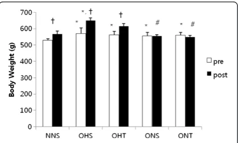
Figure 1 presents the comparison of weights before and after 8-weeks treatment in rats with high fat diet-induced obesity. Compared to the weights before treat-ment, there were significant increases of NNS, OHS and OHT after treatment, however ONS and ONT did not shown differences. ONS and ONT presented signifi-cantly lower post-treatment intergroup variation com-pared to post-treatment OHS (p < 0.05).
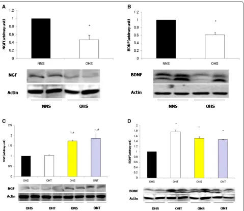
OHS displayed significantly lower expressions of hip-pocampal NGF and BDNF protein compared to NNS (p < 0.05). However, For BDNF after 8-weeks treatment,
OHT, ONS and ONT had presented significantly higher levels than OHS (p < 0.05), and for NGF, the levels of ONS and ONT were significantly higher than OHS and OHT (p < 0.05) (Figure 2).
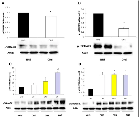
For protein expressions of p38MAPK and p-p38MAPK, the OHS group had shown significantly lower levels than NNS (p < 0.05). However, after 8-weeks of treatment, p38MAPK displayed the highest level in the ONT group (p < 0.05), and p-p38MAPK presented significantly higher levels in the OHT group, ONS group and the ONT group compared to the OHS group (p < 0.05) (Figure 3).
As shown in Figure 4, BDNF mRNA significantly de-creased in rats with high fat diet-induced obesity (p < 0.05), and TrkB significantly decreased as well (p < 0.05). How-ever, it was identified that all of NGF, BDNF, TrkA and
TrkB mRNA were significantly increased through 8-weeks exercise and dietary change (p < 0.05).
As shown in Figure 5, NGF and BDNF mRNA presented the highest levels in the ONT group that had 8-weeks combined diet control and training (p < 0.05). TrkA mRNA displayed high levels in the OHT, ONS, and the ONT group than the OHS group after treatment while TrkB mRNA and MAPK1 mRNA presented the highest levels in the ONT group (p < 0.05). Additionally, post-treatment levels of CREB mRNAand Synapsin1 mRNA were higher in the OHT, ONS and ONT groups compared to the OHS group (p < 0.05).
As shown in Figure 6, spatial learning test six times a significant interaction effect was found between group and time. In addition, there were significant differences between group and time respectively (A). As shown in
Figure 6B, the sixth time in the OHS group higher compared to the NNS, OHT and ONT significantly (p < 0.05). Also, in the ONS group higher compared to the NNS, OHT and ONT significantly (p < 0.05). We found that spatial learning reduces time to exercise.
Discussion
This study was conducted by establishing a hypothesis that regular exercise and dietary change will have posi-tive effects on the brain’s plasticity and cognitive func-tion as a result of high fat induced obesity in rats.
Induction of obesity by high fat diet was identified as having negative effects (inactivity) on all protein factors related to cognitive function. However, exercise and dietary change resulted in improvement of cognitive function protein factor and the level of mRNA (activity). In particular, regular exercise had positive effects on activation of sub signaling pathways as well as on im-provement in cognitive ability tests.
According to a recent study, it was reported that proper stress increased exercise-performing ability in young animals, while it improved learning ability in aged
animals [19]. Particularly, stress such as regular exercise had been reported to prolong the deterioration of cogni-tive function in connection to aging [20], and to reduce risks of various neuropathies [21,22]. It was considered that regular exercise activated metabolic actions so as to trigger continuous gene alterations. The environmental impact such as exercise on cognitive function, in par-ticular, causes BDNF gene alteration [23,24], and has positive effects on reduction of apoptosis by improve-ments of NGF and TrkA [25]. In addition, it was suggested that regular exercise improved the hippo-campal levels of NGF and TrkA as well as BDNF and TrkB so that it was effective for learning and cognitive abilities [9,10]. Nevertheless, BDNF and NGF are understood to be reduced or overly expressed in the state of disease [14], and stress such as high-intensity acute exercise is reported to cause damages to learning and memory [15].
From the results of this study, it was identified that the rats with high fat diet-induced obesity had signifi-cantly lower levels of cognitive function-related protein expression (NGF, BDNF, p38MAPK). This result con-firmed the findings of previous studies that obesity could have negative effects on NGF, BDNF and p38MAPK.
However due to regular 8-weeks treadmill exercise and dietary change, the protein levels of BDNF and NGF had increased significantly, and the activity of p38MAPK displayed high levels, indicating exercise and dietary control had effects on weight loss and improved cogni-tive abilities. Meanwhile, with high fat diet-induced obesity, expressions of NGF and TrkA mRNA as well as BDNF and TrkB mRNA were presented significantly low. As suggested by previous studies, obesity and dis-eases reduces the expressions of NGF [14] and BDNF [26], and it is considered that high fat diet-induced obes-ity affects hippocampal cognition-related factors. How-ever, these results were consistent with the results of previous studies [9,23] that this study had identified regular exercise and dietary change had resulted in sig-nificant increase of NGF and TrkA as well as BDNF and TrkB mRNA.
In general, NGF signaling is composed by activation of the 2-folds delivery system [27]. First, it is spread through continuous activation of mitogen activated pro-tein kinases (MAPK) by Erk phosphorylation, and sec-ondly it is spread by activation of PI3K/Akt signaling pathway as reported by Williams et al., [28]. According to a recent study, it was identified that regular exercise improves the protein levels of Akt, CREB and BDNF [29], and activates MAPK that improves synaptic activity [10]. These findings report that regular exercise had effects not only on CREB phosphorylation but also on increase of MAPK/Erk phosphorylationin the hippocam-pus of rat [30], particularly, in cases of diabetes-induced rat, it activates phosphorylation of Erk and CREB, and increases the level of NGF so as it induces activations of MAPK/Erk1/2 signaling pathway and CREB in the hippocampus even more [10]. The main results of reported by those previous studies proves that regular exercise is an important element and essential for ex-pression of brain function-related signal transmitting protein. In general, it has been known that BDNF de-cline causes excessive eating and develops obesity by in-activation of sub pathways [31], and high fat diet reduces the levels of synaptic plasticity and cognitive function, bringing negative effects on learning and mem-ory span [13]. As suggested previously, regular exercise facilitates synthesis of MAPK, CREB and synapsin 1 which are sub pathways owning to activations of BDNF and NGF [32] and activates the phosphorylation process [33] that is likely to have effects on synaptic plasticity and cognitive abilities. That means, regular exercise en-ables manifestation of extracellular activations of NGF and BDNF, which induces intracellular activations of CREB and Synapsin 1 in MAPK and the nucleus. Such mechanisms are known to have effects on emission of neurotransmitters, maintenance of synaptic connection and extension of neuritis, resulting in improvement of
learning and memory span as well as raising cognitive abilities as reported before [34,35]. Moreover, recent studies suggest that regular exercise improves hippocam-pal plasticity and learning via Akt, CREB and BDNF sig-naling [28], while the high levels of NGF and receptor TrkA improve learning and memory span [36].
Therefore, the imbalance of energy metabolism caused by high fat diet affects synaptic plasticity to be reduced and learning as well as cognition of the brain with low levels of NGF and BDNF, but with regular exercise and dietary change, it has been identified to be possible to improve the reduced synaptic plasticity and cognitive function. From the results of this study, it is indicated that the protein activations of BDNF, NGF and p38MAPK in the hippocampus of rats with high fat diet-induced obesity were inhibited resulting in significant reduction of such ac-tivations, and in addition, from the level of mRNA, the re-duction tendency of TrkA, TrkB, MAPK1, CREB and Synapsin1 had been presented. Such results indicated that high fat containing diet intake causes reduction of signaling pathways related to cognitive function prolonging the time of Morris water maze (MWM) performance, as consistent with previous studies.
However, with regular treadmill exercises for 8 weeks and dietary change, the protein levels of BDNF and NGF were presented significantly high, together with high level of p38MAPK activation, verifying exercise and diet-ary change can improve cognitive abilities. In particular, the combined treatment was identified as even more ef-fective. Additionally, the expression levels of mRNA in all of TrkA, TrkB, MAPK1, CREB and Synapsin1 were identified to be high from as a result of regular exercise, diet control and combined treatment. Given such re-sults, regular exercise and diet control not only improve cognitive function-related proteins but also increase mRNA, in addition to induce activation of sub signaling pathway, resulting in improvement of synaptic plasticity as well as cognitive abilities of brain. Furthermore, com-pared to diet control, regular exercise presented effective in facilitating muscle memory by shortening MWM time owing to the improvement of even more cognitive func-tion signaling pathways, so it thought that diet control and exercise could result in improvement of cognitive function only when they were co-performed.
Conclusion
The high fat diet-induced obesity is considered to reduce plasticity and cognitive function of brain so as to reduce muscle memory, but exercise and dietary change is con-sidered as having positive effects on synaptic plasticity and cognitive function sub pathway of brain. In particu-lar, unlike the diet control, regular exercise activates neurogenesis so that it is considered as contributing to the muscle memory effect by presenting improvement of
MWM ability that is directly associated with cognitive function.
Abbreviations
SD:Sprague Dawley; NGF: Nerve growth factor; BDNF: Brain-drived neurotrophic factor; CREB: cAMP response element-binding protein; MAPK: Mitogen activated protein kinases; MWM: Morris water maze.
Competing interests
The authors declare that they have no competing interests.
Authors’contributions
JW and SK designed the study, carried out the experiments, performed the statistical analysis. SYP carried out the experiments and KSJ collected the data. SK wrote the manuscript, and also participated in the execution and analysis of this study with KOS. All authors read and approved the final manuscript.
Acknowledgements
This work was supported by the National Research Foundation of Korea Grant funded by the Korean Government (NRF-2011-327-G00141).
Received: 29 April 2013 Accepted: 17 September 2013 Published: 8 October 2013
References
1. Rajala MW, Scherer PE:Minireview the adipocyte–at the crossroads of energy homeostasis, inflammation, and atherosclerosis.Endocrinology
2003,144:3765–3773.
2. Finkel T, Holbrook NJ:Oxidants, oxidative stress and the biology of ageing.Nature2000,408:239–247.
3. Woo J, Yeo NH, Shin KO, Lee HJ, Yoo J, Kang S:Antioxidant enzyme activities and DNA damage in children with type 1 diabetes mellitus after 12 weeks of exercise.Acta Paediatr2010,99:1263–1268. 4. Krieglstein K, Richter S, Farkas L, Schuster N, Dünker N, Oppenheim RW,
Unsicker K:Reduction of endogenous transforming growth factors beta prevents ontogenetic neuron death.Nat Neurosci2000,3:1085–1090. 5. Vecsey CG, Baillie GS, Jaganath D, Havekes R, Daniels A, Wimmer M, Huang
T, Brown KM, Li XY, Descalzi G, Kim SS, Chen T, Shang YZ, Zhuo M, Houslay MD, Abel T:Sleep deprivation impairs cAMP signalling in the
hippocampus.Nature2009,461:1122–1125.
6. Wirth MJ, Brun A, Grabert J, Patz S, Wahle P:Accelerated dendritic development of rat cortical pyramidal cells and interneurons after biolistic transfection with BDNF and NT4/5.Development2003,
130:5827–5838.
7. Cotman C, Engesser-Cesar C:Exercise enhances and protects brain function.Exerc Sport Sci Rev2002,30:75–79.
8. Woo J, Shin KO, Yoo JH, Park S, Kang S:The effects of detraining on blood adipokines and antioxidant enzyme in Korean overweight children.Eur J Pediatr2012,171:235–243.
9. Isacson O, Seo H, Lin L, Albeck D, Granholm AC:Alzheimer’s disease and Down’s syndrome: roles of APP, trophic factors and ACh.Trends Neurosci
2002,25:79–84.
10. Chae CH, Jung SL, An SH, Park BY, Wang SW, Cho IH, Cho JY, Kim HT:
Treadmill exercise improves cognitive function and facilitates nerve growth factor signaling by activating mitogen-activated protein kinase/ extracellular signal-regulated kinase1/2 in the streptozotocin-induced diabetic rat hippocampus.Neuroscience2009,164:1665–1673.
11. Wang C, Godar RJ, Billington CJ, Kotz CM:Chronic administraion of brain-derived neurotrophic factor in the hypothalamic paraventricular nucleus reverses obesity induced by high-fat diet.Am J Physiol Regul Integr Comp Physiol2010,298:R1320–R1332.
12. Yu Y, Wang Q, Huang XF:Energy-restricted pair-feeding normalizes low levels of brain-derived neurotrophic factor/tyrosine kinase B mRNA expression in the hippocampus, but not ventromedial hypothalamic nucleus, in diet-induced obese mice.Neuroscience2009,160:295–306. 13. Molteni R, Wu A, Vaynman S, Ying Z, Barnard RJ, Gomez-Pinilla F:Exercise
14. Ziegler D, Siekierka-Kleiser E, Meyer B, Schweers M:Validation of a novel screening device (NeuroQuick) for quantitative assessment of small nerve fiber dysfunction as an early feature of diabetic polyneuropathy. Diabetes Care2005,28:1169–1174.
15. Kim JJ, Song EY, Kosten TA:Stress effects in the hippocampus: synaptic plasticity and memory.Stress2006,9:1–11.
16. Chen MJ, Nguyen TV, Pike CJ, Russo-Neustadt AA:Norepinephrine induces BDNF and activates the PI-3K and MAPK cascades in embryonic hippocampal neurons.Cell Signal2006,19:114–128.
17. Neeper SA, Gómez-Pinilla F, Choi J, Cotman CW:Physical activity increases mRNA for brain-derived neurotrophic factor and nerve growth factor in rat brain.Brain Res1996,726:49–56.
18. Woo J, Shin KO, Yeo NH, Park SY, Kang S:The effects of treadmill training on neurotrophins and immediately early protein in obese rats.Korean J Life Sci2011,21:985–991.
19. Adlard PA, Engesser-Cesar C, Cotman CW:Mild stress facilitates learning and exercise improves retention in aged mice.Exp Gerontol2011,46:53–59. 20. Kramer AF, Hahn S, Cohen NJ, Banich MT, McAuley E, Harrison CR, Chason J,
Vakil E, Bardell L, Boileau RA, Colcombe A:Ageing, fitness and neurocognitive function.Nature1999,400:418–419.
21. Friedland RP, Fritsch T, Smyth KA, Koss E, Lerner AJ, Chen CH, Petot GJ, Debanne SM:Patients with Alzheimer’s disease have reduced activities in midlife compared with healthy control-group members.Proc Natl Acad Sci U S A2001,98:3440–3445.
22. Laurin D, Verreault R, Lindsay J, MacPherson K, Rockwood K:Physical activity and risk of cognitive impairment and dementia in elderly persons.Arch Neurol2001,58:498–504.
23. Gomez-Pinilla F, Zhuang Y, Feng J, Ying Z, Fan G:Exercise impacts brain-derived neurotrophic factor plasticity by engaging mechanisms of epigenetic regulation.Eur J Neurosci2011,33:383–390.
24. Sweatt JD:Experience-dependent epigenetic modifications in the central nervous system.Biol Psychiatry2009,65:191–197.
25. Uysal N, Tugyan K, Kayatekin BM, Acikgoz O, Bagriyanik HA, Gonenc S, Ozdemir D, Aksu I, Togcu A, Semin I:The effects of regular aerobic exercise in adolescent period on hippocampal neuron density, apoptosis and spatial memory.Neurosci Lett2005,383:241–245.
26. Zuccato C, Cattaneo E:Brain-derived neurotrophic factor in neurodegenerative diseases.Nat Rev Neurol2009,5:311–322. 27. Gao Y, Nikulina E, Mellado W, Filbin MT:Neurotrophins elevate cAMP to
reach a threshold required to overcome inhibition by MAG through extracellular signal-regulated kinase-dependent inhibition of phosphodiesterase.J Neurosci2003,23:11770–11777.
28. Williams B, Granholm AC, Sambamurti K:Age-dependent loss of NGF signaling in the rat basal forebrain is due to disrupted MAPK activation. Neurosci Lett2007,413:110–114.
29. Aguiar AS Jr, Castro AA, Moreira EL, Glaser V, Santos AR, Tasca CI, Latini A, Prediger RD:Short bouts of mild-intensity physical exercise improve spatial learning and memory in aging rats: involvement of hippocampal plasticity via AKT, CREB and BDNF signaling.Mech Ageing Dev2011,
132:560–567.
30. Shen H, Tong L, Balazs R, Cotman CW:Physical activity elicits sustained activation of the cyclic AMP response element-binding protein and mitogen-activated protein kinase in the rat hippocampus.Neuroscience
2001,107:219–229.
31. Lyons WE, Mamounas LA, Ricaurte GA, Coppola V, Reid SW, Bora SH, Wihler C, Koliatsos VE, Tessarollo L:Brain-derived neurotrophic factor-deficient mice develop aggressiveness and hyperphagia in conjunction with brain serotonergic abnormalities.Proc Natl Acad Sci USA1999,96:15239–15244.
32. Wang T, Xie K, Lu B:Neurotrophins promote maturation of developing neuromuscular synapses.J Neurosci1995,15:4796–4805.
33. Jovanovic JN, Czernik AJ, Fienberg AA, Greengard P, Sihra TS:Synapsins as mediators of BDNF-enhanced neurotransmitter release.Nat Neurosci
2000,3:323–329.
34. Vaynman S, Ying Z, Gomez-Pinilla F:Hippocampal BDNF mediates the efficacy of exercise on synaptic plasticity and cognition.Eur J Neurosci
2004,20:2580–2590.
35. Vaynman S, Ying Z, Gomez-Pinilla F:The select action of hippocampal calcium calmodulin protein kinase II in mediating exercise-enhanced cognitive function.Neuroscience2007,144:825–833.
36. Badowska-Szalewska E, Spodnik E, Klejbor I, Ludkiewicz B, MoryśJ:Do two models of acute and chronic stress stimulation influence the amount of nerve growth factor (NGF) and its receptor TrkA in the hippocampal neurons of middle aged rats?Brain Res2011,1384:97–109.
doi:10.1186/1476-511X-12-144
Cite this article as:Wooet al.:Effects of exercise and diet change on cognition function and synaptic plasticity in high fat diet induced obese rats.Lipids in Health and Disease201312:144.
Submit your next manuscript to BioMed Central and take full advantage of:
• Convenient online submission
• Thorough peer review
• No space constraints or color figure charges
• Immediate publication on acceptance
• Inclusion in PubMed, CAS, Scopus and Google Scholar
• Research which is freely available for redistribution