R E V I E W
Open Access
The TRAX, DISC1, and GSK3 complex in
mental disorders and therapeutic
interventions
Yu-Ting Weng
1,2, Ting Chien
1, I-I Kuan
1and Yijuang Chern
1,2*Abstract
Psychiatric disorders (such as bipolar disorder, depression, and schizophrenia) affect the lives of millions of
individuals worldwide. Despite the tremendous efforts devoted to various types of psychiatric studies and rapidly
accumulating genetic information, the molecular mechanisms underlying psychiatric disorder development remain
elusive. Among the genes that have been implicated in schizophrenia and other mental disorders, disrupted in
schizophrenia 1 (DISC1) and glycogen synthase kinase 3 (GSK3) have been intensively investigated. DISC1 binds
directly to GSK3 and modulates many cellular functions by negatively inhibiting GSK3 activity. The human DISC1
gene is located on chromosome 1 and is highly associated with schizophrenia and other mental disorders. A
recent study demonstrated that a neighboring gene of DISC1, translin-associated factor X (TRAX), binds to the
DISC1/GSK3
β
complex and at least partly mediates the actions of the DISC1/GSK3
β
complex. Previous studies also
demonstrate that TRAX and most of its interacting proteins that have been identified so far are risk genes and/or
markers of mental disorders. In the present review, we will focus on the emerging roles of TRAX and its interacting
proteins (including DISC1 and GSK3
β
) in psychiatric disorders and the potential implications for developing
therapeutic interventions.
Keywords:
TRAX, DISC1, GSK3
β
, Mental disorders, DNA damage, DNA repair, Oxidative stress, A
2AR, PKA
Background
Mental disorders (such as bipolar disorder, depression,
and schizophrenia) have recently become great concerns
because of the resultant heavy social and economic
bur-dens on societies [1–3]. Rapidly progressing genetic
technologies have provided many details regarding the
genetic nature of mental disorders. Among the genes
that have been revealed by genetic analyses of
schizo-phrenia and other mental disorders, the function of
disrupted in schizophrenia 1 (DISC1) has been
inten-sively investigated. Biochemical investigations suggest
that DISC1 is a scaffold protein that regulates various
cellular functions (including cytoskeletal processes,
intra-cellular transport, dendritic spine development activities,
neuronal development, the cAMP-signaling pathway, and
DNA repair) by interacting with various proteins [4–12].
Thus, DISC1 has been considered as a hub protein for
schizophrenia
and
possibly
other
mental
diseases
(Table
1).
Previous genetic studies have associated
DISC1
and a
neighboring gene (
translin-associated factor X
,
TSNAX
)
with multiple mental disorders (e.g., schizophrenia,
bipo-lar spectrum disorder, and major depressive disorder)
[13–15] (Table
1). TRAX was initially identified as a
binding partner of an RNA/DNA-binding protein
(trans-lin [16]). Further investigations revealed that similar to
DISC1, TRAX regulates distinct cellular functions by
selectively binding to designated partner(s). Moreover,
the list of TRAX-interacting proteins overlaps with that
of DISC1 (Table
2). Both TRAX and DISC1 are involved
in facilitating DNA repair [5]. Chien et al. demonstrated
that TRAX forms a complex with DISC1 and GSK3
β
in
the cytoplasmic region of resting neurons. Upon stresses
that cause oxidative DNA damage, inhibiting GSK3
β
causes the TRAX/DISC1/GSK3
β
complex to dissociate
* Correspondence:[email protected]1
Institute of Biomedical Sciences, Academia Sinica, 128 Sec. 2, Academia Rd. Nankang, Taipei 115, Taiwan, Republic of China
2Program in Molecular Medicine, National Yang-Ming University and
Academia Sinica, No.155, Sec.2, Linong Street, Taipei 112, Taiwan, Republic of China
and release TRAX to facilitate ATM-mediated DNA
repair [5]. Because the incomplete repair of oxidative
DNA damage may contribute to the development of
psychotic disorders [1,
17,
18] and because TRAX and
many of its interacting proteins (Table
1) are risk genes
and/or markers of mental disorders, the present review
focuses on the emerging role of TRAX/DISC1
interac-tome(s) in DNA repair as well as their potential
implica-tions in psychiatric disorders.
DNA damage, oxidative stress, and mental health
Reactive oxygen species (ROS) are usually generated
through mitochondrial oxidative reactions [19].
Exces-sive ROS levels are a source of oxidative stress, which
causes oxidative damage to DNA, proteins and lipids. ROS
can attack the nitrogenous bases and sugar-phosphate
backbone of DNA to cause single- and double-stranded
DNA breaks that ultimately lead to genetic mutations and
toxicity [20]. When cells are subjected to increased levels
of ROS and reactive nitrogen species, multiple cellular
impairments (e.g., oxidative DNA damage) occur [21].
Accumulating evidence suggests that elevated ROS levels
and the resultant oxidative damage are major factors in
human health and diseases [21–24]. Because the brain uses
approximately 20% of the total oxygen in the body and
generates significant amounts of free radicals, the brain is
more susceptible to oxidative stress than other
or-gans. Moreover, elevated ROS levels have been
impli-cated in most neurological diseases (such as mental
disorder and neurodegenerative diseases) [25–27].
Ele-vated levels of serum oxidative markers (such as
8-hydroxy-2′-deoxyguanosine,
8-OHdG)
have
also
been reported in patients with trauma or diseases of
the brain [28–31].
Ample evidence suggests that increased oxidative
stress, which may cause oxidative DNA damage and
mitochondrial dysfunction, is a common feature of
men-tal disorders in the brain. Mitochondrial dysfunction is
associated directly with elevated levels of oxidative stress
and the progression of mental disorders [18,
19,
32]. For
example, the nuclear gene expression levels of
mito-chondrial proteins, including electron transport chain
(ETC) complexes I–V, are significantly decreased in the
hippocampus and postmortem frontal cortex of patients
with bipolar disorder and schizophrenia [33–35]. It is
important to note that ETC complex I is one of the
major sources of ROS in mitochondria. Moreover, the
expression levels of NADH:Ubiquinone oxidoreductase
core subunit v2 (NDUFV2), a mitochondrial complex I
subunit gene, were decreased in lymphoblastoid cell
lines derived from patients with bipolar disorder [36].
These findings indicate that mitochondria dysfunction is
a major factor that contributes to the development of
mental disorders, including bipolar disorder and
schizo-phrenia [19]. Another important feature of the brains of
patients with mental disorders is an imbalance in the
levels of dopamine and glutamate (for a review, see
[37]). Accumulating evidence suggests that hypofunction
of NMDA receptors was observed in schizophrenia
[38,
39]. Several NMDA receptor antagonists (e.g.,
phencyclidine and ketamine) therefore have been shown
to induce schizophrenia-like symptoms [40,
41]. Other
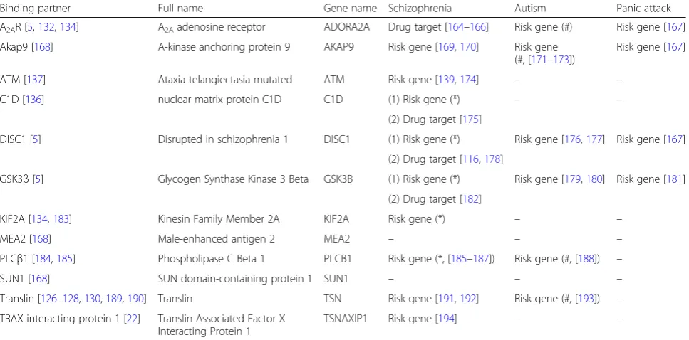
Table 1
Potential involvement of TRAX-interacting proteins in three psychiatric disorders
Binding partner Full name Gene name Schizophrenia Autism Panic attack
A2AR [5,132,134] A2Aadenosine receptor ADORA2A Drug target [164–166] Risk gene (#) Risk gene [167]
Akap9 [168] A-kinase anchoring protein 9 AKAP9 Risk gene [169,170] Risk gene (#, [171–173])
Risk gene [167]
ATM [137] Ataxia telangiectasia mutated ATM Risk gene [139,174] – –
C1D [136] nuclear matrix protein C1D C1D (1) Risk gene (*) – –
(2) Drug target [175]
DISC1 [5] Disrupted in schizophrenia 1 DISC1 (1) Risk gene (*) Risk gene [176,177] Risk gene [167]
(2) Drug target [116,178]
GSK3β[5] Glycogen Synthase Kinase 3 Beta GSK3B (1) Risk gene (*) Risk gene [179,180] Risk gene [181]
(2) Drug target [182]
KIF2A [134,183] Kinesin Family Member 2A KIF2A Risk gene (*) – –
MEA2 [168] Male-enhanced antigen 2 MEA2 – – –
PLCβ1 [184,185] Phospholipase C Beta 1 PLCB1 Risk gene (*, [185–187]) Risk gene (#, [188]) –
SUN1 [168] SUN domain-containing protein 1 SUN1 – – –
Translin [126–128,130,189,190] Translin TSN Risk gene [191,192] Risk gene (#, [193]) –
TRAX-interacting protein-1 [22] Translin Associated Factor X Interacting Protein 1
TSNAXIP1 Risk gene [194] – –
studies reported that hypofunction of synaptic NMDA
receptors are detrimental to neurons. Activation of
synap-tic NMDA receptors promotes signaling pathways that
have been implicated in neuronal survival [42]. Thus, the
enhancement of NMDA receptor function may serve as a
potential therapeutic strategy for patient with
schizophre-nia. It should be noted that excess glutamate causes
calcium influx and subsequently facilitates the generation
of ROS [43,
44].
In addition to high oxidative stress levels, impaired
DNA repair is also a pathogenic feature of mental
disor-ders [1]. Many genes involved in DNA repair or DNA
damage detection have also been implicated in mental
disorders. For example, variants of genes involved in
DNA repair, such as x-ray repair cross
complement-ing 1 (XRCC1), XRCC3, human 8-oxoguanine DNA
N-glycosylase 1 (hOGG1), and xeroderma pigmentosum
group D (XPD), have been documented in schizophrenia
pathophysiology [2]. Improving DNA repair is thus a
possible strategy for developing therapeutic interventions
for mental disorders. In the present review, the emerging
role of a new set of risk genes (DISC1, GSK3
β
, and TRAX)
for mental disorders in the repair of oxidative DNA
dam-age will be discussed.
GSK3
GSK3 was originally identified as a highly specific
serine/threonine kinase for glycogen synthase in rabbit
skeletal muscle [45]. There are two types of GSK3,
GSK3
α
and GSK3
β
, and these are encoded by two
differ-ent genes that share 83% iddiffer-entity in humans [46]. GSK3
activity can be regulated positively by the
phosphoryl-ation of GSK3
α
and GSK3
β
at Tyr
279and Tyr
216,
re-spectively [47], and negatively by the phosphorylation of
GSK3
α
and GSK3
β
at Ser
21and Ser
9, respectively [48,
49].
The phosphorylation of Tyr
279-GSK3
α
and Tyr
216-GSK3
β
are intramolecular autophosphorylation events [50],
whereas the phosphorylation of Ser
21-GSK3
α
and Ser
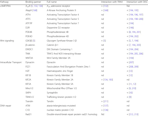
9-Table 2
Pathways interacting with DISC1 and/or TRAX
Pathway Binding partner Full name Interaction with TRAX Interaction with DISC1
cAMP/PKA A2AR [5,132,134] A2Aadenosine receptor + [132] Nd
Akap9 [168] A-Kinase Anchoring Protein 9 + [168] + [194,195]
ATF4 Activating Transcription Factor 4 nd + [194,196,197]
ATF5 Activating Transcription Factor 5 nd + [194,198–200]
ATF7IP Activating Transcription Factor 7 nd + [194]
D2R Dopamine D2 receptor nd + [146]
PDE4B Phosphodiesterase 4B nd + [8,194,201]
PDE4D Phosphodiesterase 4D nd + [194,202]
Wnt signaling GSK3β[5] Glycogen Synthase Kinase 3β + [5] + [5,7,194]
β-catenin Cateninβ-1 nd + [7,194,203]
DIXDC1 DIX Domain Containing 1 nd + [194,204]
TNIK TRAF2 And NCK Interacting Kinase nd + [194,205,206]
WNT3A Wnt Family Member 3A nd + [194]
Intracellular Transport Dynactin Dynactin nd + [207]
FEZ1 Fasciculation And Elongation Protein Zeta 1 nd + [208,209]
HZF Haematopoetic zinc finger nd + [12]
KIF1B Kinesin Family Member 1B nd + [12]
KIF2A Kinesin Family Member 2A + [134,183] nd
KIF5A Kinesin Family Member 5A nd + [11,12]
Miro1/2 Mitochondrial Rho GTPase 1/2 nd + [9,210]
SNPH Syntaphilin nd + [10]
TRAK1/2 Trafficking kinesin protein-1/2 nd + [9]
Translin Tanslin + [211] nd
DNA repair ATM ataxia-telangiectasia mutated + [137] nd
C1D nuclear matrix protein C1D + [136] nd
Rad21 Double-strand-break repair protein rad21 homolog Nd + [212,213]
GSK3
β
can be mediated by several kinases, including
AKT [51] and protein kinase A (PKA) [52]. Both GSK3
α
and GSK3
β
are expressed highly in the mouse brain [53],
whereas GSK3
β
is mainly expressed in the human brain
[54]. GSK3
β
is thus expected to play a critical role in the
brain.
As a kinase, GSK3
β
is involved in diverse biological
activities and pathways by phosphorylating its
down-stream substrates. Briefly, GSK3
β
regulates neurite
out-growth, neuronal polarization and microtubule dynamics
by phosphorylating several microtubule-associated
pro-teins (MAPs), such as tau [55], MAP1
β
[56] and
collap-sin response mediator protein-2 (CRMP-2) [57]. GSK3
β
also regulates structural synaptic plasticity. GSK3
β
phos-phorylates
β
-catenin and promotes
β
-catenin
degrad-ation [58]. GSK3
β
deletion in a subset of cortical and
hippocampal neurons results in constitutively active
β
-catenin, which reduces spine density and excitatory
synaptic neurotransmission [59]. GSK3
β
deletion in
dentate gyrus (DG) excitatory neurons also reduces the
levels of several synaptic proteins and subunits of
N-methyl-D-aspartate
(NMDA)
and
α
-amino-3-hy-droxy-5-methyl-4-
isoxazolepropionic
acid
(AMPA)
receptors and inhibits calcium/calmodulin-dependent
protein kinase II (CaMKII)/CaMKIV-cAMP response
element binding protein (CREB) signaling [60].
Further-more, GSK3
β
is involved in long-term potentiation
(LTP) and long-term depression (LTD). During LTP
induction in the DG and CA1 areas of the hippocampus,
the phosphorylation level of GSK3
β
at Ser
9is increased,
which subsequently inhibits the induction of LTD
[61–63]. GSK3
β
overexpression in the hippocampus
reduces neurotransmitter release and hyper-phosphorylates
tau, which impairs the induction of LTP and learning
[64,
65]. In addition, GSK3 inhibition rescues the
number of abnormal dendritic spines and
glutamater-gic synapses in pyramidal neurons and may improve
the psychiatric pathogenesis caused by DIXDC1/GSK3
axis impairment in mental disorders [66].
GSK3
β
is also involved in apoptotic regulation in
re-sponse to several stresses, including DNA damage [67]
and oxidative stress [68]. In response to DNA damage,
the interaction between GSK3
β
and p53 enhances the
activity of GSK3
β
and p53-mediated apoptosis via
in-creasing p21 protein levels and caspase-3 activation [67].
GSK3
β
inactivation protects hippocampal [69] and
cere-bellar granule neurons [70] from irradiation-induced
death through inhibiting p53 accumulation [71]. In
neu-rons, oxidative stress exposure for a short period of time
reduces the activity of GSK3
β
, while prolonged exposure
to ROS increases GSK3
β
activity [72,
73]. Therefore,
GSK3
β
is a redox-sensitive kinase. GSK3
β
activation in
response to oxidative stress downregulates the
nuclear-localized NF-E2-related factor 2 (NRF2), which inhibits
the expression of antioxidant genes, such as heme
oxygenase-1 (HO-1), and sensitizes neurons to oxidative
stress-induced death [72]. Furthermore, GSK3
β
activa-tion in response to oxidative stress phosphorylates and
induces the degradation of CRMP-2, a cytoskeleton
regulator involved in lithium response in bipolar
disorder patients [74], and results in axonal degeneration
and neuronal death [73,
75]. GSK3
β
inhibition is thus
expected to protect neurons from oxidative
stress-in-duced damage and death. Consistently, GSK3
β
inhibition
through activating A
2Aadenosine receptor (A
2aR) has
protective effects on oxidative stress-induced DNA
dam-age because its binding partner (TRAX) is released to
fa-cilitate DNA repair and improve survival [5].
Accumulating evidence suggests that the dysregulation
of GSK3
β
and/or its up/downstream molecules may
contribute to bipolar disorder and schizophrenia. The
inhibitory phosphorylation levels of GSK3 are lower in
the peripheral blood mononuclear cells (PBMCs) from
bipolar disorder patients than in those from healthy
controls [76], but not in platelets [77]. Interestingly,
al-though the protein levels of GSK3 are higher in PBMCs
from type 1 bipolar disorder patients than in those from
normal subjects, the amount of inhibitory GSK3
phos-phorylation shows only a decreasing trend [78].
Con-versely, the protein levels of GSK3
β
in the frontal cortex
and cerebrospinal fluid are lower in schizophrenia
patients than in normal subjects [79,
80]. However, other
studies have failed to show changes in the protein levels
or activity of GSK3
β
in patients with mental diseases
compared to normal controls [81,
82].
DISC1
DISC1 was initially identified in a large Scottish family
with a spectrum of mental diseases (including
schizo-phrenia,
recurrent
major
depression
and
bipolar
disorder) [92–95]. The N-terminal globular domain of
DISC1 contains a conserved nuclear localization signal,
and the C-terminal coiled-coil region is predicted to
me-diate its interactions with different proteins [96]. DISC1
is highly expressed in the heart, brain and placenta of
humans [94] and in the heart, brain, kidney, and testis of
mice [97]. DISC1 expression in the brain is regulated
during development; its highest level occurs during the
neonatal-infancy period and decreases gradually with
age in human brains [98]. It is important to note that
DISC1 expression may be regulated by environmental
stimuli too. For example, the activation of Toll-like
receptor 3 (TLR3) during viral infection leads to the
downregulation of DISC1 through myeloid
differenti-ation primary response gene 88 (MYD88) and
subse-quently impairs dendritic arborization and neuronal
development [99]. Such cytoarchitectural defects (e.g.,
dendritic organization) have been found in
schizo-phrenic subjects [100,
101], suggesting the importance
of DISC1 in the regulation of neuronal development at
prenatal and neonatal stages. Given the correlation
between the deficits in neuronal development and the
risk of developing schizophrenia, schizophrenia is also
referred to as a neurodevelopmental disorder.
Previous studies suggest that DISC1 functions as a
scaffold protein and mediates diverse
neurodevelop-mental processes by interacting with different proteins
(Table
2). Specifically, DISC1 regulates cytoskeletal
processes (e.g., neurite outgrowth and neuronal
mi-gration) by interacting with several proteins that are
localized to the centrosome and axonal growth cones,
including lissencephaly 1 (LIS1), nuclear distribution
nudE-Like 1 (NDEL1) [11,
102], NDE1 [103],
pericen-triolar material 1 (PCM1), and Bardet-Biedl syndrome
4 (BBS4) [104].Given that DISC1 is located at the
post-synaptic density (PSD) in the human neocortex
[105], DISC1 is likely to play an important role in
dendritic spine development and synaptic activities.
DISC1 interacts with kalirin-7 (kal-7) at the
gluta-matergic PSD and mediates the interaction between
kal-7 and PSD-95 or Rac family small GTPase 1
(Rac1) to regulate the size and number of spines [6].
Another important function of DISC1 is its regulation
of the cyclic adenosine monophosphate
(cAMP)-signal-ing pathway by bind(cAMP)-signal-ing and inhibit(cAMP)-signal-ing phosphodiesterase
4B (PDE4B; Table
2). Increased cAMP levels cause
DISC1 and PDE4B dissociation and enhance PDE4B
activity [8]. The DISC1/PDE4 complex also regulates the
PKA-mediated phosphorylation and association of a
complex (NDE1/LIS1/NDEL1) important for neuronal
development [106]. In addition, DISC1 interacts with
several key molecules in the cAMP/PKA pathway,
in-cluding an anchoring protein of PKA (A-kinase
anchor-ing protein 9 (AKAP9); Table
2), several transcription
factors (activating transcription factor 4 and 5 (ATF4
and ATF5); Table
2) that recognize the cAMP response
element, and a Gi
α
-coupled receptor that suppresses
cAMP production upon activation (D2 dopamine
recep-tor (D2R); Table
2).
DISC1 also plays an important role in intracellular
transport (Table
2). By interacting with syntaphilin
(SNPH), Mitochondrial Rho GTPase 1/2 (Miro1/2), and
Trafficking kinesin protein-1/2 (TRAK1/2), DISC1
me-diates the transport of mitochondria in the axons and
dendrites [4,
9,
10]. DISC1 is involved in the transport of
synaptic vesicles because it stabilizes the interaction
be-tween fasciculation and elongation protein zeta 1 (FEZ1)
and synaptotagmin-1 (SYT-1) in the axons [107].
More-over, DISC1 interacts with hematopoietic zinc finger
(HZF) to mediate the dendritic transport of
inositol-1,4,5--trisphosphate receptor type 1(ITRP1)
mRNA [12].
Another important interacting protein of DISC1 is
GSK3
β
, as well as several proteins involved in the Wnt
pathway (Table
2). The direct binding of DISC1 inhibits
GSK3
β
activity [7]. The interaction between DISC1 and
GSK3
β
controls the fate of neural progenitors in the
ventricular zone/subventricular zone [7] and subgranular
zone of the dentate gyrus [97]. GSK3
β
inhibition
pre-vents the phosphorylation and degradation of
β
-catenin,
its downstream target [58], resulting in increased neural
progenitor proliferation [108]. Intriguingly, the
phos-phorylation of DISC1 at Ser
710determines the affinity of
DISC1 toward its binding partners. For example,
non-phosphorylated DISC1 at Ser
710inhibits GSK3
β
and
subsequently activates
β
-catenin signaling. Conversely,
the phosphorylation of DISC1 at Ser
710increases the
af-finity of DISC1 for another binding partner, BBS protein,
which facilitates the recruitment of BBS to the
centro-some and subsequently causes the transition from
pro-genitor proliferation to neuronal migration in the
developing cortex [109]. It is interesting to note that the
PKA-mediated inhibitory phosphorylation of GSK3
β
at
Ser
9leads to the dissociation of the DISC1/GSK3
β
/
TRAX complex and facilitates TRAX-mediated DNA
repair in neurons [5]. These results collectively suggest
that phosphorylation is a key modulatory mechanism
that regulates the complex formation of DISC1 and
other interaction proteins through which a wide variety
of cellular functions are regulated.
dominant-negative C-terminal truncated DISC1 that
loses its interaction with DISC1-interacting proteins
[110,
111]. In addition, expression levels of the
DISC1-interacting proteins LIS1 and NDEL1 are decreased in
the brains of schizophrenia patients and are associated
with high-risk DISC1 SNPs [98]. To date, the DISC1
gene has been identified as a risk factor for major mental
illnesses [13,
112–120]. In contrast, some other reports
have failed to show the association between DISC1
vari-ants and mental diseases [121–123]. Further investigations
of the roles of DISC1 in mental disorders are needed.
TRAX
TRAX was first discovered as a binding partner of translin
using a yeast two-hybrid system. Amino acid sequence
alignment revealed that TRAX displays 28% identity with
translin [16]. Because the genetic removal of translin
pro-motes the degradation of TRAX, TRAX stability appears
to be controlled by its binding partner (i.e., translin [124]).
Both TRAX and translin are highly enriched in the brain.
The heteromeric complex composed of TRAX and
trans-lin shows nucleic acid binding activity in brain extracts
[125] and plays a role in dendritic RNA trafficking in
neurons [126,
127]. The heteromeric translin/TRAX
com-plex also functions as an endoribonuclease that cleaves
passenger strands of siRNA and therefore facilities siRNA
guide strand loading onto the RNA-induced silencing
complex (RISC) in
Drosophila
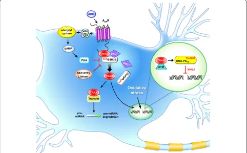
[128]. In contrast, the
TRAX/translin complex suppresses microRNA
(miR-NA)-mediated silencing in mammalian cells by degrading
pre-miRNA with mismatched stems and subsequently
reversing miRNA-mediated silencing [129] (Fig.
1). In
support of this hypothesis, TRAX/translin was recently
shown to play a critical role in regulating long-term
mem-ory by suppressing microRNA silencing at activated
synapses [130]. Given that aberrant profiles of miRNAs
and their targeted genes have been implicated in mental
disorders (such as schizophrenia, bipolar disorder and
autism) (for a review, see [131]), the role of abnormal
TRAX/translin regulation in mental disorders warrants
future investigations.
Similar to DISC1, TRAX also has many interacting
proteins with a wide variety of functions. Most of these
TRAX-interacting proteins are risk genes, markers, or
drug targets for psychotic disorders (e.g., schizophrenia,
autism, and panic disorders; Table
1). For example,
A
2AR is the binding partner of TRAX [132]. A
2AR is a
Gs
α
-coupled receptor that activates the cAMP/PKA
pathway upon stimulation [133]. A
2AR activation or
TRAX overexpression rescues the impaired neurite
outgrowth caused by p53 blockade in a neuronal cell line
(PC12) and primary hippocampal neurons. Knocking
down TRAX or preventing the interaction between
TRAX and its interacting protein (kinesin heavy chain
member 2A, KIF2A) blocks the rescue effect of A
2AR
activation [134]. Of note, KIF2A is a schizophrenia
susceptibility gene [135]. A
2AR is a risk gene for autism
and anxiety disorders, and a marker for schizophrenia
(Table
1).
Two of the TRAX-interacting proteins (C1D and
ataxia-telangiectasia mutated (ATM) kinase) are involved
directly in DNA repair. C1D is an activator of
DNA-dependent protein kinase (DNA-PK). Upon DNA
damage induced by
γ
-irradiation, TRAX increasingly
interacts with C1D in mammalian cells, suggesting that
TRAX might participate in DNA repair [136]. ATM is a
serine/threonine kinase and is activated and recruited by
DNA double-strand breaks (DSBs) to phosphorylated
proteins (e.g., histone H2A (H2AX) and p53) that are
important for DNA repair. In the absence of TRAX,
ATM fails to be recruited to DSB sites to initiate the
DNA repair machinery and subsequently causes cell
death due to insufficient DNA damage repair [5,
137].
During oxidative stress-induced DNA damage in
neu-rons, TRAX forms a complex with DISC1 and GSK3
β
in
the cytoplasmic region. A
2AR stimulation activates PKA,
which phosphorylates GSK3
β
at Ser
9and dissociates the
TRAX/DISC1/GSK3
β
complex so that TRAX can enter
the nuclei to facilitate DNA repair [5] (Fig.
1). The role
of TRAX and its interacting proteins in mental disorders
appear important because ample evidence suggests
that incomplete oxidative DNA damage repair may
contribute to the development of psychotic disorders
[1,
17,
18]. Most of the major components (including
ATM, [138,
139]) involved in TRAX-mediated DNA
repair are also risk genes of mental disorders (Table
1).
Consistent with the hypothesis that TRAX is involved
in the development of mental disorders, genetic studies
have implicated TRAX in major psychiatric diseases.
The human
TSNAX
gene is located at 1q42.1 and
adja-cent to the
DIS
C1 gene. Several
TSNAX
transcripts
con-tain the
DIS
C1 sequence at the 3’
end due to intergenic
splicing in human adult and fetal tissues [93]. A SNP
analysis revealed that 2 SNPs (i.e., rs1615409 and
rs766288) are located within intron 4 of
TSNAX
, and 2
SNPs (i.e., rs751229 and rs3738401) were found in
DISC1
in Finnish schizophrenia patients [13]. A rare
AATG haplotype comprising these 4 SNPs is positively
associated with the reaction time to visual targets and
negatively with the gray matter density in Finnish
schizophrenia patients [140]. Furthermore, another SNP
analysis identified that rs1655285 at intron 5 of
TSNAX
and a haplotype comprising rs1630250 and rs1615409
within
TSNAX
are associated with Finnish bipolar
spectrum disorder [15]. The SNP rs766288 at intron 4 of
TSNAX
has been reported to be associated with
Japanese female major depressive disorder [14]. These
studies collectively suggest that TRAX is a risk gene for
major mental diseases. It should also be noted that
TRAX and DISC1 share several interacting proteins
(e.g., GSK3
β
and AKAP9; Table
2) and functional
pathways/machineries (e.g., the cAMP/PKA pathway,
Wnt signaling, intracellular transport and DNA repair;
Table
2); thus, they may act together to regulate
import-ant pathophysiological events, including the
develop-ment of develop-mental disorders.
Regulation of the TRAX/DISC1/GSK3
β
complex and
therapeutic relevance
Although DISC1 mediates many different cellular
func-tions, it has not been implicated in DNA repair until a
recent report [5] demonstrating that DISC1 interacts
with GSK3
β
and TRAX; this complex facilitates DNA
repair by binding to ATM [137]. This finding leads to a
new mechanistic role of DISC1 in mental disorders in
which accumulating oxidative DNA damage and
in-sufficient DNA repair contribute to the pathogenesis
[1,
17,
18]. Disassembly of the TRAX/DISC1/GSK3
β
complex, followed by the release of TRAX, provides a
new means to facilitate DNA repair and ameliorate
the damage caused by unrepaired DSBs. For example,
A
2AR activation dissociates TRAX/DISC1/GSK3
β
com-plex tethering at its C terminus through a PKA-dependent
pathway and amends the DNA damage-induced apoptosis
[5]. Consistent with an important role of A
2AR in
facilitat-ing DNA repair, A
2AR activation ameliorates oxidative
DNA damage in human medium spiny neurons (MSNs)
derived from induced pluripotent stem cells (iPSCs) [141].
Interestingly, the amount of A
2AR is altered in different
brain regions of patients with schizophrenia [142,
143],
supporting that A
2AR might play an important role in
schizophrenia. Because A
2AR is an antagonistic binding
partner of the D2R and may suppress the hyperfunction
of D2R in schizophrenia [144], A
2AR agonists are
poten-tially advantageous anti-schizophrenic drugs (for a review,
see [145]). D2R is a primary target of antipsychotic drugs.
It forms complex with not only A
2AR but also DISC1 to
It is important to note that adenosine is known to
regulate the dopamine and glutamate- mediated
neuro-transmissions,
the
major
neurotransmitter
systems
involved in schizophrenia pathophysiology [147–149].
Dysfunction of purinergic system is one of the factors
that cause schizophrenia [149,
150]. Moreover, inhibition
of adenosine kinase (ADK), which controls adenosine
level, exhibits anti-psychotic-like efficacy, while
overex-pression of ADK causes changes in the sensitivity to
psychomimetic drugs in mice [151,
152]. Consistent with
the abovementioned hypothesis, increased brain
adeno-sine tone using an inhibitor of adenoadeno-sine uptake (i.e.,
dipyridamole) indirectly activates adenosine receptors
and hasWr beneficial effects on patients with
schizo-phrenia [153]. Likewise, inhibiting adenosine clearance
using ABT702 to globally increase adenosine tone also
ameliorates the psychotic and cognitive phenotypes of
schizophrenia in mice [151]. Given that adenosine is also
known to play an important role during
neurodeve-lopment. (for a review, see [154]). Augmenting the
adenosine tone in the brain using various approaches
might thus serve as a therapeutic means to treat
schizophrenia as well as to prevent the development
of schizophrenia [154,
155].
Lithium is an inhibitor of GSK3 and a common mood
stabilizer for treating mental disorders. To date, the
underlying molecular mechanism of lithium’s action
remains largely elusive [156,
157]. Accumulating
evi-dence suggests that chronic treatment with lithium
in-hibits the oxidative damage evoked by glutamate [158]
and increases the expression level of the anti-apoptotic
factor Bcl2 [159,
160]. Treatment with lithium also
pro-tects neurons by facilitating the NHEJ repair-mediated
DNA repair pathway [161]. Chronic treatment with
lith-ium results in not only the inhibition of GSK3
β
but also
the regulation of many anti-apoptotic proteins. For
example, lithium inhibits calcium influx via regulating
the NMDA receptor and reduces apoptosis by directly
inhibiting GSK3
β
[162]. Because inhibiting GSK3
β
causes the disassembly of the TRAX/DISC1/GSK3
β
complex and releases TRAX to facilitate DNA repair [5],
at least part of the actions of lithium might be mediated
by the TRAX/DISC1/GSK3
β
complex. New GSK3
β
inhibitors have been actively developed for brain diseases
[163], which may pave the way for establishing new
treatments for schizophrenia.
Conclusions
As a major gene implicated in schizophrenia and other
mental disorders, DISC1 is known to regulate various
cellular functions by interacting with proteins of
differ-ent machineries. Ample evidence suggests that DISC1 is
a hub protein for schizophrenia and possibly other
mental diseases. Emerging evidence also suggests that
TRAX, a neighboring gene of DISC1, not only physically
interacts with DISC1 but also switches binding partners
under different pathophysiological conditions as does
DISC1. Because the studies regarding TRAX are still in
their infancy, the overlapping functional pathways of
DISC1 and TRAX appear limited at this time (Table
2)
but may become more evident when more binding
part-ners of TRAX are revealed in the future. Most
import-antly, genetic evidence suggests that DISC1 and TRAX
are closely associated with several major mental
disor-ders (such as schizophrenia, autism, and anxiety
disorder; Table
1). Therefore, it is certainly worth further
exploring the role of the DISC1/TRAX complex in
psychiatric disorders. Because oxidative DNA damage
accumulation and insufficient DNA repair have been
im-plicated in the development and progression of mental
disorders, the recently reported function of the DISC1/
TRAX/GSK3
β
complex in DNA repair also warrants
fur-ther investigations of the temple and the special
regula-tion of this complex during neuronal development and
disease progression. Further understanding of when and
where the DISC1/TRAX/GSK3
β
complex is formed and
how the complex can be effectively dissembled by either
GSK3
β
inhibitors or PKA activators (such as A
2AR
ago-nists or PDE4 inhibitors) would pave the way for
devel-oping new therapeutic agents for mental disorders.
Abbreviations
8-OHdG:8-hydroxy-2'-deoxyguanosine; A2aR: A2Aadenosine receptor; ADK: Adenosine kinase; ADO: Adenosine; AKAP9: A-kinase anchoring protein 9; AMPA:α-amino-3-hydroxy-5-methyl-4- isoxazolepropionic acid;
ATF: Activating transcription factor; ATM: Ataxia-telangiectasia mutated; BBS4: Bardet-Biedl syndrome 4; C1D: Nuclear matrix protein C1D; cAMP: Cyclic adenosine monophosphate; CREB: Calcium/calmodulin-dependent protein kinase II (CaMKII)/CaMKIV-cAMP response element binding protein; CRMP-2: Collapsin response mediator protein-2; D2R: D2 Dopamine receptor; DG: Dentate gyrus; DISC1: Disrupted in schizophrenia 1; DIXDC1: DIX domain containing 1; DNA-PK: DNA- dependent protein kinase; DSBs: DNA double-strand breaks; ETC: Electron transport chain;
FEZ1: Fasciculation and elongation protein zeta 1; GSK3: Glycogen synthase kinase 3; H2AX: Histone H2A; HO-1: Heme oxygenase-1; hOGG1: Human 8-oxoguanine DNA N-glycosylase 1; HZF: Hematopoietic zinc finger; iPSCs: Induced pluripotent stem cells; ITRP1: Inositol-1,4,5-trisphosphate receptor type 1; kal-7: Kalirin-7; KIF2A: Kinesin heavy chain member 2A; LIS1: Lissencephaly 1; LTD: Long-term depression; LTP: Long-term potentiation; MAPs: Microtubule-associated proteins; MEA2: Male-enhanced antigen 2; miRNA: microRNA; Miro1/2: Mitochondrial Rho GTPase 1/2; MSNs: Medium spiny neurons; MYD88: Myeloid differentiation primary response gene 88; NDEL1: Nuclear distribution nudE-Like 1;
NDUFV2: NADH:Ubiquinone oxidoreductase core subunit V2; NHEJ: Non-homologous end joining; NMDA: N-methyl-D-aspartate; NRF2: Nuclear-localized NF-E2-related factor 2; PBMCs: Peripheral blood mononuclear cells; PCM1: Pericentriolar material 1; PCP: Phencyclidine;
Acknowledgments
We thank Mr. Tsung Hung Hung at the Medical Art Room of Institute of Biomedical Sciences, Academia Sinica for the preparation of Fig.1.
Funding
This work was supported by the Institute of the Biomedical Sciences of Academia Sinica (103-Academia Sinica Investigation Award-06).
Authors’contributions
YC outlined and wrote the manuscript. YTW wrote part of the manuscript and prepared the tables. TC wrote part of the manuscript and prepared the figure and table. IIK analyzed literatures and prepared tables. All authors read and approved the final manuscript.
Ethics approval and consent to participate Not applicable.
Consent for publication Not applicable.
Competing interests
The authors declare that they have no competing interests.
Publisher
’
s Note
Springer Nature remains neutral with regard to jurisdictional claims in published maps and institutional affiliations.
Received: 7 June 2018 Accepted: 25 September 2018
References
1. Markkanen E, Meyer U, Dianov GL. DNA Damage and Repair in Schizophrenia and Autism: Implications for Cancer Comorbidity and Beyond. Int J Mol Sci. 2016;17(6):856.
2. Odemis S, Tuzun E, Gulec H, Semiz UB, Dasdemir S, Kucuk M, Yalcinkaya N, Bireller ES, Cakmakoglu B, Kucukali CI. Association between polymorphisms of DNA repair genes and risk of schizophrenia. Genet Test Mol Biomarkers. 2016;20(1):11–7.
3. Raza MU, Tufan T, Wang Y, Hill C, Zhu MY. DNA damage in major psychiatric diseases. Neurotox Res. 2016;30(2):251–67.
4. Atkin TA, MacAskill AF, Brandon NJ, Kittler JT. Disrupted in Schizophrenia-1 regulates intracellular trafficking of mitochondria in neurons. Mol Psychiatry. 2011;16(2):122–4.
5. Chien T, Weng YT, Chang SY, Lai HL, Chiu FL, Kuo HC, Chuang DM, Chern Y. GSK3beta negatively regulates TRAX, a scaffold protein implicated in mental disorders, for NHEJ-mediated DNA repair in neurons. Mol Psychiatry. 2018. https://doi.org/10.1038/s41380-017-0007-z.
6. Hayashi-Takagi A, Takaki M, Graziane N, Seshadri S, Murdoch H, Dunlop AJ, Makino Y, Seshadri AJ, Ishizuka K, Srivastava DP, Xie Z, Baraban JM, Houslay MD, Tomoda T, Brandon NJ, Kamiya A, Yan Z, Penzes P, Sawa A. Disrupted-in-schizophrenia 1 (DISC1) regulates spines of the glutamate synapse via Rac1. Nat Neurosci. 2010;13(3):327–32.
7. Mao Y, Ge X, Frank CL, Madison JM, Koehler AN, Doud MK, Tassa C, Berry EM, Soda T, Singh KK, Biechele T, Petryshen TL, Moon RT, Haggarty SJ, Tsai LH. Disrupted in schizophrenia 1 regulates neuronal progenitor proliferation via modulation of GSK3beta/beta-catenin signaling. Cell. 2009;136(6):1017–31. 8. Millar JK, Pickard BS, Mackie S, James R, Christie S, Buchanan SR, Malloy MP, Chubb JE, Huston E, Baillie GS, Thomson PA, Hill EV, Brandon NJ, Rain JC, Camargo LM, Whiting PJ, Houslay MD, Blackwood DH, Muir WJ, Porteous DJ. DISC1 and PDE4B are interacting genetic factors in schizophrenia that regulate cAMP signaling. Science. 2005;310(5751):1187–91.
9. Norkett R, Modi S, Birsa N, Atkin TA, Ivankovic D, Pathania M, Trossbach SV, Korth C, Hirst WD, Kittler JT. DISC1-dependent regulation of mitochondrial dynamics controls the morphogenesis of complex neuronal dendrites. J Biol Chem. 2016;291(2):613–29.
10. Park C, Lee SA, Hong JH, Suh Y, Park SJ, Suh BK, Woo Y, Choi J, Huh JW, Kim YM, Park SK. Disrupted-in-schizophrenia 1 (DISC1) and Syntaphilin collaborate to modulate axonal mitochondrial anchoring. Mol Brain. 2016;9(1):69.
11. Taya S, Shinoda T, Tsuboi D, Asaki J, Nagai K, Hikita T, Kuroda S, Kuroda K, Shimizu M, Hirotsune S, Iwamatsu A, Kaibuchi K. DISC1 regulates the
transport of the NUDEL/LIS1/14-3-3epsilon complex through kinesin-1. J Neurosci. 2007;27(1):15–26.
12. Tsuboi D, Kuroda K, Tanaka M, Namba T, Iizuka Y, Taya S, Shinoda T, Hikita T, Muraoka S, Iizuka M, Nimura A, Mizoguchi A, Shiina N, Sokabe M, Okano H, Mikoshiba K, Kaibuchi K. Disrupted-in-schizophrenia 1 regulates transport of ITPR1 mRNA for synaptic plasticity. Nat Neurosci. 2015;18(5):698–707. 13. Hennah W, Varilo T, Kestila M, Paunio T, Arajarvi R, Haukka J, Parker A, Martin
R, Levitzky S, Partonen T, Meyer J, Lonnqvist J, Peltonen L, Ekelund J. Haplotype transmission analysis provides evidence of association for DISC1 to schizophrenia and suggests sex-dependent effects. Hum Mol Genet. 2003;12(23):3151–9.
14. Okuda A, Kishi T, Okochi T, Ikeda M, Kitajima T, Tsunoka T, Okumukura T, Fukuo Y, Kinoshita Y, Kawashima K, Yamanouchi Y, Inada T, Ozaki N, Iwata N. Translin-associated factor X gene (TSNAX) may be associated with female major depressive disorder in the Japanese population. NeuroMolecular Med. 2010;12(1):78–85.
15. Palo OM, Antila M, Silander K, Hennah W, Kilpinen H, Soronen P, Tuulio-Henriksson A, Kieseppa T, Partonen T, Lonnqvist J, Peltonen L, Paunio T. Association of distinct allelic haplotypes of DISC1 with psychotic and bipolar spectrum disorders and with underlying cognitive impairments. Hum Mol Genet. 2007;16(20):2517–28.
16. Aoki K, Ishida R, Kasai M. Isolation and characterization of a cDNA encoding a Translin-like protein, TRAX. FEBS Lett. 1997;401(2–3):109–12.
17. Steckert AV, Valvassori SS, Moretti M, Dal-Pizzol F, Quevedo J. Role of oxidative stress in the pathophysiology of bipolar disorder. Neurochem Res. 2010;35(9):1295–301.
18. Wang JF, Shao L, Sun X, Young LT. Increased oxidative stress in the anterior cingulate cortex of subjects with bipolar disorder and schizophrenia. Bipolar Disord. 2009;11(5):523–9.
19. Prabakaran S, Swatton JE, Ryan MM, Huffaker SJ, Huang JT, Griffin JL, Wayland M, Freeman T, Dudbridge F, Lilley KS, Karp NA, Hester S, Tkachev D, Mimmack ML, Yolken RH, Webster MJ, Torrey EF, Bahn S. Mitochondrial dysfunction in schizophrenia: evidence for compromised brain metabolism and oxidative stress. Mol Psychiatry. 2004;9(7):684–97.
20. Kavanagh JN, Redmond KM, Schettino G, Prise KM. DNA double strand break repair: a radiation perspective. Antioxid Redox Signal. 2013;18(18):2458–72. 21. Mariani E, Polidori MC, Cherubini A, Mecocci P. Oxidative stress in brain
aging, neurodegenerative and vascular diseases: an overview. J Chromatogr B Analyt Technol Biomed Life Sci. 2005;827(1):65–75.
22. Chestkov IV, Jestkova EM, Ershova ES, Golimbet VG, Lezheiko TV, Kolesina NY, Dolgikh OA, Izhevskaya VL, Kostyuk GP, Kutsev SI, Veiko NN, Kostyuk SV. ROS-induced DNA damage associates with abundance of mitochondrial DNA in white blood cells of the untreated schizophrenic patients. Oxidative Med Cell Longev. 2018;8587475:2018.
23. Hassan W, Noreen H, Castro-Gomes V, Mohammadzai I, da Rocha JB, Landeira-Fernandez J. Association of Oxidative Stress with psychiatric disorders. Curr Pharm Des. 2016;22(20):2960–74.
24. van Velzen LS, Wijdeveld M, Black CN, van Tol MJ, van der Wee NJA, Veltman DJ, Penninx B, Schmaal L. Oxidative stress and brain morphology in individuals with depression, anxiety and healthy controls. Prog Neuro-Psychopharmacol Biol Psychiatry. 2017;76:140–4.
25. Ng F, Berk M, Dean O, Bush AI. Oxidative stress in psychiatric disorders: evidence base and therapeutic implications. Int J Neuropsychopharmacol. 2008;11(6):851–76.
26. Salim S. Oxidative stress and psychological disorders. Curr Neuropharmacol. 2014;12(2):140–7.
27. Salim S. Oxidative stress and the central nervous system. J Pharmacol Exp Ther. 2017;360(1):201–5.
28. Ceylan D, Scola G, Tunca Z, Isaacs-Trepanier C, Can G, Andreazza AC, Young LT, Ozerdem A. DNA redox modulations and global DNA methylation in bipolar disorder: effects of sex, smoking and illness state. Psychiatry Res. 2018;261:589–96.
29. Chen CM, Wu YR, Cheng ML, Liu JL, Lee YM, Lee PW, Soong BW, Chiu DT. Increased oxidative damage and mitochondrial abnormalities in the peripheral blood of Huntington's disease patients. Biochem Biophys Res Commun. 2007;359(2):335–40.
30. Huang MC, Lai YC, Lin SK, Chen CH. Increased blood 8-hydroxy-2-deoxyguanosine levels in methamphetamine users during early abstinence. Am J Drug Alcohol Abuse. 2018;44(3):395–402.
DNA oxidation in bipolar I disorder. Int J Neuropsychopharmacol. 2013; 16(7):1505–12.
32. Deicken RF, Fein G, Weiner MW. Abnormal frontal lobe phosphorous metabolism in bipolar disorder. Am J Psychiatry. 1995;152(6):915–8. 33. Ben-Shachar D, Karry R. Sp1 expression is disrupted in schizophrenia; a
possible mechanism for the abnormal expression of mitochondrial complex I genes, NDUFV1 and NDUFV2. PLoS One. 2007;2(9):e817.
34. Konradi C, Eaton M, MacDonald ML, Walsh J, Benes FM, Heckers S. Molecular evidence for mitochondrial dysfunction in bipolar disorder. Arch Gen Psychiatry. 2004;61(3):300–8.
35. Sun X, Wang JF, Tseng M, Young LT. Downregulation in components of the mitochondrial electron transport chain in the postmortem frontal cortex of subjects with bipolar disorder. J Psychiatry Neurosci. 2006;31(3):189–96. 36. Washizuka S, Kakiuchi C, Mori K, Kunugi H, Tajima O, Akiyama T, Nanko S,
Kato T. Association of mitochondrial complex I subunit gene NDUFV2 at 18p11 with bipolar disorder. Am J Med Genet B Neuropsychiatr Genet. 2003;120B(1):72–8.
37. Howes O, McCutcheon R, Stone J. Glutamate and dopamine in schizophrenia: an update for the 21st century. J Psychopharmacol. 2015; 29(2):97–115.
38. Banerjee A, Wang HY, Borgmann-Winter KE, MacDonald ML, Kaprielian H, Stucky A, Kvasic J, Egbujo C, Ray R, Talbot K, Hemby SE, Siegel SJ, Arnold SE, Sleiman P, Chang X, Hakonarson H, Gur RE, Hahn CG. Src kinase as a mediator of convergent molecular abnormalities leading to NMDAR hypoactivity in schizophrenia. Mol Psychiatry. 2015;20(9): 1091–100.
39. Gao XM, Sakai K, Roberts RC, Conley RR, Dean B, Tamminga CA. Ionotropic glutamate receptors and expression of N-methyl-D-aspartate receptor subunits in subregions of human hippocampus: effects of schizophrenia. Am J Psychiatry. 2000;157(7):1141–9.
40. Farber NB. The NMDA receptor hypofunction model of psychosis. Ann N Y Acad Sci. 2003;1003:119–30.
41. Javitt DC. Glutamate and schizophrenia: phencyclidine, N-methyl-D-aspartate receptors, and dopamine-glutamate interactions. Int Rev Neurobiol. 2007;78:69–108.
42. Hardingham GE, Fukunaga Y, Bading H. Extrasynaptic NMDARs oppose synaptic NMDARs by triggering CREB shut-off and cell death pathways. Nat Neurosci. 2002;5(5):405–14.
43. Hirose K, Chan PH. Blockade of glutamate excitotoxicity and its clinical applications. Neurochem Res. 1993;18(4):479–83.
44. Zeevalk GD, Bernard LP, Sinha C, Ehrhart J, Nicklas WJ. Excitotoxicity and oxidative stress during inhibition of energy metabolism. Dev Neurosci. 1998;20(4–5):444–53.
45. Embi N, Rylatt DB, Cohen P. Glycogen synthase kinase-3 from rabbit skeletal muscle. Separation from cyclic-AMP-dependent protein kinase and phosphorylase kinase. Eur J Biochem. 1980;107(2):519–27.
46. Ali A, Hoeflich KP, Woodgett JR. Glycogen synthase kinase-3: properties, functions, and regulation. Chem Rev. 2001;101(8):2527–40.
47. Hughes K, Nikolakaki E, Plyte SE, Totty NF, Woodgett JR. Modulation of the glycogen synthase kinase-3 family by tyrosine phosphorylation. EMBO J. 1993;12(2):803–8.
48. Stambolic V, Woodgett JR. Mitogen inactivation of glycogen synthase kinase-3 beta in intact cells via serine 9 phosphorylation. Biochem J. 1994;303(Pt 3):701–4.
49. Sutherland C, Cohen P. The alpha-isoform of glycogen synthase kinase-3 from rabbit skeletal muscle is inactivated by p70 S6 kinase or MAP kinase-activated protein kinase-1 in vitro. FEBS Lett. 1994;338(1):37–42. 50. Cole A, Frame S, Cohen P. Further evidence that the tyrosine
phosphorylation of glycogen synthase kinase-3 (GSK3) in mammalian cells is an autophosphorylation event. Biochem J. 2004;377(Pt 1):249–55. 51. Cross DA, Alessi DR, Cohen P, Andjelkovich M, Hemmings BA. Inhibition of
glycogen synthase kinase-3 by insulin mediated by protein kinase B. Nature. 1995;378(6559):785–9.
52. Fang X, Yu SX, Lu Y, Bast RC Jr, Woodgett JR, Mills GB. Phosphorylation and inactivation of glycogen synthase kinase 3 by protein kinase A. Proc Natl Acad Sci U S A. 2000;97(22):11960–5.
53. Yao HB, Shaw PC, Wong CC, Wan DC. Expression of glycogen synthase kinase-3 isoforms in mouse tissues and their transcription in the brain. J Chem Neuroanat. 2002;23(4):291–7.
54. Lau KF, Miller CC, Anderton BH, Shaw PC. Expression analysis of glycogen synthase kinase-3 in human tissues. J Pept Res. 1999;54(1):85–91.
55. Cho JH, Johnson GV. Glycogen synthase kinase 3beta phosphorylates tau at both primed and unprimed sites. Differential impact on microtubule binding. J Biol Chem. 2003;278(1):187–93.
56. Lucas FR, Goold RG, Gordon-Weeks PR, Salinas PC. Inhibition of GSK-3beta leading to the loss of phosphorylated MAP-1B is an early event in axonal remodelling induced by WNT-7a or lithium. J Cell Sci. 1998; 111(Pt 10):1351–61.
57. Yoshimura T, Kawano Y, Arimura N, Kawabata S, Kikuchi A, Kaibuchi K. GSK-3beta regulates phosphorylation of CRMP-2 and neuronal polarity. Cell. 2005;120(1):137–49.
58. Aberle H, Bauer A, Stappert J, Kispert A, Kemler R. Beta-catenin is a target for the ubiquitin-proteasome pathway. EMBO J. 1997;16(13):3797–804. 59. Ochs SM, Dorostkar MM, Aramuni G, Schon C, Filser S, Poschl J, Kremer A,
Van Leuven F, Ovsepian SV, Herms J. Loss of neuronal GSK3beta reduces dendritic spine stability and attenuates excitatory synaptic transmission via beta-catenin. Mol Psychiatry. 2015;20(4):482–9.
60. Liu E, Xie AJ, Zhou Q, Li M, Zhang S, Li S, Wang W, Wang X, Wang Q, Wang JZ. GSK-3beta deletion in dentate gyrus excitatory neuron impairs synaptic plasticity and memory. Sci Rep. 2017;7(1):5781.
61. Cai F, Wang F, Lin FK, Liu C, Ma LQ, Liu J, Wu WN, Wang W, Wang JH, Chen JG. Redox modulation of long-term potentiation in the hippocampus via regulation of the glycogen synthase kinase-3beta pathway. Free Radic Biol Med. 2008;45(7):964–70.
62. Hooper C, Markevich V, Plattner F, Killick R, Schofield E, Engel T, Hernandez F, Anderton B, Rosenblum K, Bliss T, Cooke SF, Avila J, Lucas JJ, Giese KP, Stephenson J, Lovestone S. Glycogen synthase kinase-3 inhibition is integral to long-term potentiation. Eur J Neurosci. 2007;25(1):81–6.
63. Peineau S, Taghibiglou C, Bradley C, Wong TP, Liu L, Lu J, Lo E, Wu D, Saule E, Bouschet T, Matthews P, Isaac JT, Bortolotto ZA, Wang YT, Collingridge GL. LTP inhibits LTD in the hippocampus via regulation of GSK3beta. Neuron. 2007;53(5):703–17.
64. Gomez de Barreda E, Perez M, Gomez Ramos P, de Cristobal J, Martin-Maestro P, Moran A, Dawson HN, Vitek MP, Lucas JJ, Hernandez F, Avila J. Tau-knockout mice show reduced GSK3-induced hippocampal degeneration and learning deficits. Neurobiol Dis. 2010;37(3):622–9. 65. Zhu LQ, Wang SH, Liu D, Yin YY, Tian Q, Wang XC, Wang Q, Chen JG, Wang
JZ. Activation of glycogen synthase kinase-3 inhibits long-term potentiation with synapse-associated impairments. J Neurosci. 2007;27(45):12211–20. 66. Martin PM, Stanley RE, Ross AP, Freitas AE, Moyer CE, Brumback AC, Iafrati J,
Stapornwongkul KS, Dominguez S, Kivimae S, Mulligan KA, Pirooznia M, McCombie WR, Potash JB, Zandi PP, Purcell SM, Sanders SJ, Zuo Y, Sohal VS, Cheyette BNR. DIXDC1 contributes to psychiatric susceptibility by regulating dendritic spine and glutamatergic synapse density via GSK3 and Wnt/beta-catenin signaling. Mol Psychiatry. 2018;23(2):467–75.
67. Watcharasit P, Bijur GN, Zmijewski JW, Song L, Zmijewska A, Chen X, Johnson GV, Jope RS. Direct, activating interaction between glycogen synthase kinase-3beta and p53 after DNA damage. Proc Natl Acad Sci U S A. 2002;99(12):7951–5.
68. Wang Z, Ge Y, Bao H, Dworkin L, Peng A, Gong R. Redox-sensitive glycogen synthase kinase 3beta-directed control of mitochondrial permeability transition: rheostatic regulation of acute kidney injury. Free Radic Biol Med. 2013;65:849–58.
69. Yazlovitskaya EM, Edwards E, Thotala D, Fu A, Osusky KL, Whetsell WO Jr, Boone B, Shinohara ET, Hallahan DE. Lithium treatment prevents neurocognitive deficit resulting from cranial irradiation. Cancer Res. 2006; 66(23):11179–86.
70. Cross DA, Culbert AA, Chalmers KA, Facci L, Skaper SD, Reith AD. Selective small-molecule inhibitors of glycogen synthase kinase-3 activity protect primary neurones from death. J Neurochem. 2001;77(1):94–102.
71. Thotala DK, Hallahan DE, Yazlovitskaya EM. Glycogen synthase kinase 3beta inhibitors protect hippocampal neurons from radiation-induced apoptosis by regulating MDM2-p53 pathway. Cell Death Differ. 2012;19(3):387–96. 72. Rojo AI, Sagarra MR, Cuadrado A. GSK-3beta down-regulates the
transcription factor Nrf2 after oxidant damage: relevance to exposure of neuronal cells to oxidative stress. J Neurochem. 2008;105(1):192–202. 73. Wakatsuki S, Furuno A, Ohshima M, Araki T. Oxidative stress-dependent
phosphorylation activates ZNRF1 to induce neuronal/axonal degeneration. J Cell Biol. 2015;211(4):881–96.
Hunsberger J, Dorsett L, Duerr C, Basa RCB, McCarthy MJ, Udeshi ND, Mertins P, Carr SA, Rouleau GA, Mastrangelo L, Li J, Gutierrez GJ, Brill LM, Venizelos N, Chen G, Nye JS, Manji H, Price JH, McClung CA, Akiskal HS, Alda M, Chuang DM, Coyle JT, Liu Y, Teng YD, Ohshima T, Mikoshiba K, Sidman RL, Halpain S, Haggarty SJ, Goshima Y, Snyder EY. Probing the lithium-response pathway in hiPSCs implicates the phosphoregulatory set-point for a cytoskeletal modulator in bipolar pathogenesis. Proc Natl Acad Sci U S A. 2017;114(22):E4462–71.
75. Wakatsuki S, Saitoh F, Araki T. ZNRF1 promotes Wallerian degeneration by degrading AKT to induce GSK3B-dependent CRMP2 phosphorylation. Nat Cell Biol. 2011;13(12):1415–23.
76. Polter A, Beurel E, Yang S, Garner R, Song L, Miller CA, Sweatt JD, McMahon L, Bartolucci AA, Li X, Jope RS. Deficiency in the inhibitory serine-phosphorylation of glycogen synthase kinase-3 increases sensitivity to mood disturbances. Neuropsychopharmacology. 2010;35(8):1761–74. 77. de Sousa RT, Zanetti MV, Talib LL, Serpa MH, Chaim TM, Carvalho AF,
Brunoni AR, Busatto GF, Gattaz WF, Machado-Vieira R. Lithium increases platelet serine-9 phosphorylated GSK-3beta levels in drug-free bipolar disorder during depressive episodes. J Psychiatr Res. 2015;62:78–83. 78. Li X, Liu M, Cai Z, Wang G, Li X. Regulation of glycogen synthase kinase-3
during bipolar mania treatment. Bipolar Disord. 2010;12(7):741–52. 79. Kozlovsky N, Belmaker RH, Agam G. Low GSK-3beta immunoreactivity in
postmortem frontal cortex of schizophrenic patients. Am J Psychiatry. 2000; 157(5):831–3.
80. Kozlovsky N, Regenold WT, Levine J, Rapoport A, Belmaker RH, Agam G. GSK-3beta in cerebrospinal fluid of schizophrenia patients. J Neural Transm. 2004;111(8):1093–8.
81. Beasley C, Cotter D, Everall I. An investigation of the Wnt-signalling pathway in the prefrontal cortex in schizophrenia, bipolar disorder and major depressive disorder. Schizophr Res. 2002;58(1):63–7.
82. Ide M, Ohnishi T, Murayama M, Matsumoto I, Yamada K, Iwayama Y, Dedova I, Toyota T, Asada T, Takashima A, Yoshikawa T. Failure to support a genetic contribution of AKT1 polymorphisms and altered AKT signaling in schizophrenia. J Neurochem. 2006;99(1):277–87.
83. Klein PS, Melton DA. A molecular mechanism for the effect of lithium on development. Proc Natl Acad Sci U S A. 1996;93(16):8455–9.
84. Ryves WJ, Harwood AJ. Lithium inhibits glycogen synthase kinase-3 by competition for magnesium. Biochem Biophys Res Commun. 2001;280(3): 720–5.
85. Beaulieu JM, Sotnikova TD, Yao WD, Kockeritz L, Woodgett JR, Gainetdinov RR, Caron MG. Lithium antagonizes dopamine-dependent behaviors mediated by an AKT/glycogen synthase kinase 3 signaling cascade. Proc Natl Acad Sci U S A. 2004;101(14):5099–104.
86. Dandekar MP, Valvassori SS, Dal-Pont GC, Quevedo J. Glycogen synthase kinase-3beta as a putative therapeutic target for bipolar disorder. Curr Drug Metab. 2018;19(8):663–73.
87. Hur EM, Zhou FQ. GSK3 signalling in neural development. Nat Rev Neurosci. 2010;11(8):539–51.
88. Saraswati AP, Ali Hussaini SM, Krishna NH, Babu BN, Kamal A. Glycogen synthase kinase-3 and its inhibitors: potential target for various therapeutic conditions. Eur J Med Chem. 2018;144:843–58.
89. Shim SS, Stutzmann GE. Inhibition of glycogen synthase Kinase-3: an emerging target in the treatment of traumatic brain injury. J Neurotrauma. 2016;33(23):2065–76.
90. Kozikowski AP, Gaisina IN, Yuan H, Petukhov PA, Blond SY, Fedolak A, Caldarone B, McGonigle P. Structure-based design leads to the identification of lithium mimetics that block mania-like effects in rodents. Possible new GSK-3beta therapies for bipolar disorders. J Am Chem Soc. 2007;129(26): 8328–32.
91. Kalinichev M, Dawson LA. Evidence for antimanic efficacy of glycogen synthase kinase-3 (GSK3) inhibitors in a strain-specific model of acute mania. Int J Neuropsychopharmacol. 2011;14(8):1051–67.
92. Blackwood DH, Fordyce A, Walker MT, St Clair DM, Porteous DJ, Muir WJ. Schizophrenia and affective disorders--cosegregation with a translocation at chromosome 1q42 that directly disrupts brain-expressed genes: clinical and P300 findings in a family. Am J Hum Genet. 2001;69(2):428–33.
93. Millar JK, Christie S, Semple CA, Porteous DJ. Chromosomal location and genomic structure of the human translin-associated factor X gene (TRAX; TSNAX) revealed by intergenic splicing to DISC1, a gene disrupted by a translocation segregating with schizophrenia. Genomics. 2000;67(1):69–77.
94. Millar JK, Wilson-Annan JC, Anderson S, Christie S, Taylor MS, Semple CA, Devon RS, St Clair DM, Muir WJ, Blackwood DH, Porteous DJ. Disruption of two novel genes by a translocation co-segregating with schizophrenia. Hum Mol Genet. 2000;9(9):1415–23.
95. St Clair D, Blackwood D, Muir W, Carothers A, Walker M, Spowart G, Gosden C, Evans HJ. Association within a family of a balanced autosomal translocation with major mental illness. Lancet. 1990;336(8706):13–6. 96. Taylor MS, Devon RS, Millar JK, Porteous DJ. Evolutionary constraints on the
disrupted in schizophrenia locus. Genomics. 2003;81(1):67–77. 97. Ma L, Liu Y, Ky B, Shughrue PJ, Austin CP, Morris JA. Cloning and
characterization of Disc1, the mouse ortholog of DISC1 (disrupted-in-schizophrenia 1). Genomics. 2002;80(6):662–72.
98. Lipska BK, Peters T, Hyde TM, Halim N, Horowitz C, Mitkus S, Weickert CS, Matsumoto M, Sawa A, Straub RE, Vakkalanka R, Herman MM, Weinberger DR, Kleinman JE. Expression of DISC1 binding partners is reduced in schizophrenia and associated with DISC1 SNPs. Hum Mol Genet. 2006;15(8):1245–58. 99. Chen CY, Liu HY, Hsueh YP. TLR3 downregulates expression of
schizophrenia gene Disc1 via MYD88 to control neuronal morphology. EMBO Rep. 2017;18(1):169–83.
100. Akbarian S, Kim JJ, Potkin SG, Hetrick WP, Bunney WE Jr, Jones EG. Maldistribution of interstitial neurons in prefrontal white matter of the brains of schizophrenic patients. Arch Gen Psychiatry. 1996;53(5):425–36. 101. Glantz LA, Lewis DA. Decreased dendritic spine density on prefrontal
cortical pyramidal neurons in schizophrenia. Arch Gen Psychiatry. 2000;57(1):65–73.
102. Brandon NJ, Handford EJ, Schurov I, Rain JC, Pelling M, Duran-Jimeniz B, Camargo LM, Oliver KR, Beher D, Shearman MS, Whiting PJ. Disrupted in schizophrenia 1 and Nudel form a neurodevelopmentally regulated protein complex: implications for schizophrenia and other major neurological disorders. Mol Cell Neurosci. 2004;25(1):42–55.
103. Bradshaw NJ, Christie S, Soares DC, Carlyle BC, Porteous DJ, Millar JK. NDE1 and NDEL1: multimerisation, alternate splicing and DISC1 interaction. Neurosci Lett. 2009;449(3):228–33.
104. Kamiya A, Tan PL, Kubo K, Engelhard C, Ishizuka K, Kubo A, Tsukita S, Pulver AE, Nakajima K, Cascella NG, Katsanis N, Sawa A. Recruitment of PCM1 to the centrosome by the cooperative action of DISC1 and BBS4: a candidate for psychiatric illnesses. Arch Gen Psychiatry. 2008;65(9):996–1006. 105. Kirkpatrick B, Xu L, Cascella N, Ozeki Y, Sawa A, Roberts RC. DISC1 immunoreactivity at the light and ultrastructural level in the human neocortex. J Comp Neurol. 2006;497(3):436–50.
106. Bradshaw NJ, Soares DC, Carlyle BC, Ogawa F, Davidson-Smith H, Christie S, Mackie S, Thomson PA, Porteous DJ, Millar JK. PKA phosphorylation of NDE1 is DISC1/PDE4 dependent and modulates its interaction with LIS1 and NDEL1. J Neurosci. 2011;31(24):9043–54.
107. Flores R 3rd, Hirota Y, Armstrong B, Sawa A, Tomoda T. DISC1 regulates synaptic vesicle transport via a lithium-sensitive pathway. Neurosci Res. 2011;71(1):71–7.
108. Chenn A, Walsh CA. Regulation of cerebral cortical size by control of cell cycle exit in neural precursors. Science. 2002;297(5580):365–9.
109. Ishizuka K, Kamiya A, Oh EC, Kanki H, Seshadri S, Robinson JF, Murdoch H, Dunlop AJ, Kubo K, Furukori K, Huang B, Zeledon M, Hayashi-Takagi A, Okano H, Nakajima K, Houslay MD, Katsanis N, Sawa A. DISC1-dependent switch from progenitor proliferation to migration in the developing cortex. Nature. 2011;473(7345):92–6.
110. Kamiya A, Kubo K, Tomoda T, Takaki M, Youn R, Ozeki Y, Sawamura N, Park U, Kudo C, Okawa M, Ross CA, Hatten ME, Nakajima K, Sawa A. A schizophrenia-associated mutation of DISC1 perturbs cerebral cortex development. Nat Cell Biol. 2005;7(12):1167–78.
111. Ozeki Y, Tomoda T, Kleiderlein J, Kamiya A, Bord L, Fujii K, Okawa M, Yamada N, Hatten ME, Snyder SH, Ross CA, Sawa A. Disrupted-in-Schizophrenia-1 (DISC-1): mutant truncation prevents binding to NudE-like (NUDEL) and inhibits neurite outgrowth. Proc Natl Acad Sci U S A. 2003; 100(1):289–94.
112. Deng D, Jian C, Lei L, Zhou Y, McSweeney C, Dong F, Shen Y, Zou D, Wang Y, Wu Y, Zhang L, Mao Y. A prenatal interruption of DISC1 function in the brain exhibits a lasting impact on adult behaviors, brain metabolism, and interneuron development. Oncotarget. 2017;8(49):84798–817.