University of New Orleans University of New Orleans
ScholarWorks@UNO
ScholarWorks@UNO
University of New Orleans Theses and
Dissertations Dissertations and Theses
12-17-2010
A Web Service for Protein Refinement and Refinement of
A Web Service for Protein Refinement and Refinement of
Membrane Proteins
Membrane Proteins
Kapil Pothakanoori
University of New Orleans
Follow this and additional works at: https://scholarworks.uno.edu/td
Recommended Citation Recommended Citation
Pothakanoori, Kapil, "A Web Service for Protein Refinement and Refinement of Membrane Proteins" (2010). University of New Orleans Theses and Dissertations. 102.
https://scholarworks.uno.edu/td/102
This Thesis-Restricted is protected by copyright and/or related rights. It has been brought to you by
ScholarWorks@UNO with permission from the rights-holder(s). You are free to use this Thesis-Restricted in any way that is permitted by the copyright and related rights legislation that applies to your use. For other uses you need to obtain permission from the rights-holder(s) directly, unless additional rights are indicated by a Creative Commons license in the record and/or on the work itself.
A Web Service for Protein Refinement and Refinement of Membrane Proteins
A Thesis
Submitted to the Graduate Faculty of the University of New Orleans
In partial fulfillment of the Requirements for the degree of
Master of Science In
Computer Science
By
Kapil Pothakanoori
B.E (I.T) Osmania University, 2008
Acknowledgments
I acknowledge my advisor Dr. Christopher Summa. His problem solving approach
accompanied with immense knowledge, expertise and commitment has been a great source
of inspiration for me. Thank you for all the patience throughout the entire process. Under
his guidance, I have not only completed my thesis work but have also learned so much that
will help me in my future academic and professional life.
My study at UNO would not have been possible without the support of my family, Thanks
Mom, Dad and my brothers. I take this opportunity to thank all the people who supported
me throughout the 2 years of my study here.
I would also like to thank Mr. Austin Ada Orgah who has been a good friend. I thank him for
his help and support at all times.
To all my friends who reside in 6239 and 6241 Wain Wright drive New Orleans during my
time here, thank you all!!
Last but not the least; I would like to render my sincere gratitude to all those who have
Table of Contents
List of Figures ... vi
List of Tables ... vii
Abstract ... viii
Chapter 1: Introduction ... 1
1.1) Introduction to Proteins ... 1
1.2) Protein Structure ... 3
1.3) Protein Folding ... 6
1.4) Protein Structure Prediction ... 7
Chapter 2: Web Service for Protein Structure Refinement ... 11
2.1) Introduction to KB/MM Structure Refinement Method ... 11
2.2) Implementation of Web Services ... 16
2.3) Tools and Methods ... 17
2.4) Results ... 19
2.5) Future Work ... 20
Chapter 3: Refinement of Near-Native Models of Membrane Proteins 3.1) Introduction ... 21
3.1.1) Membrane Protein –Importance and Difficulties ... 22
3.1.2) Generation of Membrane Protein Dataset ... 23
3.1.3) Generation of Near Native Decoys ... 24
3.2) Tools and Methods ... 25
3.2.1) Potential Energy Minimization/Refinement Process ... 25
3.2.2) Force Fields employed ... 26
3.3) Results ... 30
3.3.1) How to compare protein conformations... 30
3.3.2) Criterion 1: Energy Minimization of Native structures ... 31
3.3.3) Criterion 1: Energy Minimization of Near Native structures ... 32
3.4) Discussion ... 34
3.5) Future Work ... 35
References ... 36
List of Figures
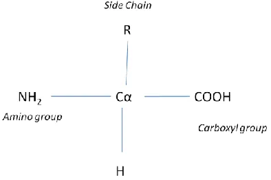
Figure 1: The general structure of an alpha amino acid ... 1
Figure 2: List of 20 different amino acids ... 2
Figure 3: Primary structure of a protein ... 3
Figure 4: Alpha Helix with hydrogen bonds ... 4
Figure 5: The Beta Sheet Structure ... 5
Figure 6: Tertiary structure showing alpha helix and beta sheet ... 5
Figure 7: Protein folding... 6
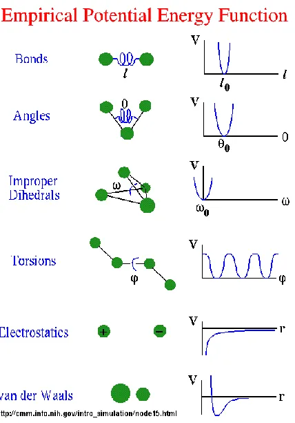
Figure 8: Different terms in Potential energy function ... 12
Figure 9: Equation for calculating potential energy of a protein (left); Modified energy function having Knowledge based term ... 14
Figure 10: Energy profiles of few atoms showing KB PMF ... 15
Figure 11: Work flow of web server ... 16
Figure 12: Xgrid Architecture ... 18
Figure 13: Home page for summa protein refinement server ... 19
Figure 14: Webpage showing the results of the refined protein ... 20
Figure 15: Cumulative growth of membrane proteins structural data ... 23
Figure 16: Work flow sequence diagram protein refinement process ... 29
Figure 17: Root means square deviation ... 30
Figure 18: Minimization of native membrane proteins ... 31
Figure 19: Minimization of near native membrane proteins ... 32
Figure 20: Minimization of near native membrane proteins ... 33
List of Tables
Abstract
The structures obtained from homology modeling methods are of intermediate resolution
1-3Å from true structure. Energy minimization methods allow us to refine the proteins and
obtain native like structures. Previous work shows that some of these methods performed
well on soluble proteins. So we extended this work on membrane proteins. Prediction of
membrane protein structures is a particularly important, since they are important
biological drug targets, and since their number is vanishingly small, as a result of the
inherent difficulties in working with these molecules experimentally. Hence there is a
pressing need for alternative computational protein structure prediction methods. This
work tests the ability of common molecular mechanics potential functions (AMBER99/03)
and a hybrid knowledge-based potential function (KB_0.1) to refine near-native structures
of membrane proteins in vacuo.
A web based utility for protein refinement has been developed and deployed based on the
KB_0.1 potential to refine proteins.
CHAPTER 1: INTRODUCTION
Proteins are the micro machines on which biological systems are based. Understanding
protein’s structure and functionality helps us design new drugs and understand the
underlying mechanism of human disease. Protein structure prediction and protein folding
are considered fundamental problems of modern computational/ molecular biology,
There has been research progress from the past several decades in understanding the
underlying biophysical interactions in proteins either by simulating the proteins or by
studying their behavior using experimental techniques.
1.1) Introduction to Proteins
Proteins are composed of individual units called amino acids. These amino acids are linked
by peptide bonds. There are twenty different naturally occurring amino acids which differ
in the chemical nature of their side chains. The nature of the side chain influences the
properties and structure of the proteins.
Each amino acid consists of
A central carbon atom usually referred
as Alpha Carbon (C) atom.
an amino group
a carboxyl group
a side chain
Proteins are intimately involved in nearly every cell activity, from replication of genetic
code to transporting oxygen, and are generally responsible for regulating the cellular
machinery and determining the phenotype of an organism. Diseases like transmissible
spongiform encephalopathies [24], are usually caused due to improper folding of protein
which may result from genetic and/or environmental influences. Examples of proteins
include hemoglobin, thyroid hormone, insulin, and myosin.
There are 20 possibilities of amino acids based on the R group side chain associated with
the amino acids
These amino acids are
classified as acidic (negatively
charged) , basic (Positively
charged), polar( side chains
with pure hydro carbon alkyl
groups or aromatic),
non-polar(Side chains with
functional groups acids,
amides, alcohols, and amines),
Hydrophobic (un likely to be
in contact with the aqueous
environment), hydrophilic
(likely to be in contact with
the aqueous
environment)based on
chemical and structural
behavior of their side chains.
1.2) Protein Structure
Primary Structure
The primary structure refers to the sequence of amino acid of the protein .The amino acids
form covalent bonds (peptide bonds) between each other during the protein biosynthesis.
The polypeptide chain thus formed has two ends a carboxyl terminus (C-terminus) and
amino terminus (N- terminus).The counting of residues starts from the N-terminus. The
sequence of the protein defines its structure and functions [41].
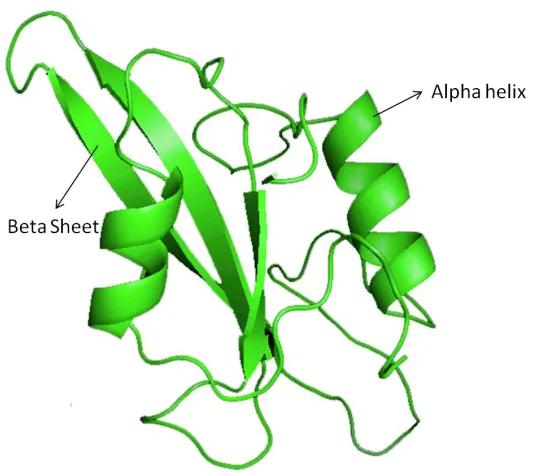
Secondary Structure in Proteins
The secondary structure of a protein is the local spatial arrangement of its C carbons
(those atoms that are not part of the side chain, often referred to as the main chain)
[IUPAC-IUB, 1970]. There are commonly three secondary structures in proteins, namely -
helices, -sheets, and turns. The other structures are usually classified as random coil, or
other.
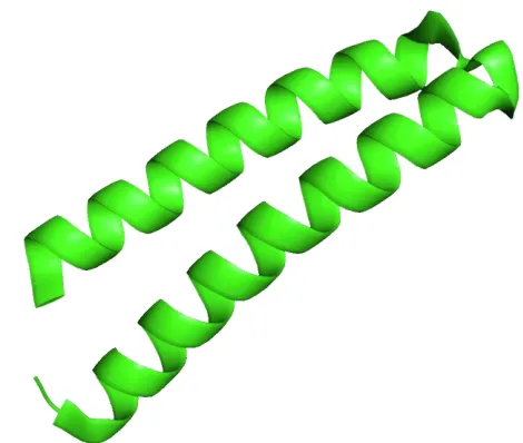
Alpha helix
The alpha helix (α-helix) is a
major part of secondary structure.
The polypeptide chain turns about
itself to form a spring like structure
with each of the poly peptide bond,
hydrogen bonded with other
peptide bonds on the chain thus
forming alpha-helix [34].
It can be either a right-handed or
left-handed coiled conformation.
This secondary structure is also sometimes called a classic Pauling-Corey-Branson alpha
helix [19].
Beta sheet
The secondary structure has another major
element called beta-sheet; it has several
individual beta strands. A beta strand is chain
of 3-10 amino acids connected by polypeptide
bonds. These beta strands are connected to
one another by two or more hydrogen bonds
forming a pleated sheet [33].
Figure 4: Two alpha helixes in protein 1ROP.
Tertiary Structure of Proteins
Tertiary structure is the final geometric shape a protein assumes. The bonding interactions
between the side chains of each amino acids will result in several folds and bends in the
protein chain [20]. These interactions finally stabilize to form tertiary structure. The
tertiary structure consists of several secondary structure elements i.e. α-helices and beta
sheets.
Quaternary Structure
Quaternary structure is the assembly of two or more protein chains – if the protein chains
have the same sequence, then this is known as a homooligomers – if they have different
sequences, then the protein is a heterooligomer. The quaternary structure can be assumed
as a structure consisting of stable tertiary structures as subunits which are stabilized by
variety of interactions like non-covalent forces, disulfide bonds, hydrogen bonding and salt
bridges [27, 28].
1.3) Protein Folding Theory
Protein folding is a physical process where a random coil (sequence of amino acids)
undergoes hydrophobic collapse and eventually comes to a stable 3-dimensional structure
(native structure). It is puzzling that in the protein folding process a protein undergoes a
spontaneous self assembly of amino acids and forms a unique three dimensional structure
despite the possibility of enormous conformational spaces [29][43]. It is assumed that if we
understand the underlying physical mechanisms involved in protein folding, we can model
them on computers and use algorithms to predict native structures from their amino acid
sequences. Science magazine in 2005 listed the protein folding problem as one of the 125
biggest unsolved problems in science [15].
Figure 7: Protein Folding [initial state (left) Final folded state (Right)][37] reprinted from Wikipedia
1.4) Protein Structure Prediction
One of the most challenging problems of computational molecular biology is the prediction
of the proteins spatial conformation from its primary structure. Scientists and researchers
have been trying to find the protein's 3-dimensional structure which helps us to
understand its functionality and provides means for planning experiments and drug design.
Why is predicting 3D structure so important?
The gene is the basic unit of hereditary. It is comprised of DNA (genetic information), and
its gene products, which through an extremely complex series of interactions with the
products of other genes, play a large role in determining the appearance and behavior of an
organism. The DNA in the gene is transcribed into messenger RNA which is translated in to
sequence of amino acids. This sequence of amino acids in turn folds up in to a three
dimensional structure to form a protein. This protein now interacts with other proteins
(lock and key arrangements, etc.) and this interaction mediates the functions of the
organism [16].
“In fact, the 3D interactions between proteins and substrates are essentially the
organism. We cannot completely understand (any predictions about) the phenotype of the
organism without knowing the 3D structure of the proteins in a genome.” -Ram Samudrala
[39].
The classical method of solving the protein structure is done by using X-ray
Crystallography and Nuclear magnetic resonance (NMR). These methods give good
techniques is that these techniques are very expensive and take quite a long time to
determine the structure.
In 1973 Prof B. Christian Anfinsen has proposed that the information determining
the tertiary structure of a protein resides in the chemistry of its amino acid sequence [41].
His research sets a new challenge for many researchers to start predicting tertiary
structure from the amino acid sequence.
After the discovery of a protein’s propensity to fold into its unique native state
without any additional genetic mechanisms, there has been a great deal of research over
the past 25 years on the prediction of 3D structure from sequence alone, without further
experimental data. Sequencing of proteins is relatively fast, simple and inexpensive.
Despite significant efforts, the protein folding and protein structure prediction problem
remains as an unsolved problem. Due to several genomic projects increasing over time
around the world, it is evident that there is a large gap between the number of known
sequences and number of known three-dimensional structures. There are a few
contemporary approaches toward protein structure prediction that can be roughly divided
into three categories of increasing difficulty.
Homology/Comparative Modeling
The homology Modeling is based on the fact that evolutionarily related proteins with
similar sequences usually exhibit similar structures. For example, two sequences that have
just 35% sequence identity usually have the same overall fold. Proteins which are
homologous have similar protein structure [17]. By evolution three dimensional structure of
protein is more conserved than the sequence itself [17].
Threading Methods
The threading methods compare the unknown protein sequence against a library of
structural templates thus producing a list of scores for each template in the library [30].
These scores are sorted, the template structure with the best score is assumed to be
adopted by the unknown protein. The threading method, along with the comparative
modeling method, use the structures of already solved proteins as templates.
Ab initio Method
The ab initio method is based on thermodynamic hypothesis which states that “The native
state of a protein is the one for which free energy achieves the global minimum” [30]. This
approach does not depend on prior information from any other proteins. It is clearly the
most difficult approach and arguably the most useful approach. But there could be some
unresolved issues with this approach.
This method requires two things, a search algorithm to explore the protein
conformational space and an energy function which evaluates whether a state is a native or
not. It is extremely complex to find a useful energy function and a useful search algorithm
to traverse the conformational space [30].
It can be understood that under normal physiological conditions there is a
possibility of one and only one conformation which has low energy than any other
fashion until the conformation with lowest energy is found. The time complexity involved
searching all the conformations is extremely high. Decreasing the time complexity of the
folding process is an extremely complex task, as our algorithm should be capable of
calculating the heuristic methods of finding a kinetic pathway by escaping the irrelevant
conformations and finally lead us to one conformation which has lower energy than any
CHAPTER 2: WEB SERVICE FOR PROTEIN STRUCTURE REFINEMENT
Protein structure refinement is the process of improving the structures of protein models
to make them more like the native structures. The models obtained from comparative
modeling and threading have a resolution within 1-to 3Å root means square deviation
range with true structure [1].The root mean square deviation (RMSD) is the measure of the
average distance between the backbones of superimposed proteins. It is extremely
challenging problem to minimize this rmsd from near native structure to true structure.
The energy minimization methods help us refine the proteins models at such low resolution [1].
Recently, there has been some very encouraging progress toward solving this problem by
using techniques that involve optimization of new potential functions [1]. Dr. Christopher
Summa and Dr. Levitt have tested whether Potential energy minimization (PEM) could be
applied for refinement and they proved it worked. The web server is uses their method for
refinement of proteins over internet. This allows any user to upload his model (either pdb,
ent) and obtain a refined structure.
2.1) Introduction to KB/MM Structure Refinement Method
The quest for proteins modeling has given rise to modeling of protein based on its
energetics. There has been some progress in developing a molecular mechanics potential
energy function, which model a protein as a collection of atoms connected by springs that
hold bold lengths and angles [16]. These molecular mechanics potential energy functions
(MMPEF) have two types of terms: “bonded” and “non-bonded” [16]. These functions treat
atoms as spheres, and bonds as springs. Thus bond stretching, bends, twists are modeled
The bonded terms also include a torsional potential contributed by torsional angle
rotation between atoms that are adjacent. The non bonded interactions between the atoms
are due to van der Waals attractions, steric repulsion and electrostatic attraction/repulsion
depending on their distance from each other[16, 31]. These van der Waals attractions and
electrostatic forces are modeled based on the Lennard-Jones function and Coulomb’s Law
respectively. All these bonded and non-bonded interactions of the Molecular mechanics
potential energy functions are derived from several sources either based on quantum
mechanics, thermodynamic data, or some combination of the two [4, 5].
These functions are largely
used in protein folding simulation, template free modeling methods, and are also used to
refine X-ray crystal structures [32]. The molecular dynamics simulation is a computer
simulation where models of atoms and molecules are allowed to interact with each other
and the forces between them are approximated by solving Newton’s classical laws of
motion. So during the refinement process if a force is applied on the protein, the effect of
force on one atom is calculated and additional effect of displacement of this atom due to the
force affects other atoms (either bonded or non bonded ) is calculated, thus resulting in
displacement of atoms resulting in new positions and velocities of the atoms.
The energy functions which are derived based on the statistics of the known native
proteins are known as “knowledge based”. These energy functions derive statistics based
on the probabilities of pair wise appearance of residues in a specific geometry [16]. These
probabilities are converted into potential energy using the Boltzmann equation:
ΔG = –RT ln (Pobserved /Pexpected), [6, 11, 16]
Where Pobserved is the probability of finding a particular structural element [6, 11, 16],
Pexpected is the expected probability of finding that structural element based on chance [6,
11, 16].
The advantage of these energy functions is that they can model behavior seen in the
protein database .The disadvantage is that they can’t predict new behavior out of scope of
database. [16]. Dr. Christopher Summa and Dr. Levitt introduced Knowledge Based
/Molecular Mechanics (KB/MM). The equation representing the potential energy of the
The equation has energy terms from both the bonded and non bonded interactions .The
bond angle bends, bond stretches, bond torsion angle twists contribute to the bonded
interactions. The van der Walls forces and columbic forces contribute to the non bonded
interactions. The energy equation was transformed in to a new equation by replacing the
non-bonded interactions and all partial charges with a knowledge based statistically
calculated term fKB .The term fKB was calculated based on the atomic pairwise Potential
Mean Force (PMF) .For all the 167 atoms types which were used a pairwise PMF was
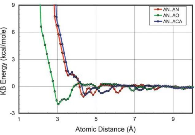
They used three different bins (0.5Å, 0.2Å, 0.1Å) .For generating the energies they have
used Lu and Skolnick derivation [9]. Depending on the bins they made a histogram and
when the counts go to zero they have added a repulsive part to account for steric over lap
[1] These curves are smoothened by fitting a quintic spline function [1][9][10]. Using this
probabilities of occurances of atoms they finally derieved an energy term which contribute
non-bonded interactions(fKB).
The KB/MM Hybrid potential is used to perform the refinement of proteins on the
webserver
Copyright Dr. Christopher Summa & Dr. Michael Levitt Reprinted from [1]
Figure 10: Energy profiles for : AN (alanine backbone nitrogen), AO (alanine backbone
carbonyl oxygen) and ACA is the alanine carbon.The symbols shown are the energies from
2.2) Implementation of Web Services
Summa Protein Refinement Server
The main idea of the web server is to provide a utility where users can refine their proteins. The web server takes a protein model as an input and returns the user with a refined protein.
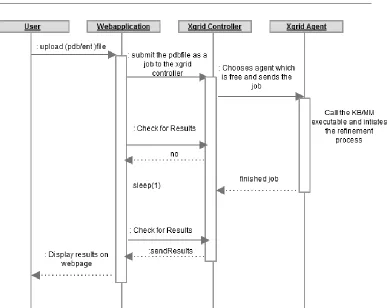
Work Flow:
The web application work flow is as follows
The user uploads a protein database file (pdb/ent). The web application cautions if
the file uploaded is a pdb/ent file for security reasons.
The web application creates a temporary directory and stores the uploaded file in to
that folder.
The web application gives response to the user that the file has been uploaded and
then calls the script file which creates the job on the grid.
The job is created and submitted to the Xgrid controller by the web application , then
the controller checks for the agent which is idle and sends the job to that agent.
The agent now starts to execute the job by calling encadv6lg.exe (is the main
executable which handles the refinement process) under the environment variables
of software ENCAD[44] (Dr. Michael Levitt).
The XGgrid agent executes the job and generates the results.
The web application keeps on querying the controller for results. The controller
finally fetches the results from the agent and stores them in the temporary directory
that it generates in the first place.
The results thus generated are displayed as links on the webpage.
The links for the output folder are generated on the webpage.
2.3) Tools & Methods
The basic idea of the web application is to use the executable (encadv6lg.exe) to minimize
the protein file. The web application is built on PHP (Pre Hyper text Processor), Apple
Xgrid Technology, Unix shell scripts.
PHP (Hypertext Preprocessor)
PHP is widely used general purpose scripting language which was designed for developing
gets interpreted by php web server. PHP is available as an open source and it supports
mostly all widely used operating systems.
Apple XGrid Technology
The field of bioinformatics and computational biology are emerging as an important
discipline for research and industrial applications. The grid computing techniques are very
useful to reduce time complexity involved in huge genomic data projects and large scale
distributed applications. Apple has introduced the XGrid software (a proprietary software
which implements distributed computing protocol) which was developed by Advanced
Computation Group under supervision of Apple. XGrid was used for the jobs as each of
them were time consuming. The XGrid architecture is outlined in Figure 12.
The XGrid works as follows,
A client submits a job to the controller.
The controller looks for the agent with status “available” .The “available” status
indicates that the agent has free processors and is ready to work on the job.
The job is then sent to that agent by the controller.
The agent completes the job and stores the results in the XGrid folder, which can be
obtained from XGrid command line utility.
2.4) Results
The web application was able to take the input file pbd/ent files. There is a validation
system that only accepts pdb/ent files while rejecting other extensions. The input pdb file
is sent to XGrid to perform minimization. The screen shot for the web server is as follows.
2.5) Future Work
The web application can be extended with some user friendly features. The application is
now capable of generating results on the webpage. The user must wait for the results for
some time (until the minimization is done). In few cases if the grid is filled the job has to
wait for a substantial amount of time. The other problems are connection problems, if the
client waiting for execution of the minimization process has some network problems then
the client fails to get results, though the job has successfully completed. The solution for
both of these problems is to have an email system implemented on server side where the
user inputs his pdb to the web server and provides his email id to the server. Later after the
job gets completed the server emails the results to the user.
CHAPTER 3: REFINEMENT OF NEAR-NATIVE MODELS OF
MEMBRANE PROTEINS
3.1) Introduction
The computational prediction of the structures of proteins that span the cellular membrane
is still in its infancy relative to prediction of the structures of water-soluble proteins.
Prediction of membrane protein structures is a particularly important endeavor, since they
are important biological drug targets, and since the number of experimental structures of
membrane proteins relative to water soluble proteins is vanishingly small, as a result of the
inherent difficulties in working with these molecules experimentally. Computational
prediction methods represent an alternative to expensive, time consuming, and often
difficult experimental methods. In this work we test the ability of common molecular
mechanics potential functions (AMBER99 and AMBER03) and a hybrid knowledge-based
potential function (KB_0.1) to refine near-native structures of membrane proteins in vacuo.
We employ the technique of potential energy minimization to a set of 88 native membrane
protein structures to determine the extent to which they are perturbed away from their
native, experimental structures and show that, for the majority of the proteins in our
dataset, this technique does not significantly alter the structure, even in the absence of
treatment of explicit or implicit membrane. As a more stringent test, large sets of
near-native decoys were generated for each of the 88 membrane proteins using the technique of
normal-mode perturbation. This technique was employed to sample the configuration
space around the native state as evenly as possible.
The mean percentage improvement in Ca-rmsd (root mean square distance of the
was 4.50% for KB_0.1, 3.97% for AMBER99 and 4.75% for AMBER03. We conclude that,
while all three potentials are able to generate a modest improvement of the decoys, they
clearly are able to draw near-native structures toward the native state rather than away
from it for most examples we tested even in in vacuo simulations. More robust search
methods can be used to greater improvement values, but also represent a significant
increase in computational cost.
3.1.1) Membrane Proteins – Importance and Difficulties
Integral membrane proteins are defined by their ability to associate with, and span the
plasma membrane of cells. They differ from water soluble proteins in the nature of the
amino acid sidechains on their exterior surface – water soluble proteins have a marked
tendency to display polar, hydrophilic amino acids which interact favorably with water,
whereas, in the transmembrane regions of integral membrane proteins, the side chains
displayed are non-polar and hydrophobic, in order to interact with the non-polar
hydrocarbon chains at the interior of the phospholipid bilayer. This propensity to be
associated with the bilayer makes it very difficult to work with these proteins
experimentally, and the relative dearth of structural information for integral membrane
proteins is the result.
There are currently ~63,000 experimental structures of water-soluble proteins in the
Figure 16 shows the count of membrane proteins structures over the last 25 years. The
graph indicates that there has not been a considerable growth in number of structures
signifying the fact that these structures are hard to work with and there is a great need of
research to be done in this domain.
3.1.2) Generation of Membrane Protein Dataset (Data Collection)
The Membrane protein dataset was generated by collecting membrane protein files from
the Stephen Whites Lab. Stephen Whites lab website has several membrane protein
structures solved either by diffraction or NMR methods. These protein files are in the form
of PDB file format. The PDB file stores the 3-dimensional structural information of the
protein (The pdb data is derived from X-ray crystallography, Nuclear Magnetic Resonance,
and theoretical simulation. The pdb stores all the 3-dimesional co-ordinates of atoms in the
proteins). The proteins initially collected were 301 membrane proteins. We have selected
1AP9.pdb 1JGJ.pdb 1QD5.pdb 2A65.pdb 2JMM.pdb 2ZFG.pdb
1AT9.pdb 1K24.pdb 1QFG.pdb 2B2F.pdb 2JO1.pdb 2ZIY.pdb
1BRX.pdb 1KMO.pdb 1QHJ.pdb 2B6O.pdb 2JQY.pdb 3B9W.pdb
1BXW.pdb 1LKF.pdb 1QJ8.pdb 2BRD.pdb 2K4T.pdb 3B9Y.pdb
1BY3.pdb 1MM4.pdb 1QJP.pdb 2C3E.pdb 2K73.pdb 3C02.pdb
1C3W.pdb 1N9P.pdb 1QKP.pdb 2CFQ.pdb 2NR9.pdb 3DWO.pdb
1C8R.pdb 1NQE.pdb 1SOR.pdb 2D1U.pdb 2O7L.pdb 3EFC.pdb
1E12.pdb 1OKC.pdb 1SU4.pdb 2D57.pdb 2O9J.pdb 3EFM.pdb
1FI1.pdb 1ORM.pdb 1T5S.pdb 2F1C.pdb 2OMF.pdb 3EMN.pdb
1FQY.pdb 1P49.pdb 1THQ.pdb 2F2B.pdb 2OQO.pdb 3F3A.pdb
1FX8.pdb 1P4T.pdb 1XIO.pdb 2GUF.pdb 2POR.pdb 3FWM.pdb
1G90.pdb 1PNZ.pdb 1XQF.pdb 2H8A.pdb 2QDZ.pdb 3GD8.pdb
1H68.pdb 1PRN.pdb 1YC9.pdb 2IC8.pdb 2QEI.pdb 3GJD.pdb
1IH5.pdb 1PW4.pdb 1YGM.pdb 2JK4.pdb 2QJU.pdb
1J4N.pdb 1Q9F.pdb 1YMG.pdb 2JLN.pdb 2UUH.pdb
Table 1: Final set of pdb files used for minimization
3.1.3) Generation of Near-Native Decoys
What are decoys?
In the protein’s conformational space there are lots of possible conformations a protein can
assume. Based on the proteins conformational space and conformations we try to make
similar conformations of proteins using computer which have some characteristics of
native proteins called decoys [8].
Generating all or few of the possible protein conformations by using several algorithms is
called as decoy generation (Samudrala et al, 1999a).The decoy data sets, consists of a
solved protein structure and numerous alternative native-like structures. Decoys are used
for the testing, development of scoring functions and refinement process in protein
structure prediction [8, 14]. The possible protein conformations are infinite, using validation
There are few packages available to generate decoys like Decoys R Urs (Ram Samudrala
and Michael Levitt) [8], ENCAD[44] packages. We generated the decoy sets using ENCAD
package.
We use the Tirion3 method to calculate the low frequency normal modes of motion for the
native structures. We then perturb the native structure along those low frequency normal
modes.
The total decoys sets were 97, one for each pdb file and a mean of ~504 near native
structure decoys per set were generated.
3.2) Methods/Tools
3.2.1) Potential Energy Minimization / Refinement Process
The Potential energy minimization is one of the earliest methods used for refinement of
protein structures [35], the structures obtained from several modeling applications are
refined to obtain a native like structures. The PEM is based on thermodynamic hypothesis
“The native state of a protein is the one where its free energy achieves the global minimum”
[1].
Test Criteria:
We test with various kinds of force fields if they are applied on the native proteins they
should not perturb them as if the force field was a perfect energy function of protein then
its global minimum should match with proteins native state. The idea of comparison and
test criteria is based on the idea of Dr.Christopher Summa and Dr. Michael Levitt‘s
“Near-native structure refinement using in vacuo energy minimization”. Their work was on
membrane proteins. We compare and contrast the ability of different force fields to move
the near native structure towards the native state [1]. To simplify this comparison we
perform single refinement technique i.e., PEM in vacuo [1].
To setup comparison criteria between the force fields in vacuo we test
The refinement process should not significantly perturb the native structure [1].
The refinement process should result in movement of near native structures towards
native [1].
Considering the test criteria 1, it is weak because when a force field is applied on the native
structure, it may reach a local minimum and stop there, here we cannot strongly say if the
force field really doing a good job. But if we consider the criteria 2 we can have an idea of
the movement near the native state, whether it is towards the native or away thus we could
at least analyze if our force field is trying to make structures more like native or deteriorate
them. In criteria 2 we can get a global picture of shape of the curve at the native state [1].
3.2.2) Force fields Employed/Tools
GROMACS:
GROMACS stands for GROningen MAchine for Chemistry Simulation. Gromacs is a package
which performs molecular dynamics by simulating the Newtonian equations of motion for
systems with hundreds to millions of particles [42]. Gromacs is considered as optimized and
fast software for molecular dynamics simulations. Gromacs is a command line utility for
AMBER 03/99 Force Field
Molecular dynamics is a computer simulation which calculates how a molecular system
behaves over a time span [25]. Gromacs is a package which performs molecular dynamics
simulation. These packages have built-in routines for energy calculations and minimization
[19]. AMBER stands for Assisted Model Building with Energy Refinement.
The amber force field package is a set of molecular mechanics force fields which can be
applied on the bio molecules. For implementing the refinement process the force fields
amber03, amber99 are applied on the proteins and observe whether these force fields have
moved these structures towards the native structure or away from native structure.
Work flow of the refinement Process
The data used in this project was collected from Stephen Whites Lab .There were 97 pdbs
and for each pdb decoys were generated and a mean count of decoys was ~504.Now based
on criteria 2 we perform refinement process on these decoys. The refinement process
involved writing lot of scripts in perl and c-shell which are explained in detail in the
following workflow.
1) For every pdb, decoys are generated and are stored in respective folder named with
“pdbname_decoys”.
2) The script files takes inputs as pdbfoldername and creates a job and submits to the
XGrid
3) XGrid checks if there are any agents available in the grid and sends the job for execution
4) The agent starts executing the job. The script file initializes environment and calls
minimize.pl to collect all pdbs and submit each one to the actual gromacs minimization
script.
5) The gromacs minimization script calls stripNonProtein.pl script to remove non protein
elements from the pdb file.
6) The gromacs script calls a set of executables to perform minimization .Initially pdb2gmx
is executed with arguments amber03/99 and pdb decoy name, pdb2gmx outputs .top, .gro
which serve as input grompp, the outputs from grompp serve as input to trjconv. Finally
after the execution of all the three the minimized output is obtained.
7) After the minimization is done the minimized file is copied back to the decoys folder.
According to criterion 2 the force fields are applied on the decoys to see their
improvement/deterioration .According to criterion 1 way the same force fields are applied
on the native structures to see how much the force filed is deteriorating the native
Work Flow of the Refinement Process
3.2.3)Knowledge Based /Molecular Mechanics Force Field
The Knowledge based molecular mechanics potential derived from the equations proposed
by Dr. Christopher Summa and Dr. Levitt in Figure 10 is used to refine proteins. The
knowledge based molecular mechanics hybrid model was discussed in chapter 2 , we used
the same method here to perform the energy minimization process.
The workflow of the refinement process is similar to the amber03/99 refinement process
but here the executable encadv6lg.exe is used to minimize the proteins. The executable
encadv6lg.exe implements the Knowledge Based /Molecular Mechanics hybrid Force Field.
3.3) Results
3.3.1) How to compare protein conformations?
There should be a method to compare our initial native conformation with refined
conformation so that we know how much we have improved or deteriorated the structure.
The measure used for such purpose is root mean square deviation [19].
“The root mean square deviation (RMSD) is the measure of the average distance between the
backbones of superimposed proteins.” [38] The rmsd for a protein is calculated for the C-α
atomic co-ordinate and is represented by the equation.
Figure 17: Root means square deviation equation (Reprinted from Wikipedia)
3.3.2) Criterion 1: Energy Minimization of the Native Structures
For testing criterion 1 the energy minimization is applied on the native structures as
starting point and find how much the native structure is affected. Each point in the graph
(Figure 18) represents the native structure of one of the proteins in our dataset, and the
Ca-RMSD after minimization using a particular potential function is shown. A value of 0.0
for Ca-RMSD indicates that the minimized structure is exactly the same as the native,
experimental structure, which is the ideal case. Lower values indicate better performance
than higher values. The AMBER99, AMBER03, KB_0.1 potentials applied on the native
structures show following results.
The mean deviation in rmsd for AMBER03 is 0.55 Å rmsd, AMBER99 is 0.53 Å rmsd and
KB_0.1 is 0.54Å rmsd. This indicates that these force fields have not significantly
deteriorated the structures.
3.3.3) Criterion 2: Energy Minimization of the Near Native Structures
For testing criterion 2 the energy minimization is applied on the near native structures as
starting point and sees how much they have improved/ deteriorated when compared to the
native structure.
The graph (Figure 20) represents the mean percentage improvement in Cα-RMSD for the
near native structure model sets .If the graph shows the bar in the left side from 0.0% for
a protein it means that the structure has been improved, if it’s the right side then the
structure has been degraded.
The overall percentage improvement in Cα (c-alpha) rsmd with KB/MM was -4.50%
Amber99 had a -3.97% while Amber03 had -4.75%.
The top and worse performers with respect to the force fields are listed in the following
tables
The top performers with respect to force fields
KB Amber99 Amber03
1H68 -27.41834 2UUH -24.11730 1JGJ -21.73361 1P49 -19.06765 2POR -17.28706
1BRX -36.0340 1QKP -32.9541 2ZIY -23.9361 2C3E -23.1384 1XQF-20.2731
1BRX -37.8618 1QKP -34.9888 2ZIY -31.0247 2C3E -23.2400 1XQF-21.2627 Table 2: Top performers with each of the force field
The worst performers with respect to force fields
KB Amber99 Amber03
1QFG 3.039096 1YGM 3.810338 2K73 4.442954 1K24 5.646301 2D1U 18.607957
3FWM 3.75568 2QDZ 6.30944 2JLN 9.18768 2BRD 23.28000 2UUH 38.59200
3FWM 4.23802 2QDZ 6.75380 2JLN 21.47155 2BRD 23.27045 2UUH 38.72682
Table 3: Worst performers with each of the force field
Structures improved:
KB has improved 72 of 88 protein structures
Amber99 has improved 67 of 88 protein structures
3.4) Discussion
The results show that three potential functions tested were able to refine the membrane
proteins in our dataset using potential energy minimization. Interestingly, the KB_0.1
potential worked as well as, an in many cases better than, the traditional molecular
mechanics force fields, despite having been derived using interatomic distance statistics
from a dataset of water soluble proteins only. The study suggests that if a low resolution
membrane protein fold has been found then we can use either traditional or knowledge
based techniques to refine the membrane proteins.
3.5) Future Work
In addition to our work in this project we intended to test other energy functions
(CHARMM, ENCAD, GROMACS, implicit solvent models) and test other algorithms for
searching (local backbone moves, simulated annealing). Improvements in decoys set
generation such that each decoy is diverse to every other in a decoy set in a range of
REFERENCES
[1] Summa CM and Levitt M. Near Native Structure Refinement Using in vacuo Energy Minimization. Proceedings of the National Academy of Sciences USA 2007 Feb
27;104(9):3177-82.
[2] Yu Xiaa and Michael Levitt ,Extracting knowledge-based energy functions from protein structures by error rate minimization: Comparison of methods using lattice model (Department of Structural Biology, Stanford University School of Medicine, Stanford, California 94305)
[3]Lazaridis, T. "Effective energy function for proteins in lipid membranes", Proteins, 52:176-192 (2003)
[4]. Mackerell, A. D., Jr. (2004). Empirical force fields for biological macromolecules: overview and issues. J. Comput. Chem. 25, 1584-604.
[5]. Jorgensen, W. L. & Tirado-Rives, J.Potential energy functions for atomic-level simulations of water and organic and bio-molecular systems. (2005). Proc.Natl. Acad. Sci. USA 102, 6665-70.
[6]Thomas PD, Dill KA.Statistical potentials extracted from protein structures: how accurate are they?
J Mol Biol. 1996 Mar 29; 257(2):457-69.
[7] Filip Jagodzinski. Guest Lecture, GROMACS, MD Tutorial, Smith College, CS 334, Bioinformatics .16 October 2008
[8] RAM SAMUDRALA and MICHAEL LEVITT, Decoys ‘R’ Us: A database of incorrect conformations to improve protein structure prediction.
Department of Structural Biology, Stanford University School of Medicine, Stanford, California 94305
[9] Lu H, Skolnick J (2001) Proteins 44:223–232.
[10] Samudrala R, Moult J (1998), J Mol Biol 275:895–916.
[11] Dehouck, Y, Gilis, D. & Rooman, M. (2006). A new generation of statistical Potentials for proteins. Biophys J. 90, 4010-7.
[12] Kortemme, T., Joachimiak, L. A., Bullock, A. N., Schuler, A. D., Stoddard, B. L. & Baker, D. (2004). Computational redesign of protein-protein interaction specificity. Nat. Struct. Mol. Biol. 11, 371-9.
D. (2003). Design of a novel globular protein fold with atomic-level accuracy.
Science 302, 1364-8.
[14] Kai Wang, Boris Fain, Michael Levitt andRam Samudrala
Improved protein structure selection using decoy-dependent discriminatory functions
BMC Structural Biology 2004, 4:8doi:10.1186/1472-6807-4-8
[15] Editorial: So much more to know. Science 2005, 309:78-102.
[16]F. Edward Boas, Physics-based design of protein ligand binding [Doctor of philosophy Thesis] Stanford University May 2008.Chapter 2, P.16, 17
[17] Kaczanowski S and Zielenkiewicz P (2010). Why similar protein sequences encode similar three-dimensional structures? Theoretical Chemistry Accounts 125:543-50
[18]Ab Initio Protein Structure Prediction Using a Combined Hierarchical Approach [Ram Samudrala, Yu Xia, Enoch Huang and Michael Levitt ]
[19] Zhijun Wu, Lecture notes on computational structural biology.[Internet]
(Iowa State University, USA) p.88.
[20] Elmhurst College: Elmhurst, Illinois [Internet][18th Oct 2010]
Available from: http://www.elmhurst.edu/~chm/vchembook/567tertprotein.html
[21] Birk Beck Crystallography, The Ramachandran Plot [Internet] [4th Feb 1996,18th Oct 2010] Available from : http://www.cryst.bbk.ac.uk/PPS2/course/section3/rama.html [22] College of Saint Benedict Saint John's University [Internet]
Available from:
http://employees.csbsju.edu/hjakubowski/classes/ch331/protstructure/olunderstandcon fo.html
[23] http://bioinsilico.blogspot.com/2008/11/secondary-structure-prediction_25.html
[24] Protein Folding, Structure and Function
Heinrich Roder, Ph.D.,Hong Cheng, Ph.D.,Harvey H. Hensley, Ph.D.,Dharmaraj Samuel, Ph.D. Paul W. Riley, B.S., Jayme Staub,* B.S.,Colin M. Hayden,*
[25] Swiss EMBnet node server,Theory of Molecular Dynamcis Simulation Avialble from : http://www.ch.embnet.org/MD_tutorial/pages/MD.Part1.html
[26] Center for Molecular Modeling [Internet] The Empirical Potential Energy Function
[27] Protein Structure –Wikipedia the free encyclopedia [Internet] [Oct 15 2010: Oct 16
2010] Available from: http://en.wikipedia.org/wiki/Protein_structure
[28] Elmhurst College: Elmhurst, Illinois [Internet][18th Oct 2010]
Available from : http://www.elmhurst.edu/~chm/vchembook/567quatprotein.html
[29] Ken A Dill, S Banu Ozkan, Thomas R Weikl, John D Choderaand Vincent A Voelz The protein folding problem: when will it be solved?
[30] Anna Bernasconi and Alberto M. Segre, Ab Initio Methods for Protein Structure Prediction: A New Technique based on Ramachandran Plots.
[31] Suzanne W. Slayden, Molecular Mechanics Theory in Brief
Available from: http://classweb.gmu.edu/sslayden/Chem350/manual/docs/MM.pdf
[32] F Edward Boas, Pehr B Harbury, Potential energy functions for protein design.Current Opinion in Structural Biology 2007, 17:199–204
[33] Beta sheet –Wikipedia the free encyclopedia [Internet] [Oct 15 2010: Oct 16 2010] Available from: http://en.wikipedia.org/wiki/Beta_sheet
[34]Bruce Alberts, Dennies Bray, Julian Lewis, Martin Raff, Keith Roberts, James D.Watson.
Molecular Biology of the Cell. Newyork and London: Garland Publishing Inc; 1983.
P.114-115.
[35] Refinement of Protein Conformations using a Macromolecular Energy Minimization Procedure , Micheal Levitt and Shneior Lifson, Weixmann Institute of Science Rehovot, Israel (29 July, 1969) J. Mol. Biol. (1969) 46, 269-279
[36] Luong, P.2009. Basic Principles of Genetics. Connexions,[Internet] [July 2, 2009: Oct
19 2010] Available from: http://cnx.org/content/m26565/1.1/.
[37]Protein Folding –Wikipedia the free encyclopedia [Internet] [Oct 15 2010: Oct 16
2010] Available from: http://en.wikipedia.org/wiki/proteinfolding
[38] Zhang Y. 2009. Protein Structure Prediction: Is It Useful? PubMed Central [Internet]. 19(2): 1-17[cited 2010 Oct 19]. Available from:
[39] Ram Samudrala [Internet] University of Washington in Seattle. Primer on protein
folding problem. [Internet] Available from : http://www.ram.org/research/pfp.html
[40]Krieger E, Koraimann G, Vriend G (2002) Proteins 47:393–402.
[41]Anfinsen, C., Principles that govern the folding of protein chains. Science, 1973. 181: p. 223-30.
[42]Gromacs .About Gromacs[Internet][Oct 19 2010] Available from : http://www.gromacs.org/About_Gromacs
[43] Robert Zwanzig, Attila Szabo, and Biman Bagchi, Levinthal's paradox
Laboratory of Chemical Physics, National Institutes of Health, Bethesda, MD 20892.
VITA
Kapil Pothakanoori was born in Medchal, Ranga Reddy Dist, AP, India. He received his
Bachelor of Engineering in Information Technology degree from Osmania University,
![Figure 2: List of 20 different amino acids[36],Reprinted from [36]](https://thumb-us.123doks.com/thumbv2/123dok_us/8946407.1855775/10.612.74.378.301.688/figure-list-different-amino-acids-reprinted.webp)
![Figure 3: Primary structure of protein [35]; the figure shows Sequence of amino acids forming a protein](https://thumb-us.123doks.com/thumbv2/123dok_us/8946407.1855775/11.612.167.437.298.453/figure-primary-structure-protein-figure-sequence-forming-protein.webp)
![Figure 7: Protein Folding [initial state (left) Final folded state (Right)][37] reprinted from Wikipedia](https://thumb-us.123doks.com/thumbv2/123dok_us/8946407.1855775/14.612.119.496.343.507/figure-protein-folding-initial-final-folded-reprinted-wikipedia.webp)
![Figure 9: Equation for calculating potential energy of a protein (left); Modified energy function having Knowledge based term, Reprinted from [1]](https://thumb-us.123doks.com/thumbv2/123dok_us/8946407.1855775/22.612.80.510.75.223/figure-equation-calculating-potential-modified-function-knowledge-reprinted.webp)