Downloaded from ht
tp://www.jr
i.ir
Original Article
J Reprod Infertil. 2019;20(4):237-243
Evaluation of IL-17 and IL-35 Serum Levels in Patients with Preeclampsia
Atefeh Batebi 1, 2, Bahia Namavar-Jahromi 1, 2, Mohammad Ali-Hassanzadeh 3, 4, Moslem Ahmadi 3, Mahsa Sadat Hosseini 3, Behrouz Gharesi-Fard 2, 3
1-Department of Gynecology and Obstetrics, School of Medicine, Shiraz University of Medical Sciences, Shiraz, Iran 2-Infertility Research Center, School of Medicine, Shiraz University of Medical Sciences, Shiraz, Iran
3-Department of Immunology, School of Medicine, Shiraz University of Medical Sciences, Shiraz, Iran 4-Department of Immunology, School of Medicine, Jiroft University of Medical Sciences, Jiroft, Iran
Abstract
Background: Pre-eclampsia (PE) is the most common pregnancy complication
af-fecting 2-8% of all pregnancies. PE could lead to maternal and prenatal morbidity. Imbalanced cytokine network and altered levels of several inflammatory and anti-inflammatory cytokines have been reported in PE. Because of scare information re-garding the roles of IL-17 and IL-35 in PE, the current study aimed to investigate the serum level of these cytokinesin a group of Iranian women suffering from PE.
Methods: Serum samples were collected from 100 pre-eclamptic and 100 healthy
pregnant women. Patients and controls were matched for age, ethnicity and body mass index. The level of IL-35 and IL-17were evaluated by ELISA technique. T test and one-way ANOVA with Tukey Post-Hoc test were used for analysis and p<0.05 were assumed significant.
Results: The serum level of IL-35 was increased in pre-eclamptic subjects as
com-pared with healthy pregnant women (p<0.001). There was no significant difference in the serum level of IL-17 between pre-eclamptic and healthy pregnant women (p=0.73). Moreover, the results of the present study also showed that the pregnant women with severe pre-eclampsia had higher level of IL-35 in their sera when com-pared to those with mild form of the disease (p<0.001). In addition, the serum level of IL-35 was significantly elevated in women with higher proteinuria (p<0.001).
Conclusion: Based on the our results, it seems that elevated levels of IL-35 in sera
of eclamptic women might work as a marker to evaluate the severity of the pre-eclampsia.
Keywords: Interleukin-17, Interleukin-35, Pre-eclampsia, Pregnancy, Proteinuria.
To cite this article: Batebi A, Namavar-Jahromi B, Ali-Hassanzadeh M, Ahmadi M,
Hosseini MS, Gharesi-Fard B. Evaluation of IL-17 and IL-35 Serum Levels in Patients with Preeclampsia. J Reprod Infertil. 2019;20(4):237-243.
Introduction
re-eclampsia (PE) is the most common preg-nancy complication affecting about 2-8% of pregnancies. PE is the main cause of preg- nancy related death in developed countries. In addition to morbidity and mortality in pregnant women, PE could lead to prenatal death, preterm birth and also intrauterine growth restriction (1). Clinically, PE is defined by gestational hyperten-sion after 20 weeks of gestation along with pro-teinuria or along with significant end-organ dys-function (2). Although the exact etiology for the disease is not well known but there is no doubt
that PE is a disease of placenta. In women with PE, reduced trophoblast invasion and defective uterine spiral artery remodeling are detected (3). Immune system plays a major role in establish-ment of a normal pregnancy. The presence of im-mune cells in feto-maternal interface and their roles in a normal pregnancy have been clarified in details (4). In case of PE, several studies have shown modified frequencies of immune cells in pre-eclamptic women compared to those with a normal pregnancy. T lymphocytes and their de-rived cytokines play important role in providing a
* Corresponding Authors:
Bahia Namavar-Jahromi, Department of Gynecology and Obstetrics, Infertility Research Center, School of Medicine, Shiraz University of Medical Sciences, Shiraz, Iran
Behrouz Gharesi-Fard, Department of Immunolo-gy, Infertility Research Center, School of Medicine, Shiraz University of Medi-cal Sciences, Shiraz, Iran, Po BOX: 71345-1798
E-mail:
Downloaded from ht
tp://www.jr
i.ir
IL-17 and IL-35 serum levels in preeclampsia
JRI
J Reprod Infertil, Vol 20, No 4, Oct-Dec 2019 proper microenvironment for normal placentation
and pregnancy. In line with the role of T cells in PE, increased frequencies of Th17, Th1 and de-creased frequencies of Th2, Treg cells have been reported in pre-eclamptic women (5, 6). Cyto-kines are the main mediators for communication in the immune system. In the pregnancy period, cytokines act in a network manner and fine tuning of this network is crucial for a normal pregnancy. Imbalanced cytokine production has been reported in several pregnancy related disorders including PE (7). Dysregulation of inflammatory to anti-inflammatory cytokines in both feto-maternal tis-sues and maternal sera have been observed in pre-eclamptic patients (8). IL-17 and IL-35 are two cytokines with antagonist effects secreted by vari-ous immune or non-immune cells (9, 10). IL-17 family consists of 6 different cytokines (A to F) which are mainly produced by Th17 cells and generally play inflammatory roles. Among these cytokines, IL-17A is the founding member and the most studied one (11). In normal pregnancy, trophoblast cells express controlled level of IL-17 which seems to be critical for maintaining im-mune hemostasis in feto-maternal interface (12). In addition, elevated level of IL-17 in third tri-mester occurs along with normal term labor but uncontrolled deviation of Th17 and in turn uncon-trolled expression of Il-17 is associated with sev-eral pregnancy complications such as preterm birthandPE(13, 14).ElevatedproportionofTh17 cells in feto-maternal interface is considered as one of the main features of PE (15). Moreover, in-creasedlevelofIL-17inbothseraandfeto-matern- al tissues of peclamptic women has been re-ported (16).
IL-35 as a member of IL-12 family was first in-troduced as an anti-inflammatory cytokine mainly produced by Tregs (17). Recently, it has been shown that trophblast cells continuously produce IL-35 during normal gestation which is a critical cytokine to maintain feto-maternal tolerance (18). As mentioned, elevated expression of IL-35 dur-ing pregnancy is in favor of healthy and normal gestation while reduced level of its expression is conducive to pregnancy complications such as abortion (19). Worthy of note that two recently published investigations on preeclampsia reported reduced production of IL-35 in pre-eclamptic women (20, 21). Additionally, no studies have been launched to investigate the level of these cy-tokines in Iranian patients with pre-eclampsia. Ac-cording to mentioned data, the current study
aim-ed to investigate the serum level of 17 and IL-35inagroupofIranianwomenwithpre-eclampsia.
Methods
Subjects and sampling: This study was conducted
on 100 women diagnosed with PE as cases and 100 healthy pregnant women as the control group. Cases were selected among women who referred to Hafez and Zynabiyeh gynecology hospitals af-filiated to Shiraz University of Medical Sciences, Shiraz, Iran. Inclusion criteria for cases were the presence of at least 1+ dipstick or 0.3 gr protein in
24-hour urine along with at least 140 mmHg sys-tolic or 90 mmHg diastolic blood pressure.
More-over, based on the blood pressure and proteinuria, twogroupsofcaseswereselected.54womenwith at least 5 gr proteinuria and blood pressure above
160/110 mmHg were selected as severe PE group
and46werelabeledasmildPEgroup.Womenwith history of blood pressure before pregnancy, auto-immunedisease,malignancy, andactiveinfection were excluded from the study. Inclusion criteria for control group were the same as cases except for PE symptoms. It should be noted that allthe patientshadreceivedmedications.Thisstudy was approved by the local Ethics Committee of Shiraz University of Medical Sciences, Iran (IR.SUMS. REC.1395.S513). After signing the informed con-sents,2mlofperipheralbloodwerecollectedfrom
all participants. Sera were separated and stored in aliquots at -70°C till performing the ELISA tests.
Cytokine assay: In order to evaluate the serum
level of IL-17 and IL-35, enzyme-linked immuno-sorbent assay (ELISA) method was utilized. IL-17 was assessed using Human IL-17A (homodimer) ELISA Ready-SET-Go (eBioscience, USA, Cali-fornia.) according to manufacturer’s instructions and recommended concentrations. Briefly, 100 µl
of serum samples were added to intended wells and incubated overnight at room temperature. Af-ter that plate was washed and then biotin-antibody was added and incubated for an hour at 37oC. Up-on a triplicate washing, HRP-avidin was added to wells and incubated for another hour at 37oC. Af-ter that, wells were washed 5 times and substrate-TMB was added to the wells and incubated in dark at 37oC for 15 min. At final step, stop solu-tion was added to wells and the optical density of each well was checked at 450 nm using ELISA
reader. The sensitivity of the IL-17A kit was 1
pg/ml. In case of IL-35, human IL-35 ELISA kit
(CUSABIO, USA, Texas) was used according to manufacturer’s instructions. The whole process
Downloaded from ht
tp://www.jr
i.ir
Batebi A, et al.
JRI
J Reprod Infertil, Vol 20, No 4, Oct-Dec 2019 was similar to IL-17A evaluation with one
exception where after adding the serum samples, plates were incubated for 2 hr at 37oC. The sensi-tivity of IL-35 kit was 15.6 pg/ml.
Statistical analysis: SPSS statistical software, ver-sion 16 (SPSS Inc, Chicago, IL, USA) and one-way ANOVA with proper post hoc tests and stu-dent T test were used for data analysis and P-values less than 0.05 were considered statistically significant. Moreover, graphs were designed using ghraphPad PRISM software version 5 (GraphPad Software Inc, USA, San Diego).
Results
Demographic data for all participants are pre-sented in table 1. As shown, there were no statisti-cal differences between cases and controls regard-ing age (30.02±5.88 vs. 29.11±5.56). Moreover,
there were no significant differences between se-vere and mild preeclamptic women regarding age (29.65±6.1 vs 30.36±5.6). Based on the level of
blood pressure and proteinuria, 54 and 46 cases were diagnosed as severe and mild preeclampsia, respectively. The level of blood pressure was sig-nificantly different between cases and controls as well as between severe and mild group (Table 1).
Evaluation of IL-35 concentration indicated that the mean level of IL-35 was significantly higher in pre-eclamptic women as compared with the controlgroup(729±335and483.9±242, respective-ly, p<0.001 (Table2)). Moreover, the level of this cytokine was significantly higher in subjects with severe preeclampsia when compared to healthy controls (927±332 and 483.9±242, respectively, p<0.001, (Table 2)). However, the difference be-tween mild preeclampsia and controls was not
statistically different (558±228 and 483.9±242, res-pectively, p=0.02 (Table 2)). Interestingly, a sig-nificant difference between severe and mild pre-eclampsia was observed regarding IL-35 level. Women with severe preeclampsia expressed high-er level of IL-35 as compared with those with the mild form (927±332 and 558±228, respectively, p<0.001 (Table 2)).
The mean serum levels of IL-17A in pregnant women with preeclampsia and healthy pregnant women were 2.79±1.26 and 2.86±1.69, respec-tively. There was no significant difference in the level of IL-17A among women with mild (3±1.2) or severe preeclampsia (2.5±1.29) when compared with control group (p=0.14, p=0.32, respectively (Table 2)). The results also showed that there was no significant difference in the serum level of IL-17A between women with mild and severe preeclampsia (3±1.2 and 2.5±1.29, respectively, p=0.46 (Table 2)).
In the next step, the relationship between the levels of IL-35 and IL-17A with blood pressure
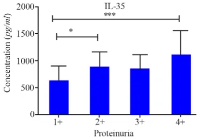
Figure 1. Comparison of IL-35 serum levels in pre-eclamptic patients with different proteinuria levels.
* and *** represent p<0.05 and p<0.001, respectively.
Table 1. Demographic and clinical characteristics of all studied groups
Parameters Healthy pregnant women PE p-value Mild PE Sever PE p-value
Age (years) 29.11±5.56 30.02±5.88 0.55 30.36±5.65 29.65±6.1 0.45
SBP (mmHg) 81.8±1.9 89.89±5.4 0.001 88.11±4.82 91.95±5.42 0.001
DBP (mmHg) 123±2.7 145±8.7 0.001 141.60±3.63 149.56±10.74 0.001
SBP= Systolic blood pressure, DBP= Diastolic blood pressure, PE= Pre-eclampsia
Table 2. IL-35 and IL-17 serum levels in pre-eclamptic patients (Severe and mild) as compared to control group Parameters Healthy pregnant women
Group 1(n=100)
PE Group 2(n=100)
Mild PE Group 3(n=46)
Severe PE
Group 4(n=54)
IL-35 483.9±242* 729±335* 558±228* 927±332*
IL-17 2.86±1.69 2.79±1.26 3±1.2 2.5±1.29
*: each group is different with others (p<0.05). One-way ANOVA with post-hoc Tukey tests were used for statistical analysis.
PE: Preeclampsia, HC: Healthy control.
Downloaded from ht
tp://www.jr
i.ir
IL-17 and IL-35 serum levels in preeclampsia
JRI
J Reprod Infertil, Vol 20, No 4, Oct-Dec 2019 and proteinuria was compared in patients. Results
indicated that the mean level of IL-35 was signifi-cantly higher in patients with 4+ proteinuria as compared to those with less than 4+ proteinuria (p<0.001) (Figure 1). There were no significant differences between the levels of IL-17A with blood pressure or proteinuria. Finally, no correla-tion was found between IL-35 and IL-17A levels in both cases and controls (data not shown).
Discussion
In the current study, the serum levels of an anti-inflammatory cytokine (IL-35) and a Th17 related inflammatory cytokine (IL-17) were evaluated in a group of Iranian women with pre-eclampsia. The results of the present study indicated that the level of IL-35 was significantly increased within sera samples from pre-eclamptic women and also the increase was related to the severity of the dis-ease. In addition, it was observed that pre-eclamp-tic women with higher level of proteinuria had elevated level of IL-35. It is well known that PE is a disease of placentation. Indeed, it is believed that a poor placentation in the beginning of preg-nancy will lead to PE after the 20th week of gesta-tion (22). To support a normal gestagesta-tion from im-plantation to parturition, immune system works to balance inflammatory to anti-inflammatory re-sponses in different time points of the pregnancy period. In the first trimester, especially at the early days of gestation, a controlled inflammatory re-sponse is needed to support implantation and pla-centation. At the second trimester, regulatory re-sponses and later in the third trimester and finally at the end of the pregnancy again, inflammatory responseswilldominatetosupportdelivery. More-over, cytokines do their function both in local and systemic manner (23). So, to investigate the im-mune responses and cytokine balances in preg-nancy, time point and sampling are two important factors that might alter the obtained results. It is well known that pre-eclamptic women are in high-er inflammatory state as compared with women with normal pregnancy. Higher expression of IL-35 during the last weeks of pregnancy, as seen in the present study in patients, may work as com-pensatory mechanisms to control the inflammato-ry responses. This finding could be explained by the results of IL-35 level as it is documented that IL-35 could suppress Th17 cells and inhibit IL-17 production (24). In line with this scenario, the level of IL-17 as an inflammatory cytokine in both cases and controls was the same. While only
a few studies have been published regarding the expression of IL-17 and IL-35 in PE, there are controversies in the reported data. Ozkan et al. reported a decreased level of both 17 and IL-35 cytokines and also IL-IL-35/IL-17 in women with PE (21). Moreover, Cao et al. studied the expres-sion of IL-17 and IL-35 at both mRNA level (PBMC) and protein level (serum) in Chinese pre-eclamptic women (20). They also reported down-regulation in expression of IL-35 and up-regula-tion for IL-17 at both mRNA and protein levels. Interestingly, both mentioned studies have report-ed unbelievable low level of IL-35 within sera. Looking for the reported IL-35 level in normal and pregnancy related disorders indicates that the mean level of this cytokine is above 120 pg/ml
while the level of this cytokine is reported in a range of 6.65-17 pg/ml by Ozkan and Coa et al.
(19). Regarding the reported levels for IL-17 in both mentioned studies, the lower level of IL-17 can be seen as compared with other published pa-pers (14). In case of IL-17, another point to be mentioned is the limitation of ELISA kit used by Ozkan et al. While they used an ELISA kit with assay range of 31-2000 pg/ml, they reported a
range between 1.8-3.57 pg/ml for IL-17. Recently,
our group have also reported declined level of IL-35 expression but in the placental tissues from pre-eclamptic women (25). In line with Cao et al.’s report, an elevated level of IL-17 was report-ed before, but only in blood samples collectreport-ed from placenta (26). Interestingly, a recent study has reported elevated expression of EBI-3 (a chain of IL-35 heterodimer) in decidua from pregnant women with preeclampsia. They also showed in-creased level of HLA-G in pre-eclamptic women which is an anti-inflammatory agent. This group concluded that these increases may contribute in PE pathophysiology or maybetheconsequenceof thedisease(27).Moreover, increased level of IL-35 has been reported in several well-known dis-eases with inflammatory basis including diabetes, and inflammatory bowel disease (IBD) (28). The latest study has interpreted this increase as a com-pensatory mechanism of immune system to atten-uate the effects of inflammation (29). The contro-versy is not limited to IL-35. For example, there are opposite reports regarding increasing or de-creasing levels of IL-10 and IL-4 in PE (30-33). These controversial data necessitates the needs for more detailed investigations on cytokine network in PE. Our data also showed an association be-tween the level of proteinuria and elevated level
Downloaded from ht
tp://www.jr
i.ir
Batebi A, et al.
JRI
J Reprod Infertil, Vol 20, No 4, Oct-Dec 2019 of IL-35. As far as searched, there was no study
investigating the effects of IL-35 on proteinuria but in accordance with our findings, a published study on SLE patients reported an association be-tween the levels of IL-35 with renal failure. How-ever, it seems that more investigations are needed to find out if IL-35 is involved in renal problems directly or not (34). Asmentioned,theresultsofthe presentstudyshowed no difference between wom-en with PE and healthy ones regarding IL-17 lev-els. This finding could be explained by the results of IL-35 levels as it is documented that IL-35 could suppress Th17 cells and inhibit IL-17 pro-duction (24). If the elevated level of IL-35 in these patients as an anti-inflammatory and compensato-ry reaction of immune system is observed, so the invariable level of IL-17 could also be attributed to this compensatory mechanism. There are some points that should be mentioned here to help for a better understanding of the current study and also other studies on PE. The first point that should be mentioned is that all patients which were enrolled in this study were under supervision of physicians and many of them received medications to control their disease. Moreover, it should be considered that the serum level of these cytokines was tested while the main scenario of pre-eclampsia patholo-gy might take place in feto-maternal interface. Last but not least, it should be noted that immune system behavior in pregnancy is completely time-dependent and this finding could just be attributed to last days of pregnancy as our patients were re-ferred to hospital for labor and delivery. Myatt et al. have published a paper on criteria and strate-gies for designing a perfect study on PE and men-tioned these points and others such as age, previ-ous disease and smoking that may influence the resultantdata(35).Investigatingtheserumlevelsof the mentioned cytokines along with other cyto-kines during all periods of pregnancy will certain-ly provide more detailed and precise data on rela-tion of these cytokines to the disease pathophysi-ology.
Conclusion
In all, although high level of IL-35 may be asso-ciated with pre-eclampsia and severity of the dis-ease but due to lots of unanswered problems, it is not suitable for evaluating the onset or severity of PE. Also, in case of its role in pathology of PE, there are lots of questions remained to be an-swered.
Conflict of Interest
Authorsdeclarenoconflictofinterest.Thisstudy was extracted form thesis written by Atefeh Ba-tebi and financially supported by Grant no. 94-10426fromShirazUniversityofMedicalSciences.
References
1. English FA, Kenny LC, McCarthy FP. Risk factors and effective management of preeclampsia. Integr Blood Press Control. 2015;8:7-12.
2. August P and Sibai BM. Preeclampsia: Clinical fea-tures and diagnosis [Internet]. Waltham, MA; 2014 [updated2019Apr25;cited2019Apr29].Available
from:https://www.uptodate.com/contents/preeclampsia -clinical-features-and-diagnosis
3. Cotechini T, Komisarenko M, Sperou A, Macdonald-
Goodfellow S, Adams MA, Graham CH. Inflam-mation in rat pregnancy inhibits spiral artery re-modeling leading to fetal growth restriction and features of preeclampsia. J Exp Med. 2014;211(1): 165-79.
4. Racicot K, Kwon JY, Aldo P, Silasi M, Mor G. Un-derstanding the complexity of the immune system during pregnancy. Am J Reprod Immunol. 2014;72 (2):107-16.
5. Vargas-Rojas MI, Solleiro-Villavicencio H, Soto-Vega E. Th1, Th2, Th17 and treg levels in umbi-lical cord blood in preeclampsia. J Matern Fetal Neonatal Med. 2016;29(10):1642-5.
6. Perez-Sepulveda A, Torres MJ, Khoury M, Illanes SE. Innate immune system and preeclampsia. Front Immunol. 2014;5:244.
7. Raghupathy R. Cytokines as key players in the pa-thophysiology of preeclampsia. Med Princ Pract. 2013;22 Suppl 1:8-19.
8. Redman CW, Sargent IL, Taylor RN. Immunology of normal pregnancy and preeclampsia. In: Taylor
RN, Roberts JM, Cunningham FG, Lindheimer M D,editors.Chesley'shypertensivedisordersin preg-nancy. USA: Elsevier; 2015. p. 161-79.
9. Teymouri M, Pirro M, Fallarino F, Gargaro M, Sahebkar A. IL-35, a hallmark of immune‐ regula-tion in cancer progression, chronic infecregula-tions and inflammatory diseases. Int J Cancer. 2018;143 (9):2105-15.
10. Keijsers RR, Joosten I, van Erp PE, Koenen HJ, van de Kerkhof PC. Cellular sources of IL-17 in psoriasis: a paradigm shift? Exp Dermatol. 2014; 23(11):799-803.
11. Monin L, Gaffen SL. Interleukin 17 family cyto-kines: signaling mechanisms, biological activities, and therapeutic implications. Cold Spring Harb Perspect Biol. 2018;10(4). pii: a028522.
Downloaded from ht
tp://www.jr
i.ir
IL-17 and IL-35 serum levels in preeclampsia
JRI
J Reprod Infertil, Vol 20, No 4, Oct-Dec 2019
12. Pongcharoen S, Somran J, Sritippayawan S, N ium-supP,ChanchanP,ButkhamchotP,etal.Interleukin- 17 expression in the human placenta. Placenta. 2007;28(1):59-63.
13. Fu B, Tian Z, Wei H. TH17 cells in human recur-rent pregnancy loss and pre-eclampsia. Cell Mol Immunol. 2014;11(6):564-70.
14. Martínez-García EA, Chávez-Robles B, Sánchez-Hernández PE, Núñez-Atahualpa L, Martín-Má-quez BT, Muñoz-Gómez A, et al. IL-17 increased in the third trimester in healthy women with term labor. Am J Reprod Immunol. 2011;65(2):99-103. 15. Cornelius DC, Wallace K, Scott JD, Campbell N,
Thomas A, Hogg JP, Moseley J, LaMarca B. [35-OR]: A role for TH17 cells and IL-17 in mediating the pathophysiology associated with preeclampsia. Pregnancy Hypertens. 2015;5(1):17.
16. Molvarec A, Czegle I, Szijártó J, Rigó J Jr. In-creased circulating interleukin-17 levels in pre-eclampsia. J Reprod Immunol. 2015;112:53-7. 17. Li X, Fang P, Yang WY, Wang H, Yang X. IL-35,
as a newly proposed homeostasis-associated mole-cular pattern, plays three major functions including anti-inflammatory initiator, effector, and blocker in cardiovasculardiseases.Cytokine. 2019;122:154076.
18. Mao H, Gao W, Ma C, Sun J, Liu J, Shao Q, et al. Human placental trophoblasts express the immuno-suppressive cytokine IL-35. Hum Immunol. 2013; 74(7):872-7.
19. Yue Cy, Zhang B, Ying CM. Elevated serum level of IL-35 associated with the maintenance of ma-ternal-fetal immune tolerance in normal pregnancy. PloS one. 2015;10(6):e0128219.
20. Cao W, Wang X, Chen T, Zhu H, Xu W, Zhao S, et al. The expression of notch/notch ligand, IL-35, IL-17, and Th17/Treg in preeclampsia. Dis Mar-kers. 2015;2015:316182.
21. Ozkan ZS, Simsek M, Ilhan F, Deveci D, Godek-merdan A, Sapmaz E. Plasma IL-17, IL-35, inter-feron-γ, SOCS3 and TGF-β levels in pregnant wo-men with preeclampsia, and their relation with se-verity of disease. J Matern Fetal Neonatal Med. 2014;27(15):1513-7.
22. Saito S, Nakashima A. A review of the mechanism for poor placentation in early-onset preeclampsia: the role of autophagy in trophoblast invasion and vascular remodeling. J Reprod Immunol. 2014; 101-102:80-8.
23. Mor G, Cardenas I, Abrahams V, Guller S. Inflam-mation and pregnancy: the role of the immune system at the implantation site. Ann N Y Acad Sci. 2011;1221(1):80-7.
24. Niedbala W, Wei XQ, Cai B, Hueber AJ, Leung BP, McInnes IB, et al. IL-35 is a novel cytokine with therapeutic effects against collagen‐induced arthritis through the expansion of regulatory T cells and suppression of Th17 cells. Eur J Immunol. 2007;37(11):3021-9.
25. Gharesi-Fard B, Mobasher-Nejad F, Nasri F. The expression of T-helper associated transcription factors and cytokine genes in pre-eclampsia. Iran J Immunol. 2016;13(4):296-308.
26. Anvari F, Dabagh-Gorjani F, Soltani-Zangbar MS, Kamali-Sarvestani E, Malek-Hosseini Z, Gharesi-Fard B. Investigating the Association of IL-17A and IL-17F with Susceptibility to Pre-eclampsia in Iranian Women. Iran J Immunol. 2015;12(2):117-28.
27. Prins JR, van der Hoorn MLP, Keijser R, Ris-Stalpers C, van Beelen E, Afink GB, et al. Higher decidual EBI3 and HLA-G mRNA expression in preeclampsia: Cause or consequence of preeclamp-sia. Hum Immunol. 2016;77(1):68-70.
28. Espes D, Singh K, Sandler S, Carlsson PO. In-creased interleukin-35 levels in patients with type 1 diabetes with remaining C-peptide. Diabetes Care. 2017;40(8):1090-5.
29. Fonseca-Camarillo G, Furuzawa-Carballeda J, Yamamoto-Furusho JK. Interleukin 35 (IL-35) and IL-37: intestinal and peripheral expression by T and B regulatory cells in patients with inflam-matory bowel disease. Cytokine. 2015;75(2):389-402.
30. Madazli R, Aydin S, Uludag S, Vildan O, Tolun N. Maternal plasma levels of cytokines in normal and preeclamptic pregnancies and their relationship with diastolic blood pressure and fibronectin le-vels. Acta Obstet Gynecol Scand. 2003;82(9):797-802.
31. Szarka A, Rigó J Jr, Lázár L, Beko G, Molvarec A. Circulating cytokines, chemokines and adhesion molecules in normal pregnancy and preeclampsia determined by multiplex suspension array. BMC Immunol. 2010;11:59.
32. Makris A, Xu B, Yu B, Thornton C, Hennessy A. Placental deficiency of interleukin-10 (IL-10) in preeclampsia and its relationship to an IL10 pro-moter polymorphism. Placenta. 2006;27(4-5):445-51.
33. Cemgil Arikan D, Aral M, Coskun A, Ozer A. Plasma IL-4, IL-8, IL-12, interferon-γ and CRP levels in pregnant women with preeclampsia, and their relation with severity of disease and fetal birth weight. J Matern Fetal Neonatal Med. 2012; 25(9):1569-73.
Downloaded from ht
tp://www.jr
i.ir
Batebi A, et al.
JRI
J Reprod Infertil, Vol 20, No 4, Oct-Dec 2019
34. He D, Liu M, Liu B. Interleukin-35 as a new biomarker of renal involvement in lupus nephritis patients. Tohoku J Exp Med. 2018;244(4):263-70. 35. Myatt L, Redman CW, Staff AC, Hansson S, Wil-
son ML, Laivuori H, et al. Strategy for standardiz-ation of preeclampsia research study design. Hy-pertension. 2014;63(6):1293-301.