PNEUMATOSIS
INTESTINALIS
Review of the Literature
with Report of 13 Cases
B)- ELLEN P. MACKENZIE, M.D. Nezi O,'leans
P NEUMATOSIS of the intestine (also called pneumatosis cystoides intestinorum
hominis, intestinal emphysema, cystic lympho-pneumatosis, peritoneal pneumatosis, entero-mesenteric bullous emphysema, and gas cysts of the abdomen) consists of the presence of gas-containing cysts of varying size within the wall of the intestinal tract,
sometimes accompanied by similar cysts on the surface of other abdominal viscera and
parietal peritoneum. It is much discussed in European literature, but largely unknown
in North America. Twelve infants and three older children were included in the world
literature on the subject prior to a recent report of 17 cases in infants.'
Intestinal pneumatosis is better known in veterinary than in human medicine ; it is common in pigs.'@4 Duvernoy observed it during dissection of a cadaver in 1747.―6
CloquetT in 1820 and Bang8 in 1876 reported finding it at autopsy. Nigrisoli9 and
Hahn'° independently reported finding pneumatosis at laparotomy ; Finney1' was the first to report it in this country. Reverdin,12 in 1924, reported its pre-operative diagnosis, based on clinical and roentgenologic observations and confirmed at laparotomy. Lerner
and Gazin'3 were the first Americans to report a nonoperative diagnosis, based on
roentgenologic evidence. Though some of the older case reports are not accepted as true pneumatosis, over 200 acceptable cases have now been reported, from practically all civilized countries. Those concerning infants and children are summarized in table 1. Pneumatosis was found at operation in the first two patients listed ; in all others it was
an autopsy finding.
The reader is referred to the bibliography for more detailed discussion of pneumatosis
intestinalis in adults. Much of the literature reviewed was found sketchy and inconclusive;
only the reports which seemed of some value and reliability are included here.
The peak incidence of pneumatosis is between 25 and 50 years, but Lyons―observed
it in a man aged 81, and case 1 3 of this series was a premature baby aged 12 days.
Two rarer conditions, pneumatosis 324 (colpohyperplasia cystica, vaginitis
emphysematosa) and pneumatosis vesicae,'3 deserve mention because of their similarity to pneumatosis intestinalis. Vaginal and intestinal pneumatosis may coexist ;25 they are be
26 to have the same pathogenesis.
Pathology: The lesion looks much like hydatidiform mole. The thin-walled cysts vary greatly in size 23,27 in infants they are less than 1 cm. across, and sessile. When com pressed, they move a short distance, or burst without collapse of surrounding cysts. Pin points or networks of serosal scars have been described near co-existent lesions or at sites of earlier lesions.'8'°
The ileum is first, the colon second, in frequency of involvement ; both may be
affected at once. Other visceral surfaces are less commonly involved, usually along with
From the Departments of Pediatrics at Tulane Medical School, the Ochsner Clinic, and the Charity
Hospital of Louisiana at New Orleans. (Received for publication April 15, 1950.)
538 ELLEN P. MACKENZIE
-; -; ‘3
- ‘3 C .@
.@ -@ -@ -@ a
C. -.@ —
‘ “3-@ @ @ @ @ C .C @ @ E
,i@ @V @‘Y E
E C.V ‘3., ‘3
@. E.a @
@ .@ ‘@E
.:@-@
@
:@ ‘@
@
@ o@bo :e@ C.
‘@
@
@
C 3@) @_
C @
@
I fi
@C
@V' C so -@ .@ .@ @ -@ @ @ U E@C @ ‘i.E @E @
h
@
@
V
@ @ P@ @ @ @.‘.2 -@ .E .@ ‘C C .@ E — .@ @-_C @Cc@ @C
,, 0
.@ -@
-,, ‘3
‘3 @C. a .:
:C @
@ 6
@
@ E ‘E@@
-@ ‘E-@
.-C .— . U
EE @ @-@ O:@ @ .@ @ @ E @@S'C @
@ .-. ‘3 —.-,
@ @“C. @ @ @E @
!
@
@
C VEV CV
@C
@ F@
@
.@ ‘@.C@- .C@
@@‘C,.. ,,@ .C_@
L'3
@ _
@ U'3@ ‘‘3 C
U -@F@
@
‘@
-J'..@ @E'c @ C.-' .0 @ VE C ‘@0 @C. U@ 0 -@‘3 .@ be UCU :@
@C -@ E ‘@ ‘3-A @ .@ 6 @ C C L@ E ‘3 p .@ 0 -E @ C -@ @ C E ‘3 Sc C .@ V 2 ?@ @‘3 2 _0@ @ ‘3 @-U @, ‘3 R ‘@ 0 .‘@ C. (‘. C 0 C .@‘3I
@,‘3@ @.0 V
@‘3 Z
0 C
so “:
C_ (@ -@ -@
‘-: ‘@ @E
@0 .0 @ 0 @ @ C @ 3 @0. @:o @
‘3E' -E @4C ‘3 .@2 @ .@ @ v@8 ‘@C @ C@ @i2
C'@ VO ,@
@ C
@
@
-E@ lo E'-'
@
>@b oE .@ @
.0@
@ .@C
so ‘?@ .C'3 C.
@ @C.
@
[email protected] ,@,,‘? .@C -u,@ VSL 0 ,@
@ @‘.s @ @ ‘3, @ @ .@ :@ :@ .@ .2'@' @ E.@ @
@ a,@o .OV
@
n,E 0,'C@ .—E ...‘@
@; @; -@ a V @ .@ I-.@ @ 0. ‘3 .-‘ C @ 0 @ 2 — ‘@ 0, -2 2 ‘C C' :., .C 2 U 0 @ 2 2 @ ‘3 33 C @ ‘3 .@ @ ,0 @C V .C0 ‘C 0 ‘@U @ C 0 ci .C 0 2 C C C ‘C ‘S V C C 0 .@ @4 4: .C' @ ‘C .0 3- ‘3 C ‘3 U @ .@ .— C .— ‘3 E C ,‘ Sc .‘ C .4 @ 2 -C -@ 2 C.@ 2 2 C 0 C, C ‘C .@ C, C
H-
@V
@
@V to.:@
:@
@
@
.1 C C [email protected] .c@ C ‘Ca .- CV ::. :: .@ -‘@ .- — ‘@ -p CC ._
@., @© ‘@,-1 @
@ V @;
@ U@@V V
‘C
@
Ca
2
SC -‘
V C ‘C
@ 0C@0 C.
@ .2.@o @@ooo
@ .@ OS@@ @ QV 3@ @ V @ @O
@ U ‘C a'C
@ U
‘CE o
@ V
@
@ E E@
@ @ @ C @ @ @ @ @
!E!:!!
@
PNEUMATOSIS INTESTINALIS 539
.@ .0 -,-‘7 C -E .‘: @) ‘— =++ .C@ [email protected] ‘3 CU C.@ 0V U5@ CE C -;eC C C 2 C C@@.E .@ C'3 CU ‘,@ 8@ =V CE ‘@e >,‘C 0@ E@ ,,e@ .@E.@ 6@. ,@ C -I).@ C 0 --‘ o Z; E
it
@. ._0 E@ @2 -@‘3!‘@1
‘C@ @‘@8E@h
@E @.,; Eo QU ‘;@c--'
U
a33
@8-.--@‘ @-2 -“ C@“v
:@
‘@C@::@ee
@Vo'3 :@.E@-'@ .S33@'3 @jC.@H:@flC2C@
@so U'@‘—IC ‘,@—-@-‘-I——.0 U V.... Sc.C'.-.
C ‘3 ‘3 C 2 C C E 0 ‘3 2 ‘3 ‘C C. C C ‘3 ‘3 ‘C C Cl C SC C C Cl ‘3 C. ‘C Cl -@ 0 ‘C 33 2 C 0 ci ‘C 0
..@6I0@
-@E C 0 Cl C ‘C C C. ‘3 C L) CV ‘C S ,@ 3 ‘C ‘C Cl SC C I@ 2
@ ‘C p
- C !@ ‘3 Cl“C ‘C C
‘C,3 ‘ .— -C
540 ELLEN P. MACKENZIE
intestinal involvement.2l Pneumatosis is often confined to an intestinal loop constricted by adhesionsta or volvulus,3'33 sometimes to a loop containing intestinal parasites.2' a@ @
In infants, cysts are predominantly submucosal. The mucosa may show atrophy36 or edema.@' The cellular infiltrate varies, but usually indicates chronic inflammation ; giant cells are rarely seen in infants. Gas has been observed in vessel walls36 and nerve sheaths.3' The cysts have been thoroughly described by Masson ;26 he believes them to begin in lymphatics, ending as a scar ringed by elastic fibers.
The gas is a mixture of 70-90% nitrogen, 3-20% oxygen, and 0-15% carbon
53, Urban alone found methane by paracentesis on a living patient.15 These
proportions
closely resemble
those
found
by Banyai 48 hours
after
induction
of
pneumoperitoneum with pure oxygen.'7
Associated gastrointestinal disease is prominent, in adults mostly peptic ulcer with pyloric stenosis ;36 various other intestinal lesions, and cardiac and pulmonary disease, are mentioned21,33, 35,39 In infants, diarrheal disease most frequently coexists, pulmonary disease ranking next.
Pathogenesis : Little experimental work has been done relative to those disease. The theories discussed here are individually inadequate ; probably all three factors operate
in the production of cysts.
1. Infectious: Many organisms, including yeasts, have been isolated from cysts in living patients ;4@Jaeger's “Bacterium coli lymphaticum aerogenes― reproduced the dis
ease in animals but killed them within 48 hours.4' Killian23 considers pneumatosis the end
result of lymphangitis
due to low-grade, nonspecific infection. Lymphangitis
elsewhere
produces no gas cysts, and it does not explain the lack of inflammation around cysts or the occurrence of extraintestinal pneumatosis.
2. Biochemical: Pneumatosis in pigs seems related to diet;122'C such relation is not
clear in man. Absorption of free or dissolved gas is supported by Stübel'sobservation of
bubbles of oxygen formed in lymphatics of intestine placed in peroxide solution.4' Masson blames acid mucosal secretions, plus circulatory stasis.―3This does not explain extraintestinal pneumatosis, its association with peptic ulcer, or its occurrence where absorption is least active.
3. Mechanical: Air injected directly into the gut wall follows lymphatics to the mesentery and disappears within a few days.8' Box saw emphysema around a peptic ulcer after washing it with air and water.4' Miyake forced air into a gut loop after scraping the mucosa and waiting for exudate to cover it ; the resulting blebs remained 5 to 6 days.44
Tung and Ngai made an artificial stenosing peptic ulcer in a dog ; when the stomach
was filled with air and salt solution, air bubbles appeared inside and outside lymphatics,
progressing distally with peristalsis, some remaining two days in serosa and omentum.'4
Menna― injected air into the gut wall of rabbits and found the gut normal 10 days
later, hut if gas withdrawn from the animal's intestine was injected, 10 days later there
was increased density of the lymph follicles with microscopic dilatation of the surrounding subcapsular space. When a culture of B. coli was injected 20 days before air injection, small vesicles were visible in the peritoneum 1 5 days after air injection, plus adhesions and mesenteric thickening. Streptococci and staphylococci produced more adhesions. Giant cells were absent, though air injected subcutaneously is said to produce them within a few days.―3
PNEUMATOSIS INTESTINALIS 541
suffered from stenosing peptic ulcer, pneumoperitoneum and pneumatosi@ of the small
intestine. During laparotomy he vomited, and the cysts at the mesenteric border im
mediately swelled, without simultaneous distention of the intestine.
The mechanical theory does not explain why bacteria do not enter the intestinal wall, nor why pneumatosis is rare in obstruction, despite hyperperistalsis, distention and thin ning of the mucosa.
Clinical Picture: Symptoms of primary pneumatosis refer only to adults ;29 31.:38they range from acute to chronic, often mimicking appendicitis, peritonitis or intestinal ob struction. The only significant sign is a crepitant, nontender abdominal mass. Pneumo
peritoneum in a patient with peptic ulcer is 2@@to be pathognomonic of pneumatosis
if perforation can be excluded. Roentgenograms show pneumoperitoneum and cysts ; inter
position, either fixed or reversible, of small intestine between liver and diaphragm (the
Moreau-Chilaiditi syndrome) is very “3““@°Barium enema shows colonic
cysts as translucent filling defects.―1―
Pneumatosis is generally of short duration, from 2 to 40 days ; re-operations have shown only roughening and scarring of the serosa. However, it may persist a long time or
recur.'4 Submucosal cysts may occlude the lumen or initiate an intussusception ;“
subserosal
cysts may initiate a volvulus.29 Interposition alone does not cause obstruction.
Treatment:
None is indicated in the absence of intestinal obstruction. Simple laparot
omy is said to have curative effects.26 Closure without drainage suffices for primary pneu matosis ; puncture or removal of cysts is unnecessary. Treatment of associated intestinal disease follows the usual indications ; healing is normal after resection or anastomosis.
MATERIAL
Cases 1 and 2 of this series were observed by the writer in 1948 at Notre Dame Hospital,
Montreal. Cases 3 to 12 were found in the records of more than 1800 autopsies performed in
18 years at the Children's Memorial Hospital, Montreal. Case 13 was observed by Dr. James B. Arey at Charity Hospital of Louisiana at New Orleans.
Case 1 (MG., 1948), white female born of a pre-eclamptic mother after 7½ mo. gestation, was transferred to pediatric service at age 1 mo. a/c diarrhea and persistent rectal prolapse, with blood and pus in the stools. Stool cultures were negative. Pneumonia and otitis yielded to antibiotics. She died on the 50th day of life, showing fever, distention and dehydration.
Autopsy (4 hr. post mortem) showed: mastoiditis; interauricular septal defect; pneumatosis of the colon from ileo-cecal valve to sigmoid, excluding the appendix ( Fig. 1).
Microscopic examination of the colon showed submucosal cysts lined with endothelium which
in larger cysts resembled fibroblasts (Fig. 2). Lymphatics underlying a small mucosal ulcer were full
of PMN. The nerve plexi were edematous with occasional necrotic ganglion cells.
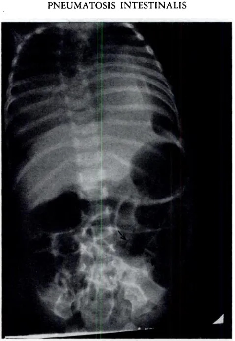
Case 2 (RB., 1948), white male aged 3 wk., was admitted a/c pneumonia of 4 days' duration. Diarrhea developed on the 5th hospital day, followed by fever, distention and intermittent vomiting.
RGs taken on the 2nd and 10th hospital days showed on postmortem review a narrow radiolucent
band outlining the distal colon (Fig. 3). He died on the 13th hospital day.
Autopsy (9 hr. post mortem) showed: closed retropleural cyst lined by gastric mucosa ; broncho pneumonia and atelectasis ; intramesenteric diverticulum beginning close to the pylorus and com municating with the jejunum 30 cm. distally; pneumatosis of the colon, exactly resembling Case 1.
Microscopic examination showed submucosal cysts, and mucosal abrasion, with edema, congestion, and infiltrate of PMN, lymphocytes and plasma cells (Fig. 4).
Case 3 (J.A., 1934), white male aged 3½ mo., was admitted a/c vomiting and athrepsia since the first few days of life. He died on the 73rd hospital day with dehydration, distention and otitis media.
5'2 ELLEN P. MACKENZIE
Microscopic examination showed submucosal md a few subserosal cysts. The smaller cysts ap peared to be lymphatic vessels without endothelial lining. The submucosa showed edema, vascular stasis, cellular infiltration, and irregular fluid-containing spaces, perhaps early cysts.
Case 4 (MW., 1935), white female aged 7 wk.. was admitted a/c diarrhea. vomiting, @tthrepsia
v,@tw
@ @@i::w;4@
-@ @
FIG. 1. Case 1.—Gross appearance of colon after fixing. Cysts were 1 to 5 mm. in diameter.
-@&@-.
FIG. 2. Case 1.—Microscopic appearance of colon, showing characteristic appearance and
submucosal situation of cysts.
and chronic otitis. She died on the 7th hospital day of aspiration pneumonia.
Autopsy ( 12 hr. post mortem) showed: marasmus; hemorrhagic alveolar pneumonia, probably streptococcal ; pneumatosis (If the cecum and 12 cm. of ascending colon. Mesenteric nodes were atrophic.
Microscopic examination showed numerous submucosal and occasional subserosal cysts, frequently endothelium-lined, with a few inflammatory and giant cells. The serosa showed edema and a thin
- PNEUMATOSIS INTESTINALIS 543
FIG. 3. Case 2.—Roentgenogram taken 1 1 days before death. Arrows indicate increased radiolucency
outlining involved distal colon.
544 ELLEN P. MACKENZIE
cellular infiltrate ; many cocci present were considered postmortem invaders because of the lack of
surrounding tissue reaction ; the epithelium was intact.
Case 5 (V.L.M., 1936), white male aged 7 wk., was admitted a/c coryza for 2 wk. and
pneumonia for 1 day. Distention was extreme by the 5th day; he died on the 1 ith hospital day. Autopsy ( 5 hr. post mortem) showed: hemorrhagic alveolar pneumonia with middle lobe atelectasis ; multiple lung abscesses and encysted empyema yielding Staph. pyogenes ; cystic fibrosis of the pancreas; moderate atrophy of mesenteric nodes; and marked distention of small intestine and proximal colon, with 4 separate areas of pneumatosis, up to 5 cm. long, along the ileum.
Microscopic examination showed submucosal and mucosal cysts, surrounded by minimal focal. inflammation; the intervening intestine was normal.
Case 6 (C.B., 1937), white -male aged 4 mo., was admitted a/c vomiting and athrepsia. He became more toxic and dehydrated, and died on the 16th hospital day.
Autopsy ( 5 hr. post mortem ) showed : underdevelopment; otitis media ; mastoiditis ; interstitial pneumonia; moderate atrophy of mesenteric nodes; and pneumatosis along 23 cm. of the ascending
colon.
Microscopic examination showed minimal inflammation around the cysts except for a loculated
abscess which resembled the cysts in outline and communicated with the intestinal lumen by 2 narrow sinus tracts lined with librino-purulent exudate. Some cysts were partially lined by endothelial-type cells; a few giant cells were seen in the cyst walls.
Case 7 (J.G., 1946), white male aged 2 mo., was admitted a/c progressive jaundice for 9 wk.,
weight loss, umbilical discharge and pale stools, Abdominal RG showed no sign of pneumatosis on later review. No pneumatosis was seen at cholecystectomy on the 9th hospital day. He grew worse from the 4th postoperative day, eviscerated on the 18th hospital day, and died following secondary closure.
Autopsy (22 hr. post mortem) showed: emaciation; marked icterus; plastic peritonitis between
transverse colon and liver; congenital atresia of the bile ducts; a mucosal ulcer 4 cm. in diameter at the jejuno-ileal junction, containing 2 tiny fresh perforations, with pneumatosis extending distally from the ulcer to involve the entire ileum and colon except the appendix.
Microscopic examination showeJ the ulcer to extenJ to the muscularis mucosae, with many submucosal and mucosal cysts around the perforations. Large submucosal cysts above and below the ulcer contained PMN and giant cells; a deep ulcer, lined by fibrin, extended to the serosa and might have been a ruptured infected cyst.
Case 8 (Ba-by C., 1946), white male aged 1 day, was admitted a/c general weakness. Atelectasis
and pneumonia yielded to routine therapy. He developed multiple staphylococcal abscesses at venepuncture and cut.down sites. He recovered well from Fredet-Ramstedt pyloromyotomy done on
the 17th hospital day, aaJ co@retion of malrotation of the colon done on the 38th hospital day. On the 63rd hospital day he developed diarrhea, with Ps. pyocyaneus on stool culture, and died on
the 66th hospital day. No evidence of pneumatosis was found at either operation, nor in the RGs, the last of which was taken on the 38th hospital day.
Autopsy (20 hr. post mortem) showed: underdevelopment; RITL pneumonia; multiple intra abdominal adhesions without peritonitis; and pneumatosis of the colon. In the cecum a small area of congestion surrounded a thrombosed mural vessel. Culture of heart, lungs and abdominal wall yielded Esch. coli.
Microscopic examination showed numerous submucosal and occasional mucosal cysts, many with a flat lining. Some contained monocytes and giant cells in the walls and lumina ; collections of similar cells, with fibroblastic proliferation, in the cecum suggested obliterated cysts. Abundant submucosal and serosal foci of chronic inflammation in the cecum diminished distally to a few
monocytes in the rectosigmoid, where cysts were numerous. Many giant cells were scattered through the intestinal wall, in the serosa perhaps representing reaction to catgut suture. The jejunum showed
only early acute inflammation, giant cells and edema.
Case 9 (L.N., 1947), white female aged 1½ mo. (premature), was admitted a/c lifelong feeding difficulty. Atelectasis appeared on the 6th hospital day, but resolved. Diarrhea began on the
1 ith day, becoming severe by the 24th day. She died on the 27th hospital day despite antibiotic
therapy.
Autopsy ( 17 hr. post mortem) showed: bilateral focal pneumonia ; bilateral otitis media ; minimal
PNEUMATOSIS INTESTINALIS 545
ileum to the ileocecal valve, with retroperitoneal and mesenteric edema and scant hydroperitoneum. Microscopic examination showed submucosal cysts with minimal inflammatory reaction, and moderate atrophy of the mesenteric LN.
Case 10 (LB., 1948), white female aged 11 mo., was admitted a/c'persistent diarrhea for 6 mo. She grew steadily worse despite extensive Rx, On the 25th hospital day, blood appeared in stools and vomitus; stool culture yielded P. morgani. She died on the 34th hospital day.
Autopsy (8 hr. post mortem) showed: extreme emaciation; purulent otitis media ; and acute ulcerative pseudomembranous ileitis and colitis with localized pneumatosis around the ileocecal valve, underlying an ulcer which was superficial, minimally indurated, and covered with mucoid substance; all other ulcers were indurated, raised and covered with adherent membrane. At the splenic flexure were a large ulcer, several hemorrhagic saccular protrusions, and small indurated b@ack nodules.
Microscopic examination showed no acute or chronic reaction around the cysts. Tne mesenteric nodes showed only moderate atrophy. Extensive bacteriologic study revealed no dysentery organisms; the enteritis was believed to be staphylococcal.
Case 11 (NC., 1948), white male born 5 wk. prematurely, had a pyloromyotomy at age 3 wk.
and was discharged 9 days later in good condition. Vomiting and diarrhea began the next day; he
was re-admitted at age 5 wk, He grew steadily worse; fresh and occult blood appeared in the stools
on the 3rd hospital day; he died on the 5th hospital day.
Autopsy ( 14 hr. post mortem-) showed: hypertrophy of pyloric muscle with adequate lumen diameter; ulcerative jejunitis, ileitis and cecitis; and pneumatosis, marked in lower jejunum and ileum, slight in the cecum wl@ere there ve@e fey. ulcers.
Microscopic examination showed no cellular reaction around the cysts ; there was moderate atrophy
of the mesenteric LN. Elaborate postmortem cultures revealed no dysentery organisms ; the enteritis
was believed staphylococcal.
Case 12 (R.R., 1949), white male aged 6 yr., had been studied at another hospital a/c rapid weight gain. He arrived at the admitting office in coma, which followed indigestion and vomiting 4 days before, and heavy breathing the day before. He died a few minutes after arrival ; the abdomen was markedly distended and tympanitic at death.
Autopsy (22 hr. post mortem) showed: marked obesity; otitis media and mastoiditis; skin petechiae; septicemia with meningococci and Str. pyogenes in the heart blood ; and 2 patches of minimal pneumatosis of the jejunum without visible mucosal defect,
Microscopic examination showed no evidence of inflammation, and only moderate atrophy of the LN.
Case 13 (Baby S., 1947). Negro male, was born after 7 mo. gestation. He went steadily down
hill for no obvious reason, and died on the 12th day of life despite transfusions and sulfonamides. Autopsy (9 days post mortem, the body having been kept in a refrigeration compartment) showed: omphalitis due to cocci ; kernicterus; focal subependymal encephalomalacia with meningeal infiltrate of microglia; rickets; hemosiderosis of liver and spleen; 5 cc. hemoperitoneum; minimal fibrinous peritonitis; superficial gastric erosions and ulcerative enterocolitis with pneumatosis of the ascending colon.
Microscopic examination showed small erosions, without inflammatory reaction, in the gastric lamina propria. The small intestine showed an area of plasma cell exudate underlying a serosal fibrin deposit, an area of deep necrosis with lymphocytic reaction, and congestion of submucosal vessels with diffuse lymphocyte and plasma cell infiltrate. The colon showed multiple irregular mucosal ulcers, mostly in the distal end. The cysts were submucosal, containing serum, lymphocytes, monocytes and giant cells. A few small vessels (veins or lymphatics) in the mesenteric LN contained fibrin and degenerated lymphocytes.
DIScUSSIoNS
546 ELLEN P. MACKENZIE
rectal prolapse, reduplication of small intestine, pancreatic fibrosis, intrahepatic biliary obstruction, and malrotation of the colon. Extraintestinal disease was absent in only one
patient (Case 1 1 ) ; of the other 1 2 patients, 10 had respiratory infections—otitis, mas
toiditis and pneumonia—with one instance (Case 5 ) of lung abscess and empyema. Case 7 had peritonitis and liver failure, Case 13 had omphalitis, kernicterus and hemosiderosis, and Case 12 had, in addition to otitis and mastoiditis, fulminating septicemia. These find ings parallel those listed in table 1.
The gastrointestinal complaints in these patients were similar to those reported else where. Seven patients had diarrhea, 6 had vomiting, 5 had abdominal distention and 3 had gross blood in the stools.
The distribution of the lesions also parallels that previously reported. It was: colon 9,
ileum 6, jejunum 2, colon and ileum 3, jejunum and ileum 1. The mesenteric lymph
nodes were examined microscopically in several cases, but showed only atrophy.
Concerning the pathogenesis, the close relation between a small mucosal defect and the immediately distal pneumatosis in Case 7 is of interest. Case 10, where the pneumatosis underlay a superficial ulcer not covered by membrane, also suggests a mucosal defect as the point of entrance of the gas. In Case 8 the pneumatosis occurred near a thrombosed vessel in the cecum, and in Case 1 1 the pneumatosis was most marked where the ulceration was most marked.
Microscopically, the cysts were predominantly submucosal, containing air and occasion ally protein-like fluid. There was no clearcut evidence that bacteria had entered the intes tinal wall along with the gas, but there was evidence, in Case 6 and perhaps also in Case
7, of secondary infection of ruptured cysts. The numerous submucosal giant cells in Case
8 were considered to be reaction to the cysts ; only the subserosal giant cells were attributed
to the presence of catgut sutures.
This is the first series of infants in which the duration of the process can be estimated.
In Case 2 the cysts were demonstrable by RG 9 days before death, in the same region
found involved at autopsy. In Case 7, nine days antemortem, and in Case 8, 20 days ante
mortem, no pneumatosis was seen on RGs or at extensive laparotomy. In Case 1 1 the
pneumatosis may have been present 20 days antemortem but not observed during pyloro
myotomy.
CONCLUSION
The chief interest of this admittedly unimportant condition is the theoretic questions it raises. No case reported in the literature holds pneumatosis responsible for death of a child or infant. However, a full explanation of the pathogenesis of pneumatosis might
give useful information about the disturbed intestinal physiology in infantile diarrhea, al
though a causal relationship cannot be definitely established. It can only be stated that diarrhea is the most frequent complaint of infants found at autopsy to have pneumatosis. The exact conditions needed to produce gas cysts, and the frequency of pneumatosis in in
fants who survive diarrhea, are not known. Present concepts of disturbed electrolyte balance
PNEUMATOSIS
INTESTINALIS
547
by the mesenteric lymphatics, and microscopic mucosal defects are frequent. In adults, removal of the peptic ulcer-or a band constricting a loop of intestine results in disappear ance of the pneumatosis. - If such change in physiology could be understood and produced deliberately in infants suffering from diarrhea, the cause of both pneumatosis and electro lyte imbalance might be rep'ioved.
- SUMMARY
Thirteen cases of pneumatosis intestinalis are reported, 12 of them in infants between 12 days and 12 months of age, one in a boy of 6 years. Review of these cases and of 32 reported cases falling within the pediatric age range discloses that the disease occurs most frequently in patients whose general condition is poor, that it is very often associated with congenital or acquired disease of the intestine, and that respiratory disease, usually infec tious, frequently co-exists. The presence of pneumatosis in pediatric patients has so far been discovered only at autopsy, but clinical diagnosis, with the aid of the typical roent
genologic findings, is feasible and may be accomplished when the disease is more widely
known.
The clinical picture and roentgenographic findings in adults are reviewed. The most acceptable theories concerning the pathogenesis are discussed, with their possible relation
to infantile diarrhea.
ACKNOWLEDGM ENT
Grateful acknowledgment is made to Dr. F. W. Wiglesworth, Director, Department of
Pathology, Children's Memorial Hospital, Montreal, for his help in locating and reviewing
case material, and to Dr. Pierre Masson and his staff at the Université de Montréal for their help in examining the autopsy specimens in the first two cases.
REFERENCES
1. Judge, N. J., Cassidy, J. E., and Rice, E. C., Intestinal emphysema in infants, Arch. Path. 48:206, 1949.
2. Edelmann, R. H., Text.Book of Meat Hygiene, ed. 8, revised by J. R. Mohler and A. Eichhorn, Philadelphia, Lea & Febiger, 1943, p. 238.
3. Andral, G., Pathological Anatomy, translated by Townsend and West, New York, Samuel Wood, 1832, p. 124.
4. Hunter, John, Works, edited by J. F. Palmer, London, Longman, 1837.
5. Duvernoy (Du Vernoi), J. G., Anatomische Beobachtungen der unter der à ussern und innern Haut der Gedarme eingeschlossenen Luft, Phys. u. med. Abhandl, d. k. Acad. d. Wissensch. in Petersb. 2:182, 1783.
6. Combalusier,cited by Sauser-Hall,J., Pneumatosiscystoidesintestini,Gastroenterologia65:193 and 313, 1940.
7. Cloquet, J., cited by DressIer, M., Ueber die Pneumatosis cystoides intestini, Helvet. med. acta 6:222, 1939.
8. Bang, B. L. F., Luftholdige Kyster i. vaggen af ileum og i. nydannet Bindevav pit sammes serosa,
Nord. med. Ark. 8:no. 18, 1876.
9. Nigrisoli, B., Sui de ciste gassosedellintestino umano, Imola, Nuovo Raccoglitore medico, 1902.
10. Hahn, E., Ueber Pneumatosis cystoides intestinorum hominis und einen durch Laparotomie behandelten Fall, Deutsche med. Wchnschr. 25:657, 1899.
11. Finney, J. M. T., Gas cysts of intestine, J.A.M.A. 51:1291, 1908.
12. Reverdin, A., Maladie kystique de l'intestin, Rev. med. de Ia Suisse Rom. 44:545, 1924.
1 3. Lerner, H. H., and Gazin, E. E., Pneumatosis intestinalis: Its roentgenologic diagnosis, Am. J.
Roentgenol. 56:464, 1946.
14. Viscontini, C.. Un caso di cisti gassose multiple dell'intestino, mesenterc e peritoneo parietale,
548 ELLEN P. MAcKENZIE
I 5. Urban, K., Ueber Pneumatosis cystoides intestinorum, Wien. med. Wchnschr. 60: 1750, 1910. 16. Muller, P., Em Fall von muskulärer Pylorusstenose bei einem Sà ugling mit melänaartigem Erbrechen und akuter Pneumatosis cystoides des Magens, Inaugural Dissertation, Giessen, Otto Kindt, 1912.
17. Schnyder, K., Gas cysts of intestine, Cor.-Bl. f. schweiz. Aerzte 47:289, 1917. 18. Moore, R. A., Intestinal pncumatosis, Am. J. Dis. Child. 38:818, 1929.
19. Botsford, T. W., and Krakower, C., Pneumatosis of intestine in infancy, J. Pediat. 13: 185, 1938.
20. Lindsay, J. W., Rice, E. C., and Selinger, M. A., Pneumatosis cystoides intestini in infant, Arch.
Path. 30:1085, 1940.
2 1. Maass, H., Demonstration eines Präparates von Emphysem des Grossen Netzes, Berl. kIm.
Wchnschr. 41:401, 1904.
22. Lyons, C., Personal communication to the author.
23. Killian, H., Pneumatopathien, Neue Deutsche Chirurgie, Stuttgart, Ferdinand Enke Verlag, 1939,
vol. 60.
24. Davis, C. H., Gynecology and Obstetrics, Hagerstown, Md., W. F. Prior Company, 1949, vol. 1,
chap. 12, p. 3.
25. Eisenlohr, W., Das interstitiel Vaginal-Darm und Blasenemphysem Zurückführtauf gasbildende
Bakterien, Beitr. z. path. Anat. u.z. allg. Path. 3:101, 1888.
26. Masson, P., La lymphopneumatose kystique, Ann. d'anat. path. 2:541, 1925.
27. Lafourcade, J., Tumeur gazeuse de l'abdomen ; ablation de Ia tumeur avec resection de 50 centi metres de l'intestin grèle; guérison, Bull. et mém.Soc. d. chirurgiens de Paris 45: 1309, 1919. 28. Yin, Y. C., Gas cysts of intestine: Report of 2 cases, Chinese M. J. 51:541, 1937.
29. DressIer, M., Ueber die Pneumatosis cystoides intestini, Helvet. med. ada 6:222, 1939.
30. Lecercle, M., Pneumatose kystique de l'intestin grèle, Mém.Acad. de chir. 66:390, 1940. 31. Sauser-Hall, P., Pneumatosis cystoides intestini, Gastroenterologia 65:193 and 313, 1940. 32. Memmi, R., Un caso di pneumatosi intestinale cistica, Policlinico (sez. chir.) 41:408, 1934.
33. Vaz, 0., Pneumatose cistica intestinal, Rev. med. m-unic.2:449, 1941.
34. Tung, P. C., and Ngai, S. E., Gas cysts of intestines, Chinese M. J. 47: 1, 1933.
35. Menna, L., Sulla patogenesi della pneumatosi cistica dell'intestino, Policlinico (sez. chir.) 47:
220, 1940.
36. Ferrandu, S., Studio anatomo-patologico e interpretazione patogenetica della pneumatosi in testinale cistica, Arch. per Ic sc. med. 60:493, 1935.
37. Banyai, A. L., Pneumoperitoneum Treatment, St. Louis, C. V. Mosby Company, 1946, p. 43. 38. Weil, M. P., La pneumatose kystique de l'intestin (étude clinique), Ann. med. 8: 1 1, 1920.
39. Mills, H. W., Gas cysts of intestine, with report of 3 cases, Surg., Gynec. & Obst. 40:387, 1925.
40. Dupraz, A., Lemphysème interstitiel des sous-muqueuses et des sous-sereuses et sa reproduction expCrimentale; étudeanatomo-pathologique et bacteriologique, Arch. de med. expér.et d'anat.
path. 9:282, 1897.
41. Jaeger, A., Das Intestinemphysem der Suiden, Arch. f. wissensch. u. prakt. Thierh. 32:4 and 410, 1906.
42. Stübel, Ada, Eine Neue Methode der Darstellung von Lymphgefässen mit Gasfullung nach
Magnus und ihre Kontrolle durch den Mikroskopischen Schnitt, Virchows Arch. f. path.
Anat. 244:287, 1923.
43. Box, cited by Nitch, C. A. R., and Shattock, S. G., Diffuse emphysema of intestinal wall with
remarks upon pneumatoses, Proc. Roy. Soc. Med. (Section on Pathology) 12:46, 1919.
44. Miyake, H., Ueber pneumatosis cystoides intestinorum insbesondere deren ätiologie,Arch. f. kIm.
Chir. 95:429, 1911.
45. Chiray, M., and Lomon, A., Pneumatose kystique de l'intestin et pneumopéritoine spontané,
Presse med. 44:1771, 1936.
46. Forfota, E., Die Pneumatosis cystoides intestini Rontgenbilde, Fortschr. a. d. Geb. d. Röntgen
strahl.53:131, 1936.
47. Urban, H., Anatomische und röntgenologische Befunde bei der Pneumatosis cystoides intestini, Fortsch. a. d. Geb. d. Röntgenstrahl. 55:231, 1937.
48. Graberger, G., Beitrag zur Kenntnis des Rontgenbildes bei Pneumatosis cystoides intestinorum.
PNEUMATOSIS
INTESTINALIS
549
49. Ruckensteiner, E., and Kux, E., Ueber die Pneuniatosis cystoides intestini und die Moglichkeit
ihrer rontgenologischen Diagnose, Fortschr. a. d. Geb. d. Röntgenstrahl. 47:661, 1933. 50. Bonnamour, B., and Beaupère, Radiologic diagnosis of gas cysts of intestine, J. de radiol. et
d'électrol.10: 164, 1926.
51. Brooke, W. S., and Gazin, A. I., Pneumatosis intestinalis: Preoperative diagnosis and manage ment, Bull. Am. CoIl. Surgeons 32:241, 1947.
52. Nitch, C. A. R., and Shattock, S. G., Diffuse emphysema of intestinal wall with remarks upon pneumatoses, Proc. Roy. Soc. Med. (Section on Pathology) 12:46, 1919.
SPANISH ABSTRACT
Pneumatosis Intestinalis; Revision de la Literatura y Reporte de 13 Casos
Se reportan 13 casos de pneumatosis intestinalis, de elba 12 en infantes de 12 dIas a 12 meses de edad, y un caso en un niñode 6 afios. La revision de estos, asI como de otros 32 casos referidos en distintas comunicaciones (todos estos pacientes menores de 14 años de edad) revela que la en fermedad ocurre másfrecuentemente en sujetos cuya condición general es pobre, o que sufren de otra enfermedad congénita 0 adquerida del intestino, o de una afección respiratoria, generalmente de tipo infecciosos. Hasta el presente, Ia pneumatosis en enfermos pediátricos ha sido d@scubierto solamente en Ia autopsia, pero el diagnóstico clmnico, con la ayuda de los rayos X (que muestran un cuadro tIpico), es posible y se hará con más frecuencia cuando Ia enfermedad sea mejor conocida.
Se revisan el cuadro clInico y los signos radiológicos en los pacientes adultos. Se discuten las teorIas más aceptables concernientes a Ia patogénesis. Se espera que elucidando Ia causa de esta enfermedad se obtenga también información en el mecanismo de producción de Ia diarrea infantil a Ia cual comunmente va asociada.