Chemical Changes in Amniotic Fluid*
AJAY S. BHATNAGAR, Ph.D.
Assistant Professor
of
Obstetrics
and
Gynecology,
Medical College of Virginia,
Richmond,
Virginia
The scope of my talk as represented by its title encompasses a very large field which time does not allow me to cover in its entirety. Therefore, I will limit my remarks to one very specific area where chemical changes in amniotic fluid could be used to predict fetal maturity.
An ever present dilemma in modern obstetrical management is the proper timing of delivery of the fetus in situations where complications either threaten its own life in utero or significantly affect maternal morbidity. In such situations the obstetrician is con-fronted with the equivocal choice between premature delivery and the high risk to the fetus of continued intrauterine existence. In addition, he is placed in a unique position where any sort of direct communi-cation with one of his "patients," namely the fetus, is nearly impossible. In the past, the delivery of obstetrical care to the fetus and the monitoring of its well-being were possible only through the agency of the maternal organism. To improve upon this situation, methods had to be found that would give a more direct and accurate reflection of fetal status. Hence, as complete a collection as possible of ac-curate indices relating to fetal maturity is of great importance to the obstetrician. Some of the notable complications of pregnancy in which these indices would be of value are diabetes, toxemia, erythro-blastosis fetalis, and previous poor obstetrical his-tory: for example, cases requiring repeat cesarean sections or abruptio placentae.
In the late fifties (Jeffccate and Scott, 1959), it was recognized that the fetus contributed metabolic products and secretions to the amniotic fluid, and this encouraged many researchers to investigate a pos-sible correlation between the status of the fetus and the amniotic fluid constituents. At that time, how-ever, the technique for amniocentesis had not been
*
Presented at the 43rd Annual McGuire Lecture Series, December 3, 1971, at the Medical College of Virginia, Richmond.MCV QUARTERLY 8(1): 37-42, 1972
generally accepted, although it has been known for over two decades, and the progress of research was slow. With the acceptance of amniocentesis as an aggressive means of diagnosis, the procedure for withdrawal of amniotic fluid led to several investi-gations into fetal maturity indices. Amongst these were the measurements of amniotic fluid creatinine (Pitkin and Zwirek, 1967; Liley, 1961; Mandel-baum, et al, 1967), bilirubin (Droegemueller, 1969), and the examination of the cytology of cells stained with Nile Blue Sulfate (Brosens and Gor-don, 1966; Anderson and Griffiths, 1968). These indices are now used in a fairly routine manner in the management of obstetrical patients, and I shall not go into any detail about them. A further and more specialized offshoot of this research has been the measurement of certain other specific amniotic fluid constituents that might lead, in the near future, to a more reliable picture of fetal well-being or distress in addition to fetal maturity.
In spite of all the advances that have been made, there still exists an appreciable degree of perinatal mortality today, the major contributor being res-piratory distress due to premature delivery. An es-timated 25,000 newborn deaths annually in the United States are attributed to this cause (Gluck, 1971). The stage of fetal lung maturity at the time of de-livery is of prime importance as it most often entirely determines the survival or death of the neo-nate. For the neonate to survive, its lung has to assume immediate function with the change from an intra to an extrauterine environment. Thus, in the case of a pregnancy complicated, for example, by the presence of maternal diabetes or toxemia, the obstetrician has to walk a tightrope where he must maintain, to the best of his ability, a critical balance between neonatal death due to respiratory distress (hyaline membrane disease) and the threat of intra-uterine demise caused by maternal disease. The frustrating decision that has to be made is, "When
is the earliest the obstetrician can deliver a healthy baby that will survive?"
At the moment, what seems to be a ray of hope on the horizon for the obstetrician has resulted from research that was started in the early fifties on the phospholipid content and its patterns in the fetal lung. As early as 1929, von Neergaard (von Neer-gaard, 1929), demonstrated that surface forces con-tribute significantly to the retractive pressure of the.
lung. At that time he postulated a lung coating to explain these observations. In 1955, Pattie (Pattie,
1955), showed that pulmonary edema foam con-tained some substance, or substances, that stabilized the tiny bubbles seen characteristically in such foam, and in 1957, Clements (Clements, 1957) and Avery and Mead (Avery and Mead, 1959) identified a sur-face tension lowering substance in lung tissue. These observations, subsequently confirmed by several workers, led to the conclusive identification of this surface tension lowering compound, called a sur-factant, as being a,,B-dipalmitoyl lecithin.
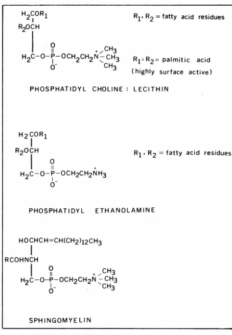
l am now going to highlight some of the sig-nificant laboratory findings that have led us to the point at which we are today. In 1961, Klaus and his co-workers (Klaus, et al, 1961) showed that bovine lung foam contained a mixture of sev-eral surface active compounds, the major constitu-ent being dipalmitoyl lecithin. The other surface active compounds he found were sphingomyelin, phosphatidyl inositol, phosphatidyl dimethylethano-lamine, and lysolecithin. The chemical structures of some of these compounds are depicted in Fig. 1. During the sixties, significant details of the surfactant system were gleaned through the work of Morgan (Morgan, et al, 1965), Fujiwara (Fu-jiwara, et al, 1968), and Gluck (Gluck, Kulovich,
et al, 1967, 1970) all working independently. Morgan and his group worked on the identification of the various components of the surface active system in alveolar and whole lung homogenates in the dog. They found that of the total lipids found in dog lung, phospholipids constituted the major group at 7 4.1 % . Of this group, phosphatidyl choline was the major component. Phosphatidyl ethanola-mine, lysophosphatidyl choline, which is lysoleci-thin, and sphingomyelin were also found, but in much smaller quantities. These are all surface active along with dipalmitoyl lecithin or phosphatidyl choline, but phosphatidyl choline seems to be by far the major component. As seen in Fig. 1, the lecithins are characterized by fatty acid groups Ri and R2 on the a and ,B positions of the
tri-glyceride backbone. Fujiwara and his associates
H2~0R1 RzOCH
I
~
. ,CH3HzC-0-~-0CHzCHzN ~ CH3
0- CH3
R1 • R2 =fatty acid residues
R1°Rz= palmitic acid (highly surface active)
PHOSPHATIDYL CHOLINE: LECITHIN
HzCOR1
I
RzOCHI
~
.
R1 • Rz = fatty acid residuesHzC-0-~_:-OCHzCHzNH3
0
PHOSPHATIDYL ETHANOLAMINE
SPHINGOMYELIN
Fig. I-Chemical structures of some phospholipids.
(Fujiwara, et al, 1968) reported on the significance of these fatty acids, which are normally C14 to C1s
amount of lecithin esterified with saturated fatty
acids. To test the hypothesis that lung maturity is
reflected by an increase in saturated lecithins, and
vice versa, Brumley and his colleagues measured
the surface tension and phospholipid content of normal lungs and lungs from infants with respira-tory distress syndrome (Brumley, et al, 1967).
Brumiey's data is shown in Table 1. As I
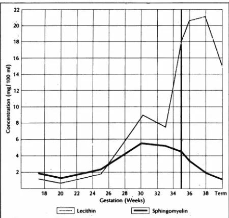
men-they parallel one another and coritiriue to do so until about the 25th week of gestation where lec-ithin starts rising slightly more rapidly than.
sphin-gomyelin. At about the 30th to 34th week there is
a sharp increase in lecithin concentration till term,
whereas sphingomyelin plateaus and starts going
dowrt again. The reason for this dramatic rise in lecithin biosynthesis at about the 32nd to 34th week
TABLE 1
Phospholipid Analysis of Lung Tissue*
Phosphatidyl choline Saturated fatty acids
Phospholipid (as % of total (as % in phdsphatidyl Surface Tension Condition (mg/g wet weight) phospholipid) choline) (dynes/cm)
Infants with respiratory
distress 10.4 46.4 64.7 23
Infants recovered from
respiratory distress 17.4 56.9 70.4 3.0
Infants without respiratory
distress 22.3 58.8 84.2 6.0
Normal adults 16.8 45.8 80.8 6.0
~ata adapted from Brumley, Hodson, and Avery, 1967.
tioned earlier, what the surfactant does is lower surface tension and thus produce more elasticity in the lung. in adults the surface tension of trachial
aspirates is somewhere of the order of 6 dynes/cm.,
and the total phospholipid content is 16.8 mg/gm wet weight. The content of phosphatidyl choline, as a percentage of total phospholipids, is about 45.8%. In the infant with no respiratory distress syndrome these values are very close to those in the normal
adult. With respiratory distress syndrome the
sur-face tension of trachial aspirates becomes much
higher. The amount of phospholipid is considerably lower compared to that of adults and infants with no respiratory distress syndrome, as are the amounts of phosphatidyl choline and saturated fatty acid. As the neonate recovers from the respiratory distress situation, these values slowly start returning to normal. This means that there is a valid justification for implicating the amount of phospholipid, speci-fically phosphatidyl cholille, in the development of fetal lung. Gluck and his group (Gluck, 1971; Gluck, Kulovich, et al, 1967, 1970) reported
extensively in the middle and late sixties on the
biochemistry of surfactant production in the lung.
They measured the amount of dipalmatoyl lecithin and sphingomyelin in amniotic fluid in humans (Gluck, 1971), with the results shown in Fig. 2. Gluck found that early in gestation, the amount of lecithin
is slightly lower than that of sphingomyelin but that
can be partially explained by the.picture of the
bio-synthetic pathways of these phosph9lipids (Fig. 3).
The lecithins are synthesized niainly by two .routes. In pathway 1 (the ch9line incorporation pathway)
an a,,B-di-substituted d~glyceride reacts with
cytidine-diphospho choline (CDP-choline) thereby incorpo-rating choline into the molecule which results in phosphatidyl choline, or lecithin: In pathway 2 (the methylatidn pathway) the a,,B-diglyceride incorporates
ethanolamine through the agency of
cytidine-diph-osphoethanolainine ( CDP-ethanolarriine) resulting in phosphatidyl ethanolamine (PE) which by subsequent methylations by S-adenosyltnethionine gives phos7 phatidyl methyl ethanolamine (PME), phosphatidyl dimethyl <;:thanolamine (PDME), and finally lecithin. These two pathways need to be studied in greater detail than they have been, but certain facets about them are known. It has been shown that the dramatic surge in lecithin at the 33rd to 34th week appears because of the activation of pathway 1, that is, the choline incorporation pathway. Gluck has evidence, primarily clinical in nature, that the methylation pathway is the primary mode of lecithin biosynthesis
in early gestation, but that on .or after about the 34th
week, the choline incorporation pathway becomes
the more important (Gluck, 1971). He found that
when he analyzed respiratory distress syndrome
cases in prematures, most of them were acidotic
acidosis nor hypothermia were associated with
clinical respiratory distress syndrome in the full-term infant. When trachial aspirates were obtained from prenatal, or frbm premature infants while they were
still hypothermic, he found little or no phosphatidyl dimethyl ethancilamine which meant that the ineth-ylation pathway was not functioning. As the infants were put into an incubator and temperature brought up, increasing amounts of PDME were found in trachial aspirates which indicated that the heat
sup-plied to them was activating this pathway. These findings suggested that the methylation pathway is vulnerable to external insult. Yet if this were the case, then some full-term infants should also show
re-spiratory distress, which they do not. Gluck's
ex-planation for this is based on his evidence that early in gestation the methylation pathway is the more important btit at about the 35th week the choline incorporation pathway takes over causing the
surge of lecithin. As the choiine incorporation path-way is not vulnerable to external insult, it is pro-ducing sufficient quantities of surfactant for the full-term infant not to have respiratory distress; Thus
the premature infant is at a disadvantage because
at around 30-34 weeks gestation when the methy-lation pathway is dominant and vulnerable to external
insult, the choline incorporation pathway has not
yet assumed maximal function, and hence, sufficient
surfactant is not being produced. The only lab find-ing that Gluck reports in support of his hypothesis is that in vitro, the methylation pathway is markedly inhibited below a pH of 7.2; in vivo, exposure to cold or hypothermia or hypoxia causes acidosis in
the neonate, which could then inhibit the me thyl-ation pathway.
With all this data, we now have the basis for developing a method for the prediction of re~
a,13-0iglyceride
@
CDP-ethanol-amine©
CDP- choline
22
20
18
16
-
-I
\
\
I
I
~
I
I
/
....,I v
-.......
__,,,
v
~,-
~ ::.---- ...~ w n M ~ n ~ n M • ~ T~m Ces .. tion (Weeki)
I ---· I lecithin I I Sphingomyelin
Fig. 2 -ConcentratiOns of sphingomyelin and lecithin in amniotic fluid during gestation. (Reprinted with permission from Gluck, Hosp. Pract. 6:45, 1971.)
spiratory distress in the neonate by measuring lec-ithin and sphingomyelin in amniotic fluid. Although lecithin and sphingomyelin are measured
individu-ally, the results are reported in terms of the-ratio
of lecithin to sphirigomyelin. Once the ratio starts getting higher than 1 or 2, one might be able to give some indicatiori about whether respiratory distress could be predicted in that fetus were it de-livered at that time. The method we have developed
and are using at present is a modification of G!uck's
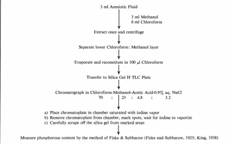
original method (Gluck, Kulovich, et al, 1971) and is -outlined in Fig. 4. Three inililiters of amniotic
fluid are taken to which are added 3 ml of methanol and 6 ml of chloroform in a centrifuge tube. The whole is extracted once, centrifuged, and the lower
Phosphatidyl choline (Lecithin)
Phosphatidyl-ethanolamine
_(PE)
Phosphatidyl -methylethanolamine
(PME)
Phosphatidyl-diinethylethanolamine
(PDME)
3 ml Amniotic Fluid
l
3 ml Methanol6 ml Chloroform
Extract once and centrifuge
i
Separate lower Chloroform: Methanol layer
i
Evaporate and reconstitute in 100 µl Chloroform
i
Transfer to Silica Gel H TLC Plate
i
Chromatograph in Chloroform-Methanol-Acetic Acid-0.93 aq. NaCl
70 :
i
25 : 4.8 3.2a) Place chromatoplate in chamber saturated with iodine vapor
b) Remove chromatoplate from chamber, mark spots, wait for iodine to vaporize
c) Carefully scrape off the silica gel from marked areas
i
Measure phosphorous content by the method of Fiske & Subbarow (Fiske and Subbarow, 1925; King, 1938)
Fig. 4- Methodologic flow sheet.
chloroform methanol layer is removed, evaporated, and reconstituted in 100 µ,l of chloroform. This is then transferred to a thin-layer plate which is run in the system, chloroform-methanol-acetic acid-0.9% aq. NaO (70:25:4.8:3.2). We found this system to be better than the old chloroform-methanoi-water
ones which are normally used for phospholipids:
Lecithin and sphingomyelin are visualized by placing the thin-layer· plate in a chamber saturated with iodine vapor. The iodine is deposited reversibly on
the areas where the lecithin and sphingomyelin are
located. The spots are then marked, scraped off, and
after the iodine has vaporized, the phosphorus
content of the spots is measured by a modification of the Fiske and Subbarow method (Fiske and Sub-barow, 1925; King, 1938). As one mole of phos-phorus corresponds to one mole of lecithin and
sphingomyelin, by measuring the amount of
phos-phorus, we can calculate the absolute amounts of lecithin and sphingomyelin and hence get the ratio of one to the other. The test we have developed takes an average technician approximately 3 hours for 6 quantitative determinations. For a quick but qualitative result, the test can be shortened by
eliminating the quantitative step, spraying the
thin-layer plate with a phosphorus specific spray
(Ditt-mer and Lester, 1964) once it comes out of its developing solvent system, and then estimating in gross terms the amounts of lecithin and sphingo-myelin by the visual intensities of their spots.
In our laboratory, we take a ratio of 2 or greater tO predict fetal lung maturity. Gluck and his group reported a few months ago that based on the results of 302 amniocenteses from 272 pregnancies using their method of monitoring ·the phospholipids, not one single case of respiratory distress occurred
(Gluck, Kulovich, et al, 1971). Our experience
here at the Medical College of Virginia is of shorter
duration. After 49 complicated pregnancies which
we have monitored through 63 amniocenteses, we
have noi encountered any instance of respiratory
distress, and among these we have had four notable cases: three of diabetes and one of Rh disease. The Rh disease patient was delivered at 331/i weeks after continuous monitoring of her lecithin-sphingomyelin ratios, and she delivered a healthy viable infant whose weight was compatible with dates. The three dia-betics were delivered at 34, 35, and 36 weeks, and all delivered healthy viable infants showing no signs of respiratory distress.
of obstetricians, it should still be used with caution because the volume of data is not sufficient to show
what would happen if one were to get a false posi-tive, if one indeed ever gets false positives. The next
big step that needs to be taken is a detailed study
of the two major biosynthetic pathways for ledthin
and a study of the characteristics and properties of
the enzymes involved. If a compound could be
found to induce one or both of these enzymes, this information would provide a much wanted tool in the hands of an obstetrician or pediatrician for
deal-ing with the fetus or neonate. Such information
would also be of great significance in cases of
res-piratory distress syndrome which are independent
of obstetrical management, such as premature labor.
In this instance, the physician is confronted with a
situation not of his own making and has to deal with
it as quickly and as best he can. At present, we are
able, with the use of the described test, to predict
the possibility of respiratory distress and thus have
the necessary facilities such as an incubator or a
respirator ready for the neonate immediately after
delivery. But of even greater benefit would be the discovery of a compound drug which would induce
the choline incorporation pathway. This, as
i
mentioned earlier, is insensitive to external insult,
and once biosynthesis of lecithin has been induced
by means of the pathway, the neonate itself can take
over the job it is supposed to do. A further goal
is the compiling of sufficient data such that a
sig-nificant statistical analysis can be made concerning
the accuracy of predicting the severity of respiratory
distress associated with specific ranges of the
lecithin/sphingomyelin ratio.
Finally, I would like to express my
apprecia-tion to our excellent housestaff who have been very
patient in supplying me with samples of amniotic
fluid as uncontaminated with blood as possible and
also my very deep gratitude to Dr. Dunn for his
constant and very enthusiastic encouragement of this
work.
REFERENCES
ANDERSON, A. B. M. AND GRIFFITHS, A. D. J. Obstet.
Gynaecol. Brit. Comm. 75:300, 1968.
AVERY, M. E. AND MEAD, J. Am. /. Dis. Child. 97:517,
1959.
BROSENS, I. A. AND GORDON, H. /. Obstet. Gynaeco/. Brit.
Comm. 73:88, 1966.
BRUMLEY, G. W., HODSON, W. A., AND AVERY, M. E.
Pediatrics 40: 13, 1967.
CLEMENTS, J. A. Proc. Soc. Exp. Biol. Med. 95: 170, 1957.
DITTMER, J.
c.
AND LESTER, R. L. /.Lipid Res. 5:126, 1964. DROEGEMUELLER, W., JACKSON, C., MAKOWSKI, E. L., AND BATTAGLIA, F. C. Am. J. Obstet. Gynec. 104:424, 1969.FISKE, C. H. AND SUBBAROW, Y. /. Biol. Chem. 66:375,
1925.
FUJIWARA, T., ADAMS, F. H., SIPOS, S., AND EL-SALAWY, A.
Am. J. Physiol. 215:375, 1968.
GLUCK, L. Hospital Practice 6:45, 1971.
GLUCK, L., KULOVICH, M. V., BORER, R. C., BRENNER,
P. H., ANDERSON, G. C., AND SPELLACY, W. N. Am. ].
Obstet. Gynec. 109:440, 1971.
GLUCK, L., KULOVIC, M. V., et al. Pediat. Res. 1:237
and 247, 1967; Gluck, et al 4:352, 1970.
JEFFCOATE, T. N. A. AND ScoTT, J. S. Canad. M.A. J. 80: 77, 1959.
KING, E. J. Biochem. ]. 26:295, 1938.
KLAUS, M. H., CLEMENTS, J. A., AND HAVEL, R. J. Proc.
Natl. Acad. Sci. (U.S. A.) 47:1858, 1961.
LILEY, A. W. Am. /. Obstet. Gynec. 82: 1359·, 1961. MANDELBAUM, B., LA CROIX, G. c., AND ROBINSON, A. R.
Obstet. Gynecol. 29:471, 1967.
MORGAN, T. E., FINLEY, T. N., AND FIALKOW, H. Biochem. Biophys. Acta 106:403, 1965.
NEERGAARD, K. VON. z. Gesamte Exp. Med. 66: 373, 1929.
PATTLE, R. E. Nature (Lond) 175:1125, 1955.
PITKIN, R. N. AND ZWIREK, S. J. Am. /. Obstet. Gynec. 98: