R E S E A R C H A R T I C L E
Open Access
Identification of functional long non-coding
RNAs in
C. elegans
Alper Akay
1,2, David Jordan
1,2, Isabela Cunha Navarro
1,2, Tomasz Wrzesinski
4, Chris P. Ponting
5, Eric A. Miska
1,2,3*and Wilfried Haerty
4*Abstract
Background:Functional characterisation of the compact genome of the model organismCaenorhabditis elegans remains incomplete despite its sequencing 20 years ago. The last decade of research has seen a tremendous increase in the number of non-coding RNAs identified in various organisms. While we have mechanistic understandings of small non-coding RNA pathways, long non-coding RNAs represent a diverse class of active transcripts whose function remains less well characterised.
Results: By analysing hundreds of published transcriptome datasets, we annotated 3392 potential lncRNAs including 143 multi-exonic loci that showed increased nucleotide conservation and GC content relative to other non-coding regions. Using CRISPR/Cas9 genome editing, we generated deletion mutants for ten long non-coding RNA loci. Using automated microscopy for in-depth phenotyping, we show that six of the long non-coding RNA loci are required for normal development and fertility. Using RNA interference-mediated gene knock-down, we provide evidence that for two of the long non-coding RNA loci, the observed phenotypes are dependent on the corresponding RNA transcripts.
Conclusions: Our results highlight that a large section of the non-coding regions of the C. elegans genome remains unexplored. Based on our in vivo analysis of a selection of high-confidence lncRNA loci, we expect that a significant proportion of these high-confidence regions is likely to have a biological function at either the genomic or the transcript level.
Keywords: C. elegans, lncRNA, lincRNA, CRISPR, Non-coding, Long non-coding RNA
Background
Transcription is not limited to the protein-coding regions of eukaryotic genomes, but instead has been observed to be pervasive in all organisms that have been studied so far. As a consequence of transcriptional activity over non-coding sections of the genomes, tens of thousands of short, < 200 nucleotides (nt) and long (> 200 nt) non-coding RNAs have now been annotated [1,2]. While much is known about the biological role of most classes of small non-coding RNAs (e.g. microRNA, Piwi-associated RNA, small nucleolar RNA, small interfering RNA) [3–5], relatively little is known about long non-coding RNAs (lncRNAs). Whether most eukaryotic lncRNAs are
functional has long been debated because of their low ex-pression levels and rapid evolutionary turnover when compared to protein-coding genes [6, 7]. However, the molecular activities of more than a hundred of such loci have now been described [8–12] including many that ap-pear to regulate the expression of protein-coding genes. Only a small proportion of these loci have been demon-strated to be fundamental to eukaryote biology from mu-tations that affect their expression or function leading to severe developmental defects or to lethal phenotypes (for example, [13,14]). While transcription of some lncRNAs has been shown to originate at promoter or enhancer ele-ments with potential DNA-dependent function [15], the activity of others depends on the RNA transcript, acting either in cisor trans, e.g. targeting protein complexes to chromatin or directly interacting with other RNAs, in-cluding mRNAs, lncRNAs, or microRNAs [16]. The pro-portions of lncRNAs belonging to each functional class
* Correspondence:[email protected];[email protected]
1Wellcome CRUK Gurdon Institute, University of Cambridge, Tennis Court
Road, Cambridge CB2 1QN, UK
4Earlham Institute, Norwich Research Park, Norwich, UK
Full list of author information is available at the end of the article
remain unknown owing to painstaking experimental vali-dations, including both knockout and knock-down assays being required.
C. elegans has been invaluable for the discovery of multiple non-coding RNA pathways and is an important model organism for genetic studies. Nevertheless, only one study has yet sought to identify and annotate lncRNAs in C. elegans, resulting in 1145 annotated loci, of which only 170 had evidence of polyadenylation [17]. Furthermore, experimental characterisation ofC. elegans
lncRNAs has been limited [18–20] and only recently a comprehensive genetic analysis of these lncRNAs have been conducted which identified 23 physiologically func-tional lncRNAs [21].
Using publicly available RNA-Seq libraries representing diverse C. elegansdevelopmental stages, we sought to ex-pand the annotated novel expressed long non-coding loci and to characterise informative features such as nucleotide composition, evolutionary conservation, transcript expres-sion and functional enrichment. To assess the physiological impact of mutations within these novel lncRNAs and thus the biological importance of these loci, we used CRISPR/ Cas9 to generate large genomic deletions for ten lncRNA loci. Six of these intergenic lncRNA loci yielded significant phenotypes upon deletion, and at least two of these have physiological functions that are RNA-dependent. Our study and associated experimental validation demonstrate that physiological lncRNA function in nematodes can be RNA-and/or transcription-dependent. Furthermore, we extrapo-late that a significant proportion of the newly identified multi-exonic non-coding loci in the C. elegans genome might be functional at the genomic or the transcript level.
Results
Long non-coding RNA annotation inC. elegans
We investigated 209 publicly available RNA-Seq datasets from diverse developmental stages (Additional file 1) to annotate de novo non-coding transcripts in C. elegans. We decided to focus on purely intergenic loci that lack any overlap with previous coding and non-coding gene annotations. After filtering for size, coding potential and overlap with existing genes (including the previously an-notated lncRNAs, see the“Methods”section), we identi-fied 3392 long (> 200 nt) non-coding RNAs expressed across C. elegans development (Additional file 2). Of these 3392 lncRNAs, 143 were multi-exonic and 3249 were mono-exonic. Previously, 1145 potential lncRNAs were identified in C. elegans [17]. Six hundred ninety-five of these were masked in our analysis due to either being individually annotated or overlapping exist-ing annotations. Only 18 multi-exonic and 179 mono-exonic lncRNAs identified in our analysis overlap with previous lncRNA annotations inC. elegans.
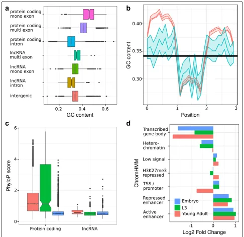
CAGE data [22] was then used to accurately annotate the transcriptional orientation of 707 mono-exonic loci if found within 100 nt of the CAGE peak summit, and ChIP-Seq [23] and CLIP-Seq [24] data were used to identify transcription factor and AGO binding sites within the loci (Additional file 3). As observed in all other model organisms, the identified lncRNA loci are smaller than annotated protein-coding genes and are expressed at significantly lower levels (Additional file 4). LncRNA exons also tend to have a GC content that is lower than protecoding sequences but higher than in-tronic sequences (Fig. 1a, b) as observed previously for other eukaryotes [25]. Inter-species sequence conserva-tion for multi-exonic lncRNAs was lower than for protein-coding genes but higher than for mono-exonic lncRNAs (Kruskal Wallis test, P= 5.73 × 10⁻⁵, Fig. 1c). Our newly annotated multi-exonic lncRNAs show se-quence features similar to the final set of 170 lncRNA reported by Nam and Bartel [17], (Kruskal Wallis test,P
= 0.79 and P= 0.15 for nucleotide conservation and composition, respectively) showing the complementarity of these lncRNA annotations.
Distinct chromatin states inferred from histone modifi-cations using ChromHMM [26] have been shown to asso-ciate with specific genomic elements (for example, transcriptional start sites and promoters, transcriptional elongation and gene bodies, enhancers, transposable element-derived sequences). Using previously published chromatin annotations inC. elegans[27,28], we assessed the functional enrichment of our newly annotated lncRNA at each of these annotated genomic elements. Enhancers, identified either by Evans et al. [27] (1.7 fold enrichment
P< 0.0001) or Daugherty et al. [28] (2.0 fold enrichment,
P< 0.0001), significantly overlapped with these lncRNA loci, but chromatin states associated with transcription elongation (“transcribed gene body”) were depleted at all developmental stages (Fig.1d, Additional file5). These re-sults could be explained by the observed low expression level of the lncRNAs. Our results are in agreement with Evans et al. [27], who showed that the chromatin states reflecting transcription elongation were associated with the most highly expressed genes in their study. Active en-hancers were particularly enriched within single exon lncRNAs at all developmental stages (2.0, 2.6 and 2.4 fold enrichment at the early embryonic, L3 and young adult stages, respectively, P< 0.001 in all comparisons, Add-itional file 5). This result is consistent with enhancer RNAs rarely being spliced [29]. In contrast, multi-exonic loci were only enriched for active enhancers during early embryonic stage (2.3 fold enrichment, P< 0.0001) which likely reflects the fewer number of multi-exonic lncRNA expressed at later stages.
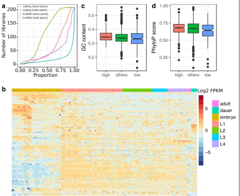
Fig. 2a). This restricted expression could reflect that many of the newly annotated loci are the result of tran-scriptional noise and therefore likely non-functional [15]. However, many of the remaining lncRNAs, most specifically multi-exonic lncRNAs, appear to be expressed in a tissue- and stage-specific manner (Fig.2b). Twenty-six lncRNAs (10 multi-exonic) were expressed
in more than 90% of the libraries (≥188 libraries). Highly reproducible loci (≥100 libraries) tended to have a significantly higher sequence conservation (Kruskal Wallis test, correctedP= 0.0077) and higher GC content (Kruskal Wallis test, P= 3.5 × 10⁻⁴
after Bonferroni cor-rection) compared with loci with limited reproducibility (< 10% libraries) (Fig. 2c, d). Highly reproducibly
a
b
c
d
expressed loci also tended to have stronger enrichment for enhancer regions identified in embryos (2.2–3.1 fold enrichment) or in L3 larvae (2.7–4.4 fold enrichment, Additional file5).
Functional characterisation of lncRNA loci
Higher sequence conservation of the 143 multi-exonic lncRNA loci, together with their higher exonic GC con-tent and their splicing, could reflect organismal function. To test this hypothesis, we used CRISPR/Cas9 genome editing to generate targeted deletions in ten of the multi-exonic lncRNA loci that each showed high se-quence conservation, high expression and evidence from multiple libraries and that are not overlapping with neighbouring coding regions. We were successful in
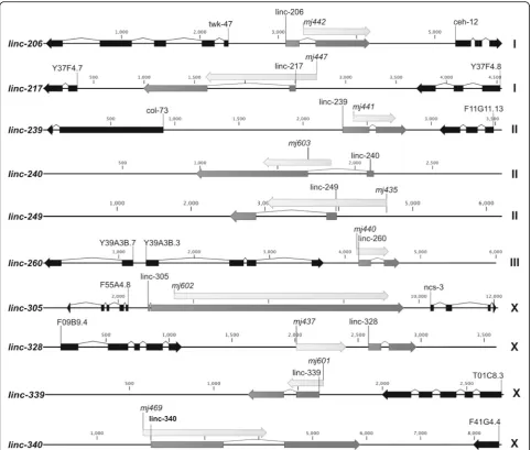
generating large genomic deletions for ten of these lncRNA loci (Table 1) out of 20 that were initially tar-geted. This success rate was due mostly to the limited efficiency of plasmid-based CRISPR/Cas9 genome edit-ing protocols that were used early in the study, as com-pared to direct protein/RNA injection methods [30]. Of these lncRNA locus deletions, nine removed at least one exonic region and one removed a region just 5′ of a lncRNA locus (Fig.3).
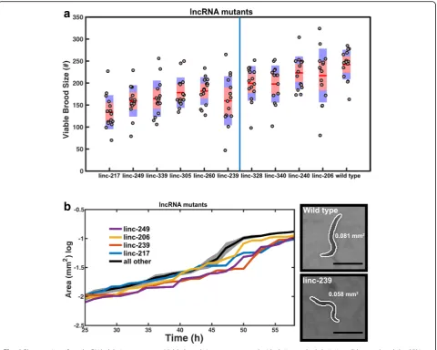
All ten lncRNA deletion mutants initially failed to dis-play overt, gross phenotypes such as sterility, embryonic lethality, larval arrest or abnormal body development. To undertake a more extensive characterisation, we captured the development of the mutant animals alongside wild type control animals using an automated microscopy
a
b
c
d
system. This system records the development of multiple animals simultaneously and permits phenotypic analysis in an unbiased manner. Two phenotypes that can influ-ence the life history and fitness of populations [31], brood size and growth rate, were selected for the automated ana-lysis. Six of ten lncRNA deletion mutants (linc-217, linc-239, linc-249, linc260, linc-305 and linc-339) yielded significant reduction in viable brood size (Fig.4a) and four of 10 mutants (linc-206, linc-217, linc-239 and linc-249) displayed reduced growth rate (slower body size increase) over development (Fig. 4b). Three mutants (linc-217, linc-239 and linc-249) showed alterations of both pheno-types (Table1, Fig.4a, b).
These phenotypes could be due to the removal of either the lncRNA transcript or of the genomic locus which, in some instances, harboured annotated transcription factor binding and enhancer sites (Additional file 3). To distin-guish between these two possibilities, we generated dsRNA expression vectors for RNAi targeting of the lncRNA transcripts in wild type animals (Table 1, Add-itional file6). Using four biological replicates per assay, we targeted six lncRNA transcripts using RNAi. We left out linc-240, linc-328, linc-260 and linc-305 because these were either lacking any phenotype or yielded only a weak phenotype when partially or fully deleted.
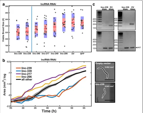
Of the four of these six lncRNA loci whose deletion yielded a reduced viable brood size phenotype, two (linc-239 and linc-339) yielded an equivalent phenotype when expression was reduced using RNAi (Fig. 5a); for one of these lncRNA loci, linc-239, equivalent reduced growth rate phenotypes were observed for both its knock-out and knock-down (Table1, Fig.5b). These results indi-cate that for these two loci, the phenotypes are caused by the disruption of their RNA transcript-dependent func-tions. We further validated the transcript expression and RNAi knock-down efficiency of linc-239 and linc-339 using RT-PCR and observed a strong reduction in
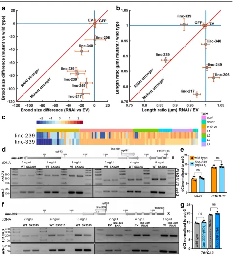
linc-239 and linc-339 transcript levels (Fig. 5c and Add-itional file 7). RNAi targeting of another lncRNA, linc-339, also showed a reduced growth rate, a phenotype that was not observed in the deletion mutant (Fig. 5b). Three additional lncRNA strains (linc-206, linc-217, linc-249) yielded discordant phenotypes when disrupted or subjected to RNAi (Table1, Figs.4and5). This would be consistent with functions of these loci being RNA-independent. Direct comparison of the phenotypes between lncRNA deletion mutants and RNAi knock-down of lncRNAs shows that RNAi knock-down phenotypes are slightly weaker or equivalent to deletion mutants (Fig.6a, b). We calculated the expression of linc-239 and linc-339 to be highest during larval development in comparison to embryogenesis (Fig. 6c). The phenotypes associated with linc-239 and linc-339 can arise from their cis regulatory function on the neighbouring genes. We analysed the ex-pression levels of genes that are in close proximity to the linc-239 and linc-339. linc-239 has two neighbouring genes,col-73and F11G11.13. In linc-239 (mj441) deletion mutants, we did not observe any change in the expression levels of col-73 and F11G11.13 using both end-point RT-PCR and qRT-PCR (Fig.6d, e). In linc-339 (mj601) de-letion mutants, we observed a small reduction in the ex-pression of its neighbouring gene T01C8.3 using RT-PCR (Fig. 6f). Both the deletion of linc-339 and its RNAi knock-down lead to comparable reduction in viable brood size. If reduction in T01C8.3 expression is required for this phenotype, then linc-339 RNAi knock-down should similarly affect the T01C8.3 expression. Instead, we ob-served that the T01C8.3 expression does not change upon linc-339 RNAi knock-down (Fig. 6 f, right hand panel). We further tested the expression of T01C8.3 using qRT-PCR (Fig. 6g) and did not observe any significant change in expression when the linc-339 is either deleted or knocked-down. Our results suggest that both linc-239 and linc-339 are likely to functionin trans.
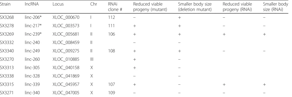
Table 1List of lncRNAs deletions and their phenotypes
Strain lncRNA Locus Chr RNAi
clone #
Reduced viable progeny (mutant)
Smaller body size (deletion mutant)
Reduced viable progeny (RNAi)
Smaller body size (RNAi)
SX3268 linc-206* XLOC_000670 I 112 – + – –
SX3278 linc-217* XLOC_003573 I 111 + + – –
SX3269 linc-239* XLOC_005681 II 106 + + + +
SX3332 linc-240 XLOC_008459 II – –
SX3340 linc-249 XLOC_009275 II 108 + + – –
SX3270 linc-260 XLOC_010885 III + –
SX3313 linc-305 XLOC_040158 X + –
SX3338 linc-328 XLOC_041869 X – –
SX3315 linc-339 XLOC_045957 X 107 + – + +
SX3271 linc-340 XLOC_047005 X 109 – – – –
Discussion
The identification of functional non-coding elements, in-cluding transcribed non-coding sequences, in genomes has long relied on computational predictions based on se-quence conservation [32], or biochemical activity [33]. However, regardless of the preferred approach to predict functional sequences, only experimental validation can truly substantiate the inferred functionality of an element.
In our study, we first provide a novel annotation of intergenic lncRNAs in C. elegans.This work expands on the previous annotations delivered by Nam and Bartel [17] as a substantially more comprehensive RNA-Seq dataset was available at the time of our study (209 vs 35). We also took advantage of existing resources to further improve the annotations for these loci. These included not
only their expression pattern and nucleotide conservation but also (i) the presence of potential functional elements within them (transcription factor binding and AGO bind-ing sites), (ii) their correlation in expression with neigh-bouring protein-coding genes and (iii) the reported mutant phenotypes for these genes. The primary aim of these comprehensive annotations was to inform the selec-tion of candidate lncRNA loci for follow-up experimental validation. Most importantly, we went beyond computa-tional predictions of funccomputa-tionality as we assessed the in vivo phenotypic effect of knocking-out a selection of ten intergenic lncRNAs using automated microscopy, and im-plemented knock-down assays to validate the observed brood size and growth rate reduction and putative transcript-mediated function of these loci.
lncRNAs ofC. elegans
Our newly annotated loci bear all the hallmarks of lncRNAs in other organisms: they tend to be shorter, expressed at lower levels and have a lower degree of conservation than protein-coding sequences [6, 7, 11]. Furthermore, the GC composition of the multi-exonic lncRNAs in C. elegans does mirror the patterns previ-ously observed in other animals, with increased GC con-tent within exons relative to introns [25]. These similarities with other animal lncRNA annotations impli-cateC. elegansas a model organism that is more broadly relevant for investigation of the molecular functions of lncRNAs and the processes through which those func-tions are conveyed. Most importantly, the wealth of re-sources available for C. elegans as a model organism
offer the opportunity to assess the in vivo impact of mu-tations within these loci.
The observation of enrichment for enhancer se-quences within our lncRNA loci emphasises that the ob-served function of a locus could be conveyed by discrete functional DNA elements located within it rather than by the RNA transcribed at this location. The former would imply that transcription at this location either re-flects or maintains open chromatin states and that the resulting transcript would likely be biologically inconse-quential, whereas the latter would imply RNA sequence-dependent functionality of the resulting tran-script [10, 34]. LncRNAs with transcript-mediated func-tion have been shown to act both in cis (Xist [35]) and
in trans (Paupar [36]), whereas those whose function is
a
b
transcription regulation related are expected to act incis
(transcriptional interference, chromatin modification at enhancers). This duality, transcription- versus transcript-mediated function, is a recurrent issue when studying lncRNAs and only the careful experimental characterisation of each locus through knockdown, and rescue can begin to deduce the functional mechanism associated with a non-coding transcript [37].
Phenotypic characterisation of lncRNAs
Historically, majority ofC. elegans genes were identified through genetic screens which concurrently provide phenotypic and functional information. Mutations
identified in non-coding regions of the genome as a re-sult of genetic screens, nevertheless, have largely remained uncatalogued. With advances in genome edit-ing methods, it is now possible to directly target non-coding regions for mutational analysis. The lncRNA annotations presented in this study, together with the detailed documentation of their expression and overlap with existing datasets, serve as a guide for the targeted analysis of these loci during animal development.
InC. elegans, many small non-coding RNA genes lack discernible phenotypes when deleted individually [38]. This is mostly due to redundancy between non-coding RNAs and the role such RNAs play as buffers in gene
a
b
c
Fig. 5RNAi-mediated knock-down of lncRNAs.aViable brood sizes are presented with their standard deviations (blue area) and the 95% confidence interval of the mean (red area). Samples were compared to“empty vector”control animals using a pairwise two-samplettest with a multiple test (Bonferroni) correction. Samples are ordered by increasingpvalue, and those found to be significant at (p≤0.05) are shown to the left of the blue line (n= 18 animals/mutant). Empty vector (EV), GFP and linc-340 RNAi are negative controls.bGrowth curves were compared to
a
c
d
f
e
g
b
expression regulation rather than being the master regu-lators. The situation may be similar for the majority of lncRNAs, because their roles in the regulation of gene expression remain incompletely understood and the phenotypic characterisation of many vertebrate lncRNAs has been challenging and has provided sometimes contradictory results [39]. The genomic deletions of the ten lncRNA sequences described in this study failed to display any gross phenotype. This is in line with another study published during the revision of our work which described a comprehensive deletion analysis of the previ-ously annotated lncRNAs inC. elegans[21]. By using au-tomated microscopy, we sought to capture the phenotypes associated with lncRNAs in an unbiased manner. While this approach gives greater power to screen larger number of individuals and accurately quan-tify phenotypes, more subtle phenotypes or phenotypes more difficult to quantify might not be captured. The observed reductions in brood size and growth rate of lncRNA loci deletion mutants greatly affect the fitness of these animals, despite their otherwise normal appear-ance. For two of the lncRNA loci, linc-239 and linc-339, the phenotypes can be recapitulated by RNAi knock-down. We thus consider these two lncRNAs as being representative of bona fide C. elegans lncRNAs. However, further experiments will be required to com-pletely rule out that these lncRNAs are not translated into functional, short polypeptides [40]. It is also import-ant to note that according to the most recent genome annotations (WS269), both linc-206 and linc-217 overlap protein-coding sequences. The same issue also exists with linc-239 which appears to partially overlap a re-cently annotated single exon coding transcript (F11G11.15) which is supported only by weak evidence and marked as potentially non-coding in Wormbase. The lack of phenotypes upon RNAi knock-down of the remaining loci could be attributed to the possibility that observed phenotypes in deletion mutants arising from the removal of DNA-dependent functional elements. It is also possible that the transcripts of these loci are
solely nuclear or expressed in neuronal tissues and thus resistant to RNAi inC. elegans[41,42].
Conclusions
In this study, we increased the current number of poten-tial lncRNAs in C. elegansfrom 801 to 4001. Together with the previously identified high-confidence lncRNA loci, in total 298 loci yield evidence for possible bio-logical functions because they display higher conserva-tion, higher expression, higher GC content and splicing. Using genome editing and RNA interference methods, we tested the functional relevance of ten of these loci and demonstrated that six yield in vivo phenotypes when deleted. Furthermore, we showed that for at least two out of these six loci, the function is likely conveyed by the RNA transcript. From our in vivo assays, we estimate by extrapolation that 40–60% of the multi-exonic lncRNAs identified in this study might have biological roles. It will be essential to employ sensitive experimen-tal approaches to decipher the fitness effect of such non-coding loci.
Methods
Intergenic lncRNA identification
A total of 209 publicly available libraries were retrieved from the SRA database (https://www.ncbi.nlm.nih.gov/ sra/, Additional file 1). Reads were mapped onto the C.
elegans (ENSEMBL release 73, WBcel235) reference
genome using TOPHAT2 (2.0.9) [43] with default pa-rameters. For each library, de novo transcripts were called using cufflinks2 (2.2.1) [44] and the coding poten-tial of all new intergenic transcripts was assessed using the Coding Potential Calculator (CPC, 0.9-r2 [45], score < 0) as well as CPAT (1.2.4 [46], score < 0.403). All of the loci for which every transcript was deemed non-coding were retained for further analyses as poten-tial intergenic lncRNAs. All lncRNAs across all libraries were merged into a single annotation file using Cuff-compare. We retained for final analyses only the loci longer than 200 nt, not overlapping any annotated gene
(See figure on previous page.)
Fig. 6Comparison of phenotypes arising from lncRNA genomic deletion mutants and phenotypes arising from RNAi-mediated knock-down of lncRNA transcript. To compare the effects of the disruption of a lncRNA genomic locus to the knock-down of the corresponding lncRNA transcript by RNAi, the mean brood size reduction compared to the control (a) and the ratio of the length at 50 h relative to the control (b) were plotted. These are shown as a scatter plot of the mean reduction (a, blue circle) or the mean ratio (b, blue circle) with the 95% confidence interval of the mean (orange lines). If the mutations or the RNAi yield an effect, data fall below the liney= 1 and to the left ofx= 1. If mutants and RNAi yield similar effects, data fall along the red line; above the red line indicates that RNAi has a greater effect, while below the red line indicates that the genomic mutation has a greater effect.cExpression (log2FPKM) acrossC. elegansdevelopment for linc-239 and linc-339.dRT-PCR analysis of neighbouring genes in linc-239
(mj441)deletion mutant. Diagram shows the relative position of the lncRNA and the surrounding protein-coding genes.Col-73expression (left panel) and F11G11.1 expression (right panel) are measured using end-point RT-PCR at three different cDNA concentrations. Actin is used as loading control.e qRT-PCR analysis ofcol-73and F11G11.13 expression in linc-239(mj441)mutants. Error bars = 95% CI, significance is tested usingttest with multiplet
and found at least 50 nucleotides away from annotated genes if located on the same strand.
We downloaded the Nam and Bartel lncRNAs [17] for further comparison with our data set. Out of the 1195 loci, we found that the 170 high-confidence Nam and Bartel lncRNAs are included in existing annotations and an additional 525 low-confidence loci overlap existing genes.
Intergenic lncRNA conservation
The nucleotide conservation of the candidate loci was assessed using the conservation tracks (PhyloP) from the UCSC database (http://hgdownload.soe.ucsc.edu/). The tracks represent the nucleotide conservation across 26 nematode species.
Chromatin modifications, transcription binding sites and enhancers associated with lncRNAs
In order to facilitate the prioritisation of lncRNAs for mutagenesis, we parsed publicly available data to further improve our annotations. We intersected our annotated loci with highly occupied target regions [47], miRNA binding sites [24], transcription factor binding sites iden-tified by modENCODE [23] and enhancers identified by Chen et al. [22]. We also computed the distance to the closest protein-coding gene as well as the correlation in expression between lncRNAs and their upstream and downstream flanking protein-coding genes. Finally, we also reported the known phenotypes for the proteins flanking lncRNAs. Genomic locations of the respective annotations were transferred to the ce11 genome assem-bly using liftOver and files available on the UCSC data-base. Enrichment analyses were performed using the Genomic Association Test (GAT) software [48].
Identification of transcriptional start sites
We used the 5′ end tag sequencing data from Chen et al. [22] to identify the putative transcriptional start sites of the intergenic lncRNAs. We applied the same ap-proach the authors previously applied to their data. Clusters with at least two reads were kept and merged if on the same strand and within 25 nucleotides of each other. We used the strand information of the CAGE peaks to inform the transcriptional orientation of mono-exonic loci found within 100 nt of a CAGE peak summit.
CRISPR/Cas9-mediated deletion of lncRNA loci
lncRNA loci were deleted using either plasmid base in-jection [49,50] or direct protein/RNA injection methods [30, 51], as previously described. gRNA sequences and the primers used for screening of the F2 generation ani-mals are given in Additional file6. Isolated deletion mu-tants were backcrossed once to wild type animals. For
lncRNA sequences and deletions, see Additional file 8; for genotyping results of deletion mutants, see Additional file9.
Cloning of RNAi vectors
Genomic sequences corresponding to the lncRNA loci (Additional file 6) were cloned using Gibson Assembly [52] into the L4440 vector and transformed into compe-tentE. colistrain HT115 [41].
RT-PCR and qRT-PCR analysis of lncRNA expression Total RNA was isolated using TriSure reagent and chloro-form extraction, followed by isopropanol precipitation. Genomic DNA was removed using TurboDNA Free kit (Thermo) according to manufacturer’s recommendations. cDNA synthesis was done using SuperScript II reverse transcriptase according to manufacturer’s instructions starting from 0.5–1 μg RNA. cDNA was directly used in PCR reactions after dilution as indicated in individual fig-ures. In Fig.5and Additional file735 cycles of PCR amp-lification was used. In Fig. 6, 25 cycles of PCR amplification was used. RT-PCR primers are listed in Add-itional file 6. qRT-PCR was done using Applied Biosys-tems PowerSYBRGreen PCR master mix and an Applied Biosystem StepOnePlus machine. Delta Ct calculations were done using the Ct values of the control geneact-3. qRT-PCR data is generated using three biological replicates.
Preparation of RNAi plates
E. coliHT115 bacteria transformed with individual RNAi expression vectors were grown in LB-ampicillin (50μg/ ml) until OD600 measurement of 0.6–0.8. Bacteria were seeded onto NGM agar plates containing a 1-mM IPTG and 25-μg/ml carbenicillin. Worms were added onto the plates as described below for growth curve and brood size measurements after at least two generations of RNAi feeding.
Automated microscopy analysis Brood size
Animals were then detected by movement using the dif-ference image between each frame and this background image and counted this way for ten additional frames. The final count was returned as the mode of these counts. This system was tested on plates with fixed numbers of animals and was accurate to within 5%, comparable to human precision. Total brood size was reported then as the sum for 3 days. For mutant strains, this experiment was done for five animals of each strain three times. For the RNAi experiment, this was done for six animals for each RNAi clone, also done three separ-ate times. Data is censored for animals that crawled off of plates. See Additional file10for comparison of auto-mated microscopy accuracy to manual counting. Raw data of brood size counts are in Additional file11.
Growth curves
Growth curves were estimated using long-term video imaging. In short, a custom camera system was used to record backlit images of C. elegans from the ex utero egg stage to the egg-laying adult stage (~ 65 h). To accomplish this, an imaging system was built, which allowed 12 video cameras (Flea3 3.2MP monochrome, Point Grey) to record in parallel. These were used to record images of 40 C. elegans nematodes in 16-mm circular arenas continuously at 1 Hz for ~ 3 days. These “mini-wells” were placed in an enclosure where temperature was maintained at 20 °C ± 20 mK. The resulting movies were analysed off-line with a custom written MATLAB script (Mathworks). Tracking was based on the Hungarian Algorithm for linear assignments [53–55] and yielded spatial trajectoriesr(n,t) and time series of attributes such as the area of the 2D projectionA(n,t) and the length along the centerlinel(n,t), wherendenotes the individual and
tdenotes the time. Growth curve data were calculated by first taking the time average at timesin a window of lengthw,An
ðsÞ ¼Aðn;s:sþwÞ. The population average in that window is then the ensemble average of the individual averagesA(s) =
〈An(s)〉. For these analysis,wwas set at 20 min. Additionally, in each window, the standard error of the mean was computed. In each set of experiments, those with mutants, and those done with RNAi bacteria, a standard growth curve was se-lected, theC. elegansN2 strain and theempty vector(ev) bac-teria, respectively. For visual clarity, any other growth curves that fell within the 99% confidence interval of the“standard” curve were combined Aj:〈{Aj(s) :p(H≠H0(Aj,Astd) < 0.01}〉.
Those that did not fall into this set were plotted individually (see Additional file 12 for raw growth rate data and Add-itional file13for length data (related to RNAi)).
Additional files
Additional file 1: List of the RNA-Seq libraries used to annotate lncRNAs (XLSX 20 kb)
Additional file 2: Annotation of the novel lncRNAs inC. elegans(GTF 878 kb)
Additional file 3: aAnnotation of the lncRNAs inC. elegans, including genomic position, multi-exonic or mono-exonic nature, distance to CAGE peak, distance to closest locus, overlap with previously annotated lncRNA, overlap with epigenomic marks and miRNA binding sites, GC proportion, nucleotide conservation, number of libraries in which the locus is expressed, median and mean expression (FPKM), stage of highest expres-sion, closest upstream and downstream protein-coding genes, correlation of expression and associatedPvalue, reported phenotype for the protein. bReported phenotypes for upstream and downstream protein-coding genes of knockout and knock-down lncRNAs (XLSX 810 kb)
Additional file 4: Comparison of protein-coding and lncRNA transcript size (PDF 66 kb)
Additional file 5: Enrichment of lncRNAs (PDF 78 kb)
Additional file 6: Primers and gRNAs used in this study. The strain name (SX), allele name for deletions (mj), lincRNA loci (XLOC), CRISPR guide RNA sequences (CRISPR gRNA), genotyping primers and RT-PCR primers are shown. Oligos are named with a prefix“M”followed by a number identifying their location in the local laboratory database. Simi-larly plasmids are identified by a prefix“pEM”followed by a number (XLSX 13 kb)
Additional file 7: RT-PCR of 2 lncRNAs, linc-239 and linc-339. End-point RT-PCR analysis of linc-239 and linc-339 in wild type and respective mu-tant animals (PDF 92 kb)
Additional file 8: Sequence of 10 lncRNAs and deletions. Full sequence information of the lincRNAs and their deletions. The legend is within the file (RTF 46 kb)
Additional file 9: Genotyping of 10 lncRNA mutants. PCR and gel electrophoresis analysis of deletion mutants for the described lincRNAs (PDF 341 kb)
Additional file 10: Extended methods for automated microscopy and phenotyping (PDF 197 kb)
Additional file 11: Raw brood size measurement data (CSV 7 kb)
Additional file 12: Raw data of growth rate measurements (CSV 10 kb)
Additional file 13: Raw data of length measurements (CSV 4 kb)
Acknowledgements
We thank the Gurdon Institute Media Kitchen for their support in providing reagents and media. We thank Lalana Songra for spending her student project time on this study. We thank Giulia Furlan for her help during the project.
Funding
This work was supported by Cancer Research UK grant (C13474/A18583, C6946/A14492), the Wellcome Trust grant (104640/Z/14/Z, 092096/Z/10/Z), and The European Research Council grant (ERC, grant 260688) awarded to EAM and supported AA and EAM. DJ is supported by a Herchel Smith postdoctoral fellowship. ICN is supported by Science without Borders Full PhD scholarship (CNPq, 205589/2014-6). WH and TW are supported by a BBSRC Core Strategic Programme Grant [BBS/E/T/000PR9818, BBS/E/T/ 000PR9783] and UK Medical Research Council [MR/P026028/1]. CPP is funded by the UK Medical Research Council. This research was supported in part by the NBI Computing Infrastructure for Science Group, which provides technical support and maintenance to Earlham Institute’s high-performance computing cluster and storage systems, which enabled us to develop this workflow.
Availability of data and materials
The datasets supporting the conclusions of this article are available in the NCBIU short read archive repository (https://www.ncbi.nlm.nih.gov/sra), and all accession numbers are listed in Additional file1.
Authors’contributions
analysis. DJ set up the automated microscopy system and conducted microscopy data analysis. EAM and CPP provided expertise and feedback. AA and WH wrote the manuscript with input from all authors. All authors read and approved the final manuscript.
Ethics approval and consent to participate Not applicable.
Competing interests
The authors declare that they have no competing interests. The datasets supporting the conclusions of this article are available in the NCBI SRA repository (https://www.ncbi.nlm.nih.gov/sra) and the accession numbers listed in Additional file1.
Publisher’s Note
Springer Nature remains neutral with regard to jurisdictional claims in published maps and institutional affiliations.
Author details
1
Wellcome CRUK Gurdon Institute, University of Cambridge, Tennis Court Road, Cambridge CB2 1QN, UK.2Department of Genetics, University of
Cambridge, Downing Street, Cambridge CB2 3EH, UK.3Wellcome Sanger
Institute, Wellcome Genome Campus, Hinxton CB10 1SA, UK.4Earlham
Institute, Norwich Research Park, Norwich, UK.5MRC Human Genetics Unit, Institute of Genetics and Molecular Medicine, University of Edinburgh, Edinburgh, UK.
Received: 2 December 2018 Accepted: 8 February 2019
References
1. Derrien T, Johnson R, Bussotti G, Tanzer A, Djebali S, Tilgner H, et al. The GENCODE v7 catalog of human long noncoding RNAs: analysis of their gene structure, evolution, and expression. Genome Res. 2012;22:1775–89. 2. Young RS, Marques AC, Tibbit C, Haerty W, Bassett AR, Liu J-L, et al.
Identification and properties of 1,119 candidate lincRNA loci in the
Drosophila melanogastergenome. Genome Biol Evol. 2012;4:427–42. 3. He L, Hannon GJ. MicroRNAs: small RNAs with a big role in gene regulation.
Nat Rev Genet. 2004;5:522–31.
4. Ghildiyal M, Zamore PD. Small silencing RNAs: an expanding universe. Nat Rev Genet. 2009;10:94–108.
5. Liu Q, Paroo Z. Biochemical principles of small RNA pathways. Annu Rev Biochem. 2010;79:295–319.
6. Marques AC, Ponting CP. Catalogues of mammalian long noncoding RNAs: modest conservation and incompleteness. Genome Biol. 2009;10:R124. 7. Ponjavic J, Ponting CP, Lunter G. Functionality or transcriptional noise?
Evidence for selection within long noncoding RNAs. Genome Res. 2007;17: 556–65.
8. Vance KW, Ponting CP. Transcriptional regulatory functions of nuclear long noncoding RNAs. Trends Genet. 2014;30:348–55.
9. Ponting CP, Oliver PL, Reik W. Evolution and functions of long noncoding RNAs. Cell. 2009;136:629–41.
10. Bassett AR, Akhtar A, Barlow DP, Bird AP, Brockdorff N, Duboule D, et al. Considerations when investigating lncRNA function in vivo. Elife. 2014;3: e03058.
11. Ulitsky I, Bartel DP. lincRNAs: genomics, evolution, and mechanisms. Cell. 2013;154:26–46.
12. Ulitsky I. Evolution to the rescue: using comparative genomics to understand long non-coding RNAs. Nat Rev Genet. 2016;17:601–14. 13. Grote P, Wittler L, Hendrix D, Koch F, Währisch S, Beisaw A, et al. The
tissue-specific lncRNA Fendrr is an essential regulator of heart and body wall development in the mouse. Dev Cell. 2013;24:206–14.
14. Marahrens Y, Panning B, Dausman J, Strauss W, Jaenisch R. Xist-deficient mice are defective in dosage compensation but not spermatogenesis. Genes Dev. 1997;11:156–66.
15. Kopp F, Mendell JT. Functional classification and experimental dissection of long noncoding RNAs. Cell. 2018;172:393–407.
16. Fatica A, Bozzoni I. Long non-coding RNAs: new players in cell differentiation and development. Nat Rev Genet. 2014;15:7–21.
17. Nam J-W, Bartel DP. Long noncoding RNAs inC. elegans. Genome Res. 2012; 22:2529–40.
18. Hellwig S, Bass BL. A starvation-induced noncoding RNA modulates expression of Dicer-regulated genes. Proc Natl Acad Sci U S A. 2008;105: 12897–902.
19. Essers PB, Nonnekens J, Goos YJ, Betist MC, Viester MD, Mossink B, et al. A long noncoding RNA on the ribosome is required for lifespan extension. Cell Rep. 2015.https://doi.org/10.1016/j.celrep.2014.12.029.
20. Liu W, Yu E, Chen S, Ma X, Lu Y, Liu X. Spatiotemporal expression profiling of long intervening noncoding RNAs inCaenorhabditis elegans. Sci Rep. 2017;7:5195.
21. Wei S, Chen H, Dzakah EE, Yu B, Wang X, Fu T, et al. Systematic evaluation of C. elegans lincRNAs with CRISPR knockout mutants. Genome Biol. 2019; 20:7.
22. Chen RA-J, Down TA, Stempor P, Chen QB, Egelhofer TA, Hillier LW, et al. The landscape of RNA polymerase II transcription initiation in C. elegans reveals promoter and enhancer architectures. Genome Res. 2013;23:1339–47.
23. Gerstein MB, Lu ZJ, Van Nostrand EL, Cheng C, Arshinoff BI, Liu T, et al. Integrative analysis of the Caenorhabditis elegans genome by the modENCODE project. Science. 2010;330:1775–87.
24. Grosswendt S, Filipchyk A, Manzano M, Klironomos F, Schilling M, Herzog M, et al. Unambiguous identification of miRNA: target site interactions by different types of ligation reactions. Mol Cell. 2014;54:1042–54.
25. Haerty W, Ponting CP. Unexpected selection to retain high GC content and splicing enhancers within exons of multiexonic lncRNA loci. RNA. 2015;21: 333–46.
26. Ernst J, Kellis M. ChromHMM: automating chromatin-state discovery and characterization. Nat Methods. 2012;9:215–6.
27. Evans KJ, Huang N, Stempor P, Chesney MA, Down TA, Ahringer J. Stable Caenorhabditis elegans chromatin domains separate broadly expressed and developmentally regulated genes. Proc Natl Acad Sci U S A. 2016.https:// doi.org/10.1073/pnas.1608162113.
28. Daugherty AC, Yeo RW, Buenrostro JD, Greenleaf WJ, Kundaje A, Brunet A. Chromatin accessibility dynamics reveal novel functional enhancers in C. elegans. Genome Res. 2017;27:2096–107.
29. Li W, Notani D, Rosenfeld MG. Enhancers as non-coding RNA transcription units: recent insights and future perspectives. Nat Rev Genet. 2016;17:207–23. 30. Paix A, Folkmann A, Rasoloson D, Seydoux G. High efficiency,
homology-directed genome editing in Caenorhabditis elegans using CRISPR-Cas9 ribonucleoprotein complexes. Genetics. 2015;201:47–54.
31. Hodgkin J, Barnes TM. More is not better: brood size and population growth in a self-fertilizing nematode. Proc Biol Sci. 1991;246:19–24. 32. Haerty W, Ponting CP. No gene in the genome makes sense except in the
light of evolution. Annu Rev Genomics Hum Genet. 2014;15:71–92. 33. Kellis M, Wold B, Snyder MP, Bernstein BE, Kundaje A, Marinov GK, et al.
Defining functional DNA elements in the human genome. Proc Natl Acad Sci U S A. 2014;111:6131–8.
34. Kornienko AE, Guenzl PM, Barlow DP, Pauler FM. Gene regulation by the act of long non-coding RNA transcription. BMC Biol. 2013;11:59.
35. Lee JT, Bartolomei MS. X-inactivation, imprinting, and long noncoding RNAs in health and disease. Cell. 2013;152:1308–23.
36. Vance KW, Sansom SN, Lee S, Chalei V, Kong L, Cooper SE, et al. The long non-coding RNA Paupar regulates the expression of both local and distal genes. EMBO J. 2014;33:296–311.
37. Bassett AR, Akhtar A, Barlow DP, Bird AP, Brockdorff N, Duboule D, et al. Science forum: considerations when investigating lncRNA function in vivo. Elife. 2014;3:e03058.
38. Miska EA, Alvarez-Saavedra E, Abbott AL, Lau NC, Hellman AB, SM MG, et al. Most Caenorhabditis elegans microRNAs are individually not essential for development or viability. PLoS Genet. 2007;3:e215.
39. Goudarzi M, Berg K, Pieper LM, Schier AF. Long non-coding RNAs are largely dispensable for zebrafish embryogenesis, viability and fertility. bioRxiv. 2018: 374702.https://doi.org/10.1101/374702.
40. Mackowiak SD, Zauber H, Bielow C, Thiel D, Kutz K, Calviello L, et al. Extensive identification and analysis of conserved small ORFs in animals. Genome Biol. 2015;16:179.
41. Timmons L, Court DL, Fire A. Ingestion of bacterially expressed dsRNAs can produce specific and potent genetic interference in Caenorhabditis elegans. Gene. 2001;263:103–12.
accurate alignment of transcriptomes in the presence of insertions, deletions and gene fusions. Genome Biol. 2013;14:R36.
44. Trapnell C, Hendrickson DG, Sauvageau M, Goff L, Rinn JL, Pachter L. Differential analysis of gene regulation at transcript resolution with RNA-seq. Nat Biotechnol. 2013;31:46–53.
45. Kong L, Zhang Y, Ye Z-Q, Liu X-Q, Zhao S-Q, Wei L, et al. CPC: assess the protein-coding potential of transcripts using sequence features and support vector machine. Nucleic Acids Res. 2007;35(Web Server issue):W345–9. 46. Wang L, Park HJ, Dasari S, Wang S, Kocher J-P, Li W. CPAT: Coding-Potential
Assessment Tool using an alignment-free logistic regression model. Nucleic Acids Res. 2013;41:e74.
47. Chen RA-J, Stempor P, Down TA, Zeiser E, Feuer SK, Ahringer J. Extreme HOT regions are CpG-dense promoters in C. elegans and humans. Genome Res. 2014;24:1138–46.
48. Heger A, Webber C, Goodson M, Ponting CP, Lunter G. GAT: a simulation framework for testing the association of genomic intervals. Bioinformatics. 2013;29:2046–8.
49. Friedland AE, Tzur YB, Esvelt KM, Colaiácovo MP, Church GM, Calarco JA. Heritable genome editing in C. elegans via a CRISPR-Cas9 system. Nat Methods. 2013;10:741–3.
50. Arribere JA, Bell RT, Fu BXH, Artiles KL, Hartman PS, Fire AZ. Efficient marker-free recovery of custom genetic modifications with CRISPR/Cas9 in Caenorhabditis elegans. Genetics. 2014;198:837–46.
51. Cho SW, Lee J, Carroll D, Kim J-S, Lee J. Heritable gene knockout in Caenorhabditis elegans by direct injection of Cas9–sgRNA ribonucleoproteins. Genetics. 2013;195:1177–80.
52. Gibson DG, Young L, Chuang R-Y, Venter JC, Hutchison CA 3rd, Smith HO. Enzymatic assembly of DNA molecules up to several hundred kilobases. Nat Methods. 2009;6:343–5.
53. Kuhn HW. The Hungarian method for the assignment problem. Nav Res Logist. 1955;2:83–97.
54. Jaqaman K, Loerke D, Mettlen M, Kuwata H, Grinstein S, Schmid SL, et al. Robust single-particle tracking in live-cell time-lapse sequences. Nat Methods. 2008;5:695–702.