ISSN (Online): 2348 – 3539
Brain Tumor Detection Based on Probabilistic Neural Network (PNN)
Training Process with Back Propagation Neural Network (BPNN)
Classification
S.Santhosh Kumar
1, Dr.M.M.Shanmugapriya
21Research Scholar, Karpagam University and Assistant Professor, Department of Mathematics, KG College Of
Arts and Science
2Associate Professor and Head of the Department of Mathematics, Karpagam University, Coimbatore
Abstract: The term Central Nervous System (CNS) is defined as the human brain along with the Spinal cord. In brain, the tumors are also known as abnormal neoplasms are created by uncontrolled and abnormal cell division in brain itself. Find the location of tumors is one of the signification as well as difficult process in the medical field. For the past few years, the image processing methods have been done as better process in medical field and it has become an emerging research area. This paper proposes a new efficient brain tumor detection process with the help of different image processing methods. In this work, it is considered that the different image processing methods such as preprocessing, segmentation, edge detection feature extraction, and classification. Initially, MRI scanned image as taken as input image, then it preprocessed by using Order Statistics Filters (OSF) and smoothing process is done by using Gaussian filter and Histogram Threshold based Watershed Segmentation (HTWS) methods are used for segmentation process. After the segmentation process the edge detection is done by using canny edge detection process then the features are extracted from the ROI region using GLCM and Gabor filters.Probabilistic Neural Network (PNN) is used for training process and then the testing process is done by using Back Propagation Neural Network (BPNN). This proposed work helps to analyse the complicated structure of the brain, hence can be utilized as a visual analysis. The experimental analysis shows results in term of brain tumor segmentation and classification when compared with other existing classification algorithms. The MATLAB tool is used for analysing the proposed work.
Keywords: Brain Tumor, Image Processing, MRI, Canny Edge Detection Histogram Threshold based Watershed Segmentation (HTWS), GLCM and Gabor Filters, Order Statistics Filters (OSF), Back Propagation Neural Network (BPNN).
Reference to this paper should be made as follows: S.Santhosh Kumar1, Dr.M.M.Shanmugapriya2 (2016) „ Brain Tumor Detection Based on Probabilistic Neural Network (PNN) Training Process with Back Propagation Neural Network (BPNN) Classification ‟, International Journal of Inventions in Computer Science and Engineering , Volume 3 Issue 5 May 2016 .
1 Introduction
The brain is known for complex structure and its main role is to control every part of human body and its behaviors. Additionally it plays in everyday behaviors and thoughts are notable. In the survey of National Cancer Institute (NCI) shows that the 22,070 new brain cancers and other CNS cancers were diagnosed in the USA in 2009. At the same time the American Brain Tumor Association (ABTA) illuminated this statistic by examining and diagnosing the new case of primary tumors approximately 62,930 in 2010 (V.P.GladisPushpaRathi, et al.). Basically, the brain tumor is known for abnormal progress of tissue in the central spine or brain that can interrupt proper brain function. The Specialists define that the tumor is created on activate abnormal tumor cells and whether they are benign (non-tumor) or cancerous (malignant) (Pankaj Kr. Saini, et al.). The main reason of the brain tumor is so complex, there are 120 sorts of CNS and brain tumor are applicable. The brain tumors have various sorts of symptoms from headache to stoke. These symptoms based on the tumor location and various location of the tumor causes in various kinds of functioning disorders (Dina AboulDahab, et al.).
However, diagnosing the brain tumor is one of the complex processes for surgeons and doctors. Thus, for diagnosing the accurate brain tumor process is turned to the digital image processing. Now it is a developing field in which surgeons and doctors are getting various kinds of easy pathway for examining complex diseases for example kidney stones, breast cancer, brain tumor, cancer etc. The detection of brain tumors is very difficult and challenging process in which special care is taken for all the image processing stages.
from the ROI region using GLCM and Gabor filters.Probabilistic Neural Network (PNN) is used for training process and then the testing process is done by using Back Propagation Neural Network (BPNN). This proposed work helps to analyse the complicated structure of the human brain, hence can be utilized as a visual analysis.
II. Related Work
In (Raouia Ayachi, et al.) focuses on segmentation of MRI brain image. This paper primarily considers the classification problem with the intention of differentiation between abnormal and normal pixels on the fundamental. The fundamental has different kinds of features namely, texture and intensities. After this segmentation process more exactly has done the classification process using Support Vector Machine (SVM). In the experimental study Gliomas dataset is used and this dataset has different image of intensities, sizes, locations and tumor shapes.
In (Moumen T El-Melegy, et al.) proposes a new fuzzy method for automatic segmentation process of pathological and normal brain using MRI volumetric datasets. This proposed method reformulates the well-known fuzzy-c-means (FCM) method to consider any available information of the class center. Additionally it considers the information‟s uncertainty. This information is used to regularize the clusters generated by basic FCM method. It is to improving segmentation performance under the unexpected and noisy conditions during data acquisition. Additionally, it accelerates the proposed algorithm convergence process. In this work the both real and simulated MRI images are used considerably and shows better results in term of robustness and segmentation accuracy.
In (Ayşe, et al.) proposes a new tissue segmentation algorithm which is used to segment the brain MRI images into Cerebrospinal Fluid (CSF), Gray Matter (GM), White Matter (WM), Edema, and tumor. This work is used glial with tumor‟s FLAIR MR, T2, and T1 images with 20 subjects. Before segmentation process, it develops a new algorithm for stripping the skull and the Self-Organizing Map (SOM) which is used for segmentation process and it is trained by using learning Vector Quantization (LVQ) with unsupervised learning algorithm. Additionally, this work is used with Stationary Wavelet Transform (SWT) coefficients to construct the input feature vector instead of using SOM additional network.
In (Fuyong Xing, et al.) proposes a learning-based framework for automatic, robust and nucleus segmentation with shape preservation. In this work initially generates a probability map by using Convolution Neural Network (CNN) for given nucleus image.After this process combining a local repulsive deformable model and robust selection-based sparse shape model to separate the individual nuclei which is termed as novel segmentation algorithm. In this work the three large-scale pathology image datasets are used for experimental process and the
experimental results show the superior performance of this proposed method.
In (Atiq Islam, et al.) proposes a stochastic model for characterizing the brain tumor texture using MR image. In this work mainly concentrated on tumor segmentation and feature extraction on MRI image. Because of the complex appearance of the MRI image, the texture of the brain tumor image is formulated by utilizing multiresolution-fractal model which is termed as multifractional Brownian motion (mBm). After this process the segmentation is done by using multifractal feature-based brain tumor segmentation approach is developed. This process is done by using proposed multifractal features. Additionally, the AdaBoosting algorithm is used for novel independent tumor segmentation process. Basic AdaBoosting algorithm is modified by assigning weights for component classifiers which depends on their classification ability. Here 14 patients with over 300 MRI images are used for experimental process and this process compared with low-grade glioma BRATS2012 dataset images, and the experimental results show the average and more consistent outperformance of proposed work.
III. Brain Tumor Detection Based On Pnn Training Process With Bpnn Classification
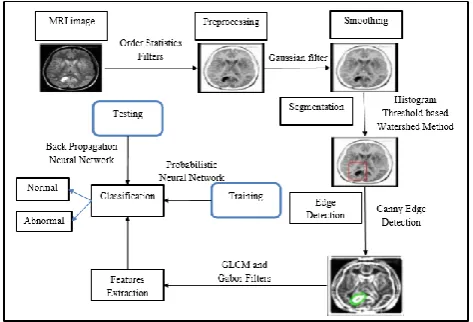
The main objective of this research work is to develop a system as diagnostic tool for recognizing the brain tumor appear in the brain. In this work the tumor classification is performed from MRI images using different types of texture features. The texture features are extracted by using the GLCM and Gabor filters before the segmentation process is done by using HistogramThreshold based Watershed Segmentation. This work performs with two stages of classifier such as PNN and BPNN. The PNN is used for training/learning process and the BPNN is used for classification /testing process. The extracted features are used for training and testing process, the PNN and BPNN based classifier are utilized to classify the types of tumor in given MRI image as abnormal or normal images which are elaborately shown in Figure 1.
Neural Network (BPNN) Classification 3
3.1 Order Statistics Filters
In this work, MRI image is preprocessed by using Order Statistics Filters (OSF) which is based on ordering the pixels comprised in given MRI image. Normally, the local statistical filter is extracted from the neighborhood of the center pixel which is used to get the information about expected values. In case of the neighborhood data are sorted (ordered), the sorted statistical information is extracted. This sorted statistics vector is employed to the Finite Impulse Response (FIR) filter is termed as OSF (A. Stella, et al.). This proposed work combines both mean filtering and median filtering to define more exact value of each and every pixels of noisy image. To find the center pixel‟s value is computed by using following equation;
Where N is defined as multiplication result of number, ROW and COLOUMN which means ( ) is defined as the set of coordinates in given image centered at point( ), ( ) and it is defined as the each pixel of the given image. For example, consider the size of given image is , gets as the average value. This order statistics filter can remove the various random noises like speckle noise and it can easily differentiate the lighted or dark pixel in given image by using the mean and median filters.
3.2 Gaussian Filter
After the noise removing process, initiate the smoothing process by using Gaussian filter is defined as;
The Gaussian filter can compute the gradient intensity changes of the image as well as minimize the Gaussian noise from given image.
The Gaussian filter can compute the gradient intensity changes of the image as well as minimize the Gaussian noise from given image.
3.3 Histogram Threshold Based Watershed
Segmentation
(HTWS) After the preprocessing, the segmentation process is done by using HTWS segmentation method.In this work user uses two fundamental approaches for segments in the
given image (T. Logeswari, et al.).Initially, compute the given image of gradient‟s local minima are selected as a marker, this process recurrent the segmentation continuously. In the second step selected regions are merged, the HTWS utilizing markers are particularly defined as the position of the marker. The gradient local minimum of the region‟s brightness is against from the background of various gray level measures. A threshold can be employed to segment the tumor region is defined as;
Where is defined as the threshold value,C (i.j) is defined as the resulting pixel at co-ordinate (i, j), and p (i,j) is defined as the pixel of the given image.The tumor parts have different positions and different shapes Morphological Operators are used for finding the different positions and shapes. The combination of morphological operators and histogram threshold are termed as the histogram threshold based watershed segmentation process. The morphological operator uses erode (Shrink) and dilating (expanding, filling) process, after this process applied in the histogram threshold to define the position of the marker.
Where, the kth band matches to the region (tumor) having pixel values in the range of TK toTK-1
where TK+1 is defined as the upper limit of gray level and TK is defined as the lower limit of gray level.
Segmentation Process
Step1:Total number of the pixels in the given image, having is obtained by utilizing the following process.
(total=bware (semented image)
Step 2:Resolution of X and Y axis is found from the image information
a=1/x_ resolution*1/y_resolution
Step 3: Area of the tumor is obtained by the following equation.
area of the tumor=total*a
3.4 GLCM and Gabor Filters
GLCM and Gabor filters. It is used for feature extraction process and the features are extracted from the segmented region. There are different types of features extracted from the segmented region presented in table 1.
Table 1. Features
3.5 PNN and BPNN
The final process of this work is a classification of PNN and BPNN which is used for training and testing process. In the training/ learning phase, the PNN classifier is used for recognizing the tumor. The given MRI image is normalized between range of -1 to 1 . Each and every feature is trained with PNN classifier, after this process the testing process is done by using BPNN classifier. To test the unknown MRI image is classified as normal and abnormal images. The final decision is made by the BPNN depends on the PNN classifier.
PNN is a kind of radial basis network classifies the distance from the input vector to the trained output vector using extracted features. The extracted features are computed by using first layer. The each and every class of vector inputs to generate as final output probabilities of vector from the second layer. All the input vectors are summed up. Finally, the complete transfer function of the output from the second layer is termed as maximum of these probabilities. This process has been done with all the images and it guaranteed to converge an optimal classifier in terms of removing or adding the training samples without extensive retraining. The hidden node stimulation is defined as;
The class output node stimulation is defined as ;
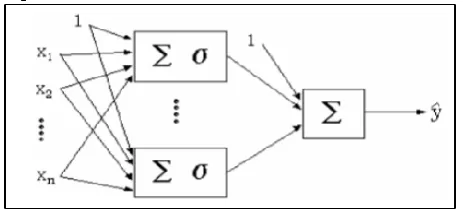
Where is defined as a smoothing factor. The smoothing factor is selected by node stimulation experiment and is defined as the hidden-node process, is defined as the total number of trained vector class. After the completion of the training process and the testing process, the help of BPNN structure is presented in figure 2.
Figure 2. BPNN Structure
The classification algorithm as follows
BPNN Classification
Step 1: Initialize the input vector weights.
Step 2: Input x1,x2,…xn transfers layer by layer and
calculate the weighted sum with input vector.
Step 3: In case preferred output = actual output is defined as back propagation error.
Step 4: Bias and Weights are updated by following equation.
IV. Results And Discussion
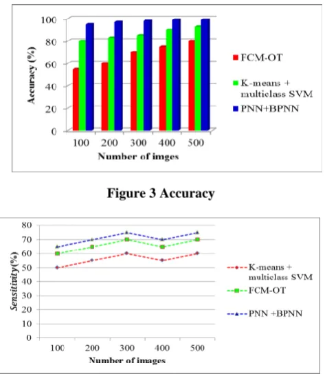
Neural Network (BPNN) Classification 5 Figure 3 shows that the comparison results in terms of
classification accuracy with Fuzzy C Means algorithm along with intelligent optimization techniques (FCM-OT) (N. Nandha, Karnan, et al.), K-means + multiclass SVM and proposed PNN with BPNN algorithm. From the results, the proposed algorithm shows the promising result in terms of accuracy when compared with other two algorithms
Figure 3 Accuracy
Figure 4 Sensitivity
Figure 4 shows that the comparison results in terms of Sensitivity with Fuzzy C Means along with intelligent optimization techniques (FCM-OT), K-means + multiclass SVM and proposed PNN with BPNN algorithm. From the results, the proposed algorithm shows the promising result in terms of Sensitivity when compared with other two algorithms.
Figure 5 Specificity
Figure 5 shows that the comparison results in terms of Specificity with Fuzzy C Means along with intelligent optimization techniques (FCM-OT), K-means + multiclass SVM and proposed PNN with BPNN algorithm. From the results, the proposed algorithm shows the promising result in terms of Specificity when compared with other two algorithms.
V. Conclusion
This paper has proposed a new efficient brain tumor detection process with the help of different image processing methods. This work is considered with the different image processing methods such as preprocessing, segmentation, edge detection, feature extraction, and classification. Initially, MRI scanned image as taken as input image, then it has been preprocessed by using OSF and smoothing process which is done by using Gaussian filter. HTWS method is used for segmentation process. After the segmentation process the edge detection has been done by using canny edge detection process, then the features are extracted from the ROI region using GLCM and Gabor filters.PNN is used for training process and then the testing process has been done by using BPNN. The experimental result has been found by using different types of metrics such as accuracy, specificity and sensitivity for analysis the proposed work and each experimental analysis shows the promising results.
References
[1] V.P.GladisPushpaRathi,S.Palani, “brain tumor MRI image classificationwith feature selection and extractionusing linear discriminant analysis”,arXiv,
1208.2128, V1, 2012
https://arxiv.org/ftp/arxiv/papers/1208/1208.2128.pdf
[2] Pankaj Kr. Saini, Mohinder Singh, “Brain Tumor
Detection In Medical Imaging Using
MATLAB”,International Research Journal of Engineering and Technology (IRJET), Vol.02 Issue: 02, 2015.
[3] Dina AboulDahab, Samy S. A. Ghoniemy, Gamal M. Selim, “Automated Brain Tumor Detection and Identification Using Image Processing and Probabilistic Neural Network Techniques”,International Journal of Image Processing and Visual Communication,Vol.1 , Issue 2, PP.1-8, 2012.
[4] RaouiaAyachi, Nahla Ben Amor, “Brain Tumor Segmentation Using Support Vector Machines”,10th European Conference, ECSQARU, Springer Berlin Heidelberg, Vol.5590, PP.736-747.
[5]Moumen T El-Melegy,Hashim M Mokhtar, “Tumor segmentation in brain MRI using a fuzzy approach with class center priors”,EURASIP Journal on Image and Video Processing, Vol. 21, PP.2-14, 2014.
Wavelets and Neural Networks”,IEEE Journal of Biomedical and Health Informatics, Vol.19, Issue: 4, PP.1451 – 1458, 2014.
[7]Fuyong Xing, YuanpuXie, Lin Yang, “An Automatic Learning-Based Framework for Robust Nucleus Segmentation”,IEEE Transactions on Medical Imaging, Vol.35, Issue.2, PP.550 – 566, 2016.
[8]Atiq Islam, Syed M. S. Reza, Khan M. Iftekharuddin, “Multifractal Texture Estimation for Detection and Segmentation of Brain Tumors”,IEEE Transactions on Biomedical Engineering, Vol.60, Issue.11, PP. 3204 – 3215,2013.
[9] A. Stella, BhushanTrivedi, “Implementation of Order Statistic Filters on Digital Image and OCT Image: A Comparative Study”, International Journal of Modern Engineering Research (IJMER),Vol.2, Issue.5, PP.3143-3145, 2012.
[10] T. Logeswari, M. Karnan, “An Enhanced Implementation of Brain Tumor Detection Using Segmentation Based on Soft Computing”,International Conference onSignal Acquisition and Processing (ICSAP), IEEE, PP. 243 – 247, 2010.