Published by Central Fisheries Research Institute (SUMAE) Trabzon, Turkey.
R E S E A R C H P A P E R
A Magnetic Bead-Based DNA Extraction Protocol Suitable for
High-Throughput Genotyping in Shrimp Breeding Programs
Cheryl K.Y. Tan
1,2, Jeff A. Cowley
1,3*
, Dean R. Jerry
1,2,41Australian Research Council (ARC) Research Hub for Advanced Prawn Breeding, James Cook University, Townsville,
QLD 4811, Australia.
2Centre for Sustainable Tropical Fisheries and Aquaculture, College of Science and Engineering, James Cook University,
Townsville, QLD 4811, Australia.
3Aquaculture, CSIRO Agriculture and Food, Queensland Bioscience Precinct, St. Lucia, QLD 4067, Australia. 4 Tropical Futures Institute, James Cook University Singapore, Singapore.
Article History
Received November 1, 2019 Accepted December 4, 2019 First Online December 6, 2019
Corresponding Author
Tel.: +61732142527 E-mail: [email protected]
Keywords
Shrimp Prawn
DNA extraction method Magnetic bead High-throughput Genetic analysis
Abstract
Due to their convenience, magnetic bead-based nucleic acid extraction kits are commonly used in shrimp genotyping and pathogen screening applications. However, in advanced breeding programs requiring the testing of many thousands of shrimp, their cost can be prohibitive. Various permutations of different Proteinase K digestion, tissue lysis and bead washing buffers as well as magnetic bead types were thus evaluated to devise a high-throughput shrimp DNA extraction (SDE) protocol capable of recovering high-purity DNA using a KingFisherTM Flex Magnetic Particle processor.
When genotyped using a MassARRAY® platform (Agena Bioscience) requiring 60-61
genome regions to be co-amplified in a single multiplexed PCR, DNA extracted from shrimp muscle tissue using either the SDE protocol or a commercial kit generated comparable single-nucleotide polymorphism (SNP) call data. The SDE protocol also extracted high-purity DNA from salmon fin clips. It thus offers potential to markedly reduce the costs of large-scale genotyping in shrimp and salmon breeding programs.
Introduction
DNA extraction methods involving various levels of sophistication continue to be developed and refined to meet yield, purity, throughput and cost requirements for genotyping, genome sequencing and pathogen screening applications across diverse species and tissue types (Chomczynski & Sacchi, 1987; Marko, Chipperfield & Birnboim, 1982; Ali et al., 2017; Dierens, Henshall & Sellars, 2014; Planella et al., 2017; Psifidi et al., 2010; Psifidi et al., 2015; Rao, Arnold & Cowley 2010; Zheng et al., 2015; Inglis et al., 2018). Since being developed in the mid-1990s (Deggerdal & Larsen, 1997; Hawkins et al., 1994; Kang et al., 2009; Levison et al., 1998; Rudi et al., 1997), magnetic bead-based methods have
increasingly been adopted due to the convenience and widespread availability of commercial kits, their ability to generate high-purity nucleic acid and their amenability to automated high-throughput instruments such as the KingFisherTM Flex Magnetic Particle
Bowen, 2009a, Dauphin et al., 2009b; Dauphin et al., 2011; Lim et al., 2018; Mertens et al., 2014). Despite this and due to most of the key reagents used in magnetic bead-based nucleic acid extraction protocols such as Proteinase K (ProK), guanidine hydrochloride (GuHCl)/thiocyanate (GuSCN) and Triton-X100 being relatively inexpensive when purchased in sizeable quantities, buffer sets that can be used to reduce extraction costs continue to be reported for different species and tissue types (Kang et al., 2009; Li et al., 2017; Mertens et al., 2014; Psifidi et al., 2010; Psifidi et al., 2015).
To grow the Black tiger shrimp (Penaeus monodon) farming industry in Australia, programs are underway to generate elite breeding lines through the use of genetic tools to manage diversity and select for desirable production traits (Alam & Pálsson, 2016; Baranski et al., 2014; Dekkers 2012; Guppy et al., 2018; Huerlimann et al., 2018; Lu et al., 2017; Robledo et al., 2017; Sellars et al., 2012; Waqairatu et al., 2012). To help reduce the high cost of genotyping and pathogen screening in these programs, described here are data leading to the development of a buffer set optimized for automated 96-well plate-based extraction of high-purity DNA from shrimp and fish tissues using a KingFisher processor.
Materials and Methods
Tissue Sources
Black tiger shrimp (Penaeus monodon) were obtained from the CSIRO Bribie Island Research Centre,
QLD, Australia. Shrimp were placed in zip-lock plastic bags, euthanized by submersion in wet ice and stored at -20°C until rapidly thawed for use. Fin clips from Atlantic salmon (Salmo salar) were obtained from an aquaculture farm in Tasmania, Australia. Individual fin clips were stored in 95% ethanol in 2 ml screw cap tubes at 4°C until used.
Trials to Define Shrimp DNA Extraction (SDE) Protocol Buffers
Initially, a set of buffers developed to extract high-purity DNA from white blood cells (WBC DNA Protocol; Psifidi et al., 2015) was compared to those used in the MagJET Genomic DNA Kit (MGD Kit, Cat. no. K2722, Thermo Scientific). As detailed in Table 1, buffer volumes used for tissue digestion and lysis and for DNA bead binding, washing and elution followed those described in Protocol E of the MGD Kit Manual (URL2). All extractions were undertaken using the automated KingFisher processor (URL1, Thermo Fisher Scientific). BindIt Software V4.0 (URL3, Thermo Scientific) was used to run the Tissue DNA KingFisherTM Flex 96 program
(KF_TissueDNA_Flex96.bdz file download available at URL4, Thermo Fisher Scientific) used to control the mechanics of each step on the KingFisher processor. Based on prior experience in using the MGD Kit to reliably obtain high-purity DNA (A260/230nm >1.95) from
shrimp muscle tissue, the Flex 96 program .bdz file was modified to include a third Wash Buffer 2 step. For the WBC DNA protocol, buffer volumes were adjusted proportionally to match those used with the MGD Kit.
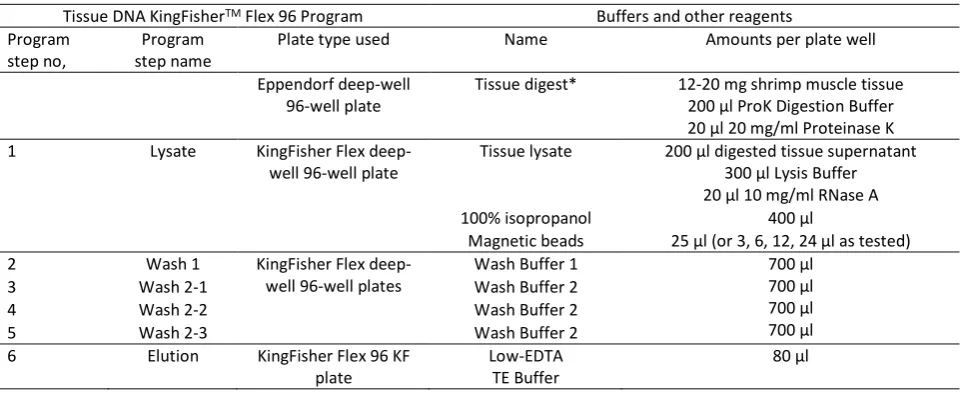
Table 1. MagJET Genomic DNA (MGD) Kit (Thermo Fisher) buffer and reagent volumes used for tissue digestion and DNA bead
binding, washing and elution on the KingFisherTM Flex Magnetic Particle processor controlled by an amended Flex 96 Program
Tissue DNA KingFisherTM Flex 96 Program Buffers and other reagents
Program step no,
Program step name
Plate type used Name Amounts per plate well
Eppendorf deep-well 96-well plate
Tissue digest* 12-20 mg shrimp muscle tissue 200 µl ProK Digestion Buffer 20 µl 20 mg/ml Proteinase K 1 Lysate KingFisher Flex
deep-well 96-deep-well plate
Tissue lysate 200 µl digested tissue supernatant 300 µl Lysis Buffer 20 µl 10 mg/ml RNase A 100% isopropanol 400 µl
Magnetic beads 25 μl (or 3, 6, 12, 24 μl as tested) 2 Wash 1 KingFisher Flex
deep-well 96-deep-well plates
Wash Buffer 1 700 µl 700 µl 700 µl 700 µl
3 Wash 2-1 Wash Buffer 2
4 Wash 2-2 Wash Buffer 2
5 Wash 2-3 Wash Buffer 2
6 Elution KingFisher Flex 96 KF plate
Low-EDTA TE Buffer
80 µl
This allowed for DNA to be extracted by both methods in the same plate under identical automated run conditions.
To develop the shrimp DNA extraction (SDE) protocol, a series of trials were run to optimize buffer compatibilities (Table 2). The SDE protocol was standardised to use 96-well plates and extract DNA from 12-20 mg tissue samples in 200 µl Tissue Digestion Buffer (ProK Digestion Buffer) containing 20 µl cut 20 mg/ml Proteinase K (ProK). To expedite digestion, the plate was shaken (200 rpm) at 56°C for 3 h using a Ratek OM11 Medium Orbital Shaking Incubator. In trials to assess various buffer permutations, larger amounts of minced muscle tissue were digested similarly in sterile 50 ml screw-cap centrifuge tubes using proportionally increased volumes of ProK Digestion Buffer. After centrifuging briefly to pellet residual particulate matter, 200 µl aliquots of clarified digest were transferred to each well of a 96-well deep-well plate containing 300 µl Lysis Buffer and 20 µl 10 mg/ml RNase A. The plate was shaken gently at room temp for 10 min using a Thermomixer (Eppendorf) before being loaded into the carousel of the KingFisher processor. Typically, 2 of 4 replicate wells of each lysate were extracted to assess DNA extraction reproducibility. To extract DNA from salmon tissue, single fin tips (35-44 mg) were digested in 1 ml ProK Digestion Buffer, with 200 µl aliquots of clarified digest then transferred to 4 replicate wells to be extracted as for shrimp tissue.
To optimise the SDE protocol, 4 different ProK Digestion Buffer formulations [ProK Buffer 1a (25 mM Tris-HCl, 25 mM Na2-EDTA, 2 M GuHCl, 5 mM CaCl2, 0.5%
Triton X-100, 1% N-Lauroylsarcosine, pH 7.5), ProK Buffer 1b (2 × concentration of 1a) (Psifidi et al., 2015), ProK Buffer 2 (30 mM Tris-HCl, 30 mM Na2-EDTA, 0.8 M
GuHCl, 0.5% Triton X-100, 5% Tween-20, pH 5.3) and ProK Buffer 3 (30 mM Tris-HCl, 10 mM Na2-EDTA, 3 M
GuHCl, 0.5% Triton X-100, 5% Tween-20, pH 8.0)] were compared to that provided in the MGD Kit. Initially these buffers were evaluated with either the MGD Kit buffers and magnetic beads or the WBC DNA protocol buffers used with Silanol magnetic beads (Table 2 Trial 1A; Table 3). As DNA was extracted at substantially higher yields and purity using the MGD Kit buffers and beads (see below), each buffer used in the WBC DNA protocol was evaluated systematically for its compatibility with the MGD Kit buffers. When DNA yields and/or purity were unacceptably low, the suspected incompatible buffer was modified and re-evaluated.
The outcome of these buffer optimization trials was a SDE protocol that used ProK Buffer 1b, SDE Lysis Buffer (50 mM Tris-HCl, 25 mM Na2-EDTA, 6 M GuSCN,
3% Triton X-100, 6% N-Lauroylsarcosine, pH 5.5), a modified SDE Wash Buffer 1 (25 mM Tris-HCl, 1.8 M GuHCl, 75% EtOH, pH 6.6) and a modified SDE Wash Buffer 2 (10 mM Tris-HCl, 100 mM NaCl, 80% EtOH, pH 6.6). To help preserve DNA integrity during long term storage, DNA was eluted from beads using low-EDTA TE
Buffer (10 mM Tris-HCl pH 7.5, 0.1 mM Na2-EDTA) rather
than water. The 4 commercially-available magnetic bead types tested for their compatibility with the SDE protocol buffers are detailed in Table 3.
DNA Yield and Purity Assessments
To assess DNA yield and relative purity, 2 µl each extract was assessed in triplicate on a Nanodrop ND8000 UV spectrophotometer (Thermo Scientific 2010; Gallagher 2011). The A260/280nm ratio provided an
estimate of levels of protein contamination and the A260/230nm ratio provided an estimate of contamination
with salts, with ratios ≥1.8 and ≥2.0, respectively, at pH 7.5 considered to represent high-purity DNA (URL5).
SNP Genotyping
To validate its amenability to downstream analyses, shrimp DNA extracted using the SDE protocol in combination with the AccuBead, Silanol or Sera-Mag bead magnetic bead types (Table 3) was genotyped using 2 MassARRAY panels (Sellars et al., 2012, URL6, Agena Bioscience) at the Australian Genome Research Facility (AGRF), Brisbane, Australia. Each MassARRAY panel was in routine use at the time for tracking pedigrees and genetic diversity within cohorts of P. monodon, and had the capacity to assign SNPs in 60 to 61 PCR products amplified in a single reaction using a highly multiplexed set of iPLEX PCR primer pairs (Henshall, Dierens, & Sellars 2014; Sellars et al., 2012, Agena Bioscience). In this analysis, each SDE protocol/bead type combination was used to extract DNA from 22-24 replicates of a bulk digest of muscle tissue from a single shrimp and was compared to DNA extracted from muscle of the same shrimp using the MGD Kit. Prior to being genotyped, the relative purity and concentration of each DNA was determined, and it was normalised to 25 ng/µl in low-EDTA TE Buffer to negate SNP call differences due to concentration-related factors.
Results and Discussion
Comparison of MGD Kit and WBC DNA Protocols
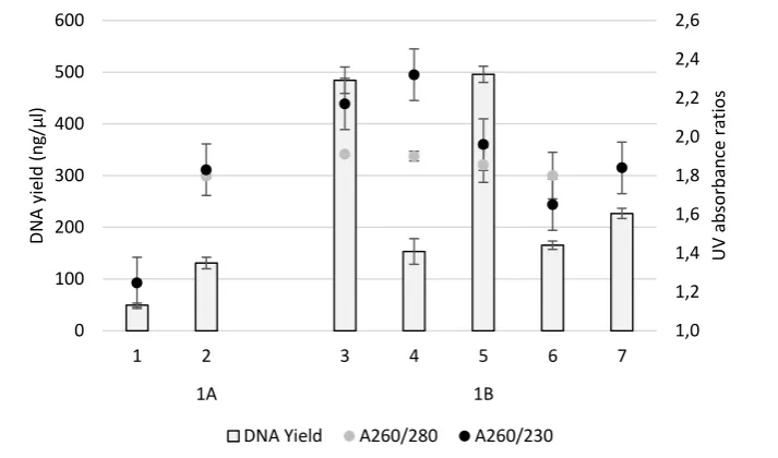
the MGD Kit (131.0 ± 6.5 ng/µl) (Figure 1A). While the A260/280 nm ratio of the WBC protocol DNA was only
slightly lower (1.80 ± 0.03) than the MGD Kit DNA (1.88 ± 0.02), its A260/230 nm ratio was non-ideal (1.25 ± 0.03)
(Figure 1A). The lower DNA yield and purity suggested WBC protocol buffer incompatibilities with shrimp muscle tissue and/or the KingFisher processor bead binding and washing program steps that either compromised DNA binding to the magnetic beads or
promoted its premature detachment and the carryover of salts (URL5). To identify which buffers were incompatible, each was evaluated systematically and modified as required.
Proteinase K Digestion Buffers
To assess the influence of different ProK Digestion Buffers, the WBC DNA ProK Buffer at 1× (ProK Buffer 1a)
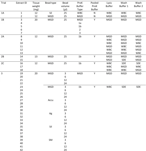
Table 2. Trials run to optimise the SDE protocol
Trial Extract ID Tissue weight
(mg)
Bead type Bead volume
(µl)
ProK Buffer
Type
Pooled ProK Buffer
Lysis Buffer
Wash Buffer 1
Wash Buffer 2
1A 1 12 Sil 25 WBC N WBC WBC WBC
2 12 MGD 25 MGD N MGD MGD MGD
1B 3 20 MGD 25 MGD Y MGD MGD MGD
4 1a
5 1b
6 2
7 3
2A 8 12 MGD 25 1b Y MGD MGD MGD
9 WBC MGD MGD
10 WBC MGD WBC
11 MGD WBC MGD
12 WBC WBC MGD
13 MGD MGD WBC
2B 14 13 MGD 25 1b Y MGD MGD MGD
15 MGD SDE MGD
2C 16 12 MGD 25 1b Y WBC SDE SDE
17 WBC MGD WBC
18 WBC WBC MGD
3 19 20 MGD 3 MGD Y MGD MGD MGD
20 6
21 12
22 24
23 MGD 3 1b Y WBC SDE SDE
24 6
25 12
26 24
27 Accu 3
28 6
29 12
30 24
31 Ag 3
32 6
33 12
34 24
35 Sil 3
36 6
37 12
38 24
39 SM 3
40 6
41 12
42 24
Abbreviations: WBC = White Blood Cell protocol (Psifidi et al., 2015); SDE = Shrimp DNA Extraction protocol; MGD = MagJET Genomic DNA protocol
Bead types: MGD = MGD Kit beads; Accu = AccuBead silica-coated beads; Sil = Silanol functional beads; SM = Sera-Mag SpeedBead Carboxylate-modified beads; Ag = Agencourt AMPure XP beads
and 2× concentration (ProK Buffer 1b) as well as 2 other buffer recipes (2 and 3) were compared against that used in the MGD Kit. For this, 50 mg shrimp muscle tissue was digested in 0.5 ml each buffer containing 50 µl 20 mg/ml ProK, with duplicate 200 µl aliquots of clarified digest then processed in the KingFisher processor using MGD Kit Lysis and Wash Buffers (Table 2 Trial 1B, Figure 1B). UV spectral analysis showed DNA yields with the MGD Kit ProK Digestion Buffer (406.2 ± 67.6 ng/µl) and ProK Buffer 1b (399.3 ± 79.4 ng/µl) to be 2- to 3-fold higher than those obtained using the other 3 buffers assessed. A260/280 nm ratios with DNA extracted
using any of the 5 buffers were >1.8, but highest with the MGD Kit buffer (1.93 ± 0.02) (Figure 1B). A260/230 nm
ratios were highest using ProK Buffer 1a (2.39 ± 0.07), but also ≥2 with DNA extracted using either the MGD Kit buffer or ProK Buffer 1b (Figure 1B). The DNA yield and purity data obtained in this trial suggested that ProK Buffer 1b, which contained the highest concentration of
GuHCl (4 M), compared well with the MGD Kit ProK buffer when used together with the MGD Kit magnetic beads and Lysis/Wash Buffers.
Lysis Buffer and Wash Buffers
As ProK Buffer 1b generated high yields of high-purity DNA when used together with MGD Kit lysis and wash buffers, it was selected as the basis for identifying compatible alternative lysis and wash buffers. To examine this, 24 mg amounts of muscle tissue from the same shrimp were digested in 400 µl ProK Buffer 1b together with 40 µl 20 mg/ml ProK, with duplicate 200 µl aliquots of clarified digest then extracted using various permutations of MGD Kit and WBC protocol lysis and wash buffers (Table 2 Trial 2A, Figure 1). Compared to extractions with the MGD Kit buffers (18.0 ± 2.1 ng/µl), a higher DNA yield was obtained with the WBC DNA protocol lysis buffer used together with the MGD
Table 3. Commercially-availablemagnetic bead types assessed
Name Vendor (Australia) Catalogue No. Cost/sample* (AUD) Silanol functionalized beads Advance Scientific
Products
PMSI-H1.0-5 $0.25
AccuBeadTM Silica coated Bioneer Pacific TA-1010-1 $0.02
Sera-Mag SpeedBead Carboxylate-modified
GE Life Sciences 65152105050250 $0.09
Agencourt AMPure XP Beckman Coulter Life Sciences
A63881 $0.02
* Approximate minimum cost based on the use of 3 µl beads/DNA extraction in 2019.
Figure 1. Yields and purity (A260/280 nm; A260/230 nm) of DNA extracted in different trials using equal amounts of shrimp
abdominal muscle tissue and various DNA extraction reagent permutations as described in Table 2. Trial 1A compared the WBC DNA (1) or MGD Kit (2) extraction protocol buffer sets. The WBC DNA protocol employed Silanol functional beads and data points represent the mean ± SD of duplicate extractions. Trial 1B compared the MGD Kit (3) extraction protocol to this protocol used with the Kit ProK buffer substituted by ProK Buffers 1a (4), 1b (5), 2 (6) or 3 (7), with data points representing the mean ± SD of duplicate extractions.
1,0 1,2 1,4 1,6 1,8 2,0 2,2 2,4 2,6
0 100 200 300 400 500 600
1 2 3 4 5 6 7
1A 1B
U
V
ab
so
rb
an
ce
r
ati
o
s
DN
A
yi
el
d
(
n
g/µl
)
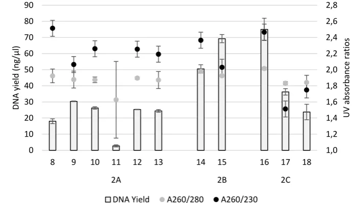
Kit wash buffers (30.0 ± 0.3 ng/µl). While DNA purity (A260/280 nm; A260/230 nm ratios) was somewhat reduced
(1.88 ± 0.15; 2.06 ± 0.11) compared to that extracted using the MGD Kit buffers (1.92 ± 0.12; 2.51 ± 0.41), they were considered acceptable. Likewise, the WBC protocol Wash Buffer 2 did not substantially compromise DNA yield or purity (MGD kit/WBC protocol A260/280 nm 1.89 ± 0.02/1.87 ± 0.15; A260/230 nm 2.25 ±
0.08/2.19 ± 0.16). However, DNA yields obtained using the WBC protocol Wash Buffer 1 were unacceptable low (2.7 ± 1.0 ng/µl).
To examine wash buffer compatibilities with the optimised ProK Buffer 1b and Lysis buffer, the propanol component of Wash Buffers 1 and 2 was replaced with higher concentrations of ethanol as specified for use in the MGD Kit wash buffers. Extraction using the revised WBC protocol Wash Buffer 1 improved DNA yield by ~40% with only a small reduction in DNA purity based on A260/230nm values (Table 2 Trial 2B, Figure 2). Extraction
using ethanol-containing Wash Buffers 1 and 2 produced markedly higher yields of high-purity DNA compared to extractions undertaken with combinations of MGD Kit and WBC protocol wash buffers (Table 2 Trial 2C, Figure 2). As DNA yields and purity were comparable to those obtained using all MKG kit buffers, this combination of buffers was adopted as the SDE protocol, albeit with the remaining need to assess its performance with alternative magnetic bead types to that used in the MGD Kit.
Magnetic Bead Optimisation
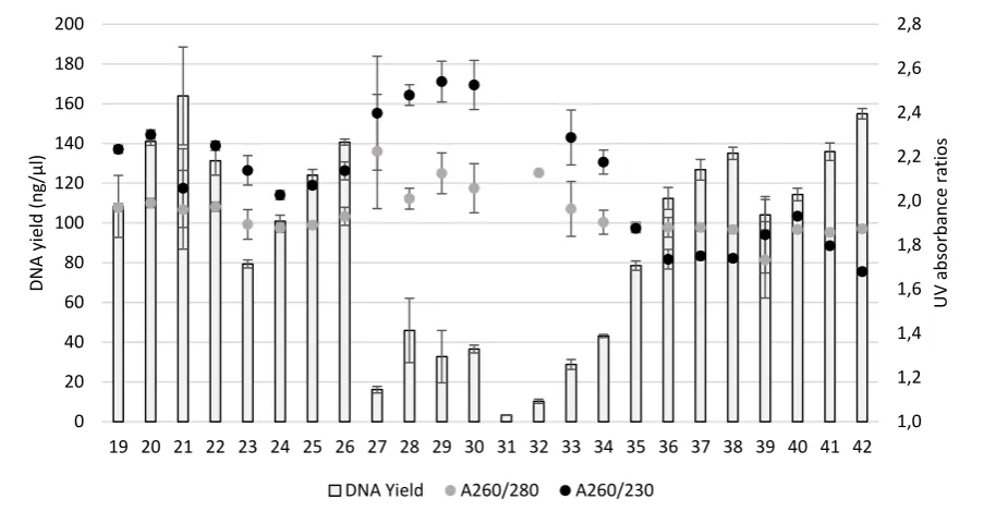
To determine DNA yields and purity using the SDE protocol buffers with any of 4 commercially-available magnetic beads, 4 replicate 200 µl aliquots of a clarified bulk digest of 300 mg shrimp muscle in 4 ml SDE ProK Buffer 1b were each added to 300 µl SDE Lysis buffer and extracted as described in Table 1 using 3 µl, 6 µl, 12 µl or 24 µl of each bead type (Table 2 Trial 3, Figure 3). As a control, an equivalent amount of shrimp muscle tissue was extracted using MGD Kit buffers and beads. DNA yields generally increased as bead amounts were increased from 3 µl and 24 µl, except with AccuBeads where DNA yields were highest using 6 µl beads and decreased using higher bead amounts. Using the SDE protocol buffer set, DNA yields with either Silanol or Sera-Mag beads equalled or bettered those obtained using the MGD Kit beads. DNA yields were generally somewhat lower using equivalent volumes of AccuBead or Agencourt AMPure-XP beads. Despite this, the purity of DNA obtained using any volume of AccuBeads (A260/280 nm values >2) surpassed that of DNA extracted using the
MGD Kit buffers and beads. While generally >1.8, A260/280 nm ratios of DNA extracted using the SDE protocol buffers
together with MGD Kit, Silanol or Sera-Mag beads were slightly inferior to those of DNA obtained using the MGD Kit buffers with equivalent amounts of each bead type (1.96 ± 0.01 to 1.99 ± 0.02).
When the bead volume used in a SDE protocol extraction resulted in an acceptable DNA yield, A260/230
Figure 2. Yields and purity (A260/280 nm; A260/230 nm) of DNA extracted in different trials using equal amounts of shrimp
abdominal muscle tissue and various DNA extraction reagent permutations as described in Table 2. Trial 2A compared use of ProK Buffer 1b in combination with the MGD Kit Lysis Buffer (8, 11, 13) and Wash Buffers 1 (8, 13) and 2 (8, 11) or with the Lysis Buffer (9, 10, 12), Wash Buffer 1 (11, 12) and Wash Buffer 2 (10, 13) replaced by buffers used in the WBC DNA protocol (Psifidi et al., 2015). Data points represent the mean ± SD of duplicate extractions. Trial 2B compared use of ProK Buffer 1b in combination with the MGD Kit lysis and wash buffers (14) or with the MGD Kit Wash Buffer 1 replaced by SDE protocol Wash Buffer 1 (15). Data points represent the mean ± SD of 4 replicate extractions. Trial 2C compared use of ProK Buffer 1b in combination with WBC DNA extraction protocol Lysis Buffer (16, 17, 18), and the various permutations of MGD Kit, WBC DNA protocol and SDE protocol Wash Buffers 1 (17, 18 and 16, respectively) and 2 (18, 17 and 16, respectively). Data points represent the mean ± SD of 4 replicate extractions.
1,0 1,2 1,4 1,6 1,8 2,0 2,2 2,4 2,6 2,8
0 10 20 30 40 50 60 70 80 90
8 9 10 11 12 13 14 15 16 17 18
2A 2B 2C
UV
ab
so
rb
an
ce
rat
io
s
DN
A
yi
el
d
(
n
g/µl
)
nm ratios of DNA obtained using the same bead volumes
were slightly lower with DNA recovered from Silanol or Sera-Mag beads (1.74 ± 0.02 to 1.68 ± 0.02) and slightly higher with DNA recovered from AccuBeads (2.40 ± 0.26 to 2.54 ± 0.09) compared to DNA recovered using the MGD Kit (2.06 ± 0.18 to 2.30 ± 0.02) (Figure 3). Overall these data indicated that any of the 4 magnetic bead types tested could be used successfully with the SDE protocol buffer set to extract DNA at acceptable yields and purity, and thus could be selected based on local availability, cost and DNA yield/purity requirements.
SNP genotyping SDE protocol DNA
DNA extracted from muscle tissue of a single shrimp using either the MGD Kit or the SDE protocol in combination with AccuBead, Silanol or Sera-Mag beads was genotyped in 2 SNP-based MassARRAY panels each employing 60-61 multiplexed pairs of iPLEX PCR primers (Sellars et al., 2012). SNP call rates were high (≥98%) irrespective of extraction method or bead type used (Table 4). Of the 121 SNPs assessed, PCR primer pairs targeting 9 failed across all 94 DNA samples irrespective of the extraction method, indicating that sequence variations existed in the shrimp tested at these genome locations. The other 112 SNPs were generally called accurately with good confidence, except for 1 DNA sample (SM-10) that failed across all 121 SNPs, suggestive of a technical issue with this sample, and for 9 other samples scattered across the different DNA extraction groups in which 1 or 2 of 3 specific SNPs were
either miscalled or not called (Table 4). As A260/280 nm and
A260/230 nm ratios were >2 for these 10 DNA samples,
issues with their purity were unlikely. As such, it is possible the aberrant SNP calls arose from the PCR primer pairs targeting these 3 SNPs being somewhat less competent at amplifying DNA and thus more prone to failure in samples where pipetting inconsistencies might have resulted in slightly less DNA template being present. Overall, DNA extracted using the SDE protocol together with any of the 3 bead types tested produced SNP call data that were indistinguishable from DNA extracted using the MGD kit, and that would be more than adequate for pedigree assignment or genetic variability analyses.
SDE Protocol Use to Extract DNA from Salmon Fin Clips
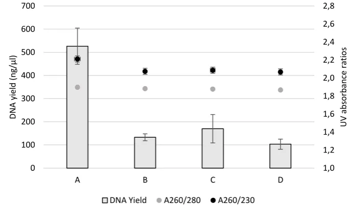
To assess how well the SDE protocol might perform with other aquaculture species highly reliant on genotyping to manage selected breeding lines, it was used to extract DNA from Atlantic salmon fin clips. When used together with AccuBead, Silanol or Sera-Mag magnetic beads, DNA yields were between 18% to 37% of DNA extracted using the MGD Kit (Figure 4). However, A260/280 nm ratios equalled or bettered those of DNA
extracted using the MGD Kit, and while A260/230 nm ratios
were slightly lower (Figure 4), all were >2 indicative of the DNA being highly pure and thus amenable to even demanding downstream processing and genetic analyses (Thermo Fisher 2010).
Figure 3. Trial 3 as described in Table 2 compared yields and purity (A260/280 nm; A260/230 nm) of DNA extracted from equal
amounts of shrimp abdominal muscle tissue using either all MGD Kit reagents in combination with 3 µl, 6 µl, 12 µl or 24 µl volumes of Kit magnetic beads (19-22) or the SDE protocol reagent set used with the same volumes of MGD Kit (23-26), AccuBeadTM Silica-coated (27-30), Agencourt AMPure XP (31-34), Silanol (35-38) or Sera-Mag SpeedBead Carboxylate-modified
magnetic beads (39-42). Data points represent the mean ± SD of 4 replicate extractions.
1,0 1,2 1,4 1,6 1,8 2,0 2,2 2,4 2,6 2,8
0 20 40 60 80 100 120 140 160 180 200
19 20 21 22 23 24 25 26 27 28 29 30 31 32 33 34 35 36 37 38 39 40 41 42
UV
ab
so
rb
an
ce
rat
io
s
DN
A
yi
el
d
(
n
g/µl
)
Conclusions
To exploit the use of the automated magnetic bead-based KingFisher™ Flex Magnetic Particle processor, a buffer set was systematically optimized for the high-throughput extraction of high-purity DNA from shrimp muscle tissue. This shrimp DNA extraction (SDE) protocol was developed to reduce the costs of using commercial DNA extraction kits (Claassen et al., 2013; Dauphin et al., 2009a, 2009b, 2011; Lim et al., 2018; Mertens et al., 2014). This is important as the costs of relying on kits can be prohibitive in breeding programs requiring many thousands of shrimp to be genotyped to assign pedigrees using SNP-based MassARRAY panels (Sellars et al., 2012, Agena Bioscience), or to interrogate large SNP numbers using technologies such as DArTSeq (Diversity Arrays Technology) to associate phenotypes
with genotypes. While broadly established from buffers developed to extract DNA from white blood cells (Psifidi et al., 2015), key to recovering high purity DNA from shrimp muscle tissue were wash buffers employing ethanol rather than isopropanol and identifying commercially-available magnetic bead types that would recover DNA at high efficiency. When the SDE protocol was used with AccuBead, Silanol or Sera-Mag magnetic bead types, the call rate for 112 SNPs assessed using a MassARRAY method (Agena Bioscience) was comparable to DNA extracted from shrimp muscle using the MGD Kit buffers and beads (Thermo Scientific) optimized for use with the KingFisher processor. The SDE protocol also proved capable of extracting high-purity DNA from salmon fin tips. Using reagent amounts purchased in sufficient quantities to prepare buffer volumes to extract 500 to >4000 tissue samples, and
Table 4. Yields and purity of DNA extracted from shrimp muscle tissue using the MGD Kit or SDE protocol in combination with 3
magnetic bead types and call rates for 121 SNPs determined in MassARRAY-based genotyping analyses
Bead Type
Tissue replicates
Plate well position
DNA yield and purity SNP call rate
DNA samples in which failed/aberrant SNP calls
were detected Mean yield
(ng/µl)
A260/280 nm A260/230 nm
MGD 1 - 24 1 - 24 171.3 ± 2.8 2.06 ± 0.00 2.28 ± 0.00 99% MGD-19A*
Accu 1 - 24 25 - 48 49.0 ± 3.3 2.07 ± 0.01 2.64 ± 0.05 98% Sil-17A, Sil-21A,B
Sil 1 - 24 49 - 72 120.3 ± 5.5 2.04 ± 0.01 2.10 ± 0.02 100%
SM 1 - 22 73 - 94 126.1 ± 1.9 2.04 ± 0.00 2.24 ± 0.01 98% SM-7A, SM-9A,B, SM-10A,
SM-12**, SM-15C, SM-16C,
SM-17C
Abbreviations: MGD = MGD Kit beads, Accu = AccuBead; Sil = Silanol bead; SM = Sera-Mag bead
* Letter codes denote same failed SNP in different DNA sample replicates (numbered sequentially 1 - 22/24) ** No data with all SNPs
Figure 4. Yields and purity (A260/280 nm; A260/230 nm) of DNA extracted from equal weights of salmon fin clip tissue using either the
(A) MGD Kit or the SDE protocol using (B) Sera-Mag SpeedBead Carboxylate-modified, (C) Silanol or (D) AccuBeadTM
Silica-coated magnetic beads. Data points represent the mean ± SD of either 4 replicate extractions using equal volumes from a single large ProK tissue digest.
1,0 1,2 1,4 1,6 1,8 2,0 2,2 2,4 2,6 2,8
0 100 200 300 400 500 600 700
A B C D
UV
ab
so
rb
an
ce
rat
io
s
DN
A
yi
el
d
(
n
g/µl
)
depending on which bead type is used, the costs per DNA extraction of using the SDE protocol were estimated to be 2- to 3-fold lower than using the MGD or other comparable kits. The SDE protocol thus offers potential to substantially reduce genotyping and pathogen screening costs in research projects and breeding programs aimed at improving the efficiency, quality and outputs of valuable aquaculture species such as shrimp and salmon.
Acknowledgements
We thank Chris Stratford for providing P. monodon and Natasha Botwright for providing salmon tissue samples, Hjinen Marcel at GE Life Sciences for providing sample Sera-Mag SpeedBeads, Martin Peet at Advanced Scientific Products for providing sample Silanol beads and Greg Manderson at Bioneer Pacific for providing sample AccuBeads. The study was funding by the ARC Industrial Transformation Research Program IH130200013.
References
Ali, N., de Cássia Pontello Rampazzo, R., Dias Tavares Costa, A., & Aurelio Krieger, M. (2017). Current nucleic acid extraction methods and their implications to point-of-care diagnostics. BioMed Research International, 9306564. https://doi.org/10.1155/2017/9306564 Alam, M.M.M. & Pálsson, S. (2016). Population structure of the
giant tiger shrimp Penaeus monodon in Bangladesh based on variation in microsatellites and immune‐ related genes. Marine Biology Research 12(7), 706-714. https://doi.org/10.1080/17451000.2016.1196820 Baranski, M., Gopikrishna, G., Robinson, N.A., Katneni, V.K.,
Shekhar, M.S., Shanmugakarthik, J., … Ponniah, A.G. (2014). The Development of a high-density linkage map for Black tiger shrimp (Penaeus monodon) based on cSNPs. PLoS One 9(1), e85413. https://doi.org/10.1371/journal.pone.0085413 Chomczynski, P., & Sacchi, N. (1987). Single-step method of
RNA isolation by acid guanidinium thiocyanate-phenol-chloroform extraction. Analytical Biochemistry 162, 156-159. https://doi.org/10.1016/0003-2697(87)90021-2 Claassen, S., du Toit, E., Kaba, M., Moodley, C., Zar, H.J., &
Nicol, M.P. (2013). A comparison of the efficiency of five different commercial DNA extraction kits for extraction of DNA from faecal samples. Journal of Microbiological
Methods 94(2):103-110.
https://doi.org/10.1016/j.mimet.2013.05.008
Dauphin, L.A., Moser, B.D., & Bowen, M.D. (2009a). Evaluation of five commercial nucleic acid extraction kits for their ability to inactivate Bacillus anthracis spores and comparison of DNA yields from spores and spiked environmental samples. Journal of Microbiological
Methods 76(1), 30-37.
https://doi.org/10.1016/j.mimet.2008.09.004
Dauphin, L.A., Hutchins, R.J., Bost, L.A., & Bowen, M.D. (2009b). Evaluation of automated and manual commercial DNA extraction methods for recovery of Brucella DNA from suspensions and spiked swabs.
Journal of Clinical Microbiology 47(12), 3920-3926.
https://doi.org/10.1128/JCM.01288-09
Dauphin, L.A., Walker, R.E., Petersen, J.M., & Bowen, M.D. (2011). Comparative evaluation of automated and manual commercial DNA extraction methods for detection of Francisella tularensis DNA from suspensions and spiked swabs by real-time polymerase chain reaction. Diagnostic Microbiology and Infectious
Disease 70(3), 299-306.
https://doi.org/10.1016/j.diagmicrobio.2011.02.010 Dekkers, J.C.M. (2012). Application of genomics tools to animal
breeding. Current Genomics 13(3), 207-212. https://doi.org/10.2174/138920212800543057 Deggerdal, A., & Larsen, F. (1997). Rapid isolation of PCR-ready
DNA from blood, bone marrow, and cultured cells, based on paramagnetic beads. BioTechniques 22, 554-557. https://doi.org/10.2144/97223pf02
Dierens, L., Henshall, J., & Sellars, M.J. (2014). An industry friendly, inexpensive DNA extraction method for Penaeid shrimp that is compatible with Sequenom(R)
iPLEX Platinum SNP pedigree genotyping platforms.
Aquaculture 433, 102-104.
https://doi.org/10.1016/j.aquaculture.2014.06.004 Gallagher, S.R. (2011). Quantitation of DNA and RNA with
absorption and fluorescence spectroscopy. Current
Protocols in Molecular Biology, Supplement 52(1),
A.4K.1-A.4K.4.
https://doi.org/10.1002/0471140864.psa04ks52 Guppy, J.L., Jones, D.B., Jerry, D.R., Wade, N.M., Raadsma,
H.W., Huerlimann, R., & Zenger, K.R. (2018). The state of "omics" research for farmed penaeids: advances in research and impediments to industry utilization.
Frontiers in Genetics 9, Article 282, 1-27.
https://doi.org/10.3389/fgene.2018.00282
Hawkins, T.L, O’Connor-Morin, T., Roy, A., & Santillan, C. (1994). DNA purification and isolation using a solid-phase. Nucleic Acids Research 22, 4543-4544. https://doi.org/10.1093/nar/22.21.4543
Henshall, J.M., Dierens, L., & Sellars, M.J. (2014). Quantitative analysis of low-density SNP data for parentage assignment and estimation of family contributions to pooled samples. Genetics Selection Evolution 46(1), 51. https://doi.org/10.1186/s12711-014-0051-y
Huerlimann, R., Wade, N.M., Gordon, L., Montenegro, J.D., Goodall, J., McWilliam, S., … Jerry, D.R. (2018). De novo assembly, characterization, functional annotation and expression patterns of the black tiger shrimp (Penaeus
monodon) transcriptome. Scientific Reports 8, 13553,
https://doi.org/10.1038/s41598-018-31148-4
Inglis, P.W., Pappas, M.d.C.R., Resende, L.V., & Grattapaglia, D. (2018). Fast and inexpensive protocols for consistent extraction of high-quality DNA and RNA from challenging plant and fungal samples for high-throughput SNP genotyping and sequencing applications. PLoS One 13, e0206085.
https://doi.org/10.1371/journal.pone.0206085 Kang, K., Choi, J., Nam, J.H., Lee, S.C., Kim, K.J., Lee, S.W., &
Chang, J.H. (2009). Preparation and characterization of chemically functionalized silica-coated magnetic nanoparticles as a DNA separator. Journal of
Physical Chemistry B 113(2), 536-43.
https://doi.org/10.1021/jp807081b.
Journal of Chromatography A, 816, 107-111. https://doi.org/10.1016/S0021-9673(98)00064-8 Li, B., Mou, X., Chen, Z., Chen, H., Deng, Y., Li, S., … He, N.
(2017). The development of a rapid high-quality universal nucleic acid extraction Kit based on magnetic separation. Science China Chemistry 60, 1602-1608. https://doi.org/10.1007/s11426-017-9061-1
Lim, M.Y., Song, E.J., Kim, S.H., Lee, J., & Nam, Y.D. (2018). Comparison of DNA extraction methods for human gut microbial community profiling. Systematic and Applied
Microbiology 41(2), 151-157.
https://doi.org/10.1016/j.syapm.2017.11.008.
Lu, X., Luan, S., Cao, B., Meng, X., Sui, J., Dai, … Kong, J. (2017). Estimation of genetic parameters and genotype-by-environment interactions related to acute ammonia stress in Pacific white shrimp (Litopenaeus vannamei) juveniles at two different salinity levels. PLoS One 12(3), e0173835. https://doi.org/10.1371/journal.pone Marko, M.A., Chipperfield, R., & Birnboim, H.C. (1982). A
procedure for the large-scale isolation of highly purified plasmid DNA using alkaline extraction and binding to glass powder. Analytical Biochemistry 12, 382-387. https://doi.org/10.1016/0003-2697(82)90497-3 Mertens, K., Freund, L., Schmoock, G., Hänsel, C., Melzer, F., &
Elschner, M.C. (2014). Comparative evaluation of eleven commercial DNA extraction kits for real-time PCR detection of Bacillus anthracis spores in spiked dairy samples. International Journal of Food Microbiology 170, 29-37.
https://doi.org/10.1016/j.ijfoodmicro.2013.10.022 Nagy, M., Otremba, P., Krüger, C., Bergner-Greiner, S., Anders,
P., Henske, B., … Roewer, L. (2005). Optimization and validation of a fully automated silica-coated magnetic beads purification technology in forensics. Forensic
Science International 152(1), 13-22.
https://doi.org/10.1016/j.forsciint.2005.02.027 Planella, L., Vera, S.H.M., García‐Marín, J-L., & Roldán, M.I.
(2017). An optimized high-quality male DNA extraction from spermatophores in open thelycum shrimp species.
Integrative Zoology 12, 421-427.
https://doi.org/10.1111/1749-4877.12250
Psifidi, A., Chrysostomos, I., Dovas, C.I., & Banosa, G. (2010). A comparison of six methods for genomic DNA extraction suitable for PCR-based genotyping applications using ovine milk samples. Molecular and Cellular Probes 24, 93-98. https://doi.org/10.1016/j.mcp.2009.11.001 Psifidi, A., Dovas, C.I., Bramis, G., Lazou, T., Russel, C.L.,
Arsenos, G., & Banos, G. (2015). Comparison of eleven methods for genomic DNA extraction suitable for large-scale whole-genome genotyping and long-term DNA banking using blood samples. PLoS One 10(1), e0115960. https://doi.org/10.1371/journal.pone.0115960 URL1
https://assets.thermofisher.com/TFS-Assets/LSG/manuals/KingFisher_Flex_User_Manual_54 00630_5400640.pdf
URL2
https://assets.thermofisher.com/TFS-Assets/LSG/manuals/MAN0012726_MagJET_Genomic_ DNA_UG.pdf
URL3
https://assets.thermofisher.com/TFS-Assets/LSG/manuals/BindIt_4_KingFisherInstrumentsUs erManual.pdf
URL4
https://www.thermofisher.com/search/results?query=k ingfisher+flex+96&sort=relevancy&refinementAction=tr ue&persona=DocSupport&focusarea=Search%20All URL5 Thermo Fisher Scientific (2015).
https://assets.thermofisher.com/TFS- Assets/CAD/Product-Bulletins/TN52646-E-0215M-NucleicAcid.pdf
URL6 http://agenabio.com/
Rao, M., Arnold, S.J., & Cowley, J.A. (2010). High-throughput DNA extraction for PCR-based genotyping of single
Penaeus monodon embryos and nauplii. Aquaculture
310, 61-65.
https://doi.org/10.1016/j.aquaculture.2010.10.001 Robledo, D., Palaiokostas, C., Bargelloni, L., Martínez, P., &
Houston, R. (2017). Applications of genotyping by sequencing in aquaculture breeding and genetics.
Reviews in Aquaculture 10, 670-682.
https://doi.org/10.1111/raq.12193.
Rudi, K., Kroken, M., Dahlberg, O.J., Deggerdal, A., Jakobsen, K.S., & Larsen, F. (1997). Rapid universal method to isolate PCR-ready DNA using magnetic beads. BioTechniques 22, 506-511. https://doi.org/10.2144/97223rr01
Sellars, M.J., Dierens, L., McWilliam, S., Little, B., Murphy, B., Coman, G.J., … Henshall, J. (2012). Comparison of microsatellite and SNP DNA markers for pedigree assignment in Black Tiger shrimp, Penaeus monodon.
Aquaculture Research 45, 417-426.
https://doi.org/10.1111/j.1365-2109.2012.03243.x. Thermo Scientific (2010). T042‐Technical Bulletin NanoDrop
Spectrophotometers, 260/280 and 260/230 Ratios. http://www.nhm.ac.uk/content/dam/nhmwww/our-
science/dpts-facilities-staff/Coreresearchlabs/nanodrop.pdf
Waqairatu, S.S., Dierens, L., Cowley, J.A., Dixon, T.J., Johnson, K.N., Barnes, A.C., & Li, Y. (2012). Genetic analysis of Black Tiger shrimp (Penaeus monodon) across its natural distribution range reveals more recent colonization of Fiji and other South Pacific islands.
Ecology and Evolution 2(8), 2057-2071.
https://doi.org/10.1002/ece3.316
Witt, S., Neumann, J., Zierdt, H., Gébel, G., & Röscheisen, C. (2012). Establishing a novel automated magnetic bead-based method for the extraction of DNA from a variety of forensic samples. Forensic Science International:
Genetics 6(5), 539-547.
https://doi.org/10.1016/j.fsigen.2012.01.002
Zheng, X., Hoegenauer, K.A., Maeda, A.B., Wang, F., Stelly, D.M., Nichols, R.L., & Jones, D.C. (2015). Non-destructive high-throughput DNA extraction and genotyping methods for cotton seeds and seedlings. Biotechniques